Salicylic acid is a key player of Arabidopsis autophagy mutant susceptibility to the necrotrophic bacterium Dickeya dadantii - Nature
←
→
Page content transcription
If your browser does not render page correctly, please read the page content below
www.nature.com/scientificreports
OPEN Salicylic acid is a key player
of Arabidopsis autophagy mutant
susceptibility to the necrotrophic
bacterium Dickeya dadantii
Martine Rigault, Sylvie Citerne, Céline Masclaux‑Daubresse & Alia Dellagi*
Autophagy is a ubiquitous vesicular process for protein and organelle recycling in eukaryotes. In
plant, autophagy is reported to play pivotal roles in nutrient recycling, adaptation to biotic and
abiotic stresses. The role of autophagy in plant immunity remains poorly understood. Several reports
showed enhanced susceptibility of different Arabidopsis autophagy mutants (atg) to necrotrophic
fungal pathogens. Interaction of necrotrophic bacterial pathogens with autophagy is overlooked. We
then investigated such interaction by inoculating the necrotrophic enterobacterium Dickeya dadantii
in leaves of the atg2 and atg5 mutants and an ATG8a overexpressing line. Overexpressing ATG8a
enhances plant tolerance to D. dadantii. While atg5 mutant displayed similar susceptibility to the WT,
the atg2 mutant exhibited accelerated leaf senescence and enhanced susceptibility upon infection.
Both phenotypes were reversed when the sid2 mutation, abolishing SA signaling, was introduced
in the atg2 mutant. High levels of SA signaling in atg2 mutant resulted in repression of the jasmonic
acid (JA) defense pathway known to limit D. dadantii progression in A. thaliana. We provide evidence
that in atg2 mutant, the disturbed hormonal balance leading to higher SA signaling is the main factor
causing increased susceptibility to the D. dadantii necrotroph by repressing the JA pathway and
accelerating developmental senescence.
Being sessile organisms, plants cannot escape the multitude of stresses they are exposed to. They can be attacked
by microbial pathogens and have the ability to protect themselves by the deployment of complex defense mecha-
nisms. Defense-related responses involve protein phosphorylation, accumulation of reactive oxygen species
(ROS), ionic fluxes and biosynthesis of phytohormones leading to transcriptional activation of genes coding
for enzymes involved in the synthesis of antimicrobial compounds such as phytoalexins or pathogenesis related
(PR) proteins1–6. To activate these defenses, plants are equipped with receptors that can detect different types
of molecules either derived from the pathogen or derived from their own t issues7–9. Hormones are strongly
involved in plant signaling during pathogen a ttack10–13. Depending on the lifestyle of the pathogen, different
hormone pathways can be more or less active14,15. For instance, plant defense against necrotrophs or insects is
generally mediated by the jasmonic acid (JA) pathway, while defense against biotrophic pathogens is mediated
by the salicylic acid (SA) pathway. Several reports indicate the existence of cross-talks between those defense
signaling pathways11,12,16. An antagonism was generally described between SA dependent defenses and JA/ET
dependent defenses17–20. The fact that the activation of one of them represses the other suggests that plants are
able to prioritize the signaling pathway they activate upon infection16. Interestingly, synergism between SA and
JA pathways was also described21.
Autophagy is an evolutionary conserved process involved in the degradation of unwanted cell material22–24.
Although the main mechanism by which autophagy contributes to cell homeostasis is thought te be the deg-
radation of cytoplasmic components, the involvement of autophagy in protein secretion is another emerging
mechanism25,26. Autophagy consists in the formation of a double membrane vesicle, named autophagosome,
that forms arround and encloses the cargoes to be degraded27–29. When cargoes are captured, autophagososmes
drive them towards the lytic vacuole for degradation27–29.
Autophagy is tightly controlled. Autophagosomes likely target specific cargoes through their interaction
with the ATG8 proteins that are lipidated and anchored to autophagosome membranes. The autophagy proteins
(ATG) involved in this complex machinery were first identified in y east30. Most of their orthologs were found in
Institut Jean‑Pierre Bourgin, UMR1318 INRA‑AgroParisTech, INRAE Centre de Versailles‑Grignon, Université Paris-
Saclay, Route de St Cyr (RD 10), 78000 Versailles Cedex, France. *email: dellagi@agroparistech.fr
Scientific Reports | (2021) 11:3624 | https://doi.org/10.1038/s41598-021-83067-6 1
Vol.:(0123456789)www.nature.com/scientificreports/
mammals and in plants. In plant, autophagy process was found to be compromised in all the atg mutants defec-
tive in the single ATG g enes23,24,31. The ATG8 protein is encoded by nine genes in Arabidopsis and although the
ATG8 protein is a key player of the autophagy core machinery, no phenotype had been reported so far for the
different atg8 single mutants isolated in Arabidopsis, possibly due to functional redundancies. The conjugation
of ATG8 to phosphatidyl-ethanolamine (PE) relies on a complex conjugation system that involves the ATG5
protein. The ATG2 protein is involved in lipid recruitment for autophagosome membrane elongation. Both
ATG5 and ATG2 are encoded by single genes and their mutants display strong senescence and limited-growth
phenotypes although phenotypes are more severe in atg2 than in atg532–35. Autophagy is post-transcriptionally
regulated by the TOR protein kinase (Target of R apamycin36–38). Autophagy genes are up-regulated under stress
conditions, amongst which plant infection by p athogens39,40.
In plants, recent studies show that autophagy is involved in plant pathogen interactions and the involvement
of autophagy machinery in disease/resistance is highly dependent on the pathosystems as well as on the plant
physiological status41–44. The fine tuning of the cell death related to the hypersensitive response (HR), which
is a strong resistance mechanism, is altered in autophagy m utants34,45. In addition, several studies show that
compromising autophagy results in an enhanced susceptibility to necrotrophic f ungi46–48. However it remains
unclear if alteration in plant immunity is directly due to the lack of autophagy degradation process or results
from indirect effects of autophagy alteration.
Dickeya dadantii is a necrotrophic plant pathogenic bacterium that causes soft rot disease on a large host range
of crops and is able to infect Arabidopsis thaliana49,50. As is is the case of many other necrotrophs, D. dadantii
produces large amounts of plant cell wall degrading enzymes that cause soft rots of plant tissues. Several lines of
defense allow the plants to limit infection by D. dadantii including the production of ROS via the disturbance
of iron homeostasis and by the membrane located NADPH oxidase50–53. Several lines of evidence show that D.
dadanti triggers JA defense p athway50,53.
The role of autophagy in plant tolerance to pathogens was mainly documented regarding the cell death
hypersensitive response to bacteria such as Pseudomonas and in response to necrotrophic fungi44,48. So far, there
is no report dealing with necrotrophic bacteria and autophagy. Here, we show that the defect of Arabidopsis
autophagy mutants’ tolerance to the bacterial necrotroph D. dadantii is not linked to the autophagy activity per
se, but is an indirect effect of impaired fine tuning of SA defense signaling.
Materials and methods
Plant material. Wild type accession of Arabidopsis thaliana Col-0 was obtained from Versailles Arabidop-
sis Stock Center (INRA Versailles France, http://publiclines.versailles.inra.fr/). The autophagy mutants atg2
(SALK_076727), atg5 (SAIL_129B07), atg2.sid2, atg5.sid2 were obtained from Yoshimoto et al. and Masclaux-
Daubresse et al.54. The sid2 mutant was kindly provided by Pr. JP Métraux. The pUBI::ATG8a::GFP overexpressor
Arabidopsis line was obtained from Chen et al.55.
Bacterial inoculation and quantification of disease severity. Inoculation experiments were per-
formed with the D. dadantii 3937 strain as described i n56. Bacteria were grown in Luria–Bertani medium. Plants
used for RNA extraction were inoculated by leaf infiltration using a syringe without a needle with a bacterial
suspension at 1 × 10⁸ Colony Forming Unit/mL (CFU) made up in 10 mM MgSO4, mock inoculated controls
consisted of leaves infiltrated with 10 mM MgSO4. Plants used for symptom scoring were inoculated by mak-
ing a small hole with a needle in the leaf limb, and then spotting 5 μL of a bacterial suspension at a density of
1 × 10⁸ CFU/mL made up in 50 mM potassium phosphate buffer (pH 7) on the top of the hole. Symptom sever-
ity scoring was performed according to the 0 to 5 severity scale described in Rigault et al.56 and indicated in
Supplementary Figure 1. Each symptom severity on inoculated leaves is scored then an average and a standard
deviation are calculated.
Monitoring plant gene expression by qRT‑PCR. RNA extractions and RT-qPCR were performed as
described in Verly et al. and Aznar et al.57,58 Leaves were harvested 24 h post inoculation (H p.i.) and then frozen
in liquid nitrogen. Total RNAs were purified with TRIzol reagent (Invitrogen, Carlsbad, CA, USA) according
to the manufacturer’s instructions. The total RNA concentration was determined using a NanoDrop ND‐1000
spectrophotometer (NanoDropTechnologies Inc., Wilmington, DE, USA). RNA samples were treated with
Turbo DNaseI (Ambion, Saint‐Aubin, France) RNase‐free to remove any DNA contamination. A total of 1 µg of
DNase‐treated RNA was reverse transcribed using the High Capacity cDNA Reverse Transcription Kit and 50 ng
of random hexamers following the supplier’s instructions. One microlitre of the 1:10 diluted cDNA was sub-
jected to real‐time quantitative PCR using SYBR Green PCR Mastermix (Applied Biosystems, Foster City, CA,
USA) and gene‐specific primers designed to amplify 100–150‐bp fragments from each gene of interest and the
reference genes APT and Clathrin. Primer sequences used for qRT-PCR are indicated in Supplementary Table 1.
Salicylic acid quantification. For each sample, 2 mg of dry powder were extracted with 0.8 mL of acetone/
water/acetic acid (80/19/1 v:v:v). Salicylic acid stable labelled isotope used as internal standard was prepared
as described in Le Roux et al.59. 1 ng of standard was added to each sample. The extract was vigorously shaken
for 1 min, sonicated for 1 min at 25 Hz, shaken for 10 min at 10 °C in a Thermomixer (EPPENDORF, and then
centrifuged at 8000g, 10 °C for 10 min). The supernatants were collected, and the pellets were re-extracted twice
with 0.4 mL of the same extraction solution, then vigorously shaken (1 min) and sonicated (1 min; 25 Hz). After
the centrifugations, the three supernatants were pooled and dried (final volume 1.6 mL).
Each dry extract was dissolved in 100 µL of acetonitrile/water (50/50 v/v), filtered, and analyzed using a
Waters Acquity ultra performance liquid chromatograph coupled to a Waters Xevo Triple quadrupole mass
Scientific Reports | (2021) 11:3624 | https://doi.org/10.1038/s41598-021-83067-6 2
Vol:.(1234567890)www.nature.com/scientificreports/
Necrotrophic pathogen Construct name Mutant/ox Phenotype Ref
B. cinerea atg5-1 Mutant Enhanced susceptibility 47
B. cinerea atg7-3 Mutant Enhanced susceptibility 47
B. cinerea atg7-2 Mutant Enhanced susceptibility 47
B. cinerea atg18a-1 Mutant Enhanced susceptibility 47
B. cinerea ATG 18a-RNAi Mutant Enhanced susceptibility 47
A. brassicicola atg5-1 Mutant Enhanced susceptibility 47
A. brassicicola atg7-3 Mutant Enhanced susceptibility 47
A. brassicicola atg7-2 Mutant Enhanced susceptibility 47
A. brassicicola atg18a-1 Mutant Enhanced susceptibility 47
A. brassicicola ATG 18a-RNAi Mutant Enhanced susceptibility 47
A. brassicicola atg5 Mutant Enhanced susceptibility 46
A. brassicicola atg10 Mutant Enhanced susceptibility 46
A. brassicicola atg18a-1 Mutant Enhanced susceptibility 46
A. brassicicola atg18a-2 Mutant Enhanced susceptibility 46
A. brassicicola atg5/ATG5 Complemented mutant Restored WT susceptibility 46
A. brassicicola atg10/ATG10 Complemented mutant Restored WT susceptibility 46
B. cinerea atg2 Mutant No effect 65
Sclerotinia atg7 Mutant No effect 66
Sclerotinia atg8e Mutant No effect 66
Sclerotinia atg12 Mutant No effect 66
B. cinerea atg5 Mutant Enhanced susceptibility 74
B. cinerea atg7 Mutant Enhanced susceptibility 74
Plectosphaerella cucumerina atg5 Mutant Enhanced susceptibility 46
Plectosphaerella cucumerina atg10 Mutant Enhanced susceptibility 46
Plectosphaerella cucumerina atg5/ATG5 Complemented mutant Restored WT susceptibility 46
Plectosphaerella cucumerina atg10/ATG10 Complemented mutant Restored WT susceptibility 46
Table 1. List of studies about autophagy involvement in disease cause by necrotrophic fungi on Arabidopsis.
spectrometer TQS (UPLC-ESI–MS/MS). The compounds were separated on a reverse-phase column (Upti-
sphere C18 UP3HDO, 100 * 2.1 mm * 3 µm particle size; Interchim, France) using a flow rate of 0.4 mL min−1
and a binary gradient: (A) acetic acid 0.1% in water (v/v) and (B) acetonitrile with 0.1% acetic acid, the column
temperature was 40 °C, we used the following binary gradient (time, % A): (0 min, 98%), (3 min, 70%), (7.5 min,
50%), (8.5 min, 5%), (9.6 min, 0%), (13.2 min, 98%), (15.7 min, 98%).
Mass spectrometry was conducted in electrospray and Multiple Reaction Monitoring scanning mode (MRM
mode), in negative ion mode. Relevant instrumental parameters were set as follows: capillary 1.5 kV (negative
mode), source block and desolvation gas temperatures 130 °C and 500 °C, respectively. Nitrogen was used to
assist the cone and desolvation (150 L h−1 and 800 L h−1, respectively), argon was used as the collision gas at a
flow rate of 0.18 mL min−1.
Results
Arabidopsis tolerance to the bacterial necrotroph D. dadantii is altered in autophagy mutants
but does not necessitate autophagy activity. The role of autophagy in Arabidopsis tolerance to necro-
trophic phytopathogens was only reported for fungal plant pathogens (Table 1), using atg mutants or transgenic
lines over-expressing ATGgenes. Consistent data were obtained in all reports showing that atg mutants display
increased susceptibility to necrotrophic fungi. It is not the case for biotrophs and hemibiotrophs for which the
role of plant autophagy depends on the pathosystem44,48. To know whether autophagy is also involved in Arabi-
dopsis tolerance to bacterial necrotrophs, we addressed the issue with the model bacterial necrotroph Dickeya
dadantii60.
For this purpose, two different atg mutants, one affected in lipid recruitment for autophagosome formation
(atg2) and the other affected in the conjugation system permitting ATG8 lipidation and anchorage to the mem-
brane of autophagosomes (atg5) were inoculated with D. dadantii. Wild type Col-0 and the two atg2 and atg5
mutants were inoculated by spotting a bacterial suspension on leaves of 6 weeks old plants. The severity of the
symptoms was scored based on 0–5 scale56. Severity of the symptoms on atg2 mutant was higher over time than
that observed on the WT plants (Fig. 1). Interestingly, the susceptibility level of the atg5 mutant was similar to
that of the WT. The absence of difference between atg5 mutant and WT was confirmed on the two atg5 mutant
allele (atg5-1 and atg5-233; data not shown). This indicates that only atg2 was more susceptible than Col-0 to D.
dadantii while the atg5 mutant known to display a less severe yellowing phenotype than atg2, was not affected
in its susceptibility to D. dadantii. Such difference between atg2 and atg5, then raised the question of the role of
Scientific Reports | (2021) 11:3624 | https://doi.org/10.1038/s41598-021-83067-6 3
Vol.:(0123456789)www.nature.com/scientificreports/
Figure 1. Disease severity on atg mutant and ATG8a overexpressing lines: indicated genotypes were inoculated
with D. dadantii and disease severity was monitored over 3 days according to the severity s cale56. Bars are
standard errors. N = 60 leaves from 20 plants. **p < 0.01, *p < 0.05 t test comparing means to the Col-0 WT.
Experiments were performed 3 times with similar results.
Figure 2. Expression of ATGgenes upon D. dadantii infection. Plants were infected or mock treated then leaves
were harvested 24 h after treatment. Transcript levels were monitored by qRT-PCR and normalized against
the transcripts of the reference genes APT and Clathrin. Bars are standard deviation, N = 4. Experiments were
performed 3 times with similar results.
the autophagy machinery in the tolerance to D. dadantii, and the potential link with a senecence status of the
mutants.
We then used an Arabidopsis line with enhanced autophagic activity consisting in the overexpression of the
ATG8a protein under the control of Ubiquitin p romoter55. Following D. dadantii inoculation, plants overexpress-
ing ATG8a displayed reduced symptom severity compared to the WT suggesting a positive role of autophagy
activity on the tolerance of Arabidopsis to D. dadantii (Fig. 1).
Autophagy genes are not up‑regulated upon D. dadantii infection. As disease symptoms observed
on the different atg lines suggested a link between plant response to D. dadantii and autophagy, we monitored
ATGgene expression following plant inoculation. RT-qPCR was performed to monitor the transcript level of 10
autophagy genes chosen for their positive response to stress in l itterature40. None of these genes was found to be
upregulated in response to infection (Fig. 2) by contrast with the defense gene marker PR1 that was upregulated
Scientific Reports | (2021) 11:3624 | https://doi.org/10.1038/s41598-021-83067-6 4
Vol:.(1234567890)www.nature.com/scientificreports/
in the same plants as expected. These data suggest that transcriptional activation of autophagy genes was not
required in the response of Arabidopsis to D. dadantii infection.
Salicylic acid dependent susceptibility of atg2 mutant to D. dadantii. Our results indicate that
functional ATG2 protein is essential to control disease symptoms in Arabidopsis upon D. dadantii infection but
that working ATG5 protein and operational autophagy is not essential. Even though autophagy genes are not
induced by infection, enhanced constitutive autophagic activity provides positive effect on plant tolerance to
D. dadantii. If autophagy machinery was directly involved in Arabidopsis tolerance to the bacterium, then any
knock-out mutant affected in autophagy should be more susceptible to the bacterium. Our results suggest that
defect in autophagy activity was not the direct cause of Arabidopsis susceptibility to D. dadantii. Indirect effects
of autophagy defects on plant tolerance to pathogens might be related to hormonal balance. It is well known that
autophagy defect triggers hormonal disorders and especially exacerbates SA production and SA signaling, then
enhancing fast and spectacular leaf senescence like symptoms41,42. Because senescence/yellowing phenotypes
observed on the atg2 mutant is stronger than in the atg5 mutant, we suspected exacerbation of SA response in
atg2 by comparison with atg5 and considered the possibility that SA accumulation in atg2 was the origin of its
enhanced susceptibility to D. dadantii.
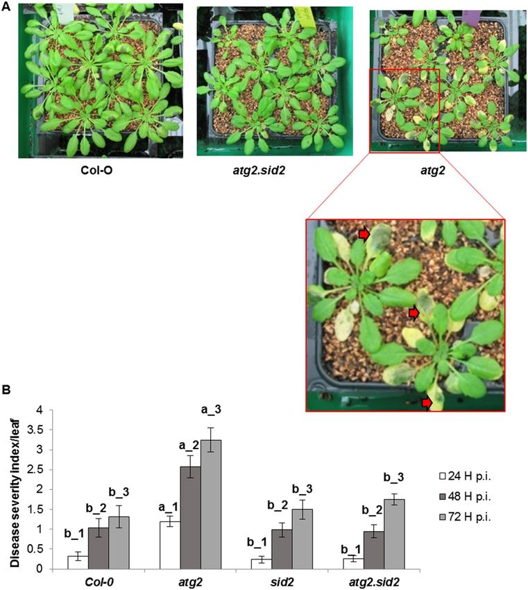
In order to determine whether SA is involved in the increased susceptibility of atg2 mutant, we monitored the
susceptibility of the atg2.sid2 double mutant defective in SA synthesis as the SID2 gene encodes an isochorismate
athogens61. Figure 3A
synthase which is involved in SA biosynthesis in Arabidopsis, in particular in response to p
shows the phenotypes of Col-0 wild type, atg2 and atg2.sid2 mutants infected with D. dadantii. Both soft rot
symptoms and leaf yellowing phenotypes were clearly more severe on atg2 leaves than on Col-0 and atg2.sid2
leaves (Fig. 3). Although atg2 was early senescing by comparison with Col-0 and atg2.sid2 plants34 (Fig. 3A), we
observed enhanced chlorosis around the D. dadantii inoculation spots which were not present in uninfected
plants or before inoculation (data not shown). This suggested that infection accelerated leaf senescence symp-
toms on atg2 plants. It can be noticed that disease severity was similar in WT, sid2 and atg2.sid2 double mutant.
These data indicate that the higher susceptibility of the atg2 mutant can be restored to the WT level when SID2
gene is non-functional, suggesting the prominent role of SA in the increased susceptibility of atg2 mutant to
D. dadantii. This confirms that autophagy degradation pathway is not essential for Arabidopsis tolerance to D.
dadantii. Nevertheless, higher constitutive autophagic activity artificially enhanced by ATG8a overexpression
may help increasing plant tolerance, possibly through positive effects on leaf longevity, meaning negative effect
on SA production.
Salicylic acid signaling rather than SA amount is involved in atg2 mutant enhanced suscepti‑
bility to D. dadantii. Previous reports indicate that the level of salicylic acid in autophagy mutants is higher
T54,62. To determine the mechanism by which high SA level could lead to enhanced susceptibility,
than that of W
we hypothesized that high amounts of SA could repress the JA signaling pathway which is known to limit D.
dadantii infection50,57. Indeed, it is commonly reported that SA signaling represses JA signaling and that ET and
JA signaling are commonly synergistic17–20,63.
To further investigate the possibility that SA and JA signaling were modified in atg2 mutants, we determined
the status of SA and JA mediated defenses in Col-0, sid2, atg2, atg5, atg2.sid2 and atg5.sid2 plants. For this pur-
pose, we monitored the expression of defense marker genes of the SA (PR1) and the JA (PDF1.2) pathways in
infected plants. Controls consisted of plants inoculated with 10 mM M gSO4 and naïve plants that were untreated.
The latter naïve plants were used to determine the initial immunity level of plants before inoculation. Transcript
levels of both the SA defense marker gene PR1 and of the JA defense marker gene PDF1.2 were increased in
WT Col-0 plants following D. dadantii infection compared to controls (Fig. 4). While no PR1 transcript was
detected in the sid2 mutant line, higher levels of PDF1.2 transcripts were detected in these plants compared to
Col-0 confirming the repressive action of the SA pathway on the JA pathway. Strikingly, in the atg2 mutant,
higher levels of PR1 transcripts were detected compared to the WT (10 times higher in atg2 compared to Col-0
infected plants) and an up-regulation of this level was observed in response to D. dadantii compared to the mock
inoculated plants. The level of PR1 transcripts in atg5 mutant was unchanged compared to the WT indicating
that SA signaling in the atg5 mutant was not affected. The expression level of PDF1.2 was reduced in the atg2
mutant compared to that of Col-0 which is consistent with a high SA signaling. The transcript level of PR1 was
undetectable in both atg2.sid2 and atg5.sid2 double mutants, indicating that SA signaling was totally abolished
by introducing the sid2 mutation. Interestingly, in naïve atg simple and double mutant plants, the level of PDF1.2
transcripts was reduced compared to that of mock inoculated plants. Our data suggest that mechanical stress
triggers JA signaling in atg mutant backgrounds, a process which is repressed by D. dadantii infection. To know
whether senescence was also involved in this interaction, the transcript level of the senescence marker gene
SAG12 was monitored. SAG12 was only expressed in atg2 mutant plants (Fig. 4). Intriguingly, it was down
regulated by mock inoculation and up-regulated by infection confirming the accelerated senescing phenotype
we observed on atg2 infected plants compared to mock atg2 plants.
To investigate the role of SA content in the response of autophagy mutants to D. dadantii, SA content was
quantified in naïve plants (Fig. 5). Both atg2 and atg5 mutants accumulate higher SA levels than Col-0 plants.
The level of SA was reduced in both double mutants atg2.sid2 and atg5.sid2 relative to single mutants confirming
the role of SID2 in SA production in Arabidopsis.
Scientific Reports | (2021) 11:3624 | https://doi.org/10.1038/s41598-021-83067-6 5
Vol.:(0123456789)www.nature.com/scientificreports/
Figure 3. Susceptibility of atg2 mutant relies on SA. (A) Pictures of 6 week-old Arabidopsis indicated genotypes
inoculated with D. dadantii. In the enlargement of atg2 mutant inoculated plants, red arrows indicate yellow
zones of early senesce. (B) Symptom severity at indicated time points on indicated genotypes. Bars represent
standard deviation. N = 30. Experiments were performed three times with similar results. Different letters in the
graph indicate statistical significance between genotypes at the same time point (one-way ANOVA with Tuckey’s
test; P < 0.05).
Discussion
The mechanisms by which plant autophagy affects immunity are complex and vary depending on the considered
pathosystems. In some cases, opposite mechanisms can take place depending on plant age or infectious stage. For
example, in young Arabidopsis leaves, autophagy has a pro-death activity enhancing HR mediated cell death45
while in older leaves, autophagy seems to counteract cell death34. Plant autophagy was shown to counteract
the bacterial pathogen Pseudomonas syringae pv. tomato which, surprisingly, secretes an effector able to trigger
autophagy64.
Several reports indicate that the susceptibility of atg mutants was increased in response to fungal necrotrophic
pathogens (Table 1). To determine the effect of atg mutations on Arabidopsis susceptibility to the bacterial necro-
troph D. dadantiii, we used an atg2 and atg5 mutants. The ATG2 and ATG5 genes are single genes in Arabidopsis
as in many plant species, and are essential for autophagic activity as their mutants cannot form autophagosomes.
The atg2 and atg5 mutants were previously characterized by many groups amongst which Pr. R. Vierstra and Pr.
K. Yoshimoto groups, and display the typical hypersensitivity to abiotic stresses described for many autophagy
Scientific Reports | (2021) 11:3624 | https://doi.org/10.1038/s41598-021-83067-6 6
Vol:.(1234567890)www.nature.com/scientificreports/
Figure 4. Expression of defense and senescence genes upon D. dadantii infection. Plants were infected or
mock treated, leaves were harvested 24 h after treatment. Transcript levels were monitored by qRT-PCR and
normalized against the transcripts of the reference genes APT and Clathrin. Bars represent standard deviation.
N = 4. Experiments were performed three times with similar results. Different letters in the graph indicate
statistical significance between treatments (one-way ANOVA with Tuckey’s test; P < 0.05). NI: Non- inoculated,
Mock: treated with MgSO4.
Figure 5. Salicylic acid content in atg mutants: Six week old plants of indicated genotypes were harvested then
SA content was monitored as indicated in Materials and Methods. Bars represent standard deviation. N = 10 to
12. Experiments were performed three times with similar results. Different letters in the graph indicate statistical
significance between treatments (one-way ANOVA with Tuckey’s test; P < 0.05).
Scientific Reports | (2021) 11:3624 | https://doi.org/10.1038/s41598-021-83067-6 7
Vol.:(0123456789)www.nature.com/scientificreports/
mutants in A rabidopsis33,34 that is related to leaf yellowing phenotypes and necrotic spots. It might be noticed
that atg2 mutant phenotypes are stronger than those of atg5 mutant. Our findings showing different behaviors
of two autophagy mutants are not surprising with regard to the literature. Indeed, it appears from the literature
that the susceptibility of atg mutants to different pathogens is not always similarly affected compared to the
WT plants. For instance, although we found that in response to D. dadantii, the atg2 mutant showed enhanced
susceptibility, the susceptibility of the same atg2 mutant to the necrotrophic fungus Botrytis cinerea has not been
found affected65. In atg7 and atg12 Arabidopsis mutants, the susceptibility to the necrotrophic fungus Sclero-
tinia sclerotiorum was shown unaffected66. All these reports indicate that although a general trend observed in
autophagy mutants is the susceptibility to necrotrophic pathogens, some mutants are likely not affected.
A common trend in necrotrophy is cell wall degrading enzyme production by pathogens. Cell wall home-
ostasis is tightly linked to membrane stability which could be in part under the control of a utophagy29,67,68.
The mechanisms explaining the higher susceptibility of some atg mutants to necrotrophic pathogens remained
unknown44,48. One hypothesis is that as a pro-survival mechanism, autophagy hampers the growth and develop-
ment of necrotrophs which proliferate preferentially on dead cells. In this situation, autophagy would ensure the
clearance of degraded cellular components thus protecting plant tissues from activating stress responses such as
the SA signaling pathway44. Another hypothesis is that the hormonal balance which is disturbed in atg mutants
favors necrotrophs. Both hypotheses are not mutually exclusive and can depend upon the the pathosystem con-
sidered. In the interaction between Arabidopsis and D. dadantii, we demonstrate that the enhanced susceptibility
of the atg2 mutant relies on SA signaling Our data also show that the senescing state of the plants correlates with
disease development since SAG12 expression and chlorotic phenotype were enhanced in atg2 plants and lost in
the atg2.sid2 mutant. It is however intriguing that there is no correlation between SA content and PR1 expression
level in atg2 and atg5 mutants. One interpretation is that senescence and SA form an amplification loop in atg2
and that both are required to trigger high s usceptibility34. Such amplification loop between senescence and SA
was indeed recently described69,70.
We showed that in atg2 higher SA content and signaling coincided with low JA signaling thus explaining
increased susceptibility, as JA signaling has been shown to contribute to Arabidopsis tolerance to D. dadantii50,57.
The disease phenotype of the atg5 mutant, which is similar to that of the WT plants, may be explained by the fact
that atg5 is senescing later than atg2 mutant. This may be due to a differentially active senescence and immunity
molecular machinery in each atg mutant. For instance, transcript levels of genes encoding transcription factors
related to immunity and senescence (WRKY, ERF and JAZ) are different in an atg5 compared to an atg9 mutant54.
Interestingly, ATG9 interacts with ATG2 in the phagophore expansion process23 suggesting that atg9 and atg2
mutants could harbor the similar immunity and senescence defects that differ from those of the atg5 mutant.
In addition to the differential expression of transcription factor encoding genes in different atg mutants, it is
possible that different proteolytic activities reside in each mutant. Indeed, it was recently shown that autophagy
deficiency in the atg5 mutant resulted in an alteration of cellular proteolytic a ctivities67. The actors of immunity
including transcription factors may be targeted by proteolysis which ensures fine tuning of adequate defense
responses71,72. It would be interesting to compare the proteolytic activities in atg2 and atg5 mutants to investigate
the lifetime of key immunity and senescence related transcription factors.
Altogether, we provide here the evidence that SA plays a pivotal role in the enhanced susceptibility of atg2
mutant to D. dadantii through the repression of JA-dependent defenses, and that autophagy degradation func-
tion is not directly involved in plant tolerance to D. dadantii. While our study argues against the involvement of
plant autophagy in the tolerance to this type of aggressors, it confirms the importance of leaf senescence status
in the susceptibility of plants and points to the hormonal balance as a key player in this process. In addition,
overexpression of ATG8 proteins, that had been shown to provide positive effects on plant tolerance to many
stresses such as drought in several plant species (Chen et al.73 for a review), significantly increased the tolerance
of our Arabidopsis plants to D. dadantii infection.
Received: 3 September 2020; Accepted: 8 January 2021
References
1. Klessig, D. F., Choi, H. W. & Dempsey, D. A. Systemic acquired resistance and salicylic acid: Past, present, and future. Mol. Plant-
Microbe Interact. 31, 871–888 (2018).
2. Piasecka, A., Jedrzejczak-Rey, N. & Bednarek, P. Secondary metabolites in plant innate immunity: Conserved function of divergent
chemicals. New Phytol. 206, 948–964 (2015).
3. Wang, W., Feng, B., Zhou, J. M. & Tang, D. Plant immune signaling: Advancing on two frontiers. J. Integr. Plant Biol. 62, 2–24
(2020).
4. Jones, J. D. G. & Dangl, J. L. The plant immune system. Nature 444, 323–329 (2006).
5. Dangl, J. L., Horvath, D. M. & Staskawicz, B. J. Pivoting the plant immune system from dissection to deployment. Science 341,
746–751 (2013).
6. Wirthmueller, L., Maqbool, A. & Banfield, M. J. On the front line: Structural insights into plant–pathogen interactions. Nat. Rev.
Microbiol. 11, 761–776 (2013).
7. Dodds, P. N. & Rathjen, J. P. Plant immunity: Towards an integrated view of plant pathogen interactions. Nat. Rev. Genet. 11,
539–548 (2010).
8. Macho, A. P. & Zipfel, C. Plant PRRs and the activation of innate immune signaling. Mol. Cell 54, 263–272 (2014).
9. Alhoraibi, H., Bigeard, J., Rayapuram, N., Colcombet, J. & Hirt, H. Plant immunity: The MTI-ETI model and beyond. Curr. Issues
Mol. Biol. 30, 39–58 (2019).
10. Robert-Seilaniantz, A., Grant, M. & Jones, J. D. G. Hormone crosstalk in plant disease and defense: More than just jasmonate-
salicylate antagonism. Annu. Rev. Phytopathol. 49, 317–343 (2011).
Scientific Reports | (2021) 11:3624 | https://doi.org/10.1038/s41598-021-83067-6 8
Vol:.(1234567890)www.nature.com/scientificreports/
11. Pieterse, C. M. J., Van der Does, D., Zamioudis, C., Leon-Reyes, A. & Van Wees, S. C. M. Hormonal modulation of plant immunity.
Annu. Rev. Cell Dev. Biol. 28, 489–521 (2011).
12. Vos, I. A., Moritz, L., Pieterse, C. M. J. & Van Wees, S. C. M. Impact of hormonal crosstalk on plant resistance and fitness under
multi-attacker conditions. Front. Plant Sci. 6, 639 (2015).
13. Verma, V., Ravindran, P. & Kumar, P. P. Plant hormone-mediated regulation of stress responses. BMC Plant Biol. 16, 86 (2016).
14. Glazebrook, J. Contrasting mechanisms of defense against biotrophic and necrotrophic pathogens. Annu. Rev. Phytopathol. 43,
205–227 (2005).
15. Andolfo, G. & Ercolano, M. R. Plant innate immunity multicomponent model. Front. Plant Sci. 6, 987 (2015).
16. Thaler, J. S., Humphrey, P. T. & Whiteman, N. K. Evolution of jasmonate and salicylate signal crosstalk. Trends Plant Sci. 5, 260–270
(2012).
17. Caarls, L. et al. Assessing the role of ETHYLENE RESPONSE FACTOR transcriptional repressors in salicylic acid-mediated sup-
pression of jasmonic acid-responsive genes. Plant Cell Physiol. 58, 266–278 (2017).
18. Koornneef, A. & Pieterse, C. M. J. Cross talk in defense signaling. Plant Physiol. 146, 839–844 (2008).
19. Leon-Reyes, A. et al. Salicylate-mediated suppression of jasmonate-responsive gene expression in Arabidopsis is targeted down-
stream of the jasmonate biosynthesis pathway. Planta 232, 1423–1432 (2010).
20. Van der Does, D. et al. Salicylic acid suppresses jasmonic acid signaling downstream of SCF COI1-JAZ by targeting GCC promoter
motifs via transcription factor ORA59. Plant Cell. 5, 744–761 (2013).
21. Mur, L. A. J., Kenton, P., Atzorn, R., Miersch, O. & Wasternack, C. The outcomes of concentration-specific interactions between
salicylate and jasmonate signaling include synergy, antagonism, and oxidative stress leading to cell death. Plant Physiol. 140,
249–262 (2006).
22. Feng, Y., He, D., Yao, Z. & Klionsky, D. J. The machinery of macroautophagy. Cell Res. 24, 24–41 (2014).
23. Avila-Ospina, L., Moison, M., Yoshimoto, K. & Masclaux-Daubresse, C. Autophagy, plant senescence, and nutrient recycling. J.
Exp. Bot. 65, 3799–3811 (2014).
24. Masclaux-Daubresse, C., Chen, Q. & Havé, M. Regulation of nutrient recycling via autophagy. Curr. Opin. Plant Biol. 39, 8–17
(2017).
25. Dupont, N. et al. Autophagy-based unconventional secretory pathway for extracellular delivery of IL-1β. EMBO J. 30, 4701–4711
(2011).
26. Cavalli, G. & Cenci, S. Autophagy and protein secretion. J. Mol. Biol. 432, 2525–2545 (2020).
27. Cui, Y. et al. Biogenesis of plant prevacuolar multivesicular bodies. Mol. Plant 9, 774–786 (2016).
28. Soto-Burgos, J., Zhuang, X., Jiang, L. & Bassham, D. C. Dynamics of autophagosome formation. Plant Physiol. 176, 219–229 (2018).
29. Wun, C. L., Quan, Y. & Zhuang, X. Recent advances in membrane shaping for plant autophagosome biogenesis. Front. Plant Sci.
11, 565 (2020).
30. Tsukada, M. & Ohsumi, Y. Isolation and characterization of autophagy-defective mutants of Saccharomyces cerevisiae. FEBS Lett.
33, 169–174 (1993).
31. Yang, X. & Bassham, D. C. New insight into the mechanism and function of autophagy in plant cells. Int. Rev. Cell Mol. Biol. 320,
1–40 (2015).
32. Doelling, J. H., Walker, J. M., Friedman, E. M., Thompson, A. R. & Vierstra, R. D. The APG8/12-activating enzyme APG7 is required
for proper nutrient recycling and senescence in Arabidopsis thaliana. J. Biol. Chem. 277, 33105–33114 (2002).
33. Thompson, A. R., Doelling, J. H., Suttangkakul, A. & Vierstra, R. D. Autophagic nutrient recycling in Arabidopsis directed by the
ATG8 and ATG12 conjugation pathways. Plant Physiol. 138, 2097–2110 (2005).
34. Yoshimoto, K. et al. Autophagy negatively regulates cell death by controlling NPR1-dependent salicylic acid signaling during
senescence and the innate immune response in arabidopsis. Plant Cell 21, 2914–2927 (2009).
35. Hanaoka, H. et al. Leaf senescence and starvation-induced chlorosis are accelerated by the disruption of an Arabidopsis autophagy
gene. Plant Physiol. 129, 1181–1193 (2002).
36. Deprost, D. et al. The Arabidopsis TOR kinase links plant growth, yield, stress resistance and mRNA translation. EMBO Rep. 8,
864–870 (2007).
37. Liu, Y. & Bassham, D. C. TOR is a negative regulator of autophagy in Arabidopsis thaliana. PLoS ONE 5, e11883 (2010).
38. Pu, Y., Luo, X. & Bassham, D. C. Tor-dependent and -independent pathways regulate autophagy in Arabidopsis thaliana. Front.
Plant Sci. 8, 1204 (2017).
39. Seay, M., Patel, S. & Dinesh-Kumar, S. P. Autophagy and plant innate immunity. Cell. Microbiol. 8, 899–906 (2006).
40. Yang, M., Bu, F., Huang, W. & Chen, L. Multiple regulatory levels shape autophagy activity in plants. Front. Plant Sci. 10, 532 (2019).
41. Leary, A. Y., Savage, Z., Tumtas, Y. & Bozkurt, T. O. Contrasting and emerging roles of autophagy in plant immunity. Curr. Opin.
Plant Biol. 52, 46–53 (2019).
42. Leary, A. Y. et al. Modulation of plant autophagy during pathogen attack. J. Exp. Bot. 69, 1325–1333 (2018).
43. Liao, C. Y. & Bassham, D. C. Combating stress: The interplay between hormone signaling and autophagy in plants. J. Exp. Bot. 71,
1723–1733 (2020).
44. Zeng, H.Y., Zheng. P., Wang, L.Y., Bao, H.N., Sahu, S.K. & Yao, N. Autophagy in plant immunity BT—autophagy regulation of
innate immunity. In (ed. Cui, J.) 23–41 (Springer Singapore, 2019).
45. Hofius, D. et al. Autophagic components contribute to hypersensitive cell death in Arabidopsis. Cell 137, 773–783 (2009).
46. Lenz, H. D. et al. Autophagy differentially controls plant basal immunity to biotrophic and necrotrophic pathogens. Plant J. 66,
818–830 (2011).
47. Lai, Z., Wang, F., Zheng, Z., Fan, B. & Chen, Z. A critical role of autophagy in plant resistance to necrotrophic fungal pathogens.
Plant J. 66, 953–968 (2011).
48. Zhou, J., Yu, J. Q. & Chen, Z. The perplexing role of autophagy in plant innate immune responses. Mol. Plant Pathol. 15, 637–645
(2014).
49. Reverchon, S. & Nasser, W. Dickeya ecology, environment sensing and regulation of virulence programme. Environ. Microbiol.
Rep. 5, 622–636 (2013).
50. Fagard, M. et al. Arabidopsis thaliana expresses multiple lines of defense to counterattack Erwinia chrysanthemi. Mol. Plant-Microbe
Interact. 20, 794–805 (2007).
51. Kieu, N. P. et al. Iron deficiency affects plant defence responses and confers resistance to Dickeya dadantii and Botrytis cinerea.
Mol. Plant Pathol. 13, 816–827 (2012).
52. Aznar, A., Chen, N. W. G., Thomine, S. & Dellagi, A. Immunity to plant pathogens and iron homeostasis. Plant Sci. 240, 90–97
(2015).
53. Taurino, M. et al. Jasmonate-dependent modifications of the pectin matrix during potato development function as a defense
mechanism targeted by Dickeya dadantii virulence factors. Plant J. 77, 418–429 (2014).
54. Masclaux-Daubresse, C. et al. Stitching together the multiple dimensions of autophagy using metabolomics and transcriptomics
reveals impacts on metabolism, development, and plant responses to the environment in Arabidopsis. Plant Cell 26, 1857–1877
(2014).
55. Chen, Q. et al. Overexpression of ATG8 in Arabidopsis stimulates autophagic activity and increases nitrogen remobilization
efficiency and grain filling. Plant Cell Physiol. 60, 343–352 (2019).
Scientific Reports | (2021) 11:3624 | https://doi.org/10.1038/s41598-021-83067-6 9
Vol.:(0123456789)www.nature.com/scientificreports/
56. Rigault, M. et al. Quantitative methods to assess differential susceptibility of Arabidopsis thaliana natural accessions to Dickeya
dadantii. Front. Plant Sci. 8, 394 (2017).
57. Verly, C. et al. Plant defense stimulator mediated defense activation is affected by nitrate fertilization and developmental stage in
Arabidopsis thaliana. Front. Plant Sci. 11, 583 (2020).
58. Aznar, A. et al. Scavenging iron: A novel mechanism of plant immunity activation by microbial siderophores. Plant Physiol. 164,
2167–2183 (2014).
59. Le Roux, C. et al. The hnRNP-Q protein LIF2 participates in the plant immune response. PLoS ONE 9, e99343 (2014).
60. Mansfield, J. et al. Top 10 plant pathogenic bacteria in molecular plant pathology. Mol. Plant Pathol. 13, 614–629 (2012).
61. Wildermuth, M. C., Dewdney, J., Wu, G. & Ausubel, F. M. Isochorismate synthase is required to synthesize salicylic acid for plant
defence. Nature 414, 562–565 (2001).
62. Yoshimoto, K. Plant autophagy puts the brakes on cell death by controlling salicylic acid signaling. Autophagy. 6, 192–193 (2010).
63. Verhage, A., van Wees, S. C. M. & Pieterse, C. M. J. Plant immunity: It’s the hormones talking, but what do they say?. Plant Physiol.
154, 536–540 (2010).
64. Üstün, S. et al. Bacteria exploit autophagy for proteasome degradation and enhanced virulence in plants. Plant Cell 30, 668–685
(2018).
65. Wang, Y., Nishimura, M. T., Zhao, T. & Tang, D. ATG2, an autophagy-related protein, negatively affects powdery mildew resistance
and mildew-induced cell death in Arabidopsis. Plant J. 68, 74–87 (2011).
66. Kabbage, M., Williams, B. & Dickman, M. B. Cell death control: The interplay of apoptosis and autophagy in the pathogenicity of
Sclerotinia sclerotiorum. PLoS Pathog. 9, e1003287 (2013).
67. Havé, M. et al. Proteomic and lipidomic analyses of the Arabidopsis atg5 autophagy mutant reveal major changes in endoplasmic
reticulum and peroxisome metabolisms and in lipid composition. New Phytol. 223, 1461–1477 (2019).
68. Zeng, Y., Li, B., Lin, Y. & Jiang, L. The interplay between endomembranes and autophagy in plants. Curr. Opin. Plant Biol. 2, 14–22
(2019).
69. Guo, P. et al. A tripartite amplification loop involving the transcription factor WRKY75, salicylic acid, and reactive oxygen species
accelerates leaf senescence. Plant Cell 29, 2854–2870 (2017).
70. Woo, H. R., Kim, H. J., Lim, P. O. & Nam, H. G. Leaf senescence: Systems and dynamics aspects. Annu. Rev. Plant Biol. 70, 347–376
(2019).
71. Copeland, C. & Li, X. Chapter two—regulation of plant immunity by the proteasome. In International Review of Cell and Molecular
Biology (ed. Galluzzi, L. B. T.-I. R. of C. and M. B.) Vol. 343, 37–63 (Academic Press, 2019).
72. Caarls, L., Pieterse, C. M. J. & Van Wees, S. C. M. How salicylic acid takes transcriptional control over jasmonic acid signaling.
Front. Plant Sci. 6, 170 (2015).
73. Chen, Q. et al. Autophagy and nutrients management in plants. Cells 8, 1426 (2019).
74. Zhou, J. et al. NBR1-mediated selective autophagy targets insoluble ubiquitinated protein aggregates in plant stress responses.
PLoS Genet. 9, e1003196 (2013).
Acknowledgements
Authors thanks Dr. Kohki Yoshimoto (Meiji University, Japan) for mutant seeds. We thank Hervé Ferry for taking
care of plants. This work has benefited from the support of IJPB’s Plant Observatory technological platforms.
The IJPB benefits from the support of Saclay Plant Sciences-SPS (ANR-17-EUR-0007).
Author contributions
M.R., S.C., A.D. performed experiments. A.D. and C.M.D. designed the work and wrote the manuscript.
Competing interests
The authors declare no competing interests.
Additional information
Supplementary Information The online version contains supplementary material available at https://doi.
org/10.1038/s41598-021-83067-6.
Correspondence and requests for materials should be addressed to A.D.
Reprints and permissions information is available at www.nature.com/reprints.
Publisher’s note Springer Nature remains neutral with regard to jurisdictional claims in published maps and
institutional affiliations.
Open Access This article is licensed under a Creative Commons Attribution 4.0 International
License, which permits use, sharing, adaptation, distribution and reproduction in any medium or
format, as long as you give appropriate credit to the original author(s) and the source, provide a link to the
Creative Commons licence, and indicate if changes were made. The images or other third party material in this
article are included in the article’s Creative Commons licence, unless indicated otherwise in a credit line to the
material. If material is not included in the article’s Creative Commons licence and your intended use is not
permitted by statutory regulation or exceeds the permitted use, you will need to obtain permission directly from
the copyright holder. To view a copy of this licence, visit http://creativecommons.org/licenses/by/4.0/.
© The Author(s) 2021
Scientific Reports | (2021) 11:3624 | https://doi.org/10.1038/s41598-021-83067-6 10
Vol:.(1234567890)You can also read


