SOFT TISSUE DEFECT RECONSTRUCTION AND LYMPHATIC COMPLICATIONS PREVENTION: THE LYMPHATIC FLOW-THROUGH (LYFT) CONCEPT - MDPI
←
→
Page content transcription
If your browser does not render page correctly, please read the page content below
medicina
Article
Soft Tissue Defect Reconstruction and Lymphatic
Complications Prevention: The Lymphatic Flow-Through
(LyFT) Concept
Mario F. Scaglioni * , Matteo Meroni and Elmar Fritsche

                                          Hand- and Plastic Surgery Department, Luzerner Kantonsspital, 6000 Lucerne, Switzerland;
                                          meroni369@gmail.com (M.M.); elmar.fritsche@luks.ch (E.F.)
                                          * Correspondence: mario.scaglioni@gmail.com

                                          Abstract: Background and Objectives: When a lymphatic-rich area is severely damaged, either af-
                                          ter trauma or a surgical procedure, both soft tissue defect reconstruction and lymphatic drainage
                                          restoration are necessary. In this setting, we aim to show the potential of the lymphatic flow-through
                                          flap (LyFT) concept, which might be an attractive new solution to reduce postoperative lymphatic
                                          complications. Materials and Methods: Between 2018 and 2021, 12 patients presenting a soft tissue
                                          defect involving damage to the lymphatic drainage pathway received a lymphatic flow-through
                                          flap for volume and lymphatic drainage restoration. Different flaps were employed: 3 pedicled
                                          superficial circumflex iliac artery perforator (SCIP) flaps, 2 free SCIP flaps, 3 pedicled deep inferior
                                          epigastric perforator (DIEP) flaps, 2 pedicled vertical posteromedial thigh (vPMT) flaps, and 2 pedi-
                                          cled anterolateral thigh (ALT) flaps. A range of 1 to 3 lymphovenous anastomosis (LVA) with flap’s
                                          veins was performed (mean 1.9). For a better dead space obliteration, an additional vastus lateralis
                                          muscle flap was performed in one case. Indocyanine green (ICG) lymphography was used in all

                                   cases to identify the lymphatic pathway, make the preoperative markings, and check the patency
Citation: Scaglioni, M.F.; Meroni, M.;    of the anastomoses. Results: In all cases, the reconstructive results were satisfactory from both the
Fritsche, E. Soft Tissue Defect           functional and aesthetic points of view. No secondary surgeries were required, and only one minor
Reconstruction and Lymphatic              complication was encountered: an infected seroma that was managed conservatively. The mean
Complications Prevention: The             follow-up was 9.9 months (range 6–14 months). Conclusions: Lymphatic flow-through flaps seem to
Lymphatic Flow-Through (LyFT)             effectively reduce the risk of lymphatic complications after the reconstruction of soft tissue defects
Concept. Medicina 2022, 58, 509.          with a compromised lymph pathway. This is a versatile solution that might be used in different body
https://doi.org/10.3390/                  regions resorting to different flap types.
medicina58040509

Academic Editor: Rytis Rimdeika           Keywords: lymphatic surgery; lymphovenous anastomosis; superficial circumflex iliac artery perforator
                                          flap; deep inferior epigastric perforator flap; anterolateral thigh flap; supermicrosurgery
Received: 4 March 2022
Accepted: 31 March 2022
Published: 2 April 2022

Publisher’s Note: MDPI stays neutral      1. Introduction
with regard to jurisdictional claims in
                                               The continuous progress of microsurgical techniques has allowed surgeons to re-
published maps and institutional affil-
                                          construct an extremely wide range of defects throughout the body. Expectations have
iations.
                                          also grown, and simple coverage is no more considered a completely satisfactory result.
                                          Nowadays, the goal of a plastic surgeon is to restore good physical and physiological
                                          function. This is a particularly critical issue when lymphatic vessels are severely damaged.
Copyright: © 2022 by the authors.
                                          Impairment of lymph drainage often leads to a series of complications, ranging from edema
Licensee MDPI, Basel, Switzerland.        to severe cellulitis, which may cause very debilitating conditions [1–3]. For this reason, vol-
This article is an open access article    ume restoration alone is often not sufficient since it is also necessary to prevent lymphatic
distributed under the terms and           sequelae [4].
conditions of the Creative Commons             Regarding dead space obliteration, many alternatives are available. In order to re-
Attribution (CC BY) license (https://     duce donor site morbidity and quicken the harvest, nowadays, perforator-based flaps are
creativecommons.org/licenses/by/          preferred, either in pedicled or free form [5]. Among these, the most employed are the
4.0/).                                    anterolateral thigh (ALT) flap [6], the superficial circumflex iliac artery perforator (SCIP)

Medicina 2022, 58, 509. https://doi.org/10.3390/medicina58040509                                          https://www.mdpi.com/journal/medicina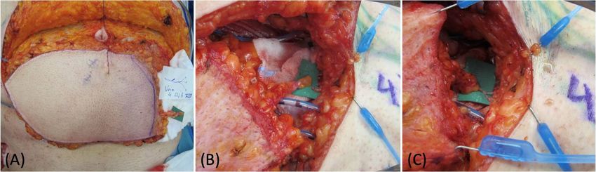
Medicina 2022, 58, 509                                                                                                                                                      2 of 9

                                       flap [7], and the deep inferior epigastric perforator (DIEP) flap [8], each of them with its
                                       own features.
                                             Different procedures aimed at the restoration of lymph drainage have been described
                                       so far, but all of them are still debated, with no clear advantages of one over the others [9].
                                       However, lymphovenous anastomosis (LVA) is gaining consistent approval to treat lympho-
                                       cele and lymphedema throughout the body. It consists of shunting the lymphatic flow into
                                       venous circulation before the impaired area, providing an alternative drainage route [10].
                                             The lymphatic flow-through (LyFT) flap is an interesting and modern concept that
                                       tries to combine both of these treatments. It not only provides healthy tissue for defect
                                       reconstruction but also allows the exploitation of the collateral veins of the flap for the LVA.
                                       This approach is particularly helpful when no suitable vessels can be found near the defect,
                                       such as after radical debulking procedures combined with radiotherapy or after severe
                                       trauma [11].
                                             In the present article, we present our experience of a 12-patient series successfully
                                       treated with LyFT flaps for the reconstruction of defects in different body regions and with
                                       various etiologies.

                                       2. Materials and Methods
                                            Twelve patients presenting a soft tissue defect involving damage to the lymphatic
                                       drainage pathway were included in this retrospective report; they received a lymphatic
                                       flow-through flap for volume and lymphatic drainage restoration (Table 1); 6 were females,
                                       and 6 were males (50:50 gender ratio). The median age was 57 years old (range 42–82);
                                       8 patients presented no comorbidities, 2 were affected by chronic arterial hypertension, and
                                       2 by diabetes mellitus.
                                       Table 1. Patient demographic and case characteristics.

                                                                                                                                                                   Functional
                                                                                                Recipient Vein                                     Follow-Up
 Patient   Gender   Age    Etiology          Location         Comorbidities       Flap                                Number LVA   Complications               Outcomes/Aesthetic
                                                                                                   for LVA                                          (Months)
                                                                                                                                                                     Result

                                                                                Pedicled
   1         M      63     Trauma           Medial thigh          DM                          Superficial Flap Vein       3           None            14         Full ROM/++
                                                                                 DIEP

                                                                                              Deep Branch Pedicle
   2         F      56     Trauma            Lower leg            HTN           Free SCIP                                 2           None            14         Full ROM/++
                                                                                                     Vein

                                                                                Pedicled                                             Infected
   3         F      76     Sarcoma             Groin             None                         Superficial Flap Vein       3                           12          Full ROM/+
                                                                                 DIEP                                                Seroma

   4         F      65     Sarcoma        Upper extremity        None           Free SCIP     Superficial Flap Vein       1           None            11          Full ROM/+

                                              Intra-
   5         M      67     Sarcoma                               None         Pedicled ALT        Pedicle Vein            3           None            11         Full ROM/++
                                         abdominal/Groin

                                              Intra-                          Pedicled ALT
   6         M      82     Sarcoma                                HTN                             Pedicle Vein            3           None            11          Full ROM/+
                                         abdominal/Groin                        + VLM

   7         F      42     Sarcoma       Groin/Medial thigh      None         Pedicled SCIP   Superficial Flap Vein       1           None            11          Full ROM/+

   8         M      45    Skin Tumor        Medial thigh         None         Pedicled SCIP   Superficial Flap Vein       2           None            11         Full ROM/++

                                                                                Pedicled
   9         M      47    Skin Tumor           Groin             None                             Pedicle Vein            1           None             6          Full ROM/+
                                                                                 vPMT

                                                                                Pedicled
   10        M      59     Sarcoma          Upper thigh           DM                          Superficial Flap Vein       2           None             6          Full ROM/+
                                                                                 DIEP

   11        F      39     Sarcoma          Upper thigh          None         Pedicled SCIP   Superficial Flap Vein       1           None             6          Full ROM/+

                                                                                Pedicled
   12        F      51     Sarcoma             Groin             None                             Pedicle Vein            1           None             6          Full ROM/+
                                                                                 vPMT

                                       DM: diabetes mellitus; HTN: hypertension; SCIP: superficial circumflex iliac artery perforator; DIEP: deep
                                       inferior epigastric artery perforator; ALT: anterolateral thigh flap; VLM: vastus lateralis muscle; vPMT: vertical
                                       posteromedial thigh; LVA: lymph venous anastomosis; ROM: range of motion; +: good aesthetic result; ++: very
                                       good aesthetic result.

                                             The cause of the defect was surgical tumor excision in 10 cases (8 because of sarcoma
                                       and 2 because of squamous cell carcinoma), while in 2 cases, the defect was due to trauma.
                                       The defect was localized as follows: 3 in the groin region, 2 in the abdomen and groin,
                                       1 in the groin and medial thigh, 2 in the medial thigh, 2 in the upper thigh, 1 in the lower
                                       leg, and 1 in the upper extremity. Different types of flaps were employed, either pedicled
                                       or free. In 3 cases, we resorted to a pedicled superficial circumflex iliac artery perforator
                                       (SCIP) flap, in 2 to a free SCIP flap, in 3 to a pedicled deep inferior epigastric perforator
                                       (DIEP) flap, in 2 to a pedicled vertical posteromedial thigh (vPMT) flap, and in 2 to an
                                       anterolateral thigh (ALT) flap. The number of lymphovenous anastomoses performed with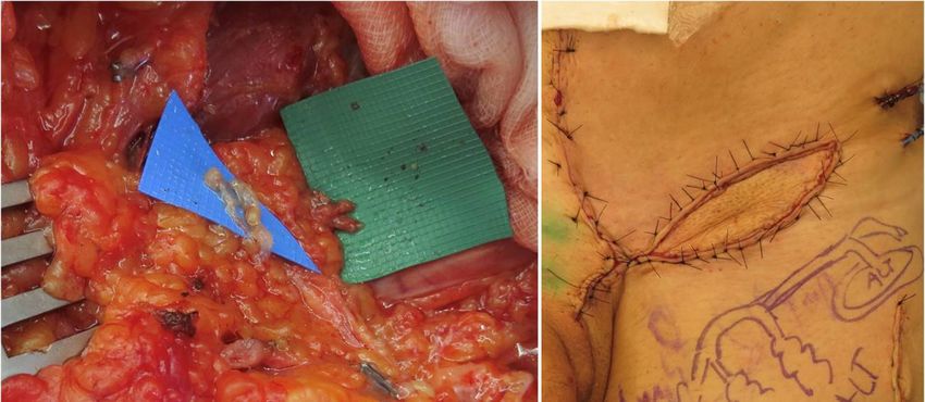
The defect was localized as follows: 3 in the groin region, 2 in the abdomen and groin, 1
in the groin and medial thigh, 2 in the medial thigh, 2 in the upper thigh, 1 in the lower
leg, and 1 in the upper extremity. Different types of flaps were employed, either pedicled
or free. In 3 cases, we resorted to a pedicled superficial circumflex iliac artery perforator
Medicina 2022, 58, 509 (SCIP) flap, in 2 to a free SCIP flap, in 3 to a pedicled deep inferior epigastric perforator
3 of 9
(DIEP) flap, in 2 to a pedicled vertical posteromedial thigh (vPMT) flap, and in 2 to an
anterolateral thigh (ALT) flap. The number of lymphovenous anastomoses performed
with flap’s veins ranged between 1 and 3 (mean 1.9). In 7 cases, we employed a superficial
flap’s veins ranged between 1 and 3 (mean 1.9). In 7 cases, we employed a superficial
flap vein, in 4 a pedicle vein, and in 1 case, we used the deep branch of the pedicle vein.
flap vein, in 4 a pedicle vein, and in 1 case, we used the deep branch of the pedicle vein.
Indocyanine green (ICG) lymphography was always performed preoperatively to
Indocyanine green (ICG) lymphography was always performed preoperatively to visualize
visualize the lymphatic pathway and intraoperatively to identify lymphatic leakages and
the lymphatic pathway and intraoperatively to identify lymphatic leakages and to confirm
to confirm the patency of LVAs. Lymphoscintigraphy was routinely performed 6 months
the patency of LVAs. Lymphoscintigraphy was routinely performed 6 months after surgery,
after surgery, confirming a sufficient lymphatic flow.
confirming a sufficient lymphatic flow.
SurgicalTechnique
Surgical Technique
Preoperativeindocyanine
Preoperative indocyanine green
green (ICG)
(ICG) lymphography
lymphography was always
was always performed
performed in orderin
order to visualize and draw the pathway of the lymphatic vessels nearby
to visualize and draw the pathway of the lymphatic vessels nearby the affected area. After the affected area.
After
the the debulking
debulking or explorative
or explorative surgery, surgery, an additional
an additional ICG scan ICG scan was
was made made to
to identify theidentify
main
the main
distal distal
leaking leaking
vessels, whichvessels,
were which were then
then carefully carefully
isolated isolated and
and prepared prepared for
for anastomosis.
anastomosis.
During the flapDuring
harvest, theparticular
flap harvest,
careparticular care in
was required was required
order to alsoinisolate
order toonealso isolate
or more
superficial reflux-free veins suitable for the LVAs. The location of these veins is veins
one or more superficial reflux-free veins suitable for the LVAs. The location of these also
is also important:
important: at this
at this stage, thestage, the should
surgeon surgeon should
know howknow how
the flap thebeflap
will will beand
managed managed
inset
and these
since inset veins
since must
thesematch
veins the
must
sitematch
of the the site of identified
previously the previously
leaking identified
lymphatics.leaking
In
lymphatics.
our experience,In we
ouralways
experience, we always
performed singleperformed
LVAs in ansingle LVAs fashion
end-to-end in an end-to-end
with nylon fashion
12-0
with nylon
stitches (Figure 12-0 stitches
1). ICG (Figure
imaging was 1). ICG performed
always imaging was afteralways performed
the anastomoses after the
to confirm
their function (Figure
anastomoses to confirm2). their function (Figure 2).
Figure1. 1.
Medicina 2022, 58, x FOR PEER REVIEW
Figure (A)(A) DIEP
DIEP flapflap harvest
harvest with with
laterallateral superficial
superficial vein isolation.
vein isolation. (B) Flap preparation
(B) Flap preparation 4 of at
at recipient 9
recipient site before and after the LVA (C).
site before and after the LVA (C).
Figure 2. Intraoperative ICG imaging to check and confirm the patency and function of the LVA.
Figure 2. Intraoperative ICG imaging to check and confirm the patency and function of the LVA.
3.3.Results
Results
InInall
allcases,
cases,the
thereconstructive
reconstructiveresults
resultswere
weresatisfactory
satisfactoryfrom
fromboth
boththe
thefunctional
functionaland and
aestheticpoints
aesthetic points ofof view,
view, with full volume
volume and andrange
rangeofofmotion
motionrestoration.
restoration.TheThe
total du-
total
ration ofofsurgery
duration surgeryranged
rangedfrom
from3:50
3:50 to
to 6:10
6:10 h. The mean follow-up
follow-up period
periodwas
was9.99.9months
months
(rangingfrom
(ranging from66toto1414months).
months).During
Duringthisthisperiod,
period,11
11patients
patientsshowed
showedno nocomplications,
complications,
while
while11patient
patient developed an aninfected
infectedseroma,
seroma,which
which was
was conservatively
conservatively treated
treated withwith
per-
percutaneous
cutaneous drainagedrainage and
and antibiotics.
antibiotics. NoNo signs
signs of of lymphocele
lymphocele nornor lymphedema
lymphedema werewereob-
observed
served ininany anyofof
thethe cases.
cases. Lymphoscintigraphy
Lymphoscintigraphy waswas routinely
routinely performed
performed 6 months
6 months after
surgery, confirming a sufficient lymphatic flow. In all cases, no secondary procedures
were required.
Case Report
A 67-year-old man presented an extremely large abdominal mass, which was diag-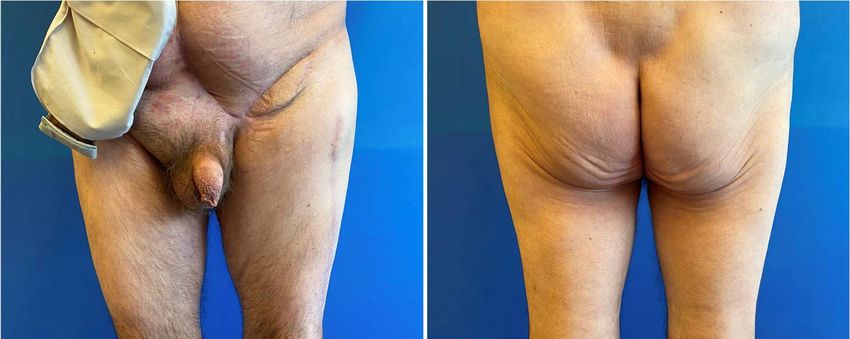
Figure 2. Intraoperative ICG imaging to check and confirm the patency and function of the LVA.
3. Results
In all cases, the reconstructive results were satisfactory from both the functional and
aesthetic points of view, with full volume and range of motion restoration. The total du-
Medicina 2022, 58, 509 ration of surgery ranged from 3:50 to 6:10 h. The mean follow-up period was 9.9 months 4 of 9
(ranging from 6 to 14 months). During this period, 11 patients showed no complications,
while 1 patient developed an infected seroma, which was conservatively treated with per-
cutaneous drainage and antibiotics. No signs of lymphocele nor lymphedema were ob-
after surgery,
served in any confirming
of the cases. a sufficient lymphatic
Lymphoscintigraphy flow.
was In all performed
routinely cases, no secondary procedures
6 months after
were required.
surgery, confirming a sufficient lymphatic flow. In all cases, no secondary procedures
were required.
Case Report
Case A
Report
67-year-old man presented an extremely large abdominal mass, which was diag-
nosedAas 67-year-old
soft tissueman presented
sarcoma afterananextremely large abdominal
open surgical biopsy. The mass, which was diag-
gastrointestinal surgeons
nosed as soft tissue sarcoma after an open surgical biopsy. The gastrointestinal
removed the whole tumor, exposing the entire bowel, and reconstructed the abdominal surgeons
removed
region the whole
bowel with tumor,
a meshexposing
(Figure the3). entire
A 25 bowel,
cm × 18 andcmreconstructed the abdominal
defect remained in the inguinal
region bowel with a mesh (Figure 3). A 25 cm × 18 cm defect remained
area, and a pedicled ALT flap was planned to fill the defect. In the inguinal in the inguinal
defect, both
area, and a pedicled ALT flap was planned to fill the defect. In the inguinal defect, both
superficial and deep lymphatics were identified and isolated with intraoperative ICG
superficial and deep lymphatics were identified and isolated with intraoperative ICG
lymphography. During the elevation of the flap, a long vein originating from the pedicle
lymphography. During the elevation of the flap, a long vein originating from the pedicle
was
was harvested
harvested andandprepared
prepared forfor anastomosis
anastomosis (Figure
(Figure 4). Using
4). Using 3 branches
3 branches of this pedicle
of this pedicle
vein, 3 LVAs were then performed, 2 with the superficial lymphatic vessels
vein, 3 LVAs were then performed, 2 with the superficial lymphatic vessels and 1 with the and 1 with the
deep
deep one. Theirpatency
one. Their patency was
was proven
proven using
using ICGICG lymphography
lymphography (Figure(Figure
5). At 65). At 6 months
months
follow-up, thereconstructive
follow-up, the reconstructive result
result was was good.
good. The The soft tissue
soft tissue coveragecoverage
was stablewaswithout
stable without
tumor
tumor recurrence,
recurrence, andandthethe lymphoscintigraphy
lymphoscintigraphy did notdidshow
not show anyofsign
any sign lymph of lymph
stasis stasis
(Figure 6).
(Figure
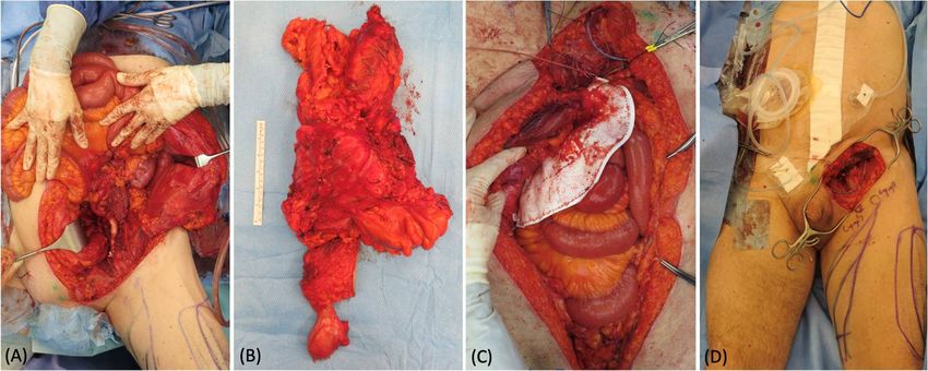
Figure 3. (A) Intraoperative picture of the extensive surgery for the sarcoma removal in the abdo-
Figure 3. (A) Intraoperative picture of the extensive surgery for the sarcoma removal in the abdomen.
men. (B) 45 × 30 cm specimen. (C) Abdomen reconstruction with a mesh. (D) Appearance5 of
Medicina 2022, 58, x FOR PEER REVIEW of the
9
(B)
groin × 30 cm
45 defect specimen.
at the end of the(C) Abdomen
abdominal reconstruction with a mesh. (D) Appearance
surgery. of the groin
defect at the end of the abdominal surgery.
Figure 4. (A) Preoperative picture of skin marking of the ALT flap with ICG-guided lymphatic ducts
Figure 4. (A) Preoperative picture of skin marking of the ALT flap with ICG-guided lymphatic ducts
detection. (B) Remaining groin defect after sarcoma removal.
detection. (B) Remaining groin defect after sarcoma removal.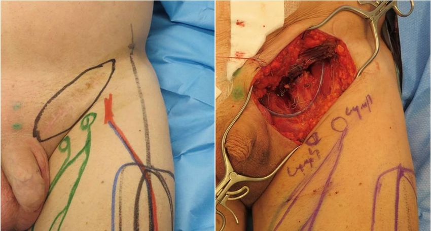
Medicina 2022, 58, 509                                                                                                            5 of 9
                                  Figure 4. (A) Preoperative picture of skin marking of the ALT flap with ICG-guided lymphatic ducts
                                  detection. (B) Remaining groin defect after sarcoma removal.

                                   Figure5.5.(A)
                                  Figure      (A)Lymphovenous
                                                 Lymphovenous anastomoses
                                                                 anastomoses of of 22 superficial
                                                                                      superficial lymphatics
                                                                                                  lymphatics and
                                                                                                             and 11 deep
                                                                                                                    deeplymphatic
                                                                                                                         lymphaticwith
                                                                                                                                   with3
                                   branches of the pedicle vein. (B) Intraoperative ICG lymphography to confirm the patency of the
                                  3 branches of the pedicle vein. (B) Intraoperative ICG lymphography to confirm the patency of the
                                   anastomoses. (C) Picture of the groin at the end of the procedure: the ALT flap was inset and 3 LVAs
 Medicina 2022, 58, x FOR PEER REVIEW                                                                                              6 of 9
                                  anastomoses. (C) Picture of the groin at the end of the procedure: the ALT flap was inset and 3 LVAs
                                   were performed with 3 pedicles branches.
                                  were performed with 3 pedicles branches.

                                  Figure6.6.(A,B)
                                 Figure      (A,B)Postoperative
                                                   Postoperativepicture
                                                                 pictureatat6 6months
                                                                                monthsfollow-up:
                                                                                       follow-up:frontal
                                                                                                   frontalview
                                                                                                           viewand
                                                                                                                andposterior
                                                                                                                    posteriorview.
                                                                                                                              view.

                                  4. Discussion
                                      Tissue defects reconstruction throughout the body represents one of the most com-
                                  mon tasks for plastic surgeons. Either pedicled or free flaps are well known as a versatile
                                  armamentarium, and they represent the most common treatment in these cases. Different
                                  types of flaps have been prosed over the years; however, the actual trend is to prefer the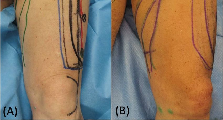
Medicina 2022, 58, 509                                                                                            6 of 9

                         4. Discussion
                               Tissue defects reconstruction throughout the body represents one of the most common
                         tasks for plastic surgeons. Either pedicled or free flaps are well known as a versatile
                         armamentarium, and they represent the most common treatment in these cases. Different
                         types of flaps have been prosed over the years; however, the actual trend is to prefer
                         the perforator-based ones since they reduce donor site morbidity and allow a quicker
                         dissection [12,13]. Among these, the typical ones are the deep inferior epigastric perforator
                         (DIEP) flap, the anterolateral thigh (ALT) flap, and the superficial circumflex iliac artery
                         perforator (SCIP) flap. When a sufficient amount of abdominal fat is present, the DIEP flap
                         is considered the gold standard for autologous breast reconstruction [14], and it is widely
                         used for soft tissue defects in many other districts [8]. The ALT flap is one of the main
                         alternatives; it has a long pedicle and low donor site morbidity and offers the advantage
                         of chimeric forms, including the vastus lateralis muscle [6]. Lately, the SCIP flap has been
                         gaining approval because of its very low donor site morbidity, versatility, and aesthetic
                         result. It also allows chimeric transfer, including muscle, nerve, and bone [15].
                               When the defects are large and compromise the lymphatic drainage network, the
                         prevention of postoperative complications is of crucial importance. Chronic lymphorrhea,
                         lymphocele, and lymphedema might develop, leading to severe discomfort for the patient,
                         with heaviness sensation in the limbs, swelling, pain, erythema, recurrent cellulitis, and
                         even range-of-motion limitations [16]. The best approach for this situation is to prevent
                         lymph stasis immediately after surgery.
                               Different techniques have been proposed to treat lymphatic sequelae, but there is
                         still a lack of consensus concerning their efficacy. The most validated options nowadays
                         are lymphovenous anastomosis (LVA) [10], vascularized lymphnode transfer (VLNT) [17],
                         and lymphatic tissue transfer [18]. LVA consists of diverting the lymph flow into the
                         venous circulation upstream of the damage, offering the lymph an alternative draining
                         route. This is performed by means of microsurgical or supermicrosurgical (when the
                         vessel’s diameter is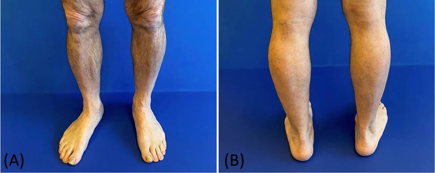
Medicina 2022, 58, 509                                                                                                  7 of 9

                         already resorted to a similar approach, employing the deep branch vein as a donor vessel
                         for LVA to prevent donor site lymphocele after SCIP flap harvest [27]. In the DIEP, we have
                         many superficial veins available, but we could also use branches coming from the pedicle.
                         Similarly, the ALT flap has a long pedicle presenting suitable venous branches.
                               From the technical point of view, it is essential to mention the role of intraoperative ICG
                         lymphography. This tool is of paramount importance to identify the leaking interrupted
                         lymphatic vessels and, hence, to shunt them into the venous circulation [28]. A feasible
                         alternative would be closing these vessels, blocking the leakage and preventing lymphocele
                         development, but it would imply a much higher risk of lymphedema. Another very
                         important examination is lymphoscintigraphy. We routinely rely on it preoperatively to
                         map the lymphatic network in the affected area and during the follow-up to confirm the
                         efficacy of the procedure. This is the most accurate method to visualize and assess lymph
                         flow restoration after the reconstruction [29,30]. A minor limitation of this procedure can
                         be the additional surgical time required to isolate the recipient vessels for the LVAs and to
                         execute the anastomoses. These procedures require about 40 to 60 min when performed by
                         an experienced surgeon
                               The number of cases described in the literature dealing with this technique is still
                         low; however, we believe that this is a very promising procedure that is worthy of further
                         study. Our results seem to confirm its efficacy, and we suggest taking it into account for the
                         reconstruction of soft tissue defects where the lymphatic network is widely compromised.
                               The most significant limitation of this report is the limited number of cases with
                         heterogenous defects and patient characteristics. This compromises the possibility of
                         gaining statistically significant results. However, as previously mentioned, this is a very
                         modern surgical approach for particularly complex defects. In this setting, it is extremely
                         difficult to gather a large cohort of patients, and further studies are necessary to confirm
                         the efficacy of this procedure.

                         5. Conclusions
                              This report is intended to show, taking into account the aforementioned limitations,
                         the potential of a modern, multi-effective approach to the reconstruction of large soft tissue
                         defects with significant lymphatic impairment. The lymphatic flow-through concept may
                         allow us to fully exploit the potential of either free or pedicled tissue transfer, combining
                         good coverage with the immediate restoration of the lymphatic drainage in order to prevent
                         immediate and long-term lymphatic complications.

                         Author Contributions: Corresponding author M.F.S. performed all surgeries and revised the manuscript
                         with E.F. Wrote the manuscript: M.M. All authors have read and agreed to the published version of
                         the manuscript.
                         Funding: This research received no external funding.
                         Institutional Review Board Statement: The report was conducted according to the guidelines of the
                         Declaration of Helsinki and approved by the Chief of the Department at the Luzerner Kantosspital.
                         The protocol was developed in accordance with the ethical standards of the Helsinki Declaration of
                         1975 and all subsequent revisions. The STROBE guidelines were adhered to. EKNZ-2022-00242.
                         Informed Consent Statement: Informed consent was obtained from all subjects involved in the
                         report. Written informed consent has been obtained from the patients to publish this paper.
                         Data Availability Statement: Data are available from the authors upon reasonable request.
                         Conflicts of Interest: The authors declare no conflict of interest.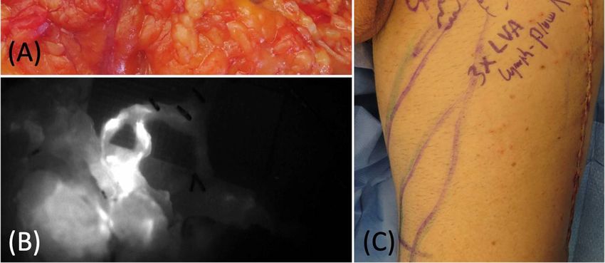
Medicina 2022, 58, 509                                                                                                                8 of 9

References
1.    Friedmann, D.; Wunder, J.S.; Ferguson, P.; O’Sullivan, B.; Roberge, D.; Catton, C.; Freeman, C.; Saran, N.; Turcotte, R.E. Incidence
      and Severity of Lymphoedema following Limb Salvage of Extremity Soft Tissue Sarcoma. Sarcoma 2011, 2011, 289673. [CrossRef]
      [PubMed]
2.    Exton, R.; Galland, R. Major Groin Complications Following the Use of Synthetic Grafts. Eur. J. Vasc. Endovasc. Surg. 2007, 34,
      188–190. [CrossRef] [PubMed]
3.    Warren, A.G.; Brorson, H.; Borud, L.J.; Slavin, S.A. Lymphedema: A comprehensive review. Ann. Plast. Surg. 2007, 59, 464–472.
      [CrossRef] [PubMed]
4.    Koshima, I.; Kawada, S.; Moriguchi, T.; Kajiwara, Y. Ultrastructural Observations of Lymphatic Vessels in Lymphedema in Human
      Extremities. Plast. Reconstr. Surg. 1996, 97, 397–405. [CrossRef]
5.    Taylor, G. The angiosomes of the body and their supply to perforator flaps. Clin. Plast. Surg. 2003, 30, 331–342. [CrossRef]
6.    Scaglioni, M.F.; Franchi, A.; Giovanoli, P. Pedicled chimeric sensitive fasciocutaneous anterolateral thigh (ALT) and vastus lateralis
      muscle (VLM) flap for groin defect reconstruction: A case report. Microsurgery 2017, 38, 423–426. [CrossRef]
7.    Koshima, I.; Nanba, Y.; Tsutsui, T.; Takahashi, Y.; Urushibara, K.; Inagawa, K.; Hamasaki, T.; Moriguchi, T. Superficial Circumflex
      Iliac Artery Perforator Flap for Reconstruction of Limb Defects. Plast. Reconstr. Surg. 2004, 113, 233–240. [CrossRef]
8.    Van Landuyt, K.; Blondeel, P.; Hamdi, M.; Tonnard, P.; Verpaele, A.; Monstrey, S. The versatile DIEP flap: Its use in lower
      extremity reconstruction. Br. J. Plast. Surg. 2005, 58, 2–13. [CrossRef]
9.    Garza, R.; Skoracki, R.; Hock, K.; Povoski, S.P. A comprehensive overview on the surgical management of secondary lymphedema
      of the upper and lower extremities related to prior oncologic therapies. BMC Cancer 2017, 17, 468. [CrossRef]
10.   Scaglioni, M.F.; Fontein, D.B.Y.; Arvanitakis, M.; Giovanoli, P. Systematic review of lymphovenous anastomosis (LVA) for the
      treatment of lymphedema. Microsurgery 2017, 37, 947–953. [CrossRef]
11.   Di Summa, P.G.; Guillier, D. The Lymphatic Flow-Through (LyFT) flap: Proof of concept of an original approach. J. Plast. Reconstr.
      Aesthetic Surg. 2020, 73, 983–1007. [CrossRef] [PubMed]
12.   Kim, J.T.; Kim, S.W. Perforator Flap versus Conventional Flap. J. Korean Med Sci. 2015, 30, 514–522. [CrossRef] [PubMed]
13.   Mohan, A.T.; Sur, Y.J.; Zhu, L.; Morsy, M.; Wu, P.S.; Moran, S.L.; Mardini, S.; Saint-Cyr, M. The Concepts of Propeller, Perforator,
      Keystone, and Other Local Flaps and Their Role in the Evolution of Reconstruction. Plast. Reconstr. Surg. 2016, 138, 710e–729e.
      [CrossRef] [PubMed]
14.   Chang, D.W. Breast Reconstruction with Microvascular MS-TRAM and DIEP Flaps. Arch. Plast. Surg. 2012, 39, 3–10. [CrossRef]
15.   Fernandez Garrido, M.; Qiu, S.S.; Vega Garcia, C.; Pons Playa, G.; Masiá Ayala, J. Chimeric Superficial Circumflex Iliac Perforator
      Flap Including External Oblique Fascia: A Refinement of Conventional Harvesting. Plast. Reconstr. Surgery Glob. Open 2016,
      4, e766. [CrossRef]
16.   Lv, S.; Wang, Q.; Zhao, W.; Han, L.; Wang, Q.; Batchu, N.; Ulain, Q.; Zou, J.; Sun, C.; Du, J.; et al. A review of the postoperative
      lymphatic leakage. Oncotarget 2017, 8, 69062–69075. [CrossRef]
17.   Schaverien, M.V.; Badash, I.; Selber, J.C.; Cheng, M.-H.; Patel, K.M. Vascularized Lymph Node Transfer for Lymphedema. Semin.
      Plast. Surg. 2018, 32, 028–035. [CrossRef]
18.   Gentileschi, S.; Servillo, M.; Garganese, G.; Fragomeni, S.; De Bonis, F.; Cina, A.; Scambia, G.; Salgarello, M. The lymphatic
      superficial circumflex iliac vessels deep branch perforator flap: A new preventive approach to lower limb lymphedema after
      groin dissection-preliminary evidence. Microsurgery 2017, 37, 564–573. [CrossRef]
19.   Scaglioni, M.F.; Uyulmaz, S. Optimizing outcomes of lymphatic-venous anastomosis (LVA) supermicrosurgery by preoperative
      identification of reflux-free vein: Choose the vein wisely. Microsurgery 2017, 38, 232–233. [CrossRef]
20.   Cigna, E.; Pierazzi, D.M.; Sereni, S.; Marcasciano, M.; Losco, L.; Bolletta, A. Lymphatico-venous anastomosis in chronic ulcer with
      venous insufficiency: A case report. Microsurgery 2021, 41, 574–578. [CrossRef]
21.   Baumeister, R.G.; Siuda, S. Treatment of lymphedemas by microsurgical lymphatic grafting: What is proved? Plast. Reconstr. Surg.
      1990, 85, 64–76. [CrossRef] [PubMed]
22.   Tourani, S.S.; Taylor, G.I.; Ashton, M.W. Vascularized Lymph Node Transfer: A Review of the Current Evidence. Plast. Reconstr.
      Surg. 2016, 137, 985–993. [CrossRef] [PubMed]
23.   Classen, D.A.; Irvine, L. Free Muscle Flap Transfer as a Lymphatic Bridge for Upper Extremity Lymphedema. J. Reconstr. Microsurg.
      2005, 21, 93–99. [CrossRef] [PubMed]
24.   Mihara, M.; Tange, S.; Hara, H.; Peng, Z.H.; Haragi, M.; Muarai, N. Modified lymph vessel flap transplantation for the treatment
      of refractory lymphedema: A case report. Microsurgery 2016, 36, 695–699. [CrossRef] [PubMed]
25.   Yan, A.; Avraham, T.; Zampell, J.C.; Aschen, S.Z.; Mehrara, B.J. Mechanisms of Lymphatic Regeneration after Tissue Transfer.
      PLoS ONE 2011, 6, e17201. [CrossRef]
26.   Fujiki, M.; Miyamoto, S.; Sakuraba, M. Flow-through anastomosis for both the artery and vein in leg free flap transfer. Microsurgery
      2015, 35, 536–540. [CrossRef]
27.   Scaglioni, M.F.; Meroni, M.; Fritsche, E. Inguinal seroma/lymphocele prevention after superficial circumflex iliac artery perforator
      (SCIP) flap harvest using the deep branch as donor vein for lymphovenous anastomosis (LVA). Microsurgery 2020, 41, 95–96.
      [CrossRef]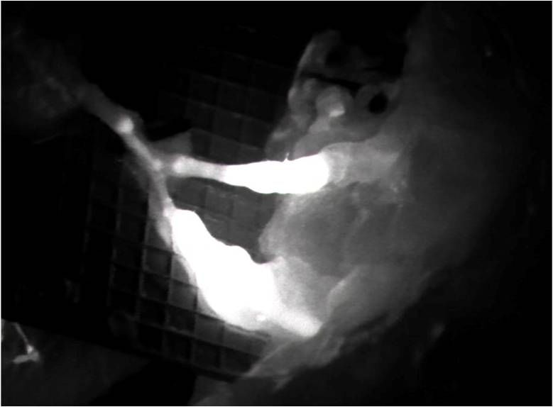
Medicina 2022, 58, 509                                                                                                       9 of 9

28.   Yamamoto, T.; Yoshimatsu, H.; Koshima, I. Navigation lymphatic supermicrosurgery for iatrogenic lymphorrhea: Supermicrosur-
      gical lymphaticolymphatic anastomosis and lymphaticovenular anastomosis under indocyanine green lymphography navigation.
      J. Plast. Reconstr. Aesthetic Surg. 2014, 67, 1573–1579. [CrossRef]
29.   Forte, A.J.; Boczar, D.; Huayllani, M.T.; Lu, X.; Ciudad, P. Lymphoscintigraphy for Evaluation of Lymphedema Treatment:
      A Systematic Review. Cureus 2019, 11, e6363. [CrossRef]
30.   De Roo, M.; Biscompte, J.P.; Verlooy, H.; Nieuborg, L.; Mortelmans, L.; Schiepers, C. Quantitative Lymphoscintigraphy for the
      Evaluation of Lymphaticovenous Anastomosis (LVA) in Treatment of Upper Limb Edema (ULE). In Radioactive Isotopes in Clinical
      Medicine and Research; Birkhäuser: Basel, Switzerland, 1995; pp. 161–166.
You can also read