Steroid Metabolites Support Evidence of Autism as a Spectrum - MDPI
←
→
Page content transcription
If your browser does not render page correctly, please read the page content below
behavioral
           sciences
Article
Steroid Metabolites Support Evidence of Autism as
a Spectrum
Benedikt Andreas Gasser 1, *, Johann Kurz 2 , Bernhard Dick 1,3 and Markus Georg Mohaupt 1,4
 1    Department of Clinical Research, University of Bern, 3010 Berne, Switzerland; bernhard.dick@gmx.ch (B.D.);
      markus.mohaupt@lindenhofgruppe.ch (M.G.M.)
 2    Intersci Research Association, Karl Morre Gasse 10, 8430 Leibnitz, Austria; john@a1.net
 3    Division of Nephrology/Hypertension, University of Bern, 3010 Berne, Switzerland
 4    Teaching Hospital Internal Medicine, Lindenhofgruppe, 3006 Berne, Switzerland
 *    Correspondence: gasser@pyl.unibe.ch

 Received: 30 March 2019; Accepted: 6 May 2019; Published: 9 May 2019

 Abstract: Objectives: It is common nowadays to refer to autism as a spectrum. Increased evidence
 of the involvement of steroid metabolites has been shown by the presence of stronger alterations
 in Kanner’s syndrome compared with Asperger syndrome. Methods: 24 h urine samples were
 collected from 20 boys with Asperger syndrome, 21 boys with Kanner’s syndrome, and identically
 sized control groups, each matched for age, weight, and height for comprehensive steroid hormone
 metabolite analysis via gas chromatography–mass spectrometry. Results: Higher levels of most steroid
 metabolites were detected in boys with Kanner’s syndrome and Asperger syndrome compared to their
 matched controls. These differences were more pronounced in affected individuals with Kanner’s
 syndrome versus Asperger syndrome. Furthermore, a specific and unique pattern of alteration
 of androsterone, etiocholanolone, progesterone, tetrahydrocortisone, and tetrahydrocortisol was
 identified in boys with Kanner’s syndrome and Asperger syndrome. Interestingly, in both matched
 samples, only androsterone, etiocholanolone, progesterone, tetrahydrocortisone, tetrahydrocortisol,
 and 5a-tetrahydrocortisol groups were positively correlated. In the Asperger syndrome group,
 all metabolites showed a positive correlation. In the Kanner’s syndrome group, 5-a tetrahydrocortisol
 with androsterone showed a positive correlation. Conclusions: Due to differences in the level of
 alteration, the premise that Asperger syndrome is on the mild side of the autism spectrum and
 that Kanner’s syndrome is on the severe side is supported, but alteration patterns yield different
 phenotypic expressions.

 Keywords: ACTH; autistic disorder; antiglucocorticoid; Kanner; Asperger

1. Introduction
     Autism spectrum disorder (ASD) comprises a group of disorders characterised by persistent
deficits in social communication and social interaction and restricted patterns of behaviour, interests
and activities [1]. The aetiology of autism is not well understood, although it is thought to involve
genetic, immunologic and environmental factors [1–12]. The diagnosis of autism is still based solely on
behavioural characteristics as there is currently no biological marker [13]. In ICD-10 (International
Classification of Diseases, 10th Revision) from the World Health Organisation (WHO), the premise
of autism as a spectrum was adopted overcoming the previously widely used diagnoses of Kanner
Syndrome respectively early-infantile autism (F84.0) and Asperger syndrome (F84.5).
     Consensus was reached that autism was regarded as a spectrum with the former diagnosis of
Asperger syndrome being a mild entity and Kanner syndrome a severe one. These former diagnoses are
nowadays given less attention, however still lots of affected individuals having these diagnoses. Clinical

Behav. Sci. 2019, 9, 52; doi:10.3390/bs9050052                                    www.mdpi.com/journal/behavsci
Behav. Sci. 2019, 9, 52 2 of 14
evidence supports the premise of Asperger’s disorder as a mild form of autism sharing a common
aetiology and developmental neuropathology with autism [14]. Involvement of epigenetic mechanisms,
linking genes with the environment, was suggested by studies conducted on lymphoblasts from siblings
with non-conforming autism diagnoses. They showed increased expression of genes associated with
steroid biosynthesis in brain development [15]. From a more comprehensive standpoint, some studies
suggest an involvement of the corticotropin-releasing hormone (CRH)-adrenocorticotropic hormone
(ACTH) system with additional alterations of adrenal gland metabolites in autistic disorders [16–21].
The regulation of proangiogenic factors was investigated in adrenocortical cells isolated from
human foetal adrenal glands for a more comprehensive understanding [22–24]. ACTH up-regulates
vascular-endothelial growth factor–A (VEGF-A) and increases mRNA encoding of angiopoietin 1
(Ang1) whereby indicating that ACTH is the primary regulator of adrenal organ growth by stimulating
VEGF and thus angiogenesis allowing to link the hypothalamic-pituitary adrenal axis (HPA-axis) with
adrenal gland development [22–24].
To broaden our understanding it should be mentioned that higher rates of clinical manifestations
of androgen-related conditions (e.g., polycystic ovary syndrome, hirsutism, acne and hormone-related
cancers) were more often reported in populations with autism spectrum conditions (as clinical correlates
of hyperandrogenism) compared to healthy non-autistic populations [25]. In particular, the relevance
of testosterone for developmental disorders was shown for children from hyperandrogenic mothers
with polycystic ovary syndrome (PCOS), increasing the risk of pervasive development disorders [26].
Especially prenatal androgen exposure was suggested to influence mental health leading to studies on
psychiatric morbidity among individuals with congenital adrenal hyperplasia (CAH, the inherited
inability to synthesie cortisol) [27]. Interestingly, individuals with 21-hydroxylase deficiency have
an increased risk of psychiatric disorders with the highest risk among those with the most severe
genotype and co-occurrence of CAH and autistic disorders [28]. Further evidence can be derived from
animal models where it was shown that maternal hyperandrogenism during pregnancy increased
the susceptibility of the foetus to autism spectrum disorder (ASD). In particular, the intrauterine
environment played an important role in the development of ASD [29]. Despite these facts, drugs
directly affecting the adrenal gland were suggested to improve clinical symptoms. Bradstreet et al.
(2006) used Spironolactone as a desirable immunologic and hormonal intervention in ASD [30]. Recent
work looking at the development of autism during gestation and childhood suggested that the placenta
is capable of synthesizing sex steroids de novo from maternal substrates (cholesterol), maternal or
foetal prohormones and steroid precursors [31]. Active steroids produced in the placental unit can
be released into both the maternal and foetal compartments to maintain pregnancy and allow foetal
development. In addition, endocrine disrupting compounds may be another source of environmentally
mediated influences on the early foetal hormonal milieu and a predisposition for the development of
autism [32]. Xu et al. (2015) studied the link between pre-term concentrations of adrenal hormones
and behaviour. The authors found that female rats treated with androgens spent less time in social
interaction in adolescence and exhibited impaired heterosexual interaction in adulthood. Furthermore,
the duration of social and heterosexual interaction of female offspring was negatively correlated
to maternal serum testosterone levels during pregnancy [29]. In summary, there seems to be clear
evidence for a link between increased gonadal adrenal sex steroids (particularly testosterone, estradiol,
dehydroepiandrosterone sulfate and androstenedione) in individuals with autism and autism-relevant
behaviour, which implies that greater postnatal testosterone levels in early infancy are predictive of
more male-typical behaviour later in life [25,33–35] Previous work on metabolite levels clearly showed
the effects of testosterone on phenotypic variability related to autism. Foetal testosterone influenced
individual differences in the typical development of eye contact, size of vocabulary, restriction of
interests, mentalizing, empathy, attention to detail, urge to systemise, and autistic traits [36].
In summary, broad evidence exists for an involvement of adrenal gland metabolites in ASD,
whereby patterns of severity might vary in individuals with Asperger compared to Kanner syndrome.
This leads us to the aim of this study, which was to analyse the degree of involvement of adrenalBehav. Sci. 2019, 9, 52 3 of 14
gland metabolites and adrenal gland enzyme activities (21-hydroxylase, 17-hydroxylase and 11
beta-hydroxylase) in children with Asperger and Kanner syndrome. We hypothesised that adrenal
gland metabolites and enzyme activities were altered to a larger extent in children with Kanner
syndrome compared to Asperger syndrome [37].
2. Materials and Methods
2.1. Participants
Twenty boys diagnosed with Asperger syndrome (BMI 18.2 ± 3.2; average age 15.3 ± 2.9 years)
with an individually pairwise matched control group (BMI 18.2 ± 3.5; average age 14.2 ± 2.4 years)
and 21 boys diagnosed with Kanner’s syndrome (BMI 20.4 ± 5.9; average age 13.6 ± 3.6 years)
with an individually pairwise matched control group (BMI 19.4 ± 3.1; average age 13.3 ± 3.1 years)
were recruited.
2.2. Study Design
All procedures performed in studies involving human participants were in accordance with the
ethical standards of the institutional and/or national research committee and with the 1964 Helsinki
declaration and its later amendments or with comparable ethical standards. The study was approved
by the governmental ethics board of Graz, Austria, and registered at ClinicalTrials.gov. Involvement
in the study was voluntary and not compensated. After study procedures had been fully explained,
the parents of the participants read and signed informed consent forms. Autistic and control boys
were recruited from the area of Leipzig (Austria). Enrolment took place from mid-2009 to mid-2012.
All participants were Caucasians. Participants were excluded if they had a neurological and psychiatric
disorder other than autism and comorbid disorders, a history of liver diseases, renal or endocrine
disorders, a current infection, or fever. Mental retardation or behavioural disorders were exclusion
criteria only for the control group but were allowed as comorbid conditions in the autistic group.
The diagnosis was given in the first years of life of the children according to the diagnostic criteria of
the DSM-IV and was cross-checked by experienced clinicians (medical doctors and/or psychologists)
during enrolment of the study. Key criteria such as the extent of language impairment were used in
order to distinguish between Kanner’s syndrome and Asperger syndrome [38](A further tool used to
increase the accuracy of diagnosis was the Marburg questionnaire for Asperger syndrome (MBAS).
2.3. Methods
Analysis of urinary steroids was conducted via gas chromatography–mass spectrometry. Urine
samples were taken between 7 a.m. and 9 a.m. in the morning after breakfast. Urine sample
preparation comprised pre-extraction, enzymatic hydrolysis, extraction from the hydrolysis mixture,
derivatization, and gel filtration [39–41]. The recovery standard was prepared by adding 2.5 µg of
medroxyprogesterone to 1.5 mL of urine. The sample was extracted on a Sep-Pak C18 column (Waters
Corp., Milford, MA, USA), dried, reconstituted in a 0.1 M acetate buffer, pH 4.6, and hydrolysed
with a powdered Helix pomatia enzyme (12.5 mg; Sigma Chemical Co., St. Louis, MI, USA) and
12.5 µL of β-glucuronidase/arylsulfatase liquid enzyme (Roche Diagnostics, Rotkreuz, Switzerland).
The resulting free steroids were extracted on a Sep-Pak C18 cartridge. A mixture of internal standards
(2.5 µg each of 5α-androstane-3α, 17α-diol, stigmasterol, and cholesterol butyrate, and 0.15 µg of
3β5β-tetrahydroaldosterone) was added to this extract, and the sample was derivatised to form the
methyloxime-trimethylsilyl ethers. Analyses were performed on a Hewlett-Packard gas chromatograph
6890 (Hewlett Packard, Palo Alto, CA, USA) with a mass selective detector 5973 by selective ion
monitoring (SIM). One characteristic ion was chosen for each compound measured. The derivatised
samples were analysed during a temperature-programmed run (210–265 ◦ C) over a 35 min period.
The calibration standard consisted of a steroid mixture containing known quantities of all steroid
metabolites to be measured. Responses and retention times were recorded regularly. In each case,Behav. Sci. 2019, 9, 52 4 of 14
the ion peak was quantified against the internal stigmasterol standard. With this method, the most
relevant metabolites of the glucocorticoid synthesis pathway were measured and presented. All results
were adjusted for creatinine in urine to check renal function.
2.4. Statistical Analysis
SPSS Version 21 and Microsoft Excel were used to analyse all data. A Student’s t-test was
performed to compare the means of data from two groups. Results where p < 0.05 were considered
significant. Data were analysed for normal distribution with a Jarque–Bera test, whereby the hypothesis
of a normal distribution, at p < 0.1 for all samples, could not be rejected. Correlations between different
metabolites and between classes of metabolites were calculated. Enzyme ratios and demographic data
are presented as standard error of mean ± standard deviation. Clinical data are presented as mean ±
SEM. Calculations were made with GraphPad Prism (GraphPad Software, Inc., La Jolla, CA, USA) and
Microsoft Excel (Microsoft Inc., Redmond, WA, USA).
3. Results
Metabolite concentrations of adrenal gland hormones differed in boys with Asperger syndrome and
Kanner’s syndrome compared to their matched control groups regarding androgens, 11-deoxycortisol,
corticosterone metabolites, and cortisone metabolites as well as oestrogens and progesterones (Table 1).
Enzyme activities were also different in boys with Asperger syndrome and those with Kanner’s
syndrome compared to their matched controls (Table 2). These differences were more pronounced in
the Kanner’s syndrome group. Bivariate correlation matrices were calculated for the most important
hormones, i.e., those that exhibited highly significant differences in order to identify varying patterns
in Asperger syndrome and Kanner’s syndrome (Table 3). Furthermore, correlations between the whole
class of metabolites (androgens and cortisol metabolites) were determined. A general dysregulation
of androgens is apparent: correlation analysis of all androgen metabolites showed a correlative
relationship of 0.959 in individuals with Asperger syndrome versus the controls and a correlative
relationship of 0.911 in individuals with Kanner’s syndrome versus the controls (Asperger syndrome
and Kanner’s syndrome correlated with 0.943, and the two control groups correlated with 0.973).
When analyzing all cortisol metabolites, markedly higher correlative relationships between the controls
and Asperger syndrome (0.987) and between the controls and Kanner’s syndrome (0.997) were
identified, further supporting the dysregulation of androgen metabolites. Special attention was paid to
androsterone and etiocholanolone, the two highly increased androgens (Figure 1a–d).Behav. Sci. 2019, 9, 52 5 of 14
Table 1. Androgen metabolites for boys with Asperger syndrome (n = 20) versus individually pairwise matched controls (n = 20) and boys with Kanner syndrome
(n = 21) versus individually pairwise matched controls (n = 21).
Asperger Kanner
Boys p-Value Controls Boys p-Value Controls
Mean SEM Mean SEM Mean SEM Mean SEM
Androgen Metabolites
Androsterone 95.43 18.36 0.008 57.7 9.79 89.29 17.4 0.001 61.45 13.67
Etiocholanolone 62.45 13.21 0.043 38.92 6.16 59.78 13.45 0.009 29.23 6.24
Androstenediol 3.35 0.66 0.056 2.2 0.41 3.58 0.7 0.087 2.88 0.6
11-Oxoetiocholanolone 26.53 3.48 0.667 24.31 2.86 30.93 6.96 0.119 19.45 2.05
11β-Hydroxyandrosterone 32.84 3.57 0.07 26.2 3.15 46.78 7.57 0.033 30.72 4.41
11β-Hydroxyetiocholanolone 18.76 3.15 0.913 19.3 2.78 23.1 4.57 0,08 14.5 1.91
Dehydroepiandrosterone 6.02 2.65 0.72 4.92 1.31 36.82 19.5 0.129 7.57 2.03
5-Androstene-3β,17β-diol 3.67 0.57 0.538 3.19 0.55 7.13 2.67 0.191 3.85 0.73
16α-Hydroxydehydroepiandrosterone 11.05 3.31 0.311 20.16 7.29 16.32 4.42 0.506 21.26 6.88
5-Androstene-3β,16α,17β-triol 18.73 4.53 0.537 23.24 5.44 19.31 4.06 0.504 23.23 5.18
5-Pregnene-3β, 16α,17β-triol 6.41 2.21 0.739 7.21 1.22 10.55 5.2 0.696 8.66 1.33
Testosterone 2.93 0.71 0.056 1.46 0.32 3.01 1 0.134 1.62 0.34
5α-Dihydrotestosterone 1.29 0.17 0.051 0.96 0.12 1.66 0.24 0.035 1.16 0.15
Oestrogen metabolites
Estriol 0.14 0.02 0.859 0.14 0.03 0.18 0.04 0.288 0.16 0.02
17β-Estradiol 0.1 0.02 0.043 0.05 0.01 0.09 0.02 0.505 0.08 0.01
Progesterone metabolites
17-Hydroxypregnanolone 5.43 0.86 0.068 3.32 0.76 7.34 1.32 0.005 3.7 0.46
Pregnanediol 10.31 0.95 0.37 8.91 2.05 13.45 1.61 0.019 9.41 1.16
Pregnanetriol 32.62 4.95 0.043 22.17 5.1 35.15 5.09 0.001 16.48 1.98
11-Oxopregnanetriol 1.41 0.29 0.482 1.1 0.25 2.01 0.57 0.108 0.99 0.16
11-Deoxycortisol Metabolite
Tetrahydrodeoxycortisol 0.48 0.04 0.588 0.44 0.07 0.68 0.09 0.031 0.47 0.03Behav. Sci. 2019, 9, 52 6 of 14
Table 1. Cont.
Asperger Kanner
Boys p-Value Controls Boys p-Value Controls
Mean SEM Mean SEM Mean SEM Mean SEM
Corticosterone metabolites
Tetrahydrodihydrocorticosterone 12.83 1.42 0.093 9.38 1.26 14.72 1.86 0.014 9.64 0.63
Tetrahydrocorticosterone 11.77 1.22 0.193 9.57 1.14 15.6 1.57 0.005 10.22 0.85
5α-Tetrahydrocorticosterone 26.8 3.73 0.096 19.67 2.13 47.71 8.2 0.015 24.66 2.2
18-Hydroxytetrahydrodihydrocorticosterone 4.53 0.51 0.364 3.34 0.58 6.24 1 0.073 3.28 0.67
Tetrahydroaldosterone 2.38 0.33 0.002 1.24 0.1 1.96 0.23 0.096 1.48 0.15
Cortisone Metabolites
Cortisone 10.8 1.34 0.592 11.86 1.18 13.74 1.91 0.884 13.42 1.46
Tetrahydrocortisone 245.62 21.04 0.005 171.72 12.04 278.77 27.31 0.007 185.14 16.29
α-Cortolone 84.51 6.91 0.002 59.54 3.65 99.76 16.05 0.031 63.1 4.78
β-Cortolone 0.69 0.07 0.001 110.39 18.11 0.73 0.1 0 137.5 25.01
20α-Dihydrocortisone 1.3 0.19 0.989 1.29 0.13 1.64 0.28 0.784 1.55 0.19
20β-Dihydrocortisone 3.79 0.49 0.849 3.92 0.39 4.88 0.79 0.932 4.96 0.61
Cortisol metabolites
Cortisol 8.72 1.4 0.209 6.33 0.65 12.72 2.22 0.044 7.36 1.02
Tetrahydrocortisol 76.84 6.13 0.016 56 4.02 106.23 9.53 0.001 63.06 6.38
5α-Tetrahydrocortisol 102.92 14.16 0.017 60.91 4.07 146 20.78 0.005 79.77 6.97
α-Cortol 16.02 1.23 0.002 10.82 0.69 22.09 2.31 0.001 12.34 0.9
β-Cortol 21.11 1.61 0.125 16.93 2.11 29.17 2.97 0.009 17.18 2.35
20α-Dihydrocortisol 3.28 0.31 0.556 3.71 0.55 5.91 1.15 0.297 4.55 0.7
6β-Hydroxycortisol 9.68 0.85 0.754 9.13 1.39 19.58 6.07 0.095 9.51 1.47
18-Hydroxycortisol 26.34 1.95 0.54 23.3 124 53.56 15.9 0.245 32.7 6.02Behav. Sci. 2019, 9, 52 7 of 14
Table 2. Enzyme activities for boys with Asperger syndrome (n = 20) versus individually pairwise matched controls (n = 20) and boys with Kanner syndrome (n = 21)
versus individually pairwise matched controls (n = 21). Data given as ratio of metabolites (mmol/L) to urine creatinine (µmol/L). (THA = Tetrahydrodehydrocorticosteron;
THB = Tetrahydrocorticosteron; 5aTHB = 5a-Tetrahydrocorticosteron; THS = Tetrahydrosubstance S; HP = 17-Hydroxypregnalonon; PT = Pregnanetriol; PT’ONE =
11-Oxo-Pregnanetriol; THDOC = Tetrahydrodoc; E = Cortison; THE = Tetrahydrocortison; F = Cortisol; THF = Tetrahydrocortisol; 5ATHF = 5-alphaTetrahydrocortisol).
Asperger Boys p< Control Boys Kanner Boys p< Control Boys
Mean SD Mean SD Mean SD Mean SD
21-Hydroxylase Deficit
17HP/(THE + THF + 5aTHF) 0.014 0.01 0.684 0.013 0.01 0.004 0.021 0.711 0.014 0.02
PT/(THE + THF + 5aTHF) 0.009 0.007 0.17 0.007 0.005 0.016 0.108 0.00 0.005 0.003
100 × PT’ONE/(THE + THF + 5aTHF) 0.003 0.004 0.000 0.357 0.377 0.002 0.001 0.00 0.532 0.481
17-Hydroxylase Deficit
(THA + THB + 5aTHB)/(THE + THF + 5aTHF) 0.121 0.035 0.000 0.238 0.098 0.085 0.159 0.00 0.259 0.085
100 × THS/(THE + THF + 5aTHF) 0.016 0.006 0.000 1.00 0.673 0.012 0.018 0.00 1.08 0.478
11-Hydroxylase Deficit
100 × THDOC/(THE + THF + 5aTHF) 0.001 0.000 0.000 7.147 5.212 0.042 0.158 0.00 7.241 3.54
11-Beta HSD Deficit
F/E 0.786 0.203 0.000 0.386 0.197 0.175 0.793 0.009 0.339 0.088
(THF + 5aTHF)/THE 21.517 14.141 0.000 2.821 4.513 19.736 9.452 0.00 1.362 0.471
(F + E)/THE + THF + 5aTHF 0.048 0.028 0.000 1.57 0.658 0.035 0.079 0.00 1.797 0.563(d) Kan. Cont.
ANDRO 1.00 0.93 0.82 −0.27 −0.35 −0.03
ETIO 0.93 1.00 0.93 −0.33 −0.42 −0.15
PT 0.82 0.93 1.00 −0.22 −0.15 −0.19
THE- −0.27 −0.33 −0.22 1.00 0.90 0.52
THF −0.35 −0.42 −0.15 0.90 1.00 0.54
Behav. Sci. 2019, 9, 52 5ATHF −0.03 −0.15 −0.19 0.52 0.54 1.00 8 of 14
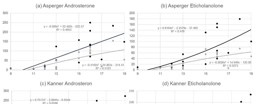
Figure 1. (a) Correlative relationship between age and androsterone (ratio of androsterone (mmol/L) to
Figure 1. (a) Correlative relationship between age and androsterone (ratio of androsterone (mmol/L)
urine creatinine (µmol/L)) for n = 20 boys with Asperger syndrome (black line) and for n = 20 pairwise
to urine creatinine (µmol/L)) for n = 20 boys with Asperger syndrome (black line) and for n = 20
matched
pairwisecontrols
matched(grey line).
controls (b)line).
(grey Correlative relationship
(b) Correlative between
relationship ageage
between andand etiocholanolone
etiocholanolone(ratio of
eticholanolone creatinine (µmol/L)) for n =
(ratio of eticholanolone (mmol/L) to urine creatinine (µmol/L)) for n = 20 boys with syndrome
(mmol/L) to urine 20 boys with Asperger Asperger (black
line)
syndrome n = 20
and for(black pairwise
line) and formatched controls
n = 20 pairwise (greycontrols
matched line). (c) Correlative
(grey relationship
line). (c) Correlative between age and
relationship
androsterone
between age (ratio of androsterone
and androsterone (ratio(mmol/L) to urine
of androsterone creatinine
(mmol/L) (µmol/L))
to urine creatinine = 21 boys
for n(µmol/L)) forwith
n = Kanner
21 boys with
syndrome Kanner
(black and n = 21
line)syndrome (black line) and
pairwise n = 21controls
matched pairwise(grey
matched
line).controls (grey line).relationship
(d) Correlative (d)
Correlative
between agerelationship between age(ratio
and etiocholanolone and etiocholanolone
of androsterone (ratio of androsterone
(mmol/L) to urine(mmol/L)
creatinineto (µmol/L))
urine for
creatinine (µmol/L)) for n = 21 boys with Kanner syndrome (black line) and n =
n = 21 boys with Kanner syndrome (black line) and n = 21 pairwise matched controls (grey line). 21 pairwise matched
controls (grey line).
Table 3. Correlation matrices depicting the relationship between the significantly different hormones
in (a) Asperger n = 20 (b) Kanner n = 21 (c) Asperger controls n = 20 (d) Kanner controls
4. Discussion
n = 21. Abbreviations: Andro = Androsterone, Etio = Etiocholanolone, PT = Progesterone, THE =
Tetrahydrocortisone, THF = Tetrahydrocortisol, 5aTHF = 5a-Tetrahydrocortisol. Dark green correlation
between 0.5 < correlation < 1, light green 0 < correlation < 0.5, red correlation is negative.
ANDRO ETIO PT THE THF 5ATHF
(a) Asperger
ANDRO 1.00 0.81 0.81 0.14 0.36 0.32
ETIO 0.81 1.00 0.71 0.39 0.39 0.39
PT 0.81 0.71 1.00 0.39 0.40 0.39
THE- 0.14 0.39 0.39 1.00 0.90 0.39
THF 0.36 0.39 0.40 0.90 1.00 0.59
5ATHF 0.32 0.39 0.39 0.39 0.59 1.00
(b) Kanner
ANDRO 1.00 0.98 0.96 −0.34 −0.43 0.20
ETIO 0.98 1.00 0.95 −0.33 −0.33 −0.33
PT 0.96 0.95 1.00 −0.42 −0.38 −0.41
THE −0.34 −0.33 −0.42 1.00 0.91 0.64
THF −0.43 −0.33 −0.38 0.91 1.00 0.72
5ATHF 0.20 −0.33 −0.41 0.64 0.72 1.00Behav. Sci. 2019, 9, 52 9 of 14
Table 3. Cont.
ANDRO ETIO PT THE THF 5ATHF
(c) Asp. Cont.
ANDRO 1.00 0.91 0.84 −0.32 −0.30 −0.11
ETIO 0.91 1.00 0.92 −0.23 −0.23 −0.23
PT 0.84 0.92 1.00 −0.12 −0.15 0.91
THE- −0.32 −0.23 −0.12 1.00 0.91 0.79
THF −0.30 −0.23 −0.15 0.91 1.00 0.72
5ATHF −0.11 −0.23 0.91 0.79 0.72 1.00
(d) Kan. Cont.
ANDRO 1.00 0.93 0.82 −0.27 −0.35 −0.03
ETIO 0.93 1.00 0.93 −0.33 −0.42 −0.15
PT 0.82 0.93 1.00 −0.22 −0.15 −0.19
THE- −0.27 −0.33 −0.22 1.00 0.90 0.52
THF −0.35 −0.42 −0.15 0.90 1.00 0.54
5ATHF −0.03 −0.15 −0.19 0.52 0.54 1.00
4. Discussion
This study investigated alterations in adrenal gland metabolites and adrenal gland enzyme
activities in boys with Asperger syndrome (F84.5) and Kanner’s syndrome (early-infantile
autism-F84.0) [42,43]. Although a relatively small cohort of affected individuals was analysed, limiting
the general validity of the results, consistent patterns can be detected. All classes of metabolites showed
differences between children with autism (Asperger syndrome and Kanner’s syndrome) and their
individually pairwise matched control groups, supporting the hypothesis of a principal involvement
of adrenal gland tissue in autism spectrum disorder. In particular, androgens, 11-deoxycortisone,
corticosterone, cortisol, cortisone, oestrogen, and progesterone metabolites varied significantly.
In principle, there is some evidence that Asperger syndrome and Kanner’s syndrome are
distinguishable entities [38,44]. For example, it was revealed that adrenotropic metabolites such as
epinephrine and norepinephrine but also chemical relatives such as tyrosine and homovanillic acid
showed different patterns in individuals with Kanner’s syndrome compared to Asperger syndrome,
clearly indicating differences in the severity of alterations [44]. In the present study, significant
differences between individuals with autism and controls were more obvious in the case of Kanner’s
syndrome compared to Asperger syndrome (e.g., 11B-hydroxyandrosterone and 5-a testosterone).
Furthermore, for 11B-hydroxyeticholanolone, 5-androstene-3β, 17β-diol, and dehydroepiandrosterone,
there were significant differences compared to matched controls in relation to Kanner’s syndrome but
not to Asperger syndrome. This is also supported by the correlation analysis of all metabolites of a
conducted class, indicating a stronger deviance from normal androgen concentrations in relation to
Kanner’s syndrome compared to the Asperger syndrome, which implies that Kanner’s syndrome is a
more severe form of autism.
The analysis of the two androgens, androsterone and etiocholanolone, showed around 50%
increased values in boys with Asperger syndrome as well as Kanner’s syndrome compared to control
groups (p = 0.06 for Asperger syndrome and p = 0.14 for Kanner’s syndrome) (Table 1). These two
metabolites depend highly on testosterone. Testosterone is converted to androstenedione first and then
to androsterone (5α-androstan-3α-ol-17-one; 5α,3α-A) and etiocholanolone (5β-androstan-3α-ol-17-one;
5β,3α-A) [45] (5α-Reductase (5α-R) and 5β-reductase (5β-R) catalyse the rate-limiting irreversible initial
steps, which are followed by sequential reductions by 3α-hydroxysteroid dehydrogenase (3α-HSD)
and 17β-hydroxysteroid dehydrogenase (17β-HSD) [45].
Although analyses in Figure 1a–d methodologically pursue a linear approach, this stands somehow
in contrast to evidence of the strong increase of androgens in boys when puberty starts [46], which we
were able to confirm with additional polynomial analysis. It is obvious from Figure 1a–d that the slope
in the Asperger syndrome group was clearly higher than in the matched controls, whereas a parallelBehav. Sci. 2019, 9, 52 10 of 14
development over time was detected in the Kanner’s syndrome group and their controls. This implies
differences in metabolic alterations over time in the two ASDs.
Androsterone and etiocholanolone showed the highest correlation in all metabolite groups
in the bivariate correlation matrices (Table 3). Both control groups had a very similar pattern,
with negative correlations of progesterone, androsterone, and etiocholanolone versus tetrahydrocortisol,
5-a tetrahydrocortisol, and tetrahydrocortisone. Interestingly, all metabolites in the Asperger syndrome
group showed positive correlations, whereas in the Kanner’s syndrome group the pattern was
similar to that of the matched controls. The absent negative correlative relationships in Asperger
syndrome for the precursor progesterone and the androgens androsterone and etiocholanolone and the
glucocorticoid groups of tetrahydrocortisol, 5-a tetrahydrocortisol, and tetrahydrocortisone stand out,
suggesting a general dysregulation of steroid pathways. Interestingly, values for androsterone and
etiocholanolone were higher in the Asperger syndrome group than in the Kanner’s syndrome group,
whereas the opposite was observed for tetrahydrocortisone, tetrahydrocortisol, 5α-tetrahydrocortisol,
and alpha-cortol.
Interestingly, in the 1970s there were some thoughts that the ratios of androsterone and
etiocholanolone discriminated between heterosexual and exclusively homosexual individuals, clearly
indicating that androsterone and etiocholanolone lead to a new sexual phenotype [47]. Looking at
enzyme activities, especially 11-beta dehydrogenase (11-beta HSD), there was a striking difference in
the ratio of (THF + 5aTHF)/THE between both Asperger syndrome (21.5) and Kanner’s syndrome
(19.7) groups and their matched controls (2.8 and 1.3, respectively) (Table 2). Values larger than 3
were regarded as pathologic [41]. The ratio of (F + E)/(THE + THF + 5aTHF) measured in the present
study indicated an alteration in beta-dehydrogenase activity. This ratio was about 15 times lower
in the Asperger syndrome group and 20 times lower in the Kanner’s syndrome group compared to
their matched controls, implying that not only adrenal gland metabolites but also enzyme activities
were affected.
When aiming for an aetiopathogenetic understanding of the dysregulation of adrenal gland
metabolites pre-term birth might provide an answer. From a morphometric point of view, pre-term
birth might be a risk factor for adrenal gland dysregulation, as some studies found that during antenatal
and neonatal periods the adrenal gland might shrink more rapidly in infants born at full term [48].
Interesting similarities can be observed when comparing our results to those of a recent study on
levels of ∆4 steroids from amniotic fluid samples. The concentration of ∆4 sex steroids (progesterone,
17α-hydroxy-progesterone, androstenedione, and testosterone) and cortisol were positively associated
with autism (Baron-Cohen et al. 2015). Furthermore, the principal component analysis confirmed that
one generalised latent steroidogenic factor was driving much of the variation in the data. The autism
group showed elevations across all hormones on this latent generalised steroidogenic factor, and this
elevation was uniform across the ICD-10 diagnostic label [34]. This work was consistent with prior
findings of interactions between the hypothalamic-pituitary adrenal axis (HPA-axis) and its known
foetal programming effects on later atypical neurodevelopmental phenotypes [49]. There are probably
more than 30 steps involved in adrenal gland metabolomics, from the first cyclised sterol lanosterol
to the most downstream product estradiol [40]. Our analyses mainly focused on the metabolic steps
for significantly different metabolites. The results presented here clearly showed an involvement of
17-hydroxylase, 21-hydroxylase, and 11β-hydroxylase in boys with Asperger syndrome as well as
those with Kanner’s syndrome, indicating a principal involvement of adrenal gland tissue in ASD.
In summary, a number of hormones and enzymes were altered in our samples from the Asperger
syndrome group and the Kanner’s syndrome group rather than the abnormality being restricted
to a specific steroid hormone. This observation suggests a dysregulation of pathways mediated by
cytochrome P450-containing enzymes that catalyse the conversion of hormones along the glucocorticoid
pathways [40]. Evidence pointing toward the importance of such enzymes was previously found via
genetic associations between autism and single-nucleotide polymorphisms in CYP17A1, CYP19A1,
and CYP11B1 genes [50]. The results of our sample of autistic children can be well embedded in theseBehav. Sci. 2019, 9, 52 11 of 14
findings, suggesting an involvement of P450 oxidoreductase deficiency and an alteration in cortisone
reductase activity.
When broadening the analysis to principal growth processes in childhood, it can be mentioned
that, for children with autism, significantly greater head circumferences and greater weights than
those in the control groups were found [51]. Furthermore, levels of insulin-like growth factor 1 (IGF-1),
insulin-like growth factor 2 (IGF-2), insulin-like growth factor binding protein (IGFBP-3) and Growth
Hormone (GH) binding protein which are all affected by cortisol, were significantly higher in autistic
children than in controls supporting the point of view of an involvement of CRH-ACTH adrenal
axis [51]. In conclusion, our data clearly showed altered adrenal gland metabolites in children with
Asperger syndrome as well as Kanner’s syndrome compared to matched control groups, suggesting
a principal involvement of adrenal gland tissue in ASD. Further evidence can be derived from the
ratios of measured metabolites, which clearly indicate a dysregulation of enzyme activities. Clear
differences between the Asperger syndrome group and the Kanner’s syndrome group emerged for the
calculated ratios of androsterone and etiocholanolone, which was further supported by the calculated
correlations of hormones with highly significant differences.
To sum up, results suggest different patterns in the alteration of adrenal gland metabolites in
relation to Asperger syndrome and Kanner’s syndrome. Steroid hormone profiles, especially those of
androgens, might be useful clinical markers that support the diagnoses of autism. The results further
imply that mild and more severe forms of autism can be distinguished. For severe forms, steroid
metabolite pathways might be a promising target for pharmaceuticals such as statins and may inhibit
the cholesterol precursor of steroid pathways.
Author Contributions: Conceptualization, J.K. and M.G.M.; Methodology, J.K., M.G.M. and B.D.; Software,
B.A.G.; Validation, B.A.G., J.K., B.D., M.G.M.; Formal Analysis, B.A.G., J.K., M.G.M.; Investigation, J.K., B.A.G.,
M.G.M.; Resources, J.K., B.A.G., M.G.M.; Data Curation, B.D. and J.K.; Writing—Original Draft Preparation,
B.A.G.; Writing—Review & Editing, B.A.G., J.K. and M.G.M.; Visualization, J.K., B.A.G., M.G.M.; Supervision, J.K.
and M.G.M.; Project Administration, B.A.G., and J.K.; Funding Acquisition, M.G.M.
Funding: This research was funded by Swiss National Foundation grant number 3200B0-113902/1, 32-135596.
Acknowledgments: We thank all families and their children, who took part in the study, for their commitment to
supporting our efforts in resolving the enigma of autism spectrum disease.
Conflicts of Interest: Funding sources had no involvement during the whole research process including the
decision for publication.
References
1. Gillberg, C.; Fernell, E.; Kočovská, E.; Minnis, H.; Bourgeron, T.; Thompson, L.; Allely, C. The role of
cholesterol metabolism and various steroid abnormalities in autism spectrum disorders: A hypothesis paper.
Autism Res. 2017. [CrossRef] [PubMed]
2. Akshoomoff, N.; Pierce, K.; Courchesne, E. The neurobiological basis of autism from a developmental
perspective. Dev. Psychopathol. 2002, 14, 613–634. [CrossRef]
3. Dziobek, I.; Gold, S.M.; Wolf, O.T.; Convit, A. Hypercholesterolemia in Asperger syndrome: Independence
from lifestyle, obsessive-compulsive behavior, and social anxiety. Psychiatry Res. 2007, 149, 321–324.
[CrossRef] [PubMed]
4. Fangmeier, T. Patogenetische Modelle. In Das Asperger-Syndrom im Erwachsenenalter; Van Elst, L.T., Ed.;
Medizinisch Wissenschaftliche Verlagsgesellschaft: Berlin, Germany, 2013.
5. Lamb, J.A.; Moore, J.; Bailey, A.; Monaco, A.P. Autism: Recent molecular genetic advances. Hum. Mol. Genet.
2000, 9, 861–868. [CrossRef] [PubMed]
6. Sasaki, T. Genetic factors in Asperger syndrome. Nihon Rinsho 2007, 65, 443–448.
7. Schwarz, E.; Guest, P.C.; Rahmoune, H.; Wang, L.; Levin, Y.; Ingudomnukul, E.; Ruta, L.; Kent, L.;
Spain, M.; Baron-Cohen, S.; et al. Sex-specific serum biomarker patterns in adults with Asperger’s syndrome.
Mol. Psychiatry 2011, 16, 1213–1220. [CrossRef] [PubMed]Behav. Sci. 2019, 9, 52 12 of 14
8. Volkmar, F.R.; Lord, C.; Bailey, A.; Schultz, R.T.; Klin, A. Autism and pervasive developmental disorders.
J. Child Psychol. Psychiatry 2004, 45, 135–170. [CrossRef] [PubMed]
9. Volkmar, F.R.; Klin, A.; Pauls, D. Nosological and genetic aspects of Asperger syndrome. J. Autism Dev. Disord.
1998, 28, 457–463. [CrossRef] [PubMed]
10. Anderson, G.M.; Gutknecht, L.; Cohen, D.J.; Brailly-Tabard, S.; Cohen, J.H.; Ferrari, P.; Roubertoux, P.L.;
Tordjman, S. Serotonin transporter promotor variants in autism: Functional effects and relationship to platelet
hyperserotonemia. Mol. Psychiatry 2002, 7, 831–836. [CrossRef]
11. Auranen, M.; Vanhala, R.; Varilo, T.; Ayers, K.; Kempas, E.; Ylisaukko-Oja, T.; Sinsheimer, J.S.; Peltonen, L.;
Järvelä, I. A genomewide screen for autism-spectrum disorders: Evidence for a major susceptibility locus on
chromosome 3q25-27. Am. J. Hum. Genet. 2002, 71, 777–790. [CrossRef] [PubMed]
12. Lee, R.W.; Tierney, E. Hypothesis: The role of sterols in autism spectrum disorder. Autism Res. Treat. 2011,
653570. [CrossRef] [PubMed]
13. Iwata, K.; Matsuzaki, H.; Miyachi, T.; Shimmura, C.; Suda, S.; Tsuchiya, K.J.; Matsumoto, K.; Suzuki, K.;
Iwata, Y.; Nakamura, K.; et al. Investigation of the serum levels of anterior pituitary hormones in male
children with autism. Mol. Autism 2011, 2, 16. [CrossRef] [PubMed]
14. Ariella Ritvo, R.; Ritvo, E.R.; Guthrie, D.; Ritvo, M.J. Clinical evidence that Asperger’s disorder is a mild
form of autism. Compr. Psychiatry 2008, 49, 1–5. [CrossRef] [PubMed]
15. Hu, V.W.; Nguyen, A.; Kim, K.S.; Steinberg, M.E.; Sarachana, T.; Scully, M.A.; Soldin, S.J.; Luu, T.; Lee, N.H.
Gene expression profiling of lymphoblasts from autistic and nonaffected sib pairs: Altered pathways in
neuronal development and steroid biosynthesis. PLoS ONE 2009, 4, e5775. [CrossRef] [PubMed]
16. Taylor, J.L.; Corbett, B.A. A review of rhythm and responsiveness of cortisol in individuals with autism
spectrum disorders. Psychoneuroendocrinology 2014, 49, 207–228. [CrossRef] [PubMed]
17. Bagnoli, F.; Mori, A.; Fommei, C.; Coriolani, G.; Badii, S.; Tomasini, B. ACTH and cortisol cord plasma
concentrations in preterm and term infants. J. Perinatol. 2013, 33, 520–524. [CrossRef] [PubMed]
18. Hamza, R.T.; Hewedi, D.H.; Ismail, M.A. Basal and adrenocorticotropic hormone stimulated plasma cortisol
levels among Egyptian autistic children: Relation to disease severity. Ital. J. Pediatr. 2010, 36, 71. [CrossRef]
19. Brosnan, M.; Turner-Cobb, J.; Munro-Naan, Z.; Jessop, D. Absence of a normal cortisol awakening response
(CAR) in adolescent males with Asperger syndrome (AS). Psychoneuroendocrinology 2009, 34, 1095–1100.
[CrossRef] [PubMed]
20. Marinović-Curin, J.; Marinović-Terzić, I.; Bujas-Petković, Z.; Zekan, L.; Skrabić, V.; Dogas, Z.; Terzić, J. Slower
cortisol response during ACTH stimulation test in autistic children. Eur. Child Adolesc. Psychiatry 2008,
17, 39–43. [CrossRef] [PubMed]
21. Tani, P.; Lindberg, N.; Matto, V.; Appelberg, B.; Nieminen-von Wendt, T.; von Wendt, L.; Porkka-Heiskanen, T.
Higher plasma ACTH levels in adults with Asperger syndrome. Psychosom. Res. 2005, 58, 533–536. [CrossRef]
22. Ishimoto, H.; Ginzinger, D.G.; Jaffe, R.B. Adrenocorticotropin preferentially up-regulates angiopoietin 2 in
the human fetal adrenal gland: Implications for coordinated adrenal organ growth and angiogenesis. J. Clin.
Endocrinol. Metab. 2006, 91, 1909–1915. [CrossRef] [PubMed]
23. Ishimoto, H.; Minegishi, K.; Higuchi, T.; Furuya, M.; Asai, S.; Kim, S.H.; Tanaka, M.; Yoshimura, Y.; Jaffe, R.B.
The periphery of the human fetal adrenal gland is a site of angiogenesis: Zonal differential expression and
regulation of angiogenic factors. J. Clin. Endocrinol. Metab. 2008, 93, 2402–2408. [CrossRef] [PubMed]
24. Hitoshi, I.; Robert, B. Development and Function of the Human Fetal Adrenal Cortex: A Key Component in
the Feto-Placental Unit. Endocr. Rev. 2011, 32, 317–355.
25. Ruta, L.; Ingudomnukul, E.; Taylor, K.; Chakrabarti, B.; Baron-Cohen, S. Increased serum androstenedione in
adults with autism spectrum conditions. Psychoneuroendocrinology 2011, 36, 1154–1163. [CrossRef] [PubMed]
26. Palomba, S.; Marotta, R.; Di Cello, A.; Russo, T.; Falbo, A.; Orio, F.; Tolino, A.; Zullo, F.; Esposito, R.; La
Sala, G.B. Pervasive developmental disorders in children of hyperandrogenic women with polycystic ovary
syndrome: A longitudinal case-control study. Clin. Endocrinol. 2012, 77, 898–904. [CrossRef] [PubMed]
27. Engberg, H.; Butwicka, A.; Nordenström, A.; Hirschberg, A.L.; Falhammar, H.; Lichtenstein, P.;
Nordenskjöld, A.; Frisén, L.; Landén, M. Congenital adrenal hyperplasia and risk for psychiatric disorders
in girls and women born between 1915 and 2010: A total population study. Psychoneuroendocrinology 2015,
60, 195–205. [CrossRef] [PubMed]Behav. Sci. 2019, 9, 52 13 of 14
28. Falhammar, H.; Butwicka, A.; Landén, M.; Lichtenstein, P.; Nordenskjöld, A.; Nordenström, A.; Frisén, L.
Increased psychiatric morbidity in men with congenital adrenal hyperplasia due to 21-hydroxylase deficiency.
J. Clin. Endocrinol. Metab. 2013, 99, E554–E560. [CrossRef] [PubMed]
29. Xu, X.J.; Zhang, H.F.; Shou, X.J.; Li, J.; Jing, W.L.; Zhou, Y.; Qian, Y.; Han, S.P.; Zhang, R.; Han, J.S. Prenatal
hyperandrogenic environment induced autistic-like behavior in rat offspring. Physiol. Behav. 2015, 138, 13–20.
[CrossRef]
30. Bradstreet, J.J.; Smith, S.; Granpeesheh, D.; El-Dahr, J.M.; Rossignol, D. Spironolactone might be a desirable
immunologic and hormonal intervention in autism spectrum disorders. Med. Hypotheses 2006, 68, 979–987.
[CrossRef] [PubMed]
31. Escobar, J.C.; Patel, S.S.; Beshay, V.E.; Suzuki, T.; Carr, B.R. The human placenta expresses CYP17 and
generates androgens de novo. J. Clin. Endocrinol. Metab. 2011, 96, 1385–1392. [CrossRef] [PubMed]
32. Lyall, K.; Schmidt, R.J.; Hertz-Picciotto, I. Maternal lifestyle and environmental risk factors for autism
spectrum disorders. Int. J. Epidemiol. 2014, 43, 443–464. [CrossRef]
33. Majewska, M.R.; Hill, M.; Urbanowicz, E.; Rok-Bujko, R.; Bien’kowski, P.; Namysłowska, I.; Mierzejewski, P.
Marked elevation of adrenal steroids, especially androgens, in saliva of prepubertal autistic children.
Eur. Child Adolesc. Psychiatry 2014, 23, 485–498. [CrossRef] [PubMed]
34. Baron-Cohen, S.; Auyeung, B.; Nørgaard-Pedersen, B.; Hougaard, D.M.; Abdallah, M.W.; Melgaard, L.;
Cohen, A.S.; Chakrabarti, B.; Ruta, L.; Lombardo, M.V. Elevated fetal steroidogenic activity in autism.
Mol. Psychiatry 2015, 20, 369–376. [CrossRef]
35. Saenz, J.; Alexander, G.M. Postnatal testosterone levels and disorder relevant behavior in the second year of
life. Biol. Psychol. 2013, 94, 152–159. [CrossRef] [PubMed]
36. Lombardo, M.V.; Ashwin, E.; Auyeung, B.; Chakrabarti, B.; Taylor, K.; Hackett, G.; Bullmore, E.T.;
Baron-Cohen, S. Fetal testosterone influences sexually dimorphic gray matter in the human brain. J. Neurosci.
2012, 32, 674–680. [CrossRef]
37. Popper, K.R. Logik der Forschung; Mohr Siebeck: Tübingen, Germany, 1969.
38. Bennett, T.; Szatmari, P.; Bryson, S.; Volden, J.; Zwaigenbaum, L.; Vaccarella, L.; Duku, E.; Boyle, M.
Differentiating autism and Asperger syndrome on the basis of language delay or impairment. J. Autism
Dev. Disord. 2008, 38, 616–625. [CrossRef] [PubMed]
39. Shackleton, C.H.L. Profiling steroid hormones and urinary steroids. J. Chromatogr. 1986, 379, 91–156. [CrossRef]
40. Shackleton, C.H.L. Role of a Disordered Steroid Metabolome in the Elucidation of Sterol and Steroid
Biosynthesis. Lipids 2012, 47, 1–12. [CrossRef] [PubMed]
41. Vogt, B.; Dick, B.; N’Gankam, V.; Frey, F.J.; Frey, B.M. Reduced 11B-hydroxysteroid dehydrogenase activity in
patients with the nephrotic syndrome. J. Clin. Endocrinol. Metab. 1999, 84, 811–814.
42. Asperger, H. Die “Autistischen Psychopathen” im Kindesalter. Archiv für Psychiatrie und Nervenkrankheiten
1944, 117, 76–136. [CrossRef]
43. Kanner, L. Autistic disturbances of affective contact. Nerv. Child 1943, 2, 217–250.
44. Gorinaa, A.S.; Kolesnichenkob, L.S.; Mikhnovichc, V.I. Catecholamine Metabolism in Children with
Asperger’s and Kanner’s Syndromes. Biomed. Chem. 2011, 5, 397–401. [CrossRef]
45. Miller, W.L.; Auchus, R.J. The Molecular Biology, Biochemistry, and Physiology of Human Steroidogenesis
and Its Disorders. Endocr. Rev. 2011, 32, 579. [CrossRef] [PubMed]
46. Ueshiba, H.; Takeda, S.; Segawa, M.; Ueshiba, H.; Miyachi, Y. Serum androsterone levels in children.
Horm. Metab. Res. 1996, 28, 190–192. [CrossRef] [PubMed]
47. Margolese, M.S.; Janiger, O. Androsterone/Etiocholanolone Ratios in Male Homosexuals. Br. Med. J. 1973,
3, 207–210. [CrossRef] [PubMed]
48. Bocian-Sobkowska, J. Morphometric study of the human suprarenal gland in the first postnatal year.
Folia Morphol. (Warsz) 2000, 58, 275–284. [PubMed]
49. Sarkar, P.; Bergman, K.; O’Connor, T.G.; Glover, V. Maternal antenatal anxiety and amniotic fluid cortisol and
testosterone: Possible implications for foetal programming. J. Neuroendocrinol. 2008, 20, 489–496. [CrossRef]Behav. Sci. 2019, 9, 52 14 of 14
50. Chakrabarti, B.; Dudbridge, F.; Kent, L.; Wheelwright, S.; Hill-Cawthorne, G.; Allison, C.; Banerjee-Basu, S.;
Baron-Cohen, S. Genes related to sex steroids, neural growth, and social-emotional behavior are associated
with autistic traits, empathy, and Asperger syndrome. Autism Res. 2009, 2, 157–177. [CrossRef]
51. Mills, J.L.; Hediger, M.L.; Molloy, C.A.; Chrousos, G.P.; Manning-Courtney, P.; Yu, K.F.; Brasington, M.;
England, L.J. Elevated levels of growth-related hormones in autism and autism spectrum disorder.
Clin. Endocrinol. 2007, 67, 230–237. [CrossRef]
© 2019 by the authors. Licensee MDPI, Basel, Switzerland. This article is an open access
article distributed under the terms and conditions of the Creative Commons Attribution
(CC BY) license (http://creativecommons.org/licenses/by/4.0/).You can also read