This is a postprint version of the following published document: Velasco, Diego; Quílez, Cristina; García, Marta; Cañizo, Juan F. del; Jorcano ...
←
→
Page content transcription
If your browser does not render page correctly, please read the page content below
This is a postprint version of the following published document:

Velasco, Diego; Quílez, Cristina; García, Marta; Cañizo, Juan F. del; Jorcano,
José L. 3D human skin bioprinting: a view from the bio side, in: Journal of
3D printing in medicine, Vol. 2, No. 3 (2018), pp. 115-127.
DOI: https://doi.org/10.2217/3dp-2018-0008

© 2018 Future Medicine Ltd.
Review
For reprint orders, please contact: reprints@futuremedicine.com
3D human skin bioprinting: a view from the
bio side
Diego Velasco1,4 , Cristina Quı́lez1 , Marta Garcia*,1,2,4 , Juan F del Cañizo3 & Jose L
Jorcano*,1,2
1
Department of Bioengineering & Aerospace Engineering, Universidad Carlos III de Madrid (UC3M), Spain
2
Department of Basic Research, Division of Epithelial Biomedicine, CIEMAT-CIBERER, Madrid, Spain
3
Department of Surgery, Universidad Complutense de Madrid, Experimental Medicine & Surgery, Hospital General Universitario
Gregorio Marañón, Madrid, Spain
4
Department of Basic Research, Instituto de Investigación Sanitaria de la Fundación Jiménez Dı́az, Madrid, Spain
*Author for correspondence: jjorcano@ing.uc3m.es
Based on the 3D printing technologies and the concepts developed in tissue engineering during the last
decades, 3D bioprinting is emerging as the most innovative and promising technology for the generation
of human tissues and organs. In the case of skin bioprinting, thanks to the research process carried out
during the last years, interfollicular skin has been printed with a structural and functional quality that
paves the way for clinical and industrial applications. This review analyzes the present achievements and
the future improvements that this area must bring about if bioprinted skin is to become widely used. We
have made an effort to integrate the technological and the biological/biomedical sides of the subject.
First draft submitted: 27 February 2018; Accepted for publication: 4 July 2018; Published online: TBC
The skin barrier: structure & functions
The skin is the largest organ of the body, typically making up 15–20% of total body weight, with an external surface
area of 1.8 m2 in adults. It is our main defense against a variety of environmental assaults such as microorganisms,
ultraviolet radiation and toxic or mechanical agents [1–5]. Probably its most important role is to prevent loss of
water (dehydration) and other bodily components. The skin also has important immune, sensory and metabolic
functions (e.g., it synthesizes vitamin D) and helps to regulate body temperature [6]. Therefore, it plays a very
relevant role in body homeostasis. In addition, skin can be the target of several prevalent disorders of different
etiology, including inflammatory and autoimmune diseases, cancer, chronic ulcers and wounds.
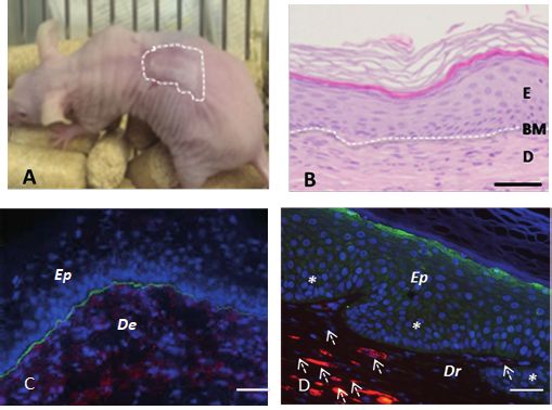
To perform these varieties of functions, skin is organized in three different layers (see Figure 1A):
• The epidermis is a rather complex stratified epithelium. It is composed primarily of keratinocytes (at least
80%) but also contains other cells: melanocytes, dendritic cells derived from the neural crest, responsible for
the production of the pigment melanin [7]; langerhans cells, dendritic cells derived from the bone marrow,
involved in a variety of immune responses; Merkel or tactile epithelial cells, oval-shaped mechanoreceptors in
contact with the nerve cells involved in tactile sensitivity. The thickness of the epidermis varies in the range of
0.05−1.5 mm from the eyelid (the thinnest layer) to the palms and soles of the feet (the thickest layers). The
epidermis is commonly divided into four layers (Figure 1C), the basal layer (stratum basale or germinativum),
the squamous layer (stratum spinosum), the granular layer (stratum granulosum) and the cornified layer (stratum
corneum) [8,9]. In the body regions exposed to friction (palms and soles) the stratum corneum is thicker and
there is an additional layer, the stratum lucidum. Skin is a dynamic organ in constant regeneration, as cells of the
outer layers are continuously lost and replaced by inner cells moving up to the surface; human skin is renewed
approximately every month. This is due to a process in which keratinocytes generated at the basal layer – the only
epidermal proliferative layer – migrate to the surface while they follow a program, called terminal differentiation
resulting in keratinization and cell death which in turn results in the formation of the horny stratum corneum, the
actual protective barrier of the skin [8]. In addition to aforementioned ‘interfollicular epidermis’, the epidermis
gives rise to three appendages: nails, hair follicles and associated sebaceous glands and sweat glands. The hairs
and the nails protect the organ. Sweat glands are involved in the control of body temperature. Hair follicles are
also particularly important for epidermal regeneration (see the section on ‘Wound healing’).
10.2217/3dp-2018-0008
C 2018 Future Medicine Ltd J. 3D Print. Med. (Epub ahead of print) ISSN 2059-4755
Review     Velasco, Quı́lez, Garcia, del Cañizo & Jorcano

                                                A                                                                 B

                                                                                     Stratum corneum
                                                                  Epidermis
                                                                                             Epidermis
                                                                 Sebaceous gland
                          Sensory
                                                                Hair follicle
                          receptors
                                                                 Dermis                         Dermis                    Papillary
                          Capillary                              Sweat gland                                              dermis
                          network                                Hipodermis
                                                                                                                          Reticular
                                                                                                                          dermis

                                                                                C

                                                                                               Stratum corneum
                                                                                               Stratum lucidum
                                                                                               Stratum granulosum

                                                                                               Stratum spinosum

                                                                                               Stratum basale

                    Figure 1. Skin structure. (A) Scheme representing the main structural components of the skin. (B) Histological
                    tinction showing in detail the structure of interfollicular skin. (C) Scheme showing the cell layers present in
                    differentiated epidermis.
                    Images adapted with permission from Servier Medical Art freeware image bank (Figure 1A & C) and
                    www.melanoma.blogsome.com and www.mrcophth.com (Figure 1B).

                    • The dermis is a layer that, contrary to the epidermis, is relatively acellular and is mostly composed of collagen
                      (types I and III) and elastic fibers (elastin). The dermis is thicker on the dorsal side of the body where it is 30–
                      40-times as thick as the overlying epidermis and in the external areas of the extremities [8]. The most abundant
                      cells are the fibroblasts, although it also contains macrophages and adipocytes. Additionally, it accommodates
                      blood vessels, nerves, glands and hair follicles.
                      The dermis comprises the bulk of the skin and provides its pliability, elasticity and tensile strength. The epidermis
                      and dermis are connected by the basal lamina, a complex structure composed of proteins such as collagen IV and
                      VII and laminins, involved in the attachment of the two skin layers. Mutations in these proteins are the basis
                      for different subtypes of epidermolysis bullosa, a group of inherited severe blistering disease [10,11]. The major
                      constituent of the dermis is type I collagen, although collagen III is found also in the matrix. Both collagens
                      are not organized in a specific manner, but the ratio between the two varies throughout the human life span
                      because of a decrease in the amount of type III with age. Loosely positioned collagen and elastic fibers are
                      found in the papillary dermis, whereas heavy bundles are present in the reticular dermis (Figure 1B). Another
                      relevant molecule in the dermal extracellular matrix (ECM) is hyaluronic acid, which has become very popular in
                      cosmetics in recent years due to its role in skin hydration [8,12,13]. In addition, this molecule plays relevant roles
                      in different processes: morphogenesis, wound healing, inflammation and angiogenesis. All these components
                      and structures provide skin with its characteristic mechanical properties and resilience.
                    • Finally, the hypodermis consists of loose connective tissue that joins skin to subjacent organs and is mainly
                      composed of adipocytes. It varies in thickness depending on the skin site. The subcutaneous tissue constitutes a
                      storehouse of energy and produces leptin, a hormone that regulates body weight by way of the hypothalamus [8].

                    Wound healing
                    In addition to skin’s complexity as an organ, we should likewise refer to its anatomical functions. Skin covers the
                    whole body, and contrary to other organs, it is in contact with the external environment. Due to this function, it
                    is subjected to injuries of a different nature and severity, which is why it is programed to repair itself. In addition

10.2217/3dp-2018-0008                                          J. 3D Print. Med. (Epub ahead of print)                future science group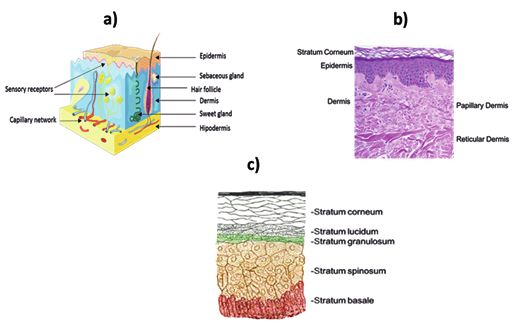
3D human skin bioprinting: a view from the bio side         Review

                                                                                                     3)
                                                  In vitro testing
    1)

                                                Human disease research:                                            4)
                                             Skin humanized mouse models

    2)
                                                 Clinical treatment
                                                                                                5)

Figure 2. Main applications of current in vitro, manually produced skin substitutes. (1) Human skin cultured in vitro
for experimental use. (2) Human skin mounted on Urgotul    R
                                                              for clinical applications. (3) Human skin differentiated in
a transwell for testing purposes. (4) Human skin produced with cells from normal or skin-diseased donors grafted to
the back of a nu mouse. (5) Human skin, as in (2), grafted to a wound site.

to this homeostatic mechanism, it is exposed to additional damages. One of the most important mechanisms from
a clinical point of view is wound healing; regeneration starts immediately after a wound occurs. The process is
regulated by different cytokines and growth factors that activate many intracellular and extracellular processes of
different types that lead to the different steps [14,15]. Because of an injury, platelets aggregate and form a plug of
fibrin, where immune cells are then attracted. After that, a new tissue matrix is produced by fibroblasts by the
generation of collagen III. Finally, re-epithelialization is carried out by keratinocytes, which eventually includes
revascularization of the wound [16]. The final phase of wound healing is matrix formation and remodeling. In this
phase, that can take several months, there is a slow accumulation of type I collagen bundles and a progressive
restoration of the mechanical properties of injured skin to resemble normal tissue architecture. Depending on the
depth of the injury, wounds can be classified into four groups: epidermal; superficial partial thickness; deep partial
thickness; and full thickness [17,18]. If the depth of the injury compromises the hair bulge neither the hair follicle
nor its corresponding glands will regenerate. Additionally, re-epithelialization of the epidermis will occur from the
edges of the wound and not from the hair follicle, which for an extensive wound will slow down the process or even
end up as a chronic wound. This is because this part of a hair follicle contains a reservoir of cells indispensable for
these processes to occur [19,20]. The wound healing process reveals that both hair follicles and fibrin clots are key in
the skin regeneration process.

In vitro engineered human skin substitutes
There is a huge demand in the development and production of in vitro-engineered substitutes that mimic human
skin, either to be used as grafts to restore the function of the skin after damage and to facilitate wound healing
or for the establishment of human-based in vitro skin models for toxicity, cosmetic and pharmaceutical testing
(Figure 2) [21–27].
   To have an idea of the dimension of the problem, we might consider the following figures: First, the WHO
estimates that nearly 11 million burn injuries per year worldwide require medical attention, with approximately
265,000 leading to death [28]. Second, the 2016 global wound management market hits $15 billion and forecasted to
be worth over $22 billion in 2024 [29–32]. Third, the tissue-engineered skin substitutes market, which is of particular
interest to this review, was valued at 958.8 million US dollars in 2014 and is projected to reach 3873.5 million US
dollars by 2023. To restore the skin after damage and to facilitate wound healing while avoiding immune rejection,
autologous grafts (autografts) obtained from the own patients donor areas are commonly used. Unfortunately, the
availability of autografts for wound coverage is insufficient when dealing with large and/or severe wounds or burns,
hence, the convenience of having in vitro methods of generating autologous skin to apply to these patients [33–35].

    future science group                                                                                                    10.2217/3dp-2018-0008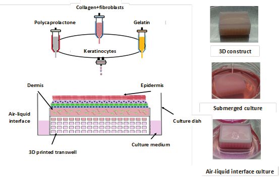
Review     Velasco, Quı́lez, Garcia, del Cañizo & Jorcano

                       In the case of in vitro toxicology testing, the market was valued at around 14.2 billion US dollars in 2016 being
                    Europe the largest market with some 6.4 billion dollars followed by North America with 4.8 and Asia with 1 [36].
                    In the case of skin testing, many drugs can not be tested directly on humans [37]. Therefore, researchers in the
                    pharmaceutical and the cosmetic industries were and are using animals to test their products, with mice being the
                    most common ones utilized. However, testing products on animals is not always predictive of responses in humans
                    and may later lead for instance to costly failures in clinical trials and other economic problems [38]. In addition,
                    testing cosmetic products and their ingredients on animals was banned in the UK in 1998 and across the EU in
                    2013 (EU Regulation 1223/2009 – Cosmetics Regulation). In this complex situation, it is however predicted that
                    the market will witness a rapid growth thanks to the increasing acceptance of in vitro methods over in vivo ones,
                    the arrival of new and promising technologies, and the advancement in new approaches as the ones discussed in
                    this review.
                       Thus, as a result of the foregoing, skin substitutes are being explored for human skin replacement therapy and for
                    human skin testing, such as acellular skin substitutes or cellular skin substitutes (containing autologous or allogeneic
                    keratinocytes, fibroblasts and adipocytes) as cellular monolayers (epidermal substitutes), cellular bilayers (dermo-
                    epidermal components) and cellular trilayers (dermo-epidermis-hypodermis components) skin substitutes [39–47].
                    Cellular bilayered skin substitutes must fulfill certain conditions: contain well-formed epidermis and dermis; the
                    dermal component should have mechanical properties similar to the dermis; include well-differentiated epidermis
                    with stratum corneum; dermis and epidermis must be properly linked by the basal layer.
                       The production of bilayered and trilayered artificial human skin usually involves the use of a scaffold composed
                    of natural and/of synthetic polymers such as alginate, collagen, chitosan, fibrin, hyaluronic acid and poly(ethylene
                    glycol), polycaprolactone (PCL), poly (vinyl alcohol) and polylactic acid and poly-L-lactic acid, respectively [48–52].
                    Among them, collagen (the main component in the ECM) and fibrin (the blood component) have been used
                    extensively to provide structural and mechanical support [53–56]. This scaffold is usually filled with fibroblasts
                    (dermal component), and keratinocytes are seeded on top of the dermal component. In the case of trilayered skin
                    substitutes, below the dermal component another scaffold is filled with adipocytes or mesenchymal cells.
                       As an example of production of autologous human artificial skin, our lab engineered a human plasma-derived
                    bilayered skin using primary human fibroblasts (hFBs) and keratinocytes (hKCs) from skin biopsies to treat burns
                    and traumatic and surgical wounds in a large number of patients in Spain and for the generation of skin-humanized
                    mouse models (Figure 2) [57–59]. In this case, skin substitutes formed by two layers, representing the dermis (the
                    lower layer) and the epidermis (the upper layer), were generated following the method developed in [57]. The lower
                    layer was a human plasma-derived fibrin matrix populated with hFBs and the upper layer was formed by hKCs,
                    seeded confluent on the top of the fibrin scaffold (Figure 3). At this point, for clinical use, the equivalents can be
                    transplanted to the patients. After grafting, the layer of epidermal cells, now in contact with the air, proliferate
                    and differentiate to form all the layers of the epidermis, including the stratum corneum. Simultaneously, growth
                    factors and chemo attractants produced by the HKCs and hFBs stimulate the growth of patient’s blood vessels and
                    in a very short time invade the transplanted skin. A very similar process occurs, when, for experimental purposes,
                    human skin equivalents are grafted onto the back of immunodeficient mice (skin-humanized mice). For in vitro
                    studies, the terminal differentiation of the epidermis is achieved by placing the culture at the air–liquid surface.
                       We have devoted an effort to explain the commercial relevance and the estructure, composition and production
                    of human bi/trilayered human skin equivalents, because as we will see in the forthcoming sections, 3D skin
                    bioprinting follows the same principles. In spite of the notorious advanced in this field, several aspects of human
                    skin equivalents need to be addressed. On the one side, they have to include other types of cells and molecules
                    found in normal skin to improve their clinical and dermatological performance. And, on the other side, the complex
                    manual and strictly regulated production lead to elevated prices. These limitations combined with a foreseen higher
                    demand for artificial human skin, have all led to an increasing need to develop new methods that offer automation,
                    standardization and reduction in time to manufacture the bilayered skin substitutes and production costs [60,61].

                    3D bioprinting
                    Three-dimensional bioprinting has emerged as a flexible tool in regenerative medicine and provides a platform
                    for addressing some of the needs described above. Three-dimensional bioprinting can be defined as the spatial
                    patterning of cells, cell aggregates, DNA, drugs, growth factors, bioactive substances, extracellular components and
                    biomaterials, referred to as bioinks, by assembling them using a computer-aided layer-by-layer deposition approach
                    for the fabrication of artificial tissues or organs or constructs for drug screening, toxicology studies and clinical

10.2217/3dp-2018-0008                                          J. 3D Print. Med. (Epub ahead of print)               future science group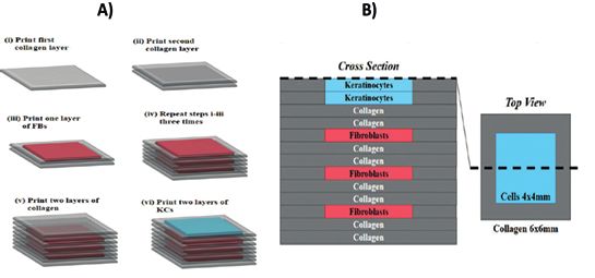
3D human skin bioprinting: a view from the bio side         Review

                             Plasma from patient           Patient                          Skin biopsy

                                                                                                      Epidermis

                                                                                                     Dermis

                                                                                                     Subcutaneous
                                                                                                     fat

                   Ca2+ induced
                    coagulation

                                                         Skin donor
    Plasma-derived
         fibrin
       hydrogels

                           Bioengineered dermis
                                                     Human fibroblasts              Fibroblasts    Keratinocytes
                                                                                     (dermis)       (epidermis)

                                                       Human keratinocytes

                   Dermo-epidermal equivalent

Figure 3. Scheme of the production of large surfaces of autologous skin equivalents from a small biopsy. From a
small (2–4 cm2 ) biopsy, human fibroblasts and primary human keratinocytes are isolated and separately expanded in
vitro using cell culture techniques well known in the field [59]. Once obtained the required high number of cells, they
are assembled into a multilayered skin equivalent. In a first step, the dermal compartment is generated by adding
calcium to human plasma and introducing the primary human fibroblasts cells at the appropriate density while fibrin
polymerization is taking place. Once the dermal hydrogel has polymerized, human keratinocytes are seeded on top of
it and allowed to grow until confluency. At this moment, they can be removed from the culture plates, properly
packed and sent to the hospital for transplantation.

use [62–73]. Although there are several definitions concerning 3D bioprinting, in this review we will consider 3D
bioprinting a process that applies 3D printing principles and technologies to the generation of human tissues using
bioinks containing fundamentally cells and the materials needed to generate scaffolds.
   Gartner, an American research and advisory company, has become a standard reference in 3D printing bench-
marking. Its 2017 3D Printing Hype Cycle provided a comprehensive analysis of the most relevant 3D printing
applications and their respective level of maturity [74]. Figure 4 shows the situation in this Hype Cycle of bioprinted
biomedical products. Three-dimensional bioprinted organs for transplants and drugs are placed in the ‘innovation
trigger phase’ (the value is triggered by the expectations raised by the disruptive innovation) with estimations of
>10 years to reach the market. Three-dimensional bioprinting for life science R&D is placed in the transition
between the ‘peak of inflated expectations’ and the ‘through disillusionment’ phases (indicating that expectations
are entering into a more realistic phase) with estimations of 5–10 years to reach the market. Three-dimensional
bioprinted human tissue has advanced to the ‘through disillusionment’ phase (indicating that difficulties are being
identified) with estimations of 5–10 years to reach the market. However, this prediction could be too pessimistic
given that in 2016 the first paper claiming to have produced a functional human tissue through 3D bioprinting
was published.
   Regarding its possible economic impact, we are not aware of any estimation of the market value of bioprinted
skin. However, according to Grand View Research, by 2022, the global 3D bioprinting market is expected to reach
1.82 billion US dollars and will include products and materials for dental, medical, analytical and food applications.
In North America, this value is around 550 million US dollars, of which around 350 million US dollars correspond
to medical applications. On the other hand, as previously presented in section 2, the wound management market

    future science group                                                                                                  10.2217/3dp-2018-0008
Review     Velasco, Quı́lez, Garcia, del Cañizo & Jorcano

                                                                                  3D printed surgical implants

                                                                                    3D bioprinting to life
                                                                                       science R&D
                                                                                     3D printing of medical
                                                                                            devices

                                                                                      3D bioprinted human
                                                                                             tissue
                                Expectations

                                                 3D bioprinted
                                               organ transplants
                                                                                                                                                   3D printing of
                                                                                                                                                  hearing devices
                                                                   3D printed
                                                                     drugs

                                                                                                                 3D printing of
                                                                                                                 dental devices

                                                                                      3DP presurgery
                                                                                     anatomical models

                                                Innovation         Peak of inflated Trough of                         Slope of                      Plateu of
                                                  trigger           expectations disillusionment                    enlightenment                  productivity

                                                                                                         Time
                           10 years

                    Figure 4. The 2017 3D Printing Hype Cycle provides a comprehensive analysis of the most relevant 3D printing
                    applications and their respective level of maturity. The figure shows selectively the situation in this Hype Cycle of
                    bioprinted biomedical products.
                    Modified or adapted with permission from [74].

                    value is expected to be worth over 22 billion US dollars in 2024, the global tissue-engineered skin substitutes market
                    is projected to reach 3873.5 million US dollars by 2023 and the global in vitro toxicology market is expected to be
                    worth 17,227 million US dollars this year. It is clear that 3D bioprinted human skin will play an important role in
                    all these markets either for clinical or testing applications.

                    Components of a 3D bioprinter
                    Three-dimensional bioprinters are printers specifically designed or adapted to work with bioinks containing living
                    cells, biomaterials and biocomponents that are very sensitive to processes such as high temperature, fluid stress, UV
                    light, compression, etc., present in conventional 3D printers. Bioprinters can either be custom made according to
                    user needs or can be acquired to one of the several companies that commercialize them. In this last case, it might
                    have to be adapted to one’s specific requisites. Although there are different types of 3D bioprinters (see section 3.2),
                    this section will describe an extrusion-based bioprinter as this type is widely used in research and is a good model
                    to use for explaining the main issues concerning any bioprinter’s architecture and functioning.
                       A bioprinter is comprised of four main components (Figure 5):

                    • First, a software system which is responsible for the communication between the bioprinter and the computer,
                      and therefore the user. It controls the ejection of the bioinks from the cartridges and the positioning of the
                      dispensers through computer-aided design/computer-aided manufacturing.
                    • Second, the extrusion module containing the cartridges, the actuators and the dispensers or nozzles. The cartridges
                      act as reservoirs containing as many bioinks as necessary and they can be one or several, depending on the number
                      and characteristics of the bioink and the bioprinting process. The actuators, under the control of the computer
                      and with great precision, pump the amount of each bioink necessary at any moment from the cartridges to the
                      nozzles. There are different methods to carry out this ejection, ranging from using mechanical energy to optical
                      energy (see section 3.2). The dispensers are involved in the extrusion and the deposition of the bioink, and are

10.2217/3dp-2018-0008                                                           J. 3D Print. Med. (Epub ahead of print)                       future science group
3D human skin bioprinting: a view from the bio side     Review

                                             Bioink 1

                                             Bioink 2

                                             Bioink 3

                                      y-movement                              Cartridge

                                                                                     Extrusion
  z-movement                     Dispenser                                            control
                                                          Actuators
                           Bed                                                                       Image
                                                                                                   acquistion
                                                           Motion
                                         x-movement
                                                           control

Figure 5.   Scheme of an extrusion-based 3D bioprinter.

  positioned in the x, y and z coordinates with the help of the positioning system. The shape, and in particular, the
  diameter of the nozzle is very important because it has to fulfill somewhat contradictory functions: not damage
  bioink, in particular cell viability, and define the resolution of the deposition. Thicker nozzles will favor bioink
  viability but decrease precision, and vice versa. On the other hand, tapered needle-type nozzles are known to
  damage fewer cells and biomaterials than cylindrical needle-type nozzles [75]. The cartridges and the dispenser
  can be integrated or independent. In this case, they are connected by sterile, flexible tubes with a diameter that
  can accommodate the liquid flow and not damage the cells and other biological components. Another function
  of the dispenser is acting as a reservoir in which two or more bioinks are mixed before being dispensed. The
  number of cartridges and dispensers can vary according to the printing requirements. For instance, the number
  of cell types and biomaterials, temperature control, etc. Additionally, it is possible to combine different extrusion
  systems to improve the versatility of the bioprinter.
• Third, the printing module consists of the positioning system and the printing bed (the place generally is a tissue
  culture plate where the deposition takes place). The bed is on a platform that can be heated, frequently at 37◦ C.
  The printing module must position the nozzle in the space under computer control. For this, the movement of
  the two module components must be coordinated. For example, if the bed moves in the x and y axes, then the
  dispenser should move in the z axis; if the dispenser moves in the x, y and z axes, the bed will be fixed.
• Fourth, the bioinks for bioprinting, defined in section 3, fundamentally contain cells and materials needed to
  generate the scaffolds [76–79]. Cells are particularly important from a biological point of view and are critical as
  they are sensitive to external conditions. Therefore, cell selection and survival are crucial points to be addressed
  when establishing the conditions of the bioprinting process (cell handling and density, residence time, buffers,
  temperature, shear stress, etc.) [80–82]. As an example, in the case of skin bioprinting, it is very important to
  control the time that keratinocytes are in the cartridge because they are known to differentiate when kept in
  suspension [83].

   Another very important issue to be considered when designing the bioprinting process of a tissue is the selection
of the scaffold. This involves taking into account parameters such as biocompatibility, biodegradability, porosity
and mechanical properties which are highly dependent on the type of tissue to be printed. For instance, in the case
of hard tissues such as bone, they require more rigid scaffolds made of ceramics, PCL, polylactic acid or mixtures
among them have been widely used. In the case of soft tissues, hydrogels are the most common scaffolds, given
that they can be defined as crosslinked 3D network structures that can absorb and retain large amounts of water
and provide a hydrated and highly permeable microenvironment, mimicking the natural ECM in this type of
tissues. They can be made of natural components (fibrin, collagen, gelatin, alginate, agarose, hyaluronic acid, etc.)
or synthetic (very frequently poly(ethylene glycol) polymers or both [84–86]. There are two parameters that need to
be particularly controlled in hydrogel design: rheological properties (especially viscosity) and gelation time. The

    future science group                                                                                              10.2217/3dp-2018-0008
Review Velasco, Quı́lez, Garcia, del Cañizo & Jorcano
Table 1. Bioinks used in 3D skin bioprinting.
†
Base material Composition Cell types Bioprinting technology Ref.
Collagen Collagen HaCaT1 , hFB2 , hKC3 Droplet based [115,116]
Collagen Collagen NIH-3T34 , HaCaT1 Laser based [123]
4 1
Human plasma Human plasma and alginate NIH-3T3 , HaCaT Laser based [126]
Fibrinogen Fibrinogen, alginate and hKC3 , hFB2 Extrusion based [93]
gelatin
Human plasma Human plasma hKC3 , hFB2 Extrusion based [109]
Collagen Polycaprolactone, gelatin and hKC3 , hFB2 Extrusion droplet based [108]
collagen
Collagen Collagen and fibrinogen hKC3 , hFB2 , HaCaT1 , AFSC5 , Droplet based (in situ) [132–135]
MSC6
Collagen Collagen hFB2 , hKC3 , hMC7 Droplet based [128]
Collagen Collagen and fibrinogen hKC3 , hFB2 , HMVEC8 Droplet based [129]
Collagen Collagen and hKC3 , hMC7 , hFB2 Droplet based [130]
polyvinylpyrrolidone
†
Ex vivo bioprinting, if not otherwise stated.
AFSC5 : Amniotic fluid-derived stem cell; HaCaT1 : Immortalized human keratinocyte; hFB2 : Primary human fibroblast; hKC3 : Primary human keratinocyte; hMC7 : Primary human melanocyte;
HMVEC8 : Human dermal microvascular endothelial cell; MSC6 : Bone marrow-derived mesenchymal stem cell; NIH-3T34 : Immortal murine fibroblast.
viscosity has to be carefully balanced. On the one hand, highly viscous bioinks will need high pressure to flow
through the nozzle which might bring about cell damage and/or require wider nozzles leading to a lower precision.
This problem is particularly relevant in the case of droplet-based bioprinting and not so much in extrusion-based
bioprinting (EBB; bioprinting technologies will be discussed in section 3.2). On the other hand, low viscosity
bioinks will tend to spread out on the receiving bed. This is particularly relevant in the case of printing structures
with a defined 3D pattern but not so much in the case where the printing is performed in a confined container,
a very common situation in soft tissues bioprinting. An efficient manner of providing viscosity at the moment of
the deposition is the use of sacrificial materials. The bioink should undergo a gelation process. If it is too rapid, it
might provoke clogging inside the nozzle, but if it is too slow it will prevent maintaining the shape and the spatial
pattern initially printed in those cases where high resolution is required. An efficient manner recently described
to overcome this problem is the inclusion of sacrificial material in the bioinks. These materials provide initial
viscosity and are washed out once the hydrogel polymerization is completed. Examples of materials used to this
end are gelatin, pluronics, etc. [87,88]. These sacrificial materials clearly provide more flexibility when designing
the bioprinting process because they allow the printing complex structures using scaffolds that themselves would
not allow it. Because of the complex physical and functional properties necessary for scaffolds, their design is a
very active area of research, and an increasing number of materials and hybrid natural/synthetic scaffolds are being
investigated (see [79,89,90]) for recent reviews. Among them, only a few have been used as bioinks for the generation
of scaffolds in skin bioprinting (Table 1).
The most commonly used are biological polymers, namely collagen and fibrin, the latter obtained either from
commercial fibrinogen or from the polymerization of human blood plasma. Collagen is the main molecular
component of normal skin dermis while fibrin constitutes the blood clot from which the process of wound healing
starts. With the existing results, it cannot be concluded which one leads to better outcomes although, the fibrin
hydrogels do not show the contraction that collagen matrices present [91,92]. Given the bilayered structure of skin,
the use of sacrificial materials has not been so far necessary in skin bioprinting, with the exception of the work
published by Pourchet et al. [93], in which the authors carried out the bioprinting without any physical confinement,
so they needed very viscous bioinks to maintain the spatial disposition with which they were deposited.
The printing modality is made based on biological needs: the necessary cells, biomaterials used and their
corresponding mechanical and chemical limitations when handling them, and the structure and characteristic of
the tissue to be constructed. In 3D bioprinting, three main methods can be distinguished: extrusion-, droplet- and
laser-based bioprinting (Figure 6); they can be home-made printers or commercially available ones [94].
10.2217/3dp-2018-0008 J. 3D Print. Med. (Epub ahead of print) future science group3D human skin bioprinting: a view from the bio side Review
Inkjet bioprinting Laser-assisted bioprinting Extrusion bioprinting
Pressurized
Piezoelectric Thermal Piston air Screw
Laser Scanning mirror
Reservoir
Bioink Print ribbon Bioink
Focusing lens Bioink inlet
Piezoelectric
Heater
actuator Printing
Absorbing nozzle
layer Bioink
Construction
platform
Figure 6. Bioprinting methods. Three main methods are currently used in 3D bioprinting: extrusion-based (extrusion bioprinting),
droplet-based (inkjet bioprinting) and laser-based (laser-assisted bioprinting) bioprinting.
Reproduced with permission from [95]. For more details, see the text.
3D bioprinting technologies
Currently three main methods are used in 3D bioprinting: extrusion-, droplet- and laser-based bioprinting. The
selection of the method will depend on the properties of the cells and biomaterials used, the precision and speed
required, and the size and characteristics of the tissue to be printed.
Extrusion-based bioprinting
The EBB is the most common modality used in bioprinting research due to its practicality and ability to fabricate
larger scale 3D constructs [96,97]. In EBB, the bioink solution is extruded through the dispenser by a pneumatic-,
mechanical- (piston or screw-driven) or solenoid-based system in form of a continuous filament.
Some of the advantages are that it has a high printing speed which can facilitate scalability and, additionally,
enable the use of a broad range of bioinks in particular cell-laden hydrogels widely used in soft tissue printing [84,98–
106]. Moreover, the technology is easy to implement, user friendly and affordable. The main disadvantages are less
accuracy and lower resolution (≥100 μm) than other techniques [107].
To our knowledge, this modality is the most used at present in skin 3D bioprinting either alone or in combination
with droplet-based bioprinting [93,108,109].
Droplet-based bioprinting
Droplet-based bioprinting has its roots in inkjet technology (Elmqvist of Siemens, 1950 which evolved into the
first inkjet 3D printer (Objet Geometries, 2000) [110]. It relies on the same principle of EBB: there is a continuous
extrusion of bioink through a dispenser, but in this case, the continuous flow is interrupted by different actuators
to form drops. These drops are then deposited on the printing bed [111].
Droplet bioprinting techniques can be classified depending on the actuator (Figure 6.). The most used modalities
are inkjet based and can be classified into piezoelectric, thermal or electrostatic according to the type of actuator
used (piezoelectric, thermal or electrostatic) [112–114]. The most important advantages of this type of impression
are: first, better resolution; drop diameters as small as 40 um have been reported in bioprinting processes although
cell viability can be affected by drop size. Second, complex geometries can easily be patterned. Third, affordable
price. Fourth, possibility of constructing hybrid printers with droplet-based+ EBB, which increases the flexibility
of the process. The main disadvantages are: first, narrow range of viscosities (3.5−12 mega pascal-second (mPa.s)
vs 30–6 × 10⁷ mPa.s in the case of EBB) which limits bioink composition. Seond, a longer time of impression as
compared with EBB, which limits the size of the printed tissue. Third, the method used by the actuator to form
the drops, in particular, the thermal modality, can affect cell viability.
This technology has been used in different studies in the generation of 3D-bioprinted skin [115,116], which will
be analyzed in more detail later.
future science group 10.2217/3dp-2018-0008Review Velasco, Quı́lez, Garcia, del Cañizo & Jorcano
Laser-based bioprinting
Laser-assisted cell printing is a nozzle-free technique. This technology is based on conventional laser-induced
forward transfer. They are two stacked slides mostly made of quartz: first, the donor slide. The lower side of this
slide is covered with a laser-absorbing layer plus a layer of bioink, containing the biomaterials and cells to be
transferred. Second, the collector slide, equivalent to the printing bed, placed at variable distances ranging from
millimeters to micrometers. A high-powered infrared laser pulse is focused onto a small area of the laser absorbing
layer of the donor slide, generating a high-pressure bubble that catapults the underlying bioink area onto the
collector slide [117,118]. To improve printing speed, several lasers can be used to scan the donor slide. There are
several variations of this method, mainly based on the composition and characteristics of the donor slide [119,120].
The main advantages of this method are its high precision and that it is smaller than the size of one cell. At present,
its main disadvantages are: its expense; it is only suitable for printing small tissue surfaces; the complex fabrication of
the donor slide; lower viscosities (1–300 mega pascal-second [mPa.s]) than EBB [121,122]. These drawbacks probably
make this technology not the most appropriate for printing interfollicular skin but, as a proof of concept, it was
used by the group [123].
3D skin bioprinting
Due to the relatively simple structure of interfollicular skin as compared with other 3D solid organs such as heart,
kidney, etc. and its demand in clinical and commercial testing uses, production of skin tissue containing dermal
and epidermal components by bioprinting is currently an important area for laboratory research and company
development. Although there are three recently published reviews on this topic [18,95,124], we focus instead on
aspects that, in our opinion, affect the structure, function and use of bioprinted human skin.
In our opinion, 3D bioprinting would be the method of choice for large-scale skin production for toxicity,
cosmetic and pharmaceutical testing. This is due to the fact that it would allow a robust, standardized and
automatized method as well as significantly reduced production costs. For this testing purpose, one can previously
generate large cell banks established from biopsies obtained from donors. One can even consider the possibility of
using immortalized human cells given rise to culture that is close enough to human skin to overcome the regulatory
requirements (Lynn-Hoffman [125]). The use of bioprinted skin for testing purposes could reach the market in the
near future.
On the other hand, the 3D bioprinting technology would not be able to reduce the time (3–4 weeks) currently
needed to obtain enough autologous cells to print large skin surfaces for the treatment of large wounds (e.g., people
with extensive burns) from small biopsies. This prolonged amplification step under very costly good manufacturing
practices conditions is one of the main factors limiting the usefulness of skin equivalents in wound healing treatment.
Therefore, the advantages of 3D bioprinting are limited in this field.
There are two main strategies concerning how to print skin for wound treatment: ex vivo and in situ bioprinting.
Ex vivo skin bioprinting
In this approach, a skin construct (usually including dermis and epidermis) is printed and, if required, matured
(e.g., differentiated) in vitro, and then analyzed in vitro or upon grafting to a patient or an experimental animal. We
will discuss the main articles published, classified according to the bioprinting method used. From the chronological
point of view, this was the first approach.
Droplet based
From a chronological point of view, this was the first technology used to print skin. In their pioneering work, Lee et
al. (2009) demonstrated the feasibility of the multilayered deposition of fibroblasts and keratinocytes in a collagen
scaffold [115]. Posteriorly, Lee et al. (2014) after optimization of the printing parameters to likewise optimize cell
density and viability, generated dermo-epidermal constructs that were exposed to the air–liquid interface to promote
the in vitro maturation and terminal differentiation and stratification of the epidermal cells [116]. These authors
deposited interspersed layers containing either collagen or collagen-containing hFBs in the dermal compartment
or hKCs in the epidermal compartment (Figure 7). In spite of its pioneering value, this alternating deposition of
cellular and acellular is unnecessary and does not resemble the skin structure. Apart from that, the main drawback of
this method is that the histology of the skin tissue does not show proper stratification and terminal differentiation as
compared with human skin. As the authors recognize, this can be, at least in part, due to the use of an immortalized
keratinocyte cell line (HaCaT cells) instead of primary hKCs. Finally, based on the data provided by the authors,
10.2217/3dp-2018-0008 J. 3D Print. Med. (Epub ahead of print) future science group3D human skin bioprinting: a view from the bio side Review
A B
(i) Print first (ii) Print second
collagen layer collagen layer
Cross section
Top view
(iii) Print one layer (iv) Repeat steps i–iii
of FBs three times
(v) Print two layers (iii) Print two layers Cells 4x4 mm
of collagen of KCs
Collagen 6x6 mm
Figure 7. Construction of 3D skin tissue using the microvalve bioprinting system with pneumatic pressure
droplet-based technology. (A) Layer-by-layer printing of the collagen matrix, human keratinocytes and human
fibroblasts to generate the dermal and epidermal compartments integrated in a single structure. (B) Schematic of the
3D-printed skin tissue showing the cross section (left) and the top view (right).
Modified with permission from [116].
the estimated printing speed allows the deposition of 1 cm2 skin per hour, which is a very slow process considering
the sizes needed for clinical or commercial applications.
Laser based
In this approach, a laser-assisted bioprinting technology was used to deposit alternating layers, containing immortal
murine fibroblasts and immortal hKCs (NIH-3T3 and HaCaT cell lines, respectively) embedded in a collagen
matrix (Figure 8) [126]. In this technique, 20 collagen sublayers containing fibroblasts were printed onto a sheet of
Matriderm R
[127] and subsequently 20 collagen sublayers containing keratinocytes were printed on top of it. The
printed equivalents are analyzed either upon in vitro differentiation at the air–liquid interface or upon grafting to
the back of nude mice using the dorsal skin full chamber method [123]. A clear problem of this work is again the use
of immortalized keratinocyte and fibroblast cell lines, in particular the NIH-3T3 murine fibroblasts that are very
different from hFBs. Moreover, the histological and immunohistochemical data presented by the authors indicated
relevant differences when comparing their transplanted skin with normal human skin. Most likely, these differences
are not attributable to the printing method but instead to the dorsal skin full chamber method that imposes a too
short time to obtain a fully differentiated epidermis. Finally, although it is difficult to make an accurate estimation
from the data provided by the authors, a caveat concerning this technology is the time it would take to print and
the effort that would be required to produce the donor slide to generate skin surface of clinical or commercial
interest (50–100 cm2 ).
In conclusion, the cited articles present several drawbacks: first, in general, they do not use human primary
fibroblasts and keratinocytes, simultaneously. The cells used might be less sensitive to the stresses of the bioprinting
process, and their proliferation and differentiation characteristics are far different from those of the cells contained
in human native skin. Second, the printed layered structures are not reminiscent of normal skin and the skin
constructs produced did not possess the structural quality of normal human skin. Probably due to these reasons,
since 2015, primary human cells have been used. Moreover, additional cell types such as human endothelial cells
and melanocytes have been introduced into the printed skin to make it more similar to normal human skin [128–130].
Extrusion based
From a practical point of view, this method is simpler and quicker than the two methods described above since,
instead of printing a high number of cellular and acellular layers to form the dermis and epidermis, it deposits
future science group 10.2217/3dp-2018-0008Review Velasco, Quı́lez, Garcia, del Cañizo & Jorcano
A B C
laser pulse
laser-absorbing layer
gel with cells
D E
Figure 8. Construction of three-dimensional (3D) skin tissue using laser assisted bioprinting. (A): Sketch of the
laser-based printing setup. The cell-containing hydrogel is propelled forward as a jet from the donor slide by the
pressure of a vapor-induced bubble generated by the energy transferred by the laser beam to the absorbing layer.
Layer-by-layer, a 3D cell pattern is generated. (B) A printed grid structure (top view) of fibroblasts (green) and
keratinocytes (red) demonstrates the micropatterning capabilities of the laser-based printing technique. (C) Seven
alternating color layers of red and green keratinocytes. Each color layer consists of four printed sublayers. (D & E)
Bilayered skin substitute produced by B laser-assisted bioprinting through the printing of fibroblasts and
keratinocytes, characterized 11 days post implantation in nude mice. (D) Histological section of the bilayered skin
substitute stained with Masson’s trichrome showing a dense epidermis (empty asterisks) and a corneal layer. The
fibroblasts, which stayed on top of the Matriderm R
, displayed an outstretched morphology being accompanied by
collagen deposition (filled asterisks). (E) Fluorescence microscopy image of the bilayered skin substitute with green
fluorescence emitted by HaCaT-eGFP cells and red fluorescence emitted by fibroblasts (NIH3T3-mCherry).
Modified with permission from [123,126].
simultaneously all the elements (hFBs, human plasma and CaCl2 ) required to form the dermis and, on top of this,
the hKCs required to form a confluent layer of epidermal cells.
Pourchet et al. reported a bioink containing bovine gelatine (10% w/v), alginate (0.5% w/v) fibrinogen (2%
w/v), and primary hFBs to produce the dermal compartment. The gelatin is used as a sacrificial, rheological
material [93]. It provides the printed bioink with temporal mechanical stability and viscosity while alginate and
fibrinogen become polymerized in the presence of calcium chloride and thrombin. The bioprinting process takes
place at low temperature (28◦ C) and the deposition occurs on cool bed (4◦ C) temperatures at which gelatin is in a
solid phase. The printed dermal equivalent is submerged in a bath containing CaCl2 and thrombin to promote the
polymerization of alginate and fibrinogen, and the temperature is then raised to 37◦ C to eliminate the gelatin. Due
to the high concentration of the components, the authors have to carefully study the rheological parameters (nozzle
length and diameter, viscosity, shear, etc.) to assure the fibroblasts viability. After a 12-day period of submerged
culture, keratinocytes are printed on top of the dermal compartment and differentiated at the air–liquid interface.
The authors performed an extensive characterization of printed skin by histological, immunohistochemical and
electron microscopy methods and concluded that the structure of the printed skin is similar to normal skin
(Figure 9). Unfortunately, these authors do not report any in vivo studies such as grafting to the back of nude mice
to further evaluate the behavior of this bioprinted skin from a preclinical point of view. The use of high viscosity
bioinks containing sacrificial components opens the possibility to bioprinting skin without confinement (for other
tissues, see for instance [88]). However, this bioprinting method is more complex and includes a long period of
maturation. The decision to use it, thus, has to be carefully balanced given that the confinement-free printing is
10.2217/3dp-2018-0008 J. 3D Print. Med. (Epub ahead of print) future science group3D human skin bioprinting: a view from the bio side Review
A B
Epidermis
DEJ
Dermis
Human skin 3D bioprinted skin
C
Figure 9. Skin bioprinting using the scaffold-free approach. (A & B) Histological and morphological characterization
of the bioprinted skin using scaffold-free approach. (A) Normal human skin. (B) Bioprinted skin after 26 days in
culture. Tissues were stained with Masson’s trichrome. (C) Fluorescence microscopy of dermal markers (fibrillin and
elastin were abundantly expressed) in the bioprinted skin.
Modified with permission from [93].
not strictly necessary for most clinical and drug-testing skin applications. Bioinks of these types might be of interest
for the 3D future printing of complex tissues and organs.
Based on our previous extensive experience in the manual production of large human skin surfaces and their
use in the treatment of extensive burns, skin genetic diseases (epidermolysis) and other types of surgical and loss
of substance wounds, as well as in the generation of humanized mice models of human cutaneous diseases, our
laboratories have developed a complete system (printer and bioinks) to print human skin for clinical and commercial
testing purposes [109]. The initial bioprinter was a very simple, open source device, which we have been constantly
modifying and improving. As described in section 2 the dermal compartment is based on a fibrin hydrogel obtained
by adding CaCl2 to human plasma (obtained from blood banks) and human primary fibroblasts. The epidermal
compartment contains human primary keratinocytes. Both cell types are isolated from biopsies collected from
patients or donors.
Our bioprinter contains four different sterile plastic syringes that are pumped by high precision stepper motors
to release the required amount of each bioink at each time. The extrusion module contained four syringes, loaded
with hFBs (a), plasma (b), CaCl2 (c) and hKCs (d), respectively (Figure 10). The contents of the syringes (a)–(c)
were continuously pumped out at the appropriate speed, mixed as they arrived at the head, extruded through the
needle and deposited on the corresponding plate type (P100 or transwell), following the trajectories dictated by
the control unit. This mixture was allowed to polymerize for 30 min at 37◦ C to form a fibroblast-containing fibrin
hydrogel, which became the dermal compartment of the skin equivalent. Immediately after this polymerization
future science group 10.2217/3dp-2018-0008Review Velasco, Quı́lez, Garcia, del Cañizo & Jorcano
i
In vitro differentiation
Bioinks
Nozzles
Plate
ii
Printing module
Stepper motors
In vivo differentiation
Figure 10. Bioprinting process. See the text for details.
step, the hKCs suspension contained in syringe (d) was similarly deposited on top of this hydrogel to form a
confluent monolayer. First, equivalents printed on transwell inserts were allowed to differentiate at the air–liquid
surface for 17 days and then analyzed. Second, equivalents printed on P100 plates were grafted on to the backs of
immunodeficient mice for 8 weeks and then analyzed.
Depending on the experimental purpose, two different approaches are followed. For in vitro use, the printing
process is performed on commercial plastic inserts. The differentiation of the epidermis is induced by placing
the cultures at the air–liquid interface for 17 days. For in vivo analysis, printing is carried out on culture plates
and the printed equivalents are transplanted to the back of nude mice and are analyzed 6–8 weeks after grafting.
In both cases, we have performed a very careful histological and immunohistochemical analysis of the obtained
tissue and compared it with hand-made and normal human skin. This analysis demonstrated the presence of
well-differentiated skin as shown by the presence of a well-developed stratum corneum and of a basal layer, a critical
structure involved in the dermo-epidermal junction (Figure 11). As in the case of [123], blood vessels attracted from
the recipient mouse wound bed were detected in the dermis. We also found a formation of rite ridges, a hallmark
of mature human skin, to our knowledge never before reported in 3D developed human skin either in vitro or in
vivo upon grafting. These encouraging results with interfollicular skin have prompted us to print a more complex
construct containing ECM molecules as well as additional cell types and structures to improve the mechanical and
functional properties of the printed dermis and epidermis.
Extrusion + droplet-based hybrid methods
Flexibility to attend diverse necessities is one of the hallmarks of 3D printing. In this regard, Kim et al. printed a
functional transwell system and human skin in the same process combining extrusion and inkjet bioprinting [108].
The bioprinter, called integrated composite tissue/organ building system (ICBS) system, was formed by six
controllable heads, which manage nine bioinks components. The process starts with the printing of the transwell
system using the extrusion module. Layer-by-layer deposition of PCL together with gelatin that acts as a sacrificial
material will generate the transwell structure (Figure 12). Then, with the use of the same module, collagen I
and human primary fibroblast are extruded to form the dermis. After gelation, human epidermal keratinocytes
are deposited with high spatial resolution of the top of the gel using inkjet-based module. The construct is
submerged in growth media at 37◦ C to reach cell confluency and remove the sacrificial material. Finally, epidermal
differentiation is induced at the liquid–air interface in the presence of differentiation medium. This methodology
adds the novelty of the in situ fabrication of a transwell system before the printing process of the skin, avoiding the
10.2217/3dp-2018-0008 J. 3D Print. Med. (Epub ahead of print) future science group3D human skin bioprinting: a view from the bio side Review
A B
C D
Figure 11. Characterization of a 3D bioprinted skin transplanted to immunodeficient mice. (A) Visual appearance of
the grafted human skin. The dotted line marks the boundary between human and mouse skin. (B) Histological
analysis (8 weeks postgrafting) of bioprinted human skin grafted to immunodeficient mice. The white dotted line in
(B) indicates the dermo-epidermal junction (basal membrane). Immunohistochemical analysis (8 weeks postgrafting)
of bioprinted human skin grafted to immunodeficient mice using antibodies against skin markers: (C) Antihuman
collagen VII (green line between dermis and epidermis) and antihuman vimentin (the red colour in the dermal
compartment) detection. (D) Antihuman filaggrin (green staining in the stratum granulosum) and smooth muscle
actin (SMA) (red staining) detection. Arrows point to some of the capillaries present in the dermal compartment.
Asterisks mark rete ridges. Nuclei were stained with DAPI (blue). For more skin differentiation markers see [109].
use of commercial inserts with a 50-times reduction in cost and with 10-times less medium used, as estimated by
the authors. In addition, it prevents the contraction of the collagen matrix which is a chief practical concern found
in this extensively used type of scaffolds. The authors justify the need for inkjet-based to obtain a high resolution
positioning keratinocytes. They claim that cell distribution and proliferation are improved when compared with
manual deposition. It may have been more interesting to compare inkjet versus EBB deposition. An interesting
result, presented in the supplementary material, is the illustration that in the EBB method the extrusion conditions
and in particular the nozzle diameters that are smaller than approximately 100 um can affect cell viability under the
printing conditions used. Finally, as the authors themselves recognized, the histological and immunohistochemical
analysis show improper epidermal differentiation and lack of stratum corneum. This is not necessarily an intrinsic
drawback of the method, but it could be due either to the experimental conditions used, for instance, a too short
time (10 days) at the air–liquid interface or to the stress imposed to the keratinocytes upon droplet formation (the
diameter of the dispenser is not explicited).
In situ skin bioprinting
The concept in situ bioprinting describes a novel mobile skin bioprinting system that includes a hand-held 3D
scanner to determine the size and topography of the wound [131]. Contrary to methods described until now that print
an ex vivo skin construct which is then transplanted to the wound site of the patient, this in situ bioprinting system
future science group 10.2217/3dp-2018-0008You can also read