One-shot analysis of translated mammalian lncRNAs with AHARIBO - eLife
←
→
Page content transcription
If your browser does not render page correctly, please read the page content below
TOOLS AND RESOURCES

                                     One-shot analysis of translated
                                     mammalian lncRNAs with AHARIBO
                                     Luca Minati1†, Claudia Firrito1†, Alessia Del Piano1†, Alberto Peretti1†,
                                     Simone Sidoli2, Daniele Peroni3, Romina Belli3, Francesco Gandolfi4,
                                     Alessandro Romanel4, Paola Bernabo1, Jacopo Zasso5, Alessandro Quattrone5,
                                     Graziano Guella6, Fabio Lauria7, Gabriella Viero7, Massimiliano Clamer1*
                                     1
                                       IMMAGINA BioTechnology, Trento, Italy; 2Department of Biochemistry, Albert
                                     Einstein College of Medicine, Bronx, United States; 3Mass Spectrometry Facility,
                                     Computational and Integrative Biology (CIBIO), University of Trento, Trento, Italy;
                                     4
                                       Laboratory of Bioinformatics and Computational Genomics, Department of
                                     Cellular, Computational and Integrative Biology (CIBIO), University of Trento,
                                     Trento, Italy; 5Laboratory of Translational Genomics, Department of Cellular,
                                     Computational and Integrative Biology (CIBIO), University of Trento, Trento, Italy;
                                     6
                                       Department of Physics, University of Trento, Trento, Italy; 7Institute of Biophysics,
                                     CNR Unit at Trento, Trento, Italy

                                     Abstract A vast portion of the mammalian genome is transcribed as long non-coding RNAs
                                     (lncRNAs) acting in the cytoplasm with largely unknown functions. Surprisingly, lncRNAs have been
                                     shown to interact with ribosomes, encode peptides, or act as ribosome sponges. These functions
                                     still remain mostly undetected and understudied owing to the lack of efficient tools for genome-
                                     wide simultaneous identification of ribosome-associated and peptide-producing lncRNAs. Here, we
                                     present AHA-mediated RIBOsome isolation (AHARIBO), a method for the detection of lncRNAs
                                     either untranslated, but associated with ribosomes, or encoding small peptides. Using AHARIBO in
*For correspondence:                 mouse embryonic stem cells during neuronal differentiation, we isolated ribosome-protected RNA
mclamer@immaginabiotech.com          fragments, translated RNAs, and corresponding de novo synthesized peptides. Besides identifying
†
                                     mRNAs under active translation and associated ribosomes, we found and distinguished lncRNAs
 These authors contributed
                                     acting as ribosome sponges or encoding micropeptides, laying the ground for a better functional
equally to this work
                                     understanding of hundreds of lncRNAs.
Competing interest: See
page 19
Funding: See page 20
Received: 25 May 2020                Introduction
Accepted: 16 February 2021           An incredibly small fraction of the mammalian genome is protein-coding (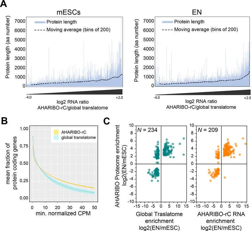
Tools and resources Cell Biology
themselves potentially or partially translated (Anderson et al., 2015; Aspden et al., 2014;
Bazin et al., 2017; Ingolia et al., 2011; Nelson et al., 2016; Ruiz-Orera et al., 2014; van Heesch
et al., 2019). As coding RNAs, lncRNAs can be associated with actively translating or translationally
silent ribosomes (Chandrasekaran et al., 2019; Chen et al., 2020; Jiao and Meyerowitz, 2010;
Kapur et al., 2017). Hence, the potential involvement of lncRNAs in translation increases the com-
plexity of the mammalian control of gene expression at the translatome and proteome level. Unfor-
tunately, classical RIBO-seq approaches barely distinguish between lncRNAs producing peptides
from those that sequester ribosomes (lncRNA bound to ribosomes without translation) and act as
ribosome sponges. Proteomics approaches, such as mass spectrometry, can help to define and
quantitatively monitor the production of peptides, but are less sensitive techniques than RNA
sequencing (Slavoff et al., 2013; van Heesch et al., 2019). Therefore, proteomics and RIBO-seq
alone cannot unravel the wide functional range of cytoplasmic lncRNAs associated with the transla-
tion machinery.
To fill this gap, we developed AHA-mediated RIBOsome isolation (AHARIBO), a combination of
protocols that simultaneously isolate RNAs and nascent proteins associated with translationally
active ribosomes. AHARIBO is based on the isolation of ribosomes trapped with their nascent pepti-
des by incorporating the non-canonical amino acid L-azidohomoalanine (AHA), followed by parallel
RNA-seq, ribosome profiling, and proteomics.
We applied AHARIBO to human and mouse cells and showed that it enables to (1) purify translat-
ing ribosomes via nascent peptide chains, (2) co-purify RNAs and proteins for transcriptome/de
novo proteome-associated studies, and (3) detect the regulatory network of lncRNAs translated or
associated with ribosomes.
Results
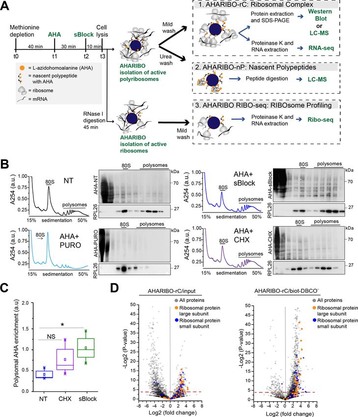
Nascent peptide labeling and separation of the ribosome complex with
AHARIBO-rC
To simultaneously purify ribosomes under active translation, associated RNAs, and corresponding
growing peptide chains, we optimized a protocol in HeLa cells (Figure 1A). Briefly, the protocol con-
sists of the following phases: (1) incubation with a methionine-depleted medium, (2) addition of the
methionine analog AHA, (3) on-ribosome anchorage of nascent peptide chains with a small mole-
cule, (4) cell lysis and AHA ‘copper-free click reaction’ (Jewett and Bertozzi, 2010) for (5) ribosome
capture with magnetic beads. We reasoned that the protocol for isolating ribosomes through AHA
can be used to obtain information about nascent peptides, constitutive components of ribosomes,
mRNAs, and lncRNAs associated with them. For this reason, we optimized several parameters from
washing steps to nuclease treatments (Figure 1A) to isolate (1) the full translational complex (AHAR-
IBO-rC, ribosomal complexes: ribosomes, ribosome-associated proteins, nascent peptides, and
RNAs), (2) the de novo synthesized proteome (AHARIBO-nP, nascent proteome), and (3) ribosome-
protected fragments (RPFs) (AHARIBO RIBO-seq: RIBOsome profiling by sequencing).
To minimize the amount of AHA-tagged and fully synthesized proteins released from ribosomes
and achieve optimal on-ribosome polypeptide stabilization, we tested multiple incubation times of
AHA exposure and compared the effect of two small molecules (namely cycloheximide [CHX] and
sBlock, an anisomycin-based reagent). Anisomycin is known to inhibit the activity of eukaryotic ribo-
somes, while keeping polypeptides bound to translating ribosomes (Garreau de Loubresse et al.,
2014; Grollman, 1967; Seedhom et al., 2016).
We observed that 30 min is the optimal incubation time for sufficient AHA incorporation and
maximum RNA recovery (Figure 1—figure supplement 1A–C). Next, we compared the efficiency of
CHX and sBlock in stabilizing the nascent peptide by co-sedimentation analysis of AHA-tagged poly-
peptides with ribosomes along the sucrose gradient (Figure 1B). As a control, cells were treated in
parallel with puromycin to cause ribosome disassembly and release of the growing peptide chains
(Figure 1B; Blobel and Sabatini, 1971; Enam et al., 2020). In agreement with literature, we found
that both CHX and sBlock are able to stabilize AHA-peptides on ribosomes and polysomes
(Biever et al., 2020; Mathias et al., 1964). The efficiency of anchoring polypeptides on ribosomes
in CHX- and sBlock-treated cells was about 50% higher compared to untreated cells, confirming that
the treatment effectively stabilizes nascent polypeptides (Figure 1C). The high signal observed in
Minati, Firrito, Del Piano, et al. eLife 2021;10:e59303. DOI: https://doi.org/10.7554/eLife.59303 2 of 25
Tools and resources                                                                                                            Cell Biology

Figure 1. L-Azidohomoalanine (AHA) labeling of nascent peptide chains and ribosome separation. (A) Schematic representation of AHA-
mediated RIBOsome isolation (AHARIBO) workflow. After methionine depletion, AHA incubation, and sBlock treatment, cell lysates can be processed
for (1) AHARIBO-rC: isolation of translational complexes (ribosomes, ribosome-associated proteins, nascent peptides, and RNAs); (2) AHARIBO-nP:
isolation of de novo synthesized proteome; and (3) AHARIBO RIBO-seq: for ribosome profiling. (B) Polysomal profiles in HeLa cells. On the right of each
Figure 1 continued on next page

Minati, Firrito, Del Piano, et al. eLife 2021;10:e59303. DOI: https://doi.org/10.7554/eLife.59303                                               3 of 25
Tools and resources                                                                                                               Cell Biology

Figure 1 continued
profile, example of SDS-PAGE of protein extracts from each fraction of the profile. Staining of the membrane was performed by biotin cycloaddition
followed by streptavidin-Horseradish peroxidase (HRP). RPL26 protein was used as a marker of the large ribosome subunit. (C) Box plot showing the
AHA signal enrichment in the polysomal fractions of the profiles in cells untreated (NT) and treated with either cycloheximide (CHX) or sBlock. Results
are shown as the median (±SE) of three independent experiments. NS: not significant. *p-value=0.05 was obtained through an unpaired t-test. (D)
Volcano plots of AHARIBO-rC-isolated proteins. Data are compared with input (AHA-containing lysate, left) or with streptavidin-coated beads without
biotin-DBCO (right). DBCO: dibenzocyclooctyne. Red line: t-test p-value
Tools and resources                                                                                                  Cell Biology

                                     AHARIBO-nP: genome-wide portray of the de novo synthesized
                                     proteome
                                     Motivated by the evidence that AHARIBO-rC can be used to isolate bona fide active ribosomes, we
                                     further tested our method genome-wide in mouse embryonic stem cells (mESCs) under basal condi-
                                     tion and after differentiation into early neurons (ENs) (Tebaldi et al., 2018; Figure 2—figure supple-
                                     ment 1A). We analyzed both AHARIBO-rC-isolated RNA and newly synthesized polypeptides
                                     associated with actively translating ribosomes by RNA-seq and LC-MS, respectively. The protocol for
                                     the isolation of the de novo synthesized polypeptides (named AHARIBO-nP) is based on urea wash-
                                     ing to remove all proteins that are not nascent peptides (Figure 2—figure supplement 1B). In paral-
                                     lel, we isolated and analyzed the global translatome by extracting the RNA after 30% sucrose
                                     cushioning of cytoplasmatic lysates (Wang et al., 2013), and then analyzed the global proteome by
                                     pulsed SILAC (pSILAC) (Schwanhäusser et al., 2009; Figure 2A).
                                         Quantitative proteomic analysis of ENs versus mESCs (EN/mESC) led to the identification of 2654
                                     differentially expressed proteins (Figure 2B, Figure 2—source data 1). As expected, differentiated
                                     cells (EN) showed a reduced turnover compared to mESCs (Figure 2—figure supplement 1C). In
                                     parallel, EN and mESC cells were analyzed by AHARIBO-nP, which captured 1365 and 2215 proteins,
                                     respectively. Of note, 74% of proteins identified through AHARIBO-nP is in common with the pSI-
                                     LAC dataset. The smaller number of proteins identified with AHARIBO-nP compared to pSILAC is
                                     most probably related to the shorter time of incubation with AHA (30 min) compared to pSILAC (24
                                     hr) and is consistent with previous observations from similar pulldown enrichment strategies
                                     (Bagert et al., 2014; Rothenberg et al., 2018). Differential expression analysis (EN/mESC) identified
                                     573 proteins (p-value
Tools and resources Cell Biology Figure 2. AHARIBO-nP and pSILAC. (A) Workflow for parallel AHARIBo-nP and pSILAC. mESCs: mouse embryonic stem cells; EN: mouse embryonic stem cells differentiated in early neurons. (B) Venn diagram representing the number of differentially expressed proteins (EN/mESCs) identified by AHARIBO-nP and pSILAC (p-value
Tools and resources                                                                                                                 Cell Biology

Figure 3. AHARIBO-rC RNA versus de novo proteome analysis. (A) Enrichment of a given transcript obtained with AHA-mediated RIBOsome
isolation (AHARIBO) versus global translatome (x-axis) as a function of the theoretical protein length (y-axis) for mouse embryonic stem cells (mESCs)
(left) and early neurons (ENs) (right). Each bar represents the number of enriched transcripts with the defined theoretical protein length. (B) Fraction of
coding genes expressed above a minimum threshold in EN. The AHARIBO-rC and global translatome group are represented in yellow and cyan,
respectively. For each group, the mean (solid line) and SD (shades) of the fractions for a given count per million (CPM) threshold are calculated over all
samples (n = 6) in that group. (C) Scatter plot of RNA fold change (global translatome on the left, AHARIBO-rC on the right) compared to protein fold
change (AHARIBO-nP) obtained by comparing EN with mESC. N: number of differentially expressed genes (DEGs) with p-value
Tools and resources                                                                                                    Cell Biology

                                        Finally, we tested whether the RNA isolated with AHARIBO-rC can predict the de novo synthe-
                                     sized proteome. After comparing differentially expressed genes (DEGs) during differentiation to the
                                     AHARIBO-nP proteome (Figure 3—source data 1), we observed that AHARIBO-rC RNA is a good
                                     proxy of the newly synthesized proteome (Pearson’s correlation r = 0.75, Figure 3C, Figure 3—fig-
                                     ure supplement 1B). In particular, we found that AHARIBO-rC RNA presents less uncoupled genes
                                     (up-RNA and down-protein or down-RNA and up-protein) than the global translatome (Figure 3—
                                     figure supplement 1C), thus faithfully recapitulating proteome changes. The correlation of the
                                     global translatome with the global protein turnover measured with pSILAC shows a Pearson’s
                                     r = 0.27 (Figure 3—figure supplement 1D, Figure 3—source data 2). This result demonstrates that
                                     AHARIBO-nP does reflect the labeling of peptides rather than completely synthesized proteins.

                                     Combined AHARIBO approaches define the functional role of lncRNAs
                                     in translation
                                     Based on the evidence that a combination of AHARIBO approaches can simultaneously detect RNAs
                                     under active translation and peptides in the process of being produced, we applied our methods to
                                     detect ribosome-associated and translated native lncRNAs.
                                         In AHARIBO-rC data, we identified a total of 687 lncRNA genes in mESCs and about 400 differen-
                                     tially expressed (DE) lncRNAs during neuronal differentiation (Figure 4—figure supplement 1A, Fig-
                                     ure 4—source data 1). Among the top five DE lncRNAs (fold change >10; p-value
Tools and resources Cell Biology Figure 4. The AHA-mediated RIBOsome isolation (AHARIBO) platform can be used to detect ribosome-interacting long non-coding RNAs (lncRNAs). (A) Linear plot illustrating the fraction of non-coding genes expressed above a minimum threshold in early neurons (EN). The AHARIBO-rC and the global translatome group are represented in yellow and cyan, respectively. For each group, the mean (solid line) and the SD (shades) of the fractions for a given count per million (CPM) threshold are calculated over all samples (n = 3) in that group. Expression values are indicated as normalized CPM. AHARIBO-rC was performed on the ribosome pellet after sucrose cushioning. (B) Venn diagram of the number of lncRNAs genes with at least 1 CPM identified by RNA-seq, AHARIBO-rC, RIBO-seq, and AHARIBO RIBO-seq. (C) Classification of lncRNAs interacting with ribosomes and relative detection through the multiple AHARIBO and standard approaches. ND: no detection of protein synthesis. (D) (Left) Schematic representation of the number of mouse embryonic stem cell (mESC) lncRNAs in common between AHARIBO RIBO-seq, AHARIBO-rC RNA, and standard RIBO-seq. These lnRNAs were validated by liquid chromatography-mass spectrometry (LC-MS). (Right) Example of an AHARIBO RIBO-seq ribosome occupancy profile of lncRNA 1810058I24Rik displaying the reads distribution along the entire transcript and the accumulation of reads at the known short open reading frame (shadow area and blue arrow on top). The online version of this article includes the following source data and figure supplement(s) for figure 4: Source data 1. A table with the list of long non-coding RNAs (lncRNAs) identified by RNA-seq by RNA-seq in mouse embryonic stem cells (mESCs). Source data 2. A table with the list of long non-coding RNAs (lncRNAs) identified by RIBO-seq in mouse embryonic stem cells (mESCs). Source data 3. A table with the list of matching peptides from AHA-mediated RIBOsome isolation’s (AHARIBO) identified long non-coding RNAs (lncRNAs). Figure supplement 1. Isolation of long non-coding RNAs (lncRNAs) with AHA-mediated RIBOsome isolation (AHARIBO). Figure 4 continued on next page Minati, Firrito, Del Piano, et al. eLife 2021;10:e59303. DOI: https://doi.org/10.7554/eLife.59303 9 of 25
Tools and resources                                                                                                  Cell Biology

Figure 4 continued
Figure supplement 2. AHA-mediated RIBOsome isolation (AHARIBO) RIBO-seq data.
Figure supplement 3. Translated long non-coding RNAs (lncRNAs).

                                     efficiency in detecting an ectopically expressed micropeptide (TUG1-BOAT) and (2) concordance
                                     with recently published data, prove that our approach could be useful to unravel translation events
                                     in lncRNAs that are misannotated as non-coding. Altogether, our data confirm that our three diverse
                                     and complementary AHARIBO approaches represent a unique method to identify ribosome-associ-
                                     ated and translated RNAs.

                                     Discussion
                                     LncRNAs localize in the nucleus or in the cytoplasm. In the nucleus, they modulate transcription, pre-
                                     mRNA splicing, or act as scaffold for protein interaction during chromatin organization (Sun et al.,
                                     2018). In the cytoplasm, the majority of lncRNAs is associated with polysomes (Carlevaro-Fita et al.,
                                     2016), where they either can or cannot produce proteins (Chen et al., 2020; Ingolia et al., 2011).
                                     Numerous lncRNAs are misannotated as non-coding but contain short ORFs encoding for micropep-
                                     tides with biological relevance in cancer (D’Lima et al., 2017; Huang et al., 2017), bone develop-
                                     ment (Galindo et al., 2007), immunity (van Solingen et al., 2018), metabolism (Magny et al., 2013;
                                     Nelson et al., 2016), and DNA repair (Slavoff et al., 2014). Different methodological approaches
                                     have been developed to quantify the variations of RNA abundance by sequencing or imaging techni-
                                     ques (Blumberg et al., 2019; Jao and Salic, 2008; Morisaki et al., 2016; Wu et al., 2016), RNA
                                     engagement with the translational machinery by RIBO-seq or polysomal profiling (Arava et al.,
                                     2003; Clamer et al., 2018; Eden et al., 2011; Taniguchi et al., 2010), and protein synthesis by
                                     mass spectrometry or metabolic labeling (Aviner et al., 2013; Dieterich et al., 2006;
                                     Schwanhäusser et al., 2009; Yan et al., 2016). Despite these advantages, available technologies
                                     hardly capture in a single experiment the dynamics of translation across multiple biological condi-
                                     tions, the translation of unannotated coding transcripts, and translation-related functions of
                                     lncRNAs. Now that it is widely accepted that a portion of the genome annotated as non-coding can
                                     result in a complex transcriptome partially engaged with ribosomes (Chen et al., 2020;
                                     Djebali et al., 2012; Iyer et al., 2015), RNA sequencing and ribosome profiling should include
                                     micropeptide detection.
                                         Our data show that AHARIBO serves as a flexible tool to detect translated RNAs, identify
                                     lncRNAs bound to elongating ribosomes, and detect de novo synthesized proteins. The intersection
                                     of standard RIBO-seq, RNA-seq, and AHARIBO approaches allowed us to identify translated
                                     lncRNAs. We demonstrated that AHARIBO is efficient in capturing short translated open reading
                                     frames, both native or ectopically expressed. Although LC-MS technologies are not as sensitive as
                                     RNA sequencing, we successfully identified a mouse-specific micropeptide reported to originate
                                     from a native lncRNA ORF, confirming the effectiveness of AHARIBO. To overcome existing limita-
                                     tions in LC-MS detection, many other translation events on lncRNAs can be predicted combining
                                     AHARIBO approaches with in silico translation of the identified leads. This approach would likely
                                     allow to selectively validate a list of still uncharacterized lncRNAs. Although the unlabeled back-
                                     ground cannot be avoided, a pre-cleaning of the cell lysate with a cushioning step can help to
                                     increase the resolution with difficult samples. Moreover, a puromycin treatment instead of sBlock
                                     could be added as control in proteomic experiments. A unique feature of AHARIBO is the possibility
                                     to simultaneously isolate ribosomes, RNA engaged with ribosomes, and the corresponding proteins
                                     produced. Besides the versatility of the method, AHA labeling has the advantage of minimal interfer-
                                     ence with protein synthesis (Hodas et al., 2012; Tom Dieck et al., 2012).
                                         The most prominent limitation of the method relies on the methionine starvation required for effi-
                                     cient AHA incorporation (Calve et al., 2016; Hodas et al., 2012; Saleh et al., 2019). This step can
                                     modify the physiological conditions of the cell and needs to be taken into consideration when plan-
                                     ning experiments requiring certain stimuli (e.g., drug treatment) during methionine depletion. The
                                     conditions used in the AHARIBO protocol give robust protein labeling, but AHA concentration can
                                     be conveniently tuned based on specific cell types or biological questions. Additionally, we observed

Minati, Firrito, Del Piano, et al. eLife 2021;10:e59303. DOI: https://doi.org/10.7554/eLife.59303                                  10 of 25
Tools and resources Cell Biology
that there are still challenges for LC-MS verification of putative lncRNA peptides identified with
AHARIBO. Of note, a potential contribution from background signal needs to be taken into consid-
eration in LC-MS and Ribo-seq analysis.
With AHARIBO we introduce a strategy for the selective isolation of active ribosomes using the
nascent peptide chain as bait for a more comprehensive interrogation of lncRNA biology and pro-
teogenomic studies. Overall, we provide evidence that AHARIBO is a comprehensive and reliable
toolkit suitable for downstream parallel RNA-seq, RIBO-seq, and LC-MS analysis, empowering scien-
tists to shed light on the functional complexity of translation.
Materials and methods
Key resources table
Reagent type (species)
or resource Designation Source or reference Identifiers Additional information
Cell line Papillomavirus-related ATCC RRID:CVCL_0030
(Homo sapiens) endocervical adenocarcinoma
Cell line 46C embryonic stem cells ATCC RRID:CVCL_Y482 Quattrone A. Lab. (CIBIO)
(Mus musculus)
Antibody Anti-b3-tubulin Promega Cat. #G712A (1:2000)
(mouse monoclonal) RRID:AB_430874
Antibody Anti-Oct4 (mouse monoclonal) Santa Cruz Cat. #SC 5279 (1:2000)
Biotechnologies RRID:AB_628051
Antibody Anti-human RPL26 Abcam Cat. #ab59567 (1:2000)
(rabbit polyclonal) RRID:AB_945306
Antibody Anti-human RPS6 Abcam Cat. #ab40820 (1:2000)
(rabbit polyclonal) RRID:AB_945319
Antibody Anti-human beta actin Abcam Cat. #ab8227 (1:2000)
(rabbit polyclonal) RRID:AB_2305186
Recombinant WT TUG1-BOAT (plasmid) PMID:32894169
DNA reagent
Recombinant D TUG1-BOAT (plasmid) This paper See ’Materials and methods
DNA reagent section: ’TUG1-BOAT ectopic
expression and qPCR’
Recombinant +1Met TUG1-BOAT (plasmid) This paper See ’Materials and methods’
DNA reagent section: ’TUG1-BOAT ectopic
expression and qPCR’
Peptide, Precision Protein BioRad Cat. #1610380 (1:5000)
recombinant protein StrepTactin-HRP Conjugate
Chemical L-Arginine-13C6,15N4 hydrochloride Sigma-Aldrich Cat. #608033
compound, drug
Chemical L-Lysine-13C6,15N2 hydrochloride Sigma-Aldrich Cat. #608041
compound, drug
Chemical L-Azidohomoalanine Invitrogen Cat. #C10102
compound, drug (Click-IT AHA)
Chemical Dibenzocyclooctyne- Sigma-Aldrich Cat. #760749SML1656
compound, drug PEG4-biotin conjugate
Chemical sBlock IMMAGINA BioTechnology Cat. #SM8
compound, drug
Chemical Puromycin Sigma-Aldrich Cat. #P8833
compound, drug
Chemical Cycloheximide Sigma-Aldrich #C4859
compound, drug
Chemical Lipofectamine 3000 Thermo Fisher Scientific. Cat. #L3000001
compound, drug Transfection Reagent
Chemical Mag-DBCO beads IMMAGINA BioTechnology Cat. #MDBCO
compound, drug
Continued on next page
Minati, Firrito, Del Piano, et al. eLife 2021;10:e59303. DOI: https://doi.org/10.7554/eLife.59303 11 of 25Tools and resources Cell Biology
Continued
Reagent type (species)
or resource Designation Source or reference Identifiers Additional information
Chemical eMagSi-cN beads IMMAGINA BioTechnology #018-eMS-001
compound, drug
commercial SMART-Seq Stranded Kit Takara Cat. #634443
assay or kit
Commercial SuperScript III Thermo Fisher Cat. #18080044
assay or kit Reverse Transcriptase
Commercial Kapa Probe Fast Kapa Biosystems #KK4702
assay or kit Universal qPCR Kit
Software, algorithm Image analysis ImageJ RRID:SCR_003070
Software, algorithm Statistical package edgeR RRID:SCR_012802
Cell culturing and treatments
For protocol development, optimization, and validation, HeLa cells were used. HeLa cells were main-
tained on adherent plates in Dulbecco’s modified Eagle’s medium (DMEM; EuroClone #ECM0728L)
supplemented with 10% fetal bovine serum, 2 mM L-glutamine, 100 units/mL penicillin, and 100 mg/
mL streptomycin at 37˚C, 5% CO2. For passaging, cells were washed with 1 Phosphate-Buffered
Saline (PBS), detached using 0.25% trypsin-EDTA, and spun down at 260 g for 5 min.
For treatments, 250,000–400,000 HeLa cells per well were seeded in six-well plates and grown to
80% confluence. At the time of treatment, culture medium was removed and cells were washed
once with warm 1 PBS. Subsequently, cells were incubated with Dulbecco’s modified Eagle’s limit-
ing medium (DMEM-LM; Thermo Scientific #30030) supplemented with 10% fetal bovine serum and
800 mM L-leucine for 40 min to deplete methionine reserves. Methionine-free medium was then sup-
plemented with L-azidohomoalanine (Click-IT AHA; Invitrogen #C10102) at a final concentration of
250 mM and incubation time (ranging from 10 min to 120 min; 30 min set as incubation time for the
protocol). Cells were then treated with 1 sBlock (IMMAGINA BioTechnology, catalog no. #RM8;
sBlock is an anisomycin-containing proprietary reagent) for 10 min. Then, six-well plates were placed
on ice, medium was removed, and cells were washed once with cold 1 PBS supplemented with 1
sBlock. After removing residual PBS with a pipette, hypotonic lysis buffer (0.01 M NaCl, 0.01 M
MgCl2, 0.01 M Tris-HCl, 1% Tx-100, 1 sBlock, 1% sodium deoxycholate, 5 units/mL DNAse I
[Thermo Scientific #89836], 200 units/mL RiboLock RNase Inhibitor [Thermo Scientific #EO0381], 1
Protease Inhibitor Cocktail [Cell Signaling Technology #5871S]) was added to each well, and cells
were lysed with the aid of a scraper. After hypotonic lysis, nuclei and cellular debris were removed
by centrifuging at 18,000 g, 4˚C for 5 min. For quantification of the total absorbance value of cell
lysates, the absorbance was measured (260 nm) using a Nanodrop ND1000 UV-VIS Spectrophotom-
eter. Lysates were aliquoted and processed directly or stored at 80˚C.
Arsenite pre-treatment was performed by adding sodium arsenite (Sigma-Aldrich #S7400) at a
final concentration of 500 mM for 1 hr.
For RNA-seq and proteomics experiments, two biological settings were assessed in triplicate
experiments: (1) undifferentiated mouse 46C embryonic stem cells (mESCs) (Ying et al., 2003) and
(2) mESCs induced to differentiate into ENs. mESCs were maintained in mESC self-renewal medium
composed of Glasgow’s MEM (Thermo Scientific #11710-035) supplemented with 1000 units/mL
ESGRO Recombinant Mouse LIF protein (Millipore #ESG1107), 10% fetal bovine serum, 55 mM 2-
mercaptoethanol, 1 mM sodium pyruvate (Thermo Scientific #11360070), MEM non-essential amino
acids (Thermo Scientific #11140050), GlutaMax (Thermo Scientific #35050061), and penicillin/strep-
tomycin. For passaging, mESCs were washed twice with 1 PBS, detached using 0.02–0.05% tryp-
sin-EDTA, and spun down at 260 g for 3 min. Pellet was resuspended in fresh medium and plated
onto 0.1% gelatin-coated culture vessels.
For treatments, 5 105 mESCs/cm2 were seeded in Petri dishes and grown to 60% confluence.
For pSILAC proteomics, 24 hr before lysis mESCs were washed twice with 1 PBS and the medium
was replaced with SILAC Advanced DMEM/F-12 Flex Medium (Thermo Scientific #A2494301), sup-
plemented with 1000 units/mL ESGRO Recombinant Mouse LIF protein, 10% dialyzed fetal bovine
Minati, Firrito, Del Piano, et al. eLife 2021;10:e59303. DOI: https://doi.org/10.7554/eLife.59303 12 of 25Tools and resources Cell Biology
serum, 4500 mg/L glucose, 17.25 mg/L proline, and penicillin/streptomycin. Either light or heavy
L-arginine (Sigma-Aldrich #608033) and L-lysine (Sigma-Aldrich #608041) were added at 84 mg/L
and 146 mg/L, respectively. For both AHA+ proteomics and RNA-seq experiments, treatments were
performed as described above for HeLa cells, with the exception that methionine-free medium was
supplemented with 1000 units/mL ESGRO Recombinant Mouse LIF protein and 10% dialyzed fetal
bovine serum. After methionine depletion, cells were treated with 250 mM AHA for 30 min. The
remaining treatment steps and hypotonic lysis were performed as detailed above.
Neuronal differentiation was performed according to a previously described protocol (Ying et al.,
2003). Briefly, 2.000 mESCs/cm2 were seeded on gelatin-coated culture vessels in N2B27 medium.
Cells were gently washed with 1 PBS, and medium was renewed every 1–2 days until 15DIV.
N2B27 medium is composed of 1:1 mix of DMEM/F-12 (Thermo Scientific #21331020) and Neuro-
basal Medium (Thermo Scientific #21103049), supplemented with 0.5% N-2 (Thermo Scientific
#17502048), 1% B-27 (Thermo Scientific #17504044), GlutaMax, and penicillin/streptomycin.
Upon differentiation, ENs were treated directly in culture vessels. For pSILAC proteomics, 24 hr
before lysis ENs were washed once with 1 PBS and the medium was replaced with SILAC
Advanced DMEM/F-12 Flex Medium, supplemented with 0.5% N2, 1% B27, 4500 mg/L glucose,
17.25 mg/L proline, and penicillin/streptomycin, 4500 mg/L glucose, 17.25 mg/L proline, and penicil-
lin/streptomycin. Either light or heavy L-arginine and L-lysine were added at 84 mg/L and 146 mg/L,
respectively. For both AHA+ proteomics and RNA-seq experiments, ENs were treated as described
above for HeLa cells, with 250 mM AHA for 30 min. The remaining treatment steps and hypotonic
lysis were performed as detailed above.
Cell lines were purchased directly from ATCC and passaged fewer than 15 times. Mus musculus
46C ES were obtained from Quattrone A. Lab (CIBIO, RRID:CVCL_Y482). All cells tested negative
for mycoplasma contamination.
Immunocytochemistry
For immunofluorescence assay, cells were fixed with 4% paraformaldehyde for 15 min at room tem-
perature, permeabilized using 0.5% Triton X-100 in 1 PBS for 15 min at room temperature, and
blocked using 5% fetal bovine serum, 0.3% Triton X-100 in 1 PBS for 2 hr at room temperature.
Cultures were then incubated overnight at 4˚C with either anti-b3-tubulin (Promega #G712A) or anti-
Oct4 (Santa Cruz Biotechnologies #SC-5279) primary antibodies diluted in 2% fetal bovine serum,
0.2% Triton X-100 in 1 PBS. Cells were then washed three times with 1 PBS and incubated with
Alexa-555 anti-mouse secondary antibodies for 2 hr. Nuclei were counterstained with Hoechst 33258
before imaging with a Zeiss Axio Observer Z1 inverted microscope equipped with a 2.83 Megapixel
AxioCam 503 mono D camera.
AHARIBO-rC/AHARIBO-nP: purification of active ribosomes for RNA/
protein isolation
For RNA-seq experiments, lysates were diluted in W-buffer (10 mM NaCl, 10 mM MgCl2, 10 mM
HEPES, 1 sBlock) to a final Nanodrop-measured absorbance (260 nm) of 1–2 a.u./mL, supple-
mented with 40 U of Superase-In RNase Inhibitor (Thermo Scientific #AM2696) and incubated with
dibenzocyclooctyne-PEG4-biotin conjugate (Sigma-Aldrich #760749; 50 mM final concentration) in a
reaction volume of 100 mL for 1 hr on a rotator in slow motion (9 rpm) at 4˚C. Lysates were then incu-
bated with 50 mL of eMagSi-cN beads (IMMAGINA BioTechnology #018-eMS-001) for 30 min at 4˚C
on the rotator in slow motion (9 rpm). Subsequently, samples were taken off the rotator and placed
on a magnetic rack on ice, and supernatants were discarded. Beads were washed two times with
500 mL of 1 PBS supplemented with 0.1% Triton-X100, 1 sBlock, and 1:10,000 RiboLock RNase
Inhibitor (Thermo Scientific #EO0381) on the rotator in slow motion at 4˚C, removing supernatants
from the tubes sitting on the magnetic rack and gently adding new washing solution each time. After
the final wash, beads were resuspended in 200 mL of W-buffer and transferred to a new vial. Then,
20 mL of 10% SDS and 5 mL of Proteinase K (Qiagen #19131) were added to each sample, and sam-
ples were incubated at 37˚C for 75 min in a water bath. Subsequently, suspensions were transferred
to a new vial, and acid phenol:chloroform:isoamyl alcohol RNA extraction was performed. Briefly, an
equal volume of acid phenol:chloroform:isoamyl alcohol (pH 4.5) was added, and samples were vor-
texed and centrifuged at 14,000 g for 5 min. Aqueous phases were then transferred to new vials,
Minati, Firrito, Del Piano, et al. eLife 2021;10:e59303. DOI: https://doi.org/10.7554/eLife.59303 13 of 25Tools and resources Cell Biology
500 mL of isopropanol and 2 mL of GlycoBlue (Thermo Scientific #AM9516) were added, samples
were mixed and incubated at room temperature for 3 min, and then stored overnight at 80˚C. The
following day samples were centrifuged at 14,000 g for 30 min, supernatants were removed, 500
mL of 70% ethanol were added to each sample, and samples were then centrifuged at 14,000 g
for 10 min. Finally, pellets were air-dried and resuspended in 10 mL of nuclease-free water. When
quality check and quantification was needed, RNA samples were run on a 2100 Bioanalyzer (Agilent)
using the Agilent RNA 6000 Nano Reagents kit (Agilent #5067–1511) and assayed on the Qubit fluo-
rometer using the Qubit RNA HS Assay Kit (Thermo Scientific #Q32852). For visualization of total
RNA patterns, samples were run on a 1% agarose gel. ImageJ software (v 1.45s) was used for the
quantitation of signal intensities of ribosomal RNA bands.
For proteomics experiments, lysates were diluted in W-buffer to a final Nanodrop-measured
absorbance (260 nm) of 1–2 a.u./mL in a final volume of 100 mL. Ribosome pulldown was performed
using Mag-DBCO beads (IMMAGINA BioTechnology #MDBCO). Lysates were incubated with 50 mL
of beads for 1 hr on a rotator in slow motion (9 rpm) at 4˚C. Supernatants were discarded after plac-
ing samples on the magnetic rack. Beads were washed three times with 500 mL of 200 mM Tris, 4%
CHAPS, 1 M NaCl, 8 M urea, and pH 8.0 at room temperature on a shaker at 1000 rpm, using the
magnetic rack to replace the washing solution. After the final wash, beads were resuspended in 30
mL of water and transferred to a new vial.
qRT-PCR analysis
Total RNA was extracted from samples processed through the AHARIBO-rC protocol as described
above. Depending on the available input material, RNA was retrotranscribed using either RevertAid
First Strand cDNA Synthesis Kit (Thermo Scientific #K1621) or SuperScript III Reverse Transcriptase
(Thermo Fisher #18080044), per manufacturer’s protocols. qPCR was run on CFX Connect Real-Time
PCR Detection System (BioRad) using Kapa Probe Fast Universal qPCR Kit (Kapa Biosystems
#KK4702). Reactions were performed in technical duplicates of biological triplicates. The following
TaqMan probes were used: Hs99999901_s1 (18S), Hs02800695_m1 (HPRT1).
For the normalization of qRT-PCR results, HPRT1 was used as housekeeping gene. The fold
change in normalized 18S RNA levels between untreated (control) and treated (arsenite) samples
was calculated. A second normalization to threshold cycles from non-AHA-treated samples was
done to account for background signal.
TUG1-BOAT ectopic expression and qPCR
We ectopically express the putative protein produced by the open reading frame of TUG1, called
TUG1-BOAT (Tug1-Bifunctional ORF and Transcript), in HeLa cells. Briefly, construct generation and
transfection was performed as in Lewandowski et al., 2020 with some minor changes to adapt the
experimental setup to the AHARIBO method. We synthesized three different constructs for human
Tug1 ORF1 (Thermo Scientific):
1. The first (called WT TUG1-BOAT) is the one reported in Lewandowski et al., 2020. It has a
non-canonical start codon and a methionine at 75 nt (25 aa) upstream of the stop codon.
2. The second (called D TUG1-BOAT) is deleted by the only methionine of the sequence present
at 75 nt from the stop codon. No methionines are present.
3. The third (called +1Met TUG1-BOAT) has an ATG start codon (methionine) instead of the non-
canonical CTG start codon e. Therefore, the third construct has two methionines, one at the N
terminal and the other at 25 aa (about 75 nt) upstream of the C-terminal.
We cloned the constructs in the pcDNA3.1(+) plasmid with HindIII and EcoRV restriction enzymes.
For transfection of TUG1-BOAT constructs, we seeded HeLa cells in a six-well plate and transfected
the cells with 2.5 mg of plasmids (pcDNA3.1(+) containing each of the inserts) using 742 Lipofect-
amine 3000 Transfection Reagent (Thermo Fisher Scientific). After 24 or 48 hr post transfection, cells
were processed with AHARIBO-rC protocol followed by RNA extraction.
We performed qPCR analysis on AHARIBO pulldowns and input for each vector to validate the
efficiency in capturing short translated ORF deriving from RNA annotated as lncRNA (TUG1). Briefly,
200 ng of DNase I-treated RNA was used as input to generate cDNA using High-Capacity cDNA
Reverse Transcription Kit (Applied Biosystems), according to the manufacturer’s protocol. qPCR was
run on CFX Connect Real-Time PCR Detection System (BioRad) using Powerup Sybr Master Mix
Minati, Firrito, Del Piano, et al. eLife 2021;10:e59303. DOI: https://doi.org/10.7554/eLife.59303 14 of 25Tools and resources Cell Biology
(Applied Biosystems) and a couple of primers design to amplify 150 nt of the CDS of TUG1-BOAT
transcript (see below). Reactions were performed in technical duplicates of biological duplicates. For
normalization, 18S was used as housekeeping gene. Ct values were analyzed using the DDCt method
(Livak and Schmittgen, 2001).
Fw PRIMER: GGCTCTTTCTCCTGCTCTGG
Rev PRIMER: CTCCTCGTCGAATCGCAAAC
Insert size: 150 nt
TUG1-BOAT sequences are listed below:
Italics: 50 UTR leader sequence; bold: canonical and non-canonical start codons; red: methionine.
>WT TUG1-BOAT
GGCCGAGCGACGCAGCCGGGACGGTAGCTGCGGTGCGGACCGGAGGAGCCATCTTGTC
TCGTCGCCGGGGAGTCAGCCCCTAAATCGAAGAAGCCCTGGCGCGCCCTCCCCCCC
TCCCGGGTCTGGTAGGGCGAAGGAACGGGCGTGCGGTCGATCGAGCGATCGG
TTGGCGGCTCTTTCTCCTGCTCTGGCATCCAGCTC
TTGGGGCGCAGGCCCGGCCGCCGCGGCGCGCGCCCGGTGGCCGTTGGCGC
TCGCGCCGCGTCTTTCTTCTCGTACGCAGAACTCGGGCGGCGGCCTATGCGTTTGCGA
TTCGACGAGGAGTCGTCCGGGTGGTCGGCGGCGGCGGGCAGCTGCTCCGCCCCGC
TCCGGGGGAGGCGGCGGCGGCAGCGGCCGCGGGATTTGGAGCGGCCGGG-
GAGGCGGGGGTGGCCGGGGCCGGCTTGGAGGCCTGGCGCCACCCTTCGGGGCC
TGCAAGGACCCAGTTGGGGGGGCAGGAGGGGGCCGGAGGATGGTTGGTTGTGGGATTTC
TACTTTGCCTTTTCCTCCTTATGCCGCCTGACTACAAAGACCATGACGGTGATTATAAAGA
TCATGACATCGACTACAAGGATGACGATGACAAGTAG
>DTUG1-BOAT
GGCCGAGCGACGCAGCCGGGACGGTAGCTGCGGTGCGGACCGGAGGAGCCATCTTGTC
TCGTCGCCGGGGAGTCAGCCCCTAAATCGAAGAAGCCCTGGCGCGCCCTCCCCCCC
TCCCGGGTCTGGTAGGGCGAAGGAACGGGCGTGCGGTCGATCGAGCGATCGG
TTGGCGGCTCTTTCTCCTGCTCTGGCATCCAGCTC
TTGGGGCGCAGGCCCGGCCGCCGCGGCGCGCGCCCGGTGGCCGTTGGCGC
TCGCGCCGCGTCTTTCTTCTCGTACGCAGAACTCGGGCGGCGGCCTATGCGTTTGCGA
TTCGACGAGGAGTCGTCCGGGTGGTCGGCGGCGGCGGGCAGCTGCTCCGCCCCGC
TCCGGGGGAGGCGGCGGCGGCAGCGGCCGCGGGATTTGGAGCGGCCGGG-
GAGGCGGGGGTGGCCGGGGCCGGCTTGGAGGCCTGGCGCCACCCTTCGGGGCC
TGCAAGGACCCAGTTGGGGGGGCAGGAGGGGGCCGGAGGATGGTTGGTTGTGGGATTTC
TACTTTGCCTTTTCCTCCTTCCGCCTGACTACAAAGACCATGACGGTGATTATAAAGATCA
TGACATCGACTACAAGGATGACGATGACAAGTAG
>+1Met TUG1-BOAT
GGCCGAGCGACGCAGCCGGGACGGTAGCTGCGGTGCGGACCGGAGGAGCCATCTTGTC
TCGTCGCCGGGGAGTCAGGCCCCTAAATCGAAGAAGCCATGGACTACAAGGATGACGA
TGACAAGGCGCGCCCTCCCCCCCTCCCGGGTCTGGTAGGGCGAAGGAACGGGCGTGCGG
TCGATCGAGCGATCGGTTGGCGGCTCTTTCTCCTGCTCTGGCATCCAGCTC
TTGGGGCGCAGGCCCGGCCGCCGCGGCGCGCGCCCGGTGGCCGTTGGCGC
TCGCGCCGCGTCTTTCTTCTCGTACGCAGAACTCGGGCGGCGGCCTATGCGTTTGCGA
TTCGACGAGGAGTCGTCCGGGTGGTCGGCGGCGGCGGGCAGCTGCTCCGCCCCGC
TCCGGGGGAGGCGGCGGCGGCAGCGGCCGCGGGATTTGGAGCGGCCGGG-
GAGGCGGGGGTGGCCGGGGCCGGCTTGGAGGCCTGGCGCCACCCTTCGGGGCC
TGCAAGGACCCAGTTGGGGGGGCAGGAGGGGGCCGGAGGATGGTTGGTTGTGGGATTTC
TACTTTGCCTTTTCCTCCTTATGCCGCCTGACTACAAAGACCATGACGGTGATTATAAAGA
TCATGACATCTAG
RNA-seq
RNA samples were subjected to library preparation for the Illumina platform using the SMART-Seq
Stranded Kit (Takara #634443) as per manufacturer’s instructions using 5 ng of RNA as starting mate-
rial. For quality check and quantification, the final libraries were run on a 2100 Bioanalyzer (Agilent)
using the Agilent DNA 1000 Kit (Agilent #5067-1504) and assayed on the Qubit fluorometer using
the Qubit dsDNA HS Assay Kit (Thermo Scientific #Q32851). Libraries were sequenced on an Illumina
HiSeq2500 by the NGS Core Facility (University of Trento).
Minati, Firrito, Del Piano, et al. eLife 2021;10:e59303. DOI: https://doi.org/10.7554/eLife.59303 15 of 25Tools and resources Cell Biology
Polysome profiling
HeLa cells were treated and lysed as described above, adding one of the following blocking drugs:
(1) sBlock (IMMAGINA BioTechnology #SM8, final concentration 1, 10 min treatment); (2) cyclohex-
imide (Sigma-Aldrich #C4859; final concentration 30 mM, 5 min treatment); (3) puromycin (Sigma-
Aldrich #P8833; final concentration 50 mM, 5 min treatment); and (4) no blocking drug. Cleared
supernatants obtained from cytoplasmic lysates were loaded on a linear 15–50% sucrose gradient
and ultracentrifuged in a SW41Ti rotor (Beckman) for 1 hr and 40 min at 180,000 g at 4˚C in a
Beckman Optima LE-80K Ultracentrifuge. After ultracentrifugation, gradients were fractionated in 1
mL volume fractions with continuous absorbance monitoring at 254 nm using an ISCO UA-6 UV
detector. Each fraction was flash-frozen in liquid nitrogen and stored at 80˚C for subsequent pro-
tein extraction.
Polysome profiles were analyzed as follows. The relative intensity of each individual fraction was
determined for both on-membrane AHA and RPL26 signals, then the AHA/RPL26 relative intensity
ratio was calculated for each fraction. For each profile, the relative intensity ratios of polysome-con-
taining fractions (fractions 8/9–10/11) were averaged and normalized to the relative intensity ratio of
the 60S fraction, which was chosen as internal baseline for background signal based on the fact that
it should be devoid of translationally active ribosomes. To assess the effect of the different blocking
drugs, averaged normalized relative intensity ratios for the profiles obtained from different blocking
drugs and from the untreated control sample were compared. ImageJ software (v 1.45s) was used
for quantitation of signal intensities of protein bands.
Sucrose cushioning for ribosome enrichment (global translatome)
HeLa cells were treated in Petri dishes and lysed as described above, adding 1 sBlock as blocking
drug. Sucrose cushioning was performed according to a modified version of a previously described
protocol (Ingolia et al., 2012). For each sample, a volume of cell lysate corresponding to 1.7 a.u.
(based on Nanodrop measurement of absorbance at 260 nm) was layered on top of 900 mL of 30%
sucrose cushion (30 mM Tris-HCl pH 7.5, 100 mM NaCl, 10 mM MgCl2, 1 M sucrose in nuclease-free
water) supplemented with 1 sBlock. Samples were ultracentrifuged at 95,000 rpm at 4˚C for 1 hr
and 40 min using a TLA100.2 rotor (Beckman). Pellets were resuspended in 100 mL of nuclease-free
water supplemented with 30 mM Tris-HCl, pH 7.5, 100 mM NaCl, 10 mM MgCl2.
Protein extraction from sucrose gradient fractions
Polysomal fractions (1 mL) or pellet/supernatant fractions from 30% sucrose cushioning (1/5th of
total amount, adjusted to 260 mL volume) were processed for methanol/chloroform protein extrac-
tion. Briefly, 600 mL of methanol and 150 mL of chloroform were added to each sample and samples
were vortexed. Then, 450 mL of deionized water were added to each sample and samples were vor-
texed again. Samples were centrifuged at 14,000 g for 1 min at room temperature, and the result-
ing aqueous phase was removed without disrupting the underlying white ring (protein interface).
Subsequently, 450 mL of methanol were added to each sample, samples were vortexed, and then
centrifuged at 14,000 g for 2 min at room temperature. After centrifugation, supernatants were
removed and pellets air-dried. Finally, pellets were resuspended in deionized water supplemented
with Pierce Lane Marker Reducing Sample Buffer (Thermo Scientific #39000) to a final volume of 15
mL and either stored at 80˚C or heated at 95˚C and directly used for SDS-PAGE.
On-membrane click chemistry
Cell lysate or protein extracts obtained from sucrose gradient fractions were supplemented with
Pierce Lane Marker Reducing Sample Buffer (Thermo Scientific #39000), heated at 95˚C for 10 min,
and separated by SDS-PAGE. Separated proteins were transferred to nitrocellulose membranes,
then membranes were blocked overnight at 4˚C in 5% milk prepared in 1 Tris-Buffered
Saline (TBS) 0.1% Tween20 supplemented with dibenzocyclooctyne-PEG4-biotin conjugate
(Sigma-Aldrich #760749; 50 mM final concentration). Membranes were washed three times in 1
TBS 0.1% Tween20 for 10 min each, then incubated with Precision Protein StrepTactin-HRP Conju-
gate (BioRad #1610380; 1:1000 in 5% milk prepared in 1 TBS 0.1% Tween20) for 1 hr at room
temperature, then washed again. Membranes were subsequently developed using Amersham ECL
Prime Western Blotting Detection Reagent (GE Healthcare #RPN2236). Images were acquired
Minati, Firrito, Del Piano, et al. eLife 2021;10:e59303. DOI: https://doi.org/10.7554/eLife.59303 16 of 25Tools and resources Cell Biology
through the ChemiDoc MP Imaging System. ImageJ software (v 1.45s) was used for quantitation of
AHA signal intensities.
Immunoblotting
Aliquots of 10–20 mL of cell lysate or protein extracts obtained from sucrose gradient fractions were
supplemented with Pierce Lane Marker Reducing Sample Buffer (Thermo Scientific #39000), heated
at 95˚C for 10 min, and separated by SDS-PAGE. Separated proteins were transferred to nitrocellu-
lose membranes, then membranes were blocked for 1 hr at room temperature in 5% milk prepared
in 1 TBS 0.1% Tween20. Membranes were subsequently incubated for 1 hr at room temperature
with the following primary antibodies, diluted in 5% milk prepared in 1 TBS 0.1% Tween20: anti-
RPL26 (Abcam #ab59567; 1:2000), anti-RPS6 (Abcam #ab40820; 1:1000), and anti-beta-actin (Abcam
#ab8227; 1:2000). Membranes were washed three times in 1 TBS 0.1% Tween20 for 10 min
each, then incubated with the appropriate HRP-conjugated secondary antibodies for 1 hr at room
temperature and washed again as before. Membranes were then developed using either Amersham
ECL Prime Western Blotting Detection Reagent (GE Healthcare #RPN2236) or SuperSignal West
Femto Maximum Sensitivity Substrate (Thermo Scientific #34095), depending on signal intensities.
Images were acquired through the ChemiDoc MP Imaging System. ImageJ software (v 1.45s) was
used for quantitation of signal intensities of protein bands.
RNA-seq data analysis
FASTQ files from Illumina HiSeq2500 were first checked for adapters and quality-base distribution
using FASTQC tool (http://www.bioinformatics.babraham.ac.uk/projects/fastqc), followed by trim-
ming with Trimmomatic-0.35 (Bolger et al., 2014) with the following settings: ILLUMINACLIP:ADAP-
TOR_FILE:2:30:8:1 LEADING:3 TRAILING:3 SLIDINGWINDOW:4:15. Prior to sequencing data
processing, technical replicates (different sequencing lanes) from the same library were merged
together generating a unique FASTQ per sample. Reads were aligned onto mm10 Mouse genome
using STAR-2.6.0a (Dobin et al., 2013) with a maximum mismatch of two and default setting for all
other parameters. Once uniquely mapped reads were selected, the GRCm38.92 mouse gene anno-
tation from Ensembl (http://www.ensembl.org) was incorporated in the HTSeq-count v0.5.4
(Anders et al., 2015) tool to obtain gene-level counts. Genes with counts per million (CPM)Tools and resources Cell Biology
Samples were analyzed using an Easy-nLC 1200 system coupled online with an Orbitrap Fusion
Tribrid mass spectrometer (both Thermo Fisher Scientific). Peptides were loaded onto a 25-cm-long
Acclaim PepMap RSLC C18 column (Thermo Fisher Scientific, 2 mm particle size, 100 Å pore size, id
75 mm) heated at 40˚C. For pSILAC samples, the gradient for peptide elution was set as follows: 5–
25% buffer-B (80% acetonitrile, 0.1% formic acid) over 90 min, 25–40% over 15 min, 40–100% over
18 min, and 100% for 17 min at a flow rate of 400 nL/min. For AHARIBO pulldown samples, the
same steps for peptide elution were set over a total gradient of 80 min. The instrument was set in a
data-dependent acquisition mode. The full MS scan was 350–1100 m/z in the orbitrap with a resolu-
tion of 120,000 (200 m/z) and an AGC target of 1 106. MS/MS was performed in the ion trap using
the top speed mode (3 s), an AGC target of 5 103, and an HCD collision energy of 30.
MS raw files were analyzed by using Proteome Discoverer (v2.2, Thermo Scientific). MS/MS spec-
tra were searched by the SEQUEST HT search engine against the human or the mouse UniProt
FASTA databases (UniProtKB 11/2018). Trypsin was specified as the digestive enzyme. Cysteine car-
bamidomethylation (+57.021 Da) was set as fixed modification, and methionine oxidation (+15.995
Da) and N-term acetylation (+42.011 Da) as variable modifications. SILAC labeling (Lys +8.014 Da,
Arg +10.008 Da) was used as quantification method for pSILAC samples. All other values were kept
as default.
Proteomics data analysis
Heteroscedastic T-test was used to assess the significant differences in peptide/protein abundance
(p-value lower than 0.05) unless stated otherwise. Data distribution was assumed to be normal, but
this was not formally tested. GO and Kyoto Encyclopedia of Genes and Genomes pathway analysis
were performed using DAVID version 6.8, PANTHER 14.1, and Enrichr (http://amp.pharm.mssm.
edu/Enrichr/).
Identification of lncRNA peptides from result spectra
Sequenced non-coding RNAs were in silico translated into amino acid sequences using the EMBOSS
Transeq tool from EMBL. Only the three forward frames were translated. Spectra obtained from the
AHARIBO enrichment of newly synthesized proteins were searched against a database of typical
contaminants like keratins, trypsin, and bovine serum albumin provided by MaxQuant (Cox and
Mann, 2008). The software utilized for database searching was Proteome Discoverer (v2.4, Thermo
Scientific); the non-fragment filter and the Top N Peaks Filter (with N = 4 per 100 Da) were also used
in the workflow to eliminate noise signals from the MS/MS spectra. The spectra not matching with
high confidence this database were searched against the human SwissProt database. Those not
matching with both databases were used to match the in silico translated database generated by
EMBOSS Transeq using semi-specific tryptic cleavage to consider also unexpected translation start
sites. We considered only those spectra that passed the 1% FDR threshold and created two distinct
groups for those peptides with an AUG ‘in-frame’ versus not in-frame.
Ribosome profiling
mESCs at 80% confluence were pre-treated with the elongation inhibitor cycloheximide before rapid
harvest on ice and cell lysis (lysis buffer, IMMAGINA BioTechnology #RL001-1). Clarified cell lysates
(1.7 total a.u., measured by Nanodrop) were treated with 1.3 U of RNase I (Thermo, #AM2295) in
W-buffer (IMMAGINA BioTechnology #RL001-4) containing 1 sBlock to digest RNA not protected
by ribosomes. Digestion was performed for 45 min at RT and then stopped with Superase-In RNase
Inhibitor (Thermo Scientific #AM2696) for 10 min on ice. Samples were then processed differentially
according to the specific approaches described below.
Standard RIBO-seq
80S ribosomes were isolated by centrifuging lysates through a 30% sucrose cushion at 95,000 rpm,
for 2 hr at 4˚C. The cushion was resuspended in W-buffer and treated with SDS 10% (final 1%) and 5
mL of proteinase K (20 mg/mL), and incubated at 37˚C in a water bath for 75 min before acid phenol:
chloroform:isoamyl alcohol (pH 4.5) RNA extraction.
Minati, Firrito, Del Piano, et al. eLife 2021;10:e59303. DOI: https://doi.org/10.7554/eLife.59303 18 of 25Tools and resources Cell Biology
AHARIBO RIBO-seq
The lysates were incubated with dibenzocyclooctyne-PEG4-biotin conjugate (Sigma-Aldrich #760749;
50 mM final concentration) in a reaction volume of 100 mL for 1 hr on a rotator in slow motion (9 rpm)
at 4˚C. Lysates were then incubated with 50 mL of eMagSi-cN beads (IMMAGINA BioTechnology
#018-eMS-001) for 30 min at 4˚C on the rotator in slow motion (9 rpm). Subsequently, samples were
taken off the rotator and placed on a magnetic rack on ice, and supernatants were discarded. Beads
were washed two times with 500 mL of 1 PBS supplemented with 0.1% Triton-X100, 1 sBlock, and
1:10,000 RiboLock RNase Inhibitor (Thermo Scientific #EO0381) on the rotator in slow motion at 4˚C,
removing supernatants from the tubes sitting on the magnetic rack and gently adding new washing
solution each time. After the final wash, beads were resuspended in 200 mL of W-buffer and trans-
ferred to a new vial. Then, 20 mL of 10% SDS and 5 mL of Proteinase K (Qiagen #19131) were added
to each sample, and samples were incubated at 37˚C for 75 min in a water bath. Subsequently, sus-
pensions were transferred to a new vial, and acid phenol:chloroform:isoamyl alcohol (pH 4.5) RNA
extraction was performed.
For both approaches, protocol steps starting from RNA extraction were performed as follows.
Briefly, an equal volume of phenol:chloroform:isoamyl alcohol was added, and samples were vor-
texed and centrifuged at 14,000 g for 5 min. Aqueous phases were then transferred to new vials,
500 mL of isopropanol and 2 mL of GlycoBlue (Thermo Scientific #AM9516) were added, samples
were mixed and incubated at room temperature for 3 min, and then stored overnight at 80˚C. The
following day samples were centrifuged at 14,000 g for 30 min, supernatants were removed, 500
mL of 70% ethanol were added to each sample, and samples were then centrifuged at 14,000 g
for 10 min. Finally, pellets were air-dried and resuspended in 10 mL of nuclease-free water. Extracted
RNA was then resolved by electrophoresis through denaturing TBE-urea gels, and fragments
between 25 nt and 35 nt were excised. Libraries were prepared using the RiboLace kit_module 2
(IMMAGINA BioTechnolgy #RL001_mod2) and sequenced on an Illumina HiSeq 2500 sequencer with
a single-end 50 bp run.
RIBO-seq data analysis
Reads were processed by removing 5’ adapters, discarding reads shorter than 20 nucleotides, and
trimming the first nucleotide of the remaining ones (using Trimmomatic v0.36). Reads mapping on
the collection of M. musculus rRNAs (from the SILVA rRNA database, release 119) and tRNAs (from
the Genomic tRNA database: gtrnadb.ucsc.edu/) were removed. Remaining reads were mapped on
the mouse transcriptome (using the Gencode M17 transcript annotations). Antisense and duplicate
reads were removed. All alignments were performed with STAR (v020201) employing default
settings.
The identification of the P-site position within the reads was performed using riboWaltz (v1.1.0)
computing the P-site offsets from the 3’ end of the reads. The percentage of P-sites falling in the
three annotated transcript regions (5’ UTR, CDS, and 3’ UTR) was computed using the function
region_psite included in riboWaltz (v1.1.0). Transcript counts were normalized using the
TMM method implemented in the edgeR Bioconductor package. Transcripts displaying 1 CPM in at
least one replicate were kept for further analyses.
Additional information
Competing interests
Luca Minati: L.M. is an employee of IMMAGINA BioTechnology S.r.l. Claudia Firrito: C.F. is an
employee of IMMAGINA BioTechnology S.r.l. Alessia Del Piano: A.D.P. is an employee of IMMA-
GINA BioTechnology S.r.l. Alberto Peretti: A.P. is an employee of IMMAGINA BioTechnology S.r.l.
Paola Bernabo: P.B. is an employee of IMMAGINA BioTechnology S.r.l. Alessandro Quattrone: A.Q.
is a shareholder of IMMAGINA BioTechnology S.r.l. Graziano Guella: G.G. is shareholders of IMMA-
GINA BioTechnology S.r.l. Gabriella Viero: G.V. is a scientific advisor of IMMAGINA BioTechnology
S.r.l. Massimiliano Clamer: M.C. is the founder of, director of, and a shareholder in IMMAGINA Bio-
Technology S.r.l., a company engaged in the development of new technologies for gene expression
analysis at the ribosomal level. The other authors declare that no competing interests exist.
Minati, Firrito, Del Piano, et al. eLife 2021;10:e59303. DOI: https://doi.org/10.7554/eLife.59303 19 of 25Tools and resources Cell Biology
Funding
Funder Grant reference number Author
Autonomous Province LP6/99 Luca Minati
of Trento Claudia Firrito
Alessia Del Piano
Alberto Peretti
Paola Bernabo
Massimiliano Clamer
The funders had no role in study design, data collection and interpretation, or the
decision to submit the work for publication.
Author contributions
Luca Minati, Conceptualization, Formal analysis, Validation, Investigation, Writing - original draft;
Claudia Firrito, Alessia Del Piano, Investigation; Alberto Peretti, Investigation, Methodology, Writing
- first workflow and methods; Simone Sidoli, Data curation, Formal analysis, Methodology; Daniele
Peroni, Data curation, Formal analysis, Investigation, Methodology; Romina Belli, Data curation, For-
mal analysis, Visualization, Methodology; Francesco Gandolfi, Data curation, Formal analysis; Ales-
sandro Romanel, Data curation, Formal analysis, Investigation, Visualization, Methodology; Paola
Bernabo, Data curation, Project administration; Jacopo Zasso, Visualization, Methodology; Alessan-
dro Quattrone, Methodology; Graziano Guella, Writing - review and editing; Fabio Lauria, Formal
analysis; Gabriella Viero, Conceptualization, Data curation, Formal analysis, Supervision, Methodol-
ogy, Writing - review and editing; Massimiliano Clamer, Conceptualization, Resources, Data curation,
Formal analysis, Supervision, Funding acquisition, Validation, Investigation, Visualization, Methodol-
ogy, Writing - original draft, Project administration, Writing - review and editing
Author ORCIDs
Daniele Peroni http://orcid.org/0000-0002-0862-266X
Romina Belli http://orcid.org/0000-0002-5690-2797
Jacopo Zasso http://orcid.org/0000-0002-3151-6443
Gabriella Viero https://orcid.org/0000-0002-6755-285X
Massimiliano Clamer https://orcid.org/0000-0001-8185-059X
Decision letter and Author response
Decision letter https://doi.org/10.7554/eLife.59303.sa1
Author response https://doi.org/10.7554/eLife.59303.sa2
Additional files
Supplementary files
. Transparent reporting form
Data availability
All data generated or analysed during this study are included in the manuscript and supporting files.
Source data files have been provided. All sequencing data are deposited in public archives and
made available upon publication.
The following datasets were generated:
Database and
Author(s) Year Dataset title Dataset URL Identifier
Minati L, Romanel 2021 One-shot analysis of translated https://www.ebi.ac.uk/
A, Peretti A, mammalian lncRNAs with pride/archive/
Gandolfi F, Clamer AHARIBO - Mass-spectrometry
M proteomics data
ProteomeXchangeConsortium,
PXD022679
Minati, Firrito, Del Piano, et al. eLife 2021;10:e59303. DOI: https://doi.org/10.7554/eLife.59303 20 of 25You can also read