Tyrosine Phosphorylation Inhibits PKM2 to Promote the Warburg Effect and Tumor Growth - Science Signaling
←
→
Page content transcription
If your browser does not render page correctly, please read the page content below
RESEARCH ARTICLE

CANCER

Tyrosine Phosphorylation Inhibits PKM2 to
Promote the Warburg Effect and Tumor Growth
Taro Hitosugi,1 Sumin Kang,1 Matthew G. Vander Heiden,2 Tae-Wook Chung,1 Shannon Elf,1
Katherine Lythgoe,1 Shaozhong Dong,1 Sagar Lonial,1 Xu Wang,1 Georgia Z. Chen,1
Jianxin Xie,3 Ting-Lei Gu,3 Roberto D. Polakiewicz,3 Johannes L. Roesel,4 Titus J. Boggon,5
Fadlo R. Khuri,1 D. Gary Gilliland,6 Lewis C. Cantley,2 Jonathan Kaufman,1 Jing Chen1*
(Published 17 November 2009; Volume 2 Issue 97 ra73)

The Warburg effect describes a pro-oncogenic metabolism switch such that cancer cells take up more
glucose than normal tissue and favor incomplete oxidation of glucose even in the presence of oxygen.
To better understand how tyrosine kinase signaling, which is commonly increased in tumors, regu-
lates the Warburg effect, we performed phosphoproteomic studies. We found that oncogenic forms
of fibroblast growth factor receptor type 1 inhibit the pyruvate kinase M2 (PKM2) isoform by direct
phosphorylation of PKM2 tyrosine residue 105 (Y105). This inhibits the formation of active, tetrameric

                                                                                                                                                                 Downloaded from http://stke.sciencemag.org/ on August 25, 2021
PKM2 by disrupting binding of the PKM2 cofactor fructose-1,6-bisphosphate. Furthermore, we found
that phosphorylation of PKM2 Y105 is common in human cancers. The presence of a PKM2 mutant in
which phenylalanine is substituted for Y105 (Y105F) in cancer cells leads to decreased cell proliferation
under hypoxic conditions, increased oxidative phosphorylation with reduced lactate production, and
reduced tumor growth in xenografts in nude mice. Our findings suggest that tyrosine phosphorylation
regulates PKM2 to provide a metabolic advantage to tumor cells, thereby promoting tumor growth.

INTRODUCTION                                                                      PKM2 is crucial for aerobic glycolysis and provides a growth advantage
Cancer cells show increased aerobic glycolysis and enhanced lactate pro-          to tumors. However, it remains unclear which tyrosine kinase pathways
duction compared to healthy cells, a phenomenon known as the Warburg              are physiologically responsible for this inhibition of PKM2 activity and
effect. Furthermore, tumor tissue accumulates more glucose than does              which protein factors undergo tyrosine phosphorylation, allowing them
healthy tissue, because cancer cells require increased amounts of glucose         to bind to and thereby inhibit PKM2. Furthermore, it is not clear whether
as a carbon source for anabolic reactions [reviewed in (1, 2)]. Cell surface      PKM2 is itself tyrosine phosphorylated in cancer cells and such a physi-
growth factor receptors, which often carry tyrosine kinase activities in their    ological modification of PKM2 promotes the switch to aerobic glycolysis
cytoplasmic domains, are overexpressed in many human cancers and are              from oxidative phosphorylation. Here, we address all of these questions.
believed to play a key role in determining cell metabolism (3). Thus, we
explored the hypothesis that tyrosine kinase signaling, which is commonly
increased in tumors, regulates the Warburg effect and contributes to tumor-       RESULTS
igenesis and maintenance of the tumor.
    Pyruvate kinase (PK), a rate-limiting enzyme during glycolysis, cat-          PKM2 is phosphorylated at Y105 and inhibited by FGFR1
alyzes the production of pyruvate and adenosine 5′-triphosphate (ATP)             in cancer cells
from phosphoenolpyruvate (PEP) and adenosine 5′-diphosphate (ADP)                 We performed a mass spectrometry (MS)–based proteomics study (11, 12)
(4–6). Four mammalian PK isoenzymes (M1, M2, L, and R) exist, which               using murine hematopoietic Ba/F3 cells stably expressing ZNF198-FGFR1,
are present in different cell types. PKM1 is a constitutively active form         a constitutively active fusion tyrosine kinase in which an N-terminal self-
of PK that is found in normal adult cells. In contrast, PKM2 is found             association motif of ZNF198 is fused to the C-terminal kinase domain of
predominantly in the fetus and also in tumor cells, where the abundance           fibroblast growth factor (FGF) receptor type 1 (FGFR1). ZNF198-FGFR1
of other isoforms of PK is low. PKM2 can exist in either active tetramers         is associated with t(8;13)(p11;q12) stem cell myeloproliferative disorder
or inactive dimers, but in tumor cells, it predominantly occurs in dimers         (MPD) (13). Ba/F3 cells require interleukin-3 (IL-3) for cell survival and
with low activity (4, 7–10).                                                      proliferation; however, constitutively active ZNF198-FGFR1 confers
    Recent studies by Christofk et al. (7, 8) demonstrated that the enzy-         IL-3–independent proliferation to Ba/F3 cells (11). We identified various
matic activity of the pyruvate kinase M2 isoform (PKM2) is inhibited              proteins that were tyrosine phosphorylated in Ba/F3 cells containing
by phosphotyrosine binding; moreover, these researchers found that                ZNF198-FGFR1 but not in control cells grown in the absence of IL-3.
                                                                                  These proteins included a group of enzymes that regulate metabolism,
1
 Winship Cancer Institute, Emory University School of Medicine, Atlanta, GA       including PKM2, lactate dehydrogenase A (LDH-A), glucose-6-phosphate
30322, USA. 2Dana Farber Cancer Institute, Beth Israel Deaconess Medical          dehydrogenase (G6PD), and malate dehydrogenase 2 (MDH2) (fig. S1A).
Center and Harvard Medical School, Boston, MA 02115, USA. 3Cell Signaling             We investigated PKM2 as a possible downstream effector of FGFR1
Technology, Inc. (CST), Danvers, MA 01923, USA. 4Novartis Pharma AG, CH-4002      because of its critical role in cancer cell metabolism. Figure 1A shows a
Basel, Switzerland. 5Department of Pharmacology, Yale University School of Med-
icine, New Haven, CT 06520, USA. 6Howard Hughes Medical Institute, Brigham
                                                                                  schematic illustration of PKM2 and the tyrosine residues identified as phos-
and Women’s Hospital and Harvard Medical School, Boston, MA 02115, USA.           phorylated in response to oncogenic FGFR1 signaling; these include Y83,
*To whom correspondence should be addressed. E-mail: jchen@emory.edu              Y105, Y148, Y175, Y370, and Y390. The MS spectrum of peptide fragments

                                                                            www.SCIENCESIGNALING.org          17 November 2009      Vol 2 Issue 97 ra73     1
RESEARCH ARTICLE
Fig. 1. PKM2 is tyrosine phospho-
rylated and inhibited by FGFR1 in
cancer cells with oncogenic or
overexpressed FGFR1. (A) Sche-
matic representation of PKM2. The
six phosphorylated tyrosine resi-
dues identified in the proteomics
studies are indicated. (B) Immuno-
blotting (WB) of 293T cell lysates
for tyrosine phosphorylation of
GST-PKM2 when coexpressed
with the constitutively active fu-
sion protein 8p11 ZNF198-FGFR1
PR/TK or with FGFR1 in the pres-
ence and absence of FGFR1 lig-
and (bFGF). (C) FGFR1 wild type
(WT) but not a kinase dead (KD)
mutant inhibits PKM2 enzyme ac-
tivity in 293T cells (*0.01 < P <

                                                                                                                                                             Downloaded from http://stke.sciencemag.org/ on August 25, 2021
0.05; n.s., not significant). The er-
ror bars represent the means ± SD
from three independent experi-
ments. Relative PKM2 activity
was normalized to that in control 293T cells. (D) Inhibition of FGFR1 by       MDA-MB-134 (FGFR1), and lung cancer H1299 cells (FGFR1) (*0.01< P <
tyrosine kinase inhibitor TKI258 (1 mM for 2 hours) results in increased       0.05; **P < 0.01). Relative PKM2 activity was normalized to that in control
PKM2 enzyme activity in leukemia KG-1a (FOP2-FGFR1), breast cancer             cells without TKI258 treatment.

of PKM2 that contained the specified phospho-Tyr residues is shown in          erate mouse PKM2 (mPKM2) wild type, Y105F, and Y390F “rescue”
fig. S1B. Previous phosphoproteomic studies have shown that PKM2               cell lines as described (7) by RNA interference–mediated stable knock-
tyrosine residues Y83, Y105, and Y370 are also phosphorylated in human         down of endogenous human PKM2 (hPKM2) and rescue expression of
leukemia KG-1a cells expressing FGFR1OP (FOP) 2-FGFR1, a consti-               Flag-tagged mPKM2 variants (Fig. 2B). Consistent with the data in Fig.
tutively active fusion tyrosine kinase associated with ins(12,8)(p11;p11p22)   2A, mPKM2 Y105F showed increased enzymatic activity in the rescue
stem cell MPD (14).                                                            cells compared with that of wild-type and Y390F mPKM2.
    Glutathione S-transferase (GST)–tagged PKM2 was tyrosine phospho-              We also generated an antibody that specifically recognizes PKM2
rylated in 293T cells co-transfected with plasmids encoding a constitutively   phospho-Y105. This antibody detected PKM2 (but not the Y105F mutant)
active mutant form of ZNF198-FGFR1, PR/TK, in which an N-terminal              in 293T cells coexpressing FGFR1 wild type but not in cells coexpres-
proline-rich (PR) domain of ZNF198 is fused to the C-terminal FGFR1            sing the KD mutant (fig. S2A). Moreover, in an in vitro kinase assay,
tyrosine kinase (TK) domain (15), and in ligand-treated cells expressing       recombinant FGFR1 (rFGFR1) phosphorylated purified GST-PKM2 at
FGFR1, but not in cells expressing GST-PKM2 without FGFR1 (Fig.                Y105, whereas phosphorylation of this site by rFGFR1 was not appar-
1B). Moreover, the presence of FGFR1 wild type, but not a kinase-dead          ent in the GST-PKM2 Y105F mutant (Fig. 2C and fig. S2B). Using a
(KD) mutant, significantly decreased the enzymatic activity of endogenous      pan-tyrosine phosphorylation antibody, pY99, we observed decreased
PKM2 in 293T cells (Fig. 1C). Overexpression of FGFR1 or its mutational        total tyrosine phosphorylation of Y105F compared with PKM2 wild
activation has been implicated in various human solid tumors, including        type in the in vitro assay (fig. S2B), suggesting that FGFR1 directly
breast cancer, pancreatic adenocarcinoma, and malignant astrocytoma            phosphorylates PKM2 at multiple sites including Y105, which may repre-
(16–19). We found that treatment with the FGFR1 inhibitor TKI258 sig-          sent a major phosphorylation site of PKM2 by FGFR1. Furthermore,
nificantly increased PKM2 enzymatic activity in human myeloid leu-             Y105 phosphorylation of PKM2 was apparent in human lung cancer
kemia KG-1a cells harboring the FOP2-FGFR1 fusion protein (14), as             H1299 cells overexpressing FGFR1 and leukemia KG-1a cells expressing
well as breast cancer MDA-MB-134 cells (20, 21) and lung cancer                FOP2-FGFR1; inhibition of FGFR1 and FOP2-FGFR1 by TKI258 re-
NCI-H1299 cells overexpressing FGFR1 (22) (Fig. 1D). Together,                 sulted in decreased phosphorylation of PKM2 at Y105 (Fig. 2D and
these data suggest that FGFR1 may directly or indirectly phosphorylate         fig. S2C, respectively).
and inhibit PKM2.
    Mutational analysis revealed that expression of GST-PKM2 wild type         Y105 phosphorylation disrupts formation of
or of several PKM2 mutants in which a Tyr residue was replaced with a          active tetrameric PKM2 by releasing cofactor
Phe to abolish phosphorylation, including Y83F, Y148F, Y175F, Y370F,           fructose-1,6-bisphosphate
and Y390F, resulted in comparable, increased PKM2 enzyme activity              To gain mechanistic insight into the role of Y105 phosphorylation in
compared with that in control 293T cells, whereas substitution of Y105         PKM2 regulation, we determined whether a phospho-Y105 peptide based
led to significantly greater PKM2 activation (Fig. 2A). To elucidate the       on the PKM2 sequence surrounding Y105 could inhibit PKM2. We incu-
role of FGFR1 in phosphorylation and inhibition of PKM2 in cancer              bated recombinant PKM2 (rPKM2) preincubated with fructose-1,6-
cells, we used FGFR1-expressing human lung cancer H1299 cells to gen-          bisphosphate (FBP) with identical amounts (final concentration, 1.5 mM)

                                                                         www.SCIENCESIGNALING.org         17 November 2009      Vol 2 Issue 97 ra73     2
RESEARCH ARTICLE
Fig. 2. FGFR1 inhibits PKM2 by
phosphorylation at Y105. (A) Mu-
tational analysis revealed that
substitution of Y105 results in a
significant increase in PKM2 ac-
tivity in 293T cells (n.s., not signif-
icant; *P < 0.05). Relative enzyme
activity was normalized to that
of cells expressing GST-PKM2
WT. (B) Left: Immunoblotting
(WB) shows shRNA-mediated
stable knockdown of endoge-
nous hPKM2 in H1299 cells by
lentiviral transduction, and “res-
cue” expression of Flag-tagged
mPKM2 proteins including WT,
Y105F, and Y390F mutants. Right:
Y105F has significantly higher cat-
alytic activity than mPKM2 WT or
Downloaded from http://stke.sciencemag.org/ on August 25, 2021
Y390F in rescue H1299 cells. Rel-
ative catalytic activity was normal-
ized to that of mPKM2 WT. (C)
GST-PKM2 WT or Y105F mutant
was incubated with active recom-
binant FGFR1 (rFGFR1) in an in
vitro kinase assay. Phosphoryla-
tion at Y105 in PKM2 was detected
by specific antibody p-PKM2
(Y 105). (D) Immunoblotting re-
vealed that inhibition of FGFR1
by TKI258 treatment in H1299
cells results in decreased Y105
phosphorylation of endogenous
PKM2.
of a phospho-Y105 peptide (SDPILpYRPVAV) or a non–phospho-Y105 FBP-bound rPKM2. Exposure of PKM2 to the phospho-Y105 peptide
peptide (SDPILYRPVAV) and followed this by dialysis and analysis of resulted in a significant decrease in the amount of [14C]FBP bound to
PKM2 enzymatic activity (23). Mock treatment without peptide and rPKM2 (Fig. 3C). PKM2 K433 is essential for phosphotyrosine binding;
treatment with a phospho-Y390 peptide (AEAAIpYHLQLF) were included a PKM2 K433E mutant is phosphotyrosine binding–deficient and resist-
as negative controls. As shown in Fig. 3A, FBP treatment resulted in a ant to inhibition mediated by tyrosine kinase signaling (8). Consistent
~65% increase in PKM2 activity compared with the mock treatment. This with this, both mPKM2 K433E and Y105F mutants are constitutively
increase was abolished by the phospho-Y105 peptide, whereas the non– active and were resistant to FGFR1-dependent inhibition in the rescue
phospho-Y105 and phospho-Y390 peptides did not affect FBP-dependent H1299 cells (Fig. 3D), even though FGFR1 phosphorylated K433E at
activation of rPKM2. Formation of PKM2 tetramers is induced by bind- Y105 (Fig. 3E). Together, these results suggest that inhibition of PKM2
ing of its cofactor FBP (6), and cross-linking revealed that incubation of by FGFR1 is predominantly mediated through phosphorylation at Y105,
PKM2 and FBP with phospho-Y105 peptide led to a marked decrease in which likely involves K433-dependent phosphotyrosine binding, release
formation of tetrameric, active PKM2 (Fig. 3B), an observation that cor- of cofactor FBP, and disruption of active PKM2 tetramers.
relates with the reduced PKM2 activity (Fig. 3A).
PKM2 activity is inhibited after phosphotyrosine binding through the PKM2 is specifically phosphorylated at Y105 in various
release of FBP from the PKM2 allosteric pocket (8). We hypothesized cancer cell lines
that, in an active PKM2 tetramer, one PKM2 molecule, when Y105 phos- We found that PKM2 was phosphorylated at Y105 in various human solid
phorylated, might act as the unidentified, “PKM2 binding partner” that tumor cell lines, including A549 and H1299 lung cancer cells, MDA-
provides the inhibitory phosphotyrosine motif that releases FBP from MB231 breast cancer cells, and PC3 and Du145 prostate cancer cells,
other sister molecules in the same tetramer in an “intermolecular” man- but not in LNCaP and 22Rv prostate cancer cells (Fig. 4A). Moreover,
ner. We thus examined the effect of phospho-Y105 peptide binding on we found that PKM2 is Y105-phosphorylated in several hematopoietic
www.SCIENCESIGNALING.org 17 November 2009 Vol 2 Issue 97 ra73 3RESEARCH ARTICLE
Fig. 3. Y 105 phosphorylation
disrupts formation of active,
tetrameric PKM2 by releasing
cofactor FBP. (A) Incubation of
a pY105 phosphopeptide atten-
uates the catalytic activity of
FBP-loaded recombinant PKM2
(rPKM2). Relative PKM2 activity
was normalized to rPKM2 with-
out preincubation with FBP. The
error bars represent the means ±
SD from three independent ex-
periments. (B) Incubation of
phospho-Y 105 peptide leads
to reduced formation of tetra-
meric, active PKM2. rPKM2
(Myc-tagged) preincubated with
FBP was incubated with each
peptide, followed by 0.025%
Downloaded from http://stke.sciencemag.org/ on August 25, 2021
glutaraldehyde cross-linking (+)
before SDS–polyacrylamide gel
electrophoresis and Western
blot analysis. Parallel samples
without cross-linking treatment
(−) were included as loading
controls. The numbers repre-
sent the relative intensity of the
specific bands of PKM2 tetra-
mers, which are normalized to
the value of control sample with-
out treatment of peptide. (C)
Phospho-Y105 peptide incuba-
tion results in release of FBP
from PKM2. [14C]FBP was incu-
bated with rPKM2 and the un-
bound FBP was dialysed away. [14C]FBP-soaked rPKM2 was incubated mutant, but not in cells expressing mPKM2 Y105F and K433E mutants
with the indicated peptides followed by dialysis to remove the unbound (*0.01 < P < 0.05; n.s., not significant). Relative PKM2 activity was normal-
FBP peptides. Retained [14C]FBP on PKM2 was measured with a scintilla- ized to WT cells without TKI258 treatment. (E) Phosphorylation of PKM2 at
tion counter. (D) Inhibition of FGFR1 by TKI258 results in increased PKM2 Y105 is detected by immunoblotting in rescue cells expressing mPKM2 WT,
enzyme activity in rescue H1299 cells expressing mPKM2 WT or Y390F Y390F, or K433E mutants, but not in cells expressing the Y105F mutant.
cancer cell lines associated with various constitutively activated tyrosine Presence of the PKM2 Y105F mutant in cancer cells
kinase mutants. These include HEL [which contains the Janus kinase 2 leads to decreased proliferation under hypoxic
(JAK2) Val617Phe mutant], KG-1a (which contains FOP2-FGFR1 conditions, increased oxidative phosphorylation,
fusion tyrosine kinase), Mo91 [which contains est variant 6 (ETV6)– and reduced tumor growth
neurotrophic tyrosine kinase, receptor, type 3 (NTRK3) fusion tyrosine We used the H1299 rescue cell lines to elucidate the role of PKM2
kinase], Molm14 [which contains fms-related tyrosine kinase 3 (FLT3)– Y105 phosphorylation in cancer cell metabolism and tumor growth. Un-
internal tandem duplication (ITD) mutant], and K562 [which contains der normoxic conditions, cells rescued with any of the mPKM2 var-
breakpoint cluster region (BCR)–ABL fusion tyrosine kinase] (Fig. iants showed a comparable rate of proliferation that was greater than
4A). We observed that inhibiting FGFR1 decreased PKM2 Y105 phos- that of parental cells, in which endogenous hPKM2 was stably knocked
phorylation in lung cancer H1299 cells and leukemia KG-1a cells (Fig. down. However, cells rescued with mPKM2 Y105F showed a signifi-
2D and fig. S2C, respectively). Furthermore, experiments using differ- cantly slower proliferation rate under hypoxic conditions than did cells
ent tyrosine kinase inhibitors [imatinib to inhibit BCR-ABL (Fig. 4B), rescued with mPKM2 wild type or mPKM2 Y390F (Fig. 5A and fig.
AG490 to inhibit JAK2 (Fig. 4C), and TKI258 to inhibit FLT3 (Fig. S4). The mPKM2 Y105F rescue cells also had a higher rate of oxygen
4D)] revealed that BCR-ABL, JAK2, and FLT3-ITD are responsible consumption than did cells rescued with mPKM2 wild type (Fig. 5B).
for phosphorylation of PKM2 at Y105 in the pertinent human cancer Moreover, under normoxia, a significant decrease in lactate production
cell lines. We also found that ABL, JAK2, and FLT3 directly phos- was apparent in the Y105F rescue cells compared with that in mPKM2
phorylated PKM2 in the in vitro kinase assays using recombinant pro- wild type and Y390F rescue cells (Fig. 5C). In addition, treatment with
teins (fig. S3). oligomycin, a specific inhibitor of mitochondrial ATP synthase, led to
www.SCIENCESIGNALING.org 17 November 2009 Vol 2 Issue 97 ra73 4RESEARCH ARTICLE
Fig. 4. PKM2 is specifically phos-
phorylated at Y105 in various can-
cer cell lines. (A) Immunoblotting
detects Y105 phosphorylation of
PKM2 in diverse lung cancer
(A549, H1299), breast cancer
(MDA-MB231), prostate cancer
(PC3, Du145), and leukemia
(HEL, KG-1a, Mo91, Molm14,
K562) cell lines, but not in two
prostate cancer cell lines, LNCaP
and 22Rv. Immunoblotting shows
that targeting BCR-ABL by imatinib
in K562 cells (B), JAK2 by AG490
in HEL cells (C), and FLT3 by
TKI258 in Molm14 cells (D) de-
creases phosphorylation of PKM2
Y105.
Downloaded from http://stke.sciencemag.org/ on August 25, 2021
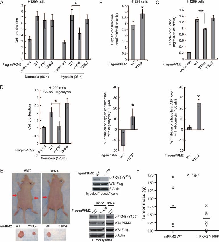
a significant decrease in the proliferation rate, oxygen consumption changes believed to be regulated by transcription factors, including hypoxia-
rate, and intracellular ATP concentration of Y105F rescue cells com- inducible factor 1 and Myc. However, the mechanism by which lactate
pared to those in cells rescued with mPKM2 wild type (Fig. 5D, left, production is increased in cancer cells harboring phospho-PKM2 with
middle, and right, respectively). Together, these data suggest that rescue low activity is unknown.
cells with a form of PKM2 that is catalytically more active (Y105F) rely It has been argued that the stoichiometry of tyrosine phosphorylation
more on oxidative phosphorylation for cell proliferation than do cells of glycolytic enzymes, including pyruvate kinase, is too low to affect their
with PKM2 wild type or the Y390F mutant. catalytic activity (8). Indeed, only a small fraction of PKM2 is phosphoryl-
We performed xenograft experiments in which we injected nude ated in FOP2-FGFR1–expressing KG-1a cells, which could not be vi-
mice with mPKM2 wild type and Y105F rescue H1299 cells. The mice sualized in isoelectric focusing (IEF) experiments (fig. S5A). However,
were injected with 10 million cells (PKM2 wild type rescue cells on the our “intermolecular”, or transprotein, FBP-release model suggests that a
left flank and Y105F cells on the right flank; n = 6) and monitored for single PKM2 molecule, when phosphorylated at Y105, can directly and
tumor growth over a 6-week period. The masses of tumors derived from transiently mediate FBP release from many PKM2 molecules, as pro-
Y105F rescue cells were significantly reduced compared to those of tumors posed by Christofk et al. (8). This would allow a small amount of phos-
formed by mPKM2 wild type rescue cells (Fig. 5, E and F); indeed, Y105F phorylated PKM2-Y105 to convert substantial amounts of PKM2 to the
rescue cells failed to form a tumor in one mouse (#874). These results dem- low-activity FBP-unbound state. However, the stoichiometry of PKM2
onstrate that the presence of PKM2 Y105F in cancer cells results in atten- tyrosine phosphorylation may vary in different cellular contexts. For ex-
uated tumor growth in vivo, suggesting that inhibitory phosphorylation at ample, our IEF experiment showed that FGFR1 wild type causes a stoi-
Y105 of PKM2 confers a proliferative advantage. choimetric shift (>90%) of PKM2 to a more phosphorylated form in 293T
cells, compared with cells expressing the FGFR1 KD control (fig. S5B).
Such high stoichiometry could potentially allow Y105 phosphorylation to
DISCUSSION
inhibit PKM2 in an “intramolecular manner,” in which Y105 phosphoryl-
Our finding that direct phosphorylation at Y105 inhibits PKM2 activity ation causes a conformational alteration within the same molecule of
provides new insight into the molecular mechanism underlying tyro- PKM2 to affect K433-dependent FBP binding. Pyruvate kinase transmits
sine kinase–dependent regulation of tumor cell metabolism. We iden- regulatory signals across large distances within a single PKM2 molecule,
tified PKM2 as a direct substrate of the oncogenic tyrosine kinase and the intersubunit interfaces are important for allosteric signal transmis-
FGFR1, which phosphorylates PKM2 at Y105. Consistent with these sion between the binding sites of the PKM2 substrate PEP and cofactor
findings, our colleagues at Cell Signaling Technologies (CST) have FBP (25, 26). Y105 is located on the interface between the A and C do-
found in phosphoproteomics-based studies that Y105 of PKM2 is phos- mains of PKM2, ~17 Å distal from FBP (fig. S5C). Because long-range
phorylated in human cancer cell lines established from different malig- allosteric regulation in PKM2 is possible, phosphorylation of Y105 could
nancies, including leukemias associated with the oncogenic tyrosine potentially transmit an allosteric signal to the FBP binding site within the
kinases BCR-ABL and FLT3, and solid tumors such as ovarian cancer, same PKM2 molecule, leading to decreased FBP binding. We hypothe-
glial tumor, lung cancer, and stomach cancer (24). Therefore, our finding size that such an allosteric signal could contribute to FBP release in
that phosphorylation of Y105 inhibits PKM2 activity may represent a PKM2 molecules that are Y105-phosphorylated and act in concert with
common, short-term molecular mechanism underlying the Warburg ef- the “intermolecular” model that may represent the predominant mecha-
fect in both leukemias and solid tumors, in addition to the long-term nism for phospho-Y105–dependent inhibition of PKM2.
www.SCIENCESIGNALING.org 17 November 2009 Vol 2 Issue 97 ra73 5RESEARCH ARTICLE
Fig. 5. Expression of PKM2 Y105F
mutant in H1299 cells leads to
decreased proliferation under
hypoxic conditions, increased
oxidative phosphorylation and
reduced tumor growth. (A) Res-
cue expression of mPKM2 Y105F
in H1299 cells results in reduced
cell proliferation under hypoxic
conditions (1%) but not at nor-
mal oxygen tension (normoxic;
17% oxygen) compared with
cells expressing PKM2 WT or
Y390F mutant. Cell proliferation
was determined by the increase
in cell number 96 hours after
seeding compared to that at
seeding for each cell line (T = 0).
Error bars represent the means ±
Downloaded from http://stke.sciencemag.org/ on August 25, 2021
SD from three independent exper-
iments. (B) Y105F rescue H1299
cells have a higher rate of oxygen
consumption than do cells with
mPKM2 WT. (C) Y105F rescue
H1299 cells show significantly
reduced lactate production un-
der normoxia. (D) Proliferation
(left), oxygen consumption rate
(middle), and intracellular ATP
concentration (right) of Y105F
rescue H1299 cells are signifi-
cantly decreased relative to cells
expressing PKM2 WT or the
Y390F mutant when treated with
oligomycin. (E) Upper left: Tu-
mors (indicated by red arrows)
in representative nude mice in-
jected with mPKM2 WT H1299
cells on the left flank and mPKM2
Y105F H1299 cells on the right
flank. Lower left: Dissected tu-
mors from the depicted nude
mice. Right: expression of Flag-
tagged mPKM2 WT and Y105F detected by immunoblotting in injected but not in tumors derived from Y105F-expressing cells. (F) H1299 cells ex-
rescue cells (upper) and tumor lysates (lower). Phosphorylation of PKM2 pressing PKM2 Y105F show significantly reduced tumor formation in xeno-
at Y105 was detected in tumors formed by cells expressing mPKM2 WT, graft nude mice (P value was determined by the paired Student’s t test).
Christofk et al. (8) proposed that binding of tyrosine-phosphorylated olism and proliferation than cells with WT mPKM2 is consistent with
proteins inhibits PKM2 by inducing the release of FBP. We found that previous observations, made by Christofk et al., when they replaced en-
FGFR1 binds to PKM2 in a tyrosine phosphorylation–dependent manner; dogenous hPKM2 with mouse PKM1 (mPKM1) in H1299 cells (8). Most
however, FGFR1 still binds to PKM2 K433E and Y105F mutants (fig. noticeably, both the PKM2 Y105F mutant and PKM1 are catalytically
S6), and both mutants are catalytically active and resistant to FGFR1- more active than PKM2 and are resistant to tyrosine kinase–dependent
dependent inhibition (Fig. 3D). This suggests that Y105 phosphorylation inhibition. These studies suggest that the physiological phosphorylation
is the predominant mechanism underlying FGFR1-dependent inhibition and dephosphorylation kinetics at Y105 of PKM2 may regulate the switch
of PKM2 through K433, and it is unlikely that the binding of FGFR1 between aerobic glycolysis and oxidative phosphorylation, perhaps by
to PKM2 affects PKM2 activity directly. Such an interaction may contrib- balancing the ratio between the active and inactive forms of PKM2. More-
ute to inhibition of PKM2 indirectly, because it may be required for over, because either knockdown of PKM2 (8) or replacement of PKM2 with
FGFR1 to phosphorylate Y105. the catalytically more active Y105F mutant (Fig. 5) or PKM1 (8) effectively
Our finding that cancer cells expressing the active mPKM2 Y105F attenuates cancer cell proliferation in vitro and in vivo, PKM2 may serve
mutant are more dependent on oxidative phosphorylation for cell metab- as an interesting therapeutic target in cancer treatment, such that either
www.SCIENCESIGNALING.org 17 November 2009 Vol 2 Issue 97 ra73 6RESEARCH ARTICLE
inhibition or activation of PKM2 may affect cancer cell metabolism and variants were infected with harvested lentivirus and were selected by pu-
cause tumor regression. romycin (2 mg/ml) for 1 week.
Antibodies
MATERIALS AND METHODS Antibodies against phospho-Tyr (pY99) and against FGFR1, c-ABL, and
FLT-3 were from Santa Cruz Biotechnology; antibodies against PKM2 and
Proteomics studies JAK2 were from Cell Signaling Technology; antibodies against GST, Flag,
Phosphopeptides were prepared with the PhosphoScan Kit (Cell Sig- and b-actin and Flag M2 beads were from Sigma. Specific antibody against
naling Technology). In brief, 2 × 10⁸ to 3 × 10⁸ Ba/F3 cells (~20 to phospho-PKM2 (Tyr105) was generated by Cell Signaling Technology.
40 mg of total protein) and cells that stably express distinct ZNF198-
FGFR1 variants were treated with IL-3 and serum withdrawal for 4 hours Purification of recombinant PKM2 proteins
before preparation of cell lysates as described (12). Protein extracts from Hexahistidine-tagged PKM2 proteins were purified by sonication of BL21
whole-cell lysates were trypsin-digested. Tyrosine-phosphorylated peptides (DE3)pLysS cells obtained from 250 ml of culture with IPTG (isopropyl-b-
were enriched by immunoaffinity purification (IAP) with antibody against D-thiogalactopyranoside) induction for 4 hours. Cell lysates were resolved
phosphotyrosine (P-Tyr-100) and analyzed by liquid chromatography by centrifugation and loaded onto a Ni-NTA column in 20 mM imidazole.
coupled with MS. Tandem mass spectra were collected in a data-dependent After washing twice, the protein was eluted with 250 mM imidazole. Pro-
manner with an LTQ ion trap mass spectrometer (ThermoFinnigan). teins were desalted on a PD-10 column and the purification efficiency was
examined by Coomassie staining and Western blotting.
Downloaded from http://stke.sciencemag.org/ on August 25, 2021
Reagents
Tyrosine kinase inhibitor (TKI258) was provided by Novartis Pharma. PKM2 enzyme assay
Short hairpin RNA (shRNA) constructs for PKM2 knockdown were Pyruvate kinase activity was measured by an LDH-coupled enzyme as-
purchased from Open Biosystems. The nonphospho- and phosphopep- say (27). The assay was carried out with 1 mg of cell lysates or 20 ng of
tides were synthesized by American Peptide Company. Murine PKM2 recombinant PKM2 with an enzyme buffer [50 mM tris-HCl, 100 mM
was Flag-tagged by polymerase chain reaction and subcloned into KCl, 5 mM MgCl2, 1 mM ADP, 0.5 mM PEP, 0.2 mM NADH (reduced
pLHCX retroviral vector. PKM2 variants were subcloned into pDEST27 form of NAD+), and 8 U of LDH]. The decrease in absorbance at 340 nm
and pET100 vectors (Invitrogen) for GST-tagged PKM2 expression in from the oxidation of NADH was measured as pyruvate kinase activity by
mammalian cells and histidine (His)-tagged PKM2 expression in bacterial a spectrophotometer. For the peptide competition assay, 1 mM recombinant
cells, respectively. Mutations Y83F, Y105F, Y148F, Y175F, Y370F, and His-PKM2 was incubated with or without 10 mM FBP (Sigma) for 30 min
Y390F were introduced into PKM2 with QuikChange-XL site-directed mu- at room temperature in a dialysis buffer containing 50 mM tris-HCl (pH
tagenesis kit (Stratagene). 7.5), 100 mM KCl, 5 mM MgCl2, and 5 % glycerol. After incubation,
samples were dialysed against 2 liters of the dialysis buffer with 10,000
Cell culture molecular weight cutoff (MWCO) dialysis cassettes (Pierce) for 7 hours.
H1299, A549, MDA-MB-134, MDA-MB231, HEL, KG-1a, Mo91, FBP-loaded samples were divided into four samples and incubated with
Molm14, and K562 cells were cultured in RPMI 1640 medium with each peptide (mock, pY390, Y105, and pY105) at the final concentration
10% fetal bovine serum (FBS). 293T and GP2-293 cells were cultured of 1.5 mM for 30 min at room temperature, and each sample was subjected
in Dulbecco’s modified Eagle’s medium (DMEM) with 10% FBS. LNCaP to the PKM2 enzyme assay as described above.
and 22Rv cells were cultured in RPMI 1640 medium with 10% FBS,
1 mM sodium pyruvate, and 10 mM Hepes. PC3 cells were cultured in [14C]FBP dialysis assay
F12 Kaighn’s medium with 5% FBS. Du145 cells were cultured in min- Recombinant His-tagged PKM2 (1 mM) was incubated with 10 mM
imum essential medium with 5% FBS, NaHCO3 (1.5 g/liter), 0.1 mM [14C]FBP (MP Biomedicals) for 30 min at room temperature in a dial-
nonessential amino acids, and 1 mM sodium pyruvate. In the cell prolif- ysis buffer containing 50 mM tris-HCl (pH 7.5), 100 mM KCl, 5 mM
eration assay, 5 × 10⁴ cells were seeded in a six-well plate and cultured at MgCl2, and 5 % glycerol. After incubation, samples were dialysed against
37°C in normoxia (5% CO2 and 95% air). Twenty-four hours after seed- 2 liters of dialysis buffer with 10,000 MWCO dialysis cassettes (Pierce) for
ing, cells used in hypoxia experiments were incubated at 37°C in a sealed 7 hours. The dialysed samples were divided into four samples and incu-
hypoxia chamber filled with 1% O2, 5% CO2, and 94% N2. Cells used for bated with each peptide (mock, pY390, Y105, and pY105) at the final con-
oligomycin treatment were incubated at 37°C under normoxic condition. centration of 1.5 mM for 30 min at room temperature, and each sample was
To generate the PKM2 rescue H1299 cell lines, Flag-tagged mouse PKM2 redialysed against 2 liters of the dialysis buffer with 10,000 MWCO dial-
wild type, Y105F, and Y390F were cloned into the retroviral vector ysis cassettes for 7 hours. After redialysis, samples were recovered and the
pLHCX (Clontech). The constructs were cotransfected with pAmpho cas- amount of [14C]FBP was measured by scintillation counting.
sette vector (Clontech) into GP2-293 cells. Retrovirus was harvested 48
hours after transfection. H1299 cells were infected with harvested retro- In vitro kinase assay
virus and were selected by hygromycin (300 mg/ml) for 2 weeks. For len- GST-PKM2 construct was transfected into 293T cells with Lipofectamine
tiviral infection to knock down endogenous hPKM2, shRNA construct 2000 (Invitrogen). Cells were lysed 24 hours after transfection, and GST-
was obtained from Open Biosystems. The sequence of shRNA used for PKM2 was pulled down by Glutathione Sepharose 4B beads (Amersham
knockdown is as follows: 5′-CCGGGCTGTGGCTCTAGACACTAA- Bioscience), followed by treatment of 50 U of YOP phosphatase (New En-
ACTCGAGTTTAGTGTCTAGAGCCACAGCTTTTTG-3′. The shRNA gland Biolab) at 30°C for 1 hour in bovine serum albumin (1 mg/ml) and
construct was cotransfected with two packaging plasmids (pCMV-VSV- 1 × YOP reaction buffer containing 50 mM tris (pH 7.0), 100 mM NaCl,
G and pCMV-dR8.2 dvpr) into 293T cells. Lentivirus was harvested 48 2 mM Na2EDTA, and 5 mM dithiothreitol (DTT). The beads were then
hours after transfection. H1299 cells stably expressing Flag-tagged PKM2 washed with PBS and subjected to FGFR1 kinase assay according to
www.SCIENCESIGNALING.org 17 November 2009 Vol 2 Issue 97 ra73 7RESEARCH ARTICLE
manufacturer’s protocol (Invitrogen). In brief, the YOP-treated beads were 9. M. Guminska, J. Ignacak, T. Kedryna, M. B. Stachurska, Tumor-specific pyruvate ki-
nase isoenzyme M2 involved in biochemical strategy of energy generation in neoplas-
incubated with 100 ng of recombinant FGFR1 for 30 min at room tempera- tic cells. Acta Biochim. Pol. 44, 711–724 (1997).
ture in FGFR1 kinase buffer [10 mM Hepes (pH 7.5), 150 mM NaCl, 5 mM 10. Y. Kumar, N. Tapuria, N. Kirmani, B. R. Davidson, Tumour M2-pyruvate kinase: A
DTT, 0.01 % Triton X-100, 10 mM MnCl2, and 200 mM ATP]. The samples gastrointestinal cancer marker. Eur. J. Gastroenterol. Hepatol. 19, 265–276 (2007).
were electrophoresed on 10% SDS–acrylamide gel, transferred onto a nitro- 11. J. Chen, D. J. Deangelo, J. L. Kutok, I. R. Williams, B. H. Lee, M. Wadleigh, N. Duclos,
S. Cohen, J. Adelsperger, R. Okabe, A. Coburn, I. Galinsky, B. Huntly, P. S. Cohen,
cellulose membrane, and then detected with antibody against phosphotyro-
T. Meyer, D. Fabbro, J. Roesel, L. Banerji, J. D. Griffin, S. Xiao, J. A. Fletcher, R. M.
sine (pY99) and specific antibody against phospho-PKM2 (Y105). Stone, D. G. Gilliland, PKC412 inhibits the zinc finger 198-fibroblast growth factor
receptor 1 fusion tyrosine kinase and is active in treatment of stem cell myeloprolifera-
Lactate production, oxygen consumption, and tive disorder. Proc. Natl. Acad. Sci. U.S.A. 101, 14479–14484 (2004).
intracellular ATP assays 12. J. Rush, A. Moritz, K. A. Lee, A. Guo, V. L. Goss, E. J. Spek, H. Zhang, X. M. Zha, R. D.
Polakiewicz, M. J. Comb, Immunoaffinity profiling of tyrosine phosphorylation in cancer
Cellular lactate production was measured under normoxia with a fluorescence- cells. Nat. Biotechnol. 23, 94–101 (2005).
based lactate assay kit (MBL). Phenol red–free RPMI medium without FBS 13. S. Xiao, S. R. Nalabolu, J. C. Aster, J. Ma, L. Abruzzo, E. S. Jaffe, R. Stone, S. M.
was added to a six-well plate of subconfluent cells and incubated for 1 hour Weissman, T. J. Hudson, J. A. Fletcher, FGFR1 is fused with a novel zinc-finger gene,
at 37°C. After incubation, 1 ml of medium from each well was assessed with ZNF198, in the t(8;13) leukaemia/lymphoma syndrome. Nat. Genet. 18, 84–87 (1998).
14. T. L. Gu, V. L. Goss, C. Reeves, L. Popova, J. Nardone, J. Macneill, D. K. Walters,
the lactate assay kit. Cell numbers were counted by a microscope (×40). The Y. Wang, J. Rush, M. J. Comb, B. J. Druker, R. D. Polakiewicz, Phosphotyrosine profiling
oxygen consumption assay was performed as described previously (8). In- identifies the KG-1 cell line as a model for the study of FGFR1 fusions in acute myeloid
tracellular ATP concentration was measured by an ATP bioluminescent so- leukemia. Blood 108, 4202–4204 (2006).
matic cell assay kit (Sigma). 15. S. Xiao, J. G. McCarthy, J. C. Aster, J. A. Fletcher, ZNF198-FGFR1 transforming
activity depends on a novel proline-rich ZNF198 oligomerization domain. Blood 96,
Downloaded from http://stke.sciencemag.org/ on August 25, 2021
699–704 (2000).
Xenograft studies 16. M. S. Kobrin, Y. Yamanaka, H. Friess, M. E. Lopez, M. Korc, Aberrant expression of
Nude mice (nu/nu, male, 6 to 8 weeks old, Charles River Laboratories) were type I fibroblast growth factor receptor in human pancreatic adenocarcinomas.
subcutaneously injected with 10 × 106 H1299 cells stably expressing Cancer Res. 53, 4741–4744 (1993).
17. Y. A. Luqmani, M. Graham, R. C. Coombes, Expression of basic fibroblast growth
mPKM2 wild type and Y105F mutant in conjunction with stable knock-
factor, FGFR1 and FGFR2 in normal and malignant human breast, and comparison
down of endogenous hPKM2 on the left and right flanks, respectively. Tu- with other normal tissues. Br. J. Cancer 66, 273–280 (1992).
mor formation was assessed every 2 to 3 days. Tumor growth was recorded 18. F. Penault-Llorca, F. Bertucci, J. Adelaide, P. Parc, F. Coulier, J. Jacquemier, D. Birnbaum,
by measuring two perpendicular diameters of the tumors over a 6-week time O. deLapeyriere, Expression of FGF and FGF receptor genes in human breast cancer.
course with the formula 4p/3 × (width/2)2 × (length/2). The tumors were Int. J. Cancer 61, 170–176 (1995).
19. R. S. Morrison, F. Yamaguchi, J. M. Bruner, M. Tang, W. McKeehan, M. S. Berger,
harvested and weighed at the experimental endpoint, and the masses of tu- Fibroblast growth factor receptor gene expression and immunoreactivity are elevated
mors (grams) derived from cells expressing mPKM2 wild type or Y105F in human glioblastoma multiforme. Cancer Res. 54, 2794–2799 (1994).
mutant in both flanks of each mouse were compared. Statistical analyses 20. J. C. Pole, C. Courtay-Cahen, M. J. Garcia, K. A. Blood, S. L. Cooke, A. E. Alsop,
were done in comparison to the control group with a paired Student’s t test. D. M. Tse, C. Caldas, P. A. Edwards, High-resolution analysis of chromosome rear-
rangements on 8p in breast, colon and pancreatic cancer reveals a complex pattern
of loss, gain and translocation. Oncogene 25, 5693–5706 (2006).
SUPPLEMENTARY MATERIALS 21. J. S. Reis-Filho, P. T. Simpson, N. C. Turner, M. B. Lambros, C. Jones, A. Mackay,
A. Grigoriadis, D. Sarrio, K. Savage, T. Dexter, M. Iravani, K. Fenwick, B. Weber,
www.sciencesignaling.org/cgi/content/full/2/97/ra73/DC1
D. Hardisson, F. C. Schmitt, J. Palacios, S. R. Lakhani, A. Ashworth, FGFR1 emerges
Fig. S1. Six tyrosine residues of PKM2 are phosphorylated in cells harboring active
as a potential therapeutic target for lobular breast carcinomas. Clin. Cancer Res. 12,
FGFR1 mutants.
6652–6662 (2006).
Fig. S2. FGFR1 directly phosphorylates PKM2.
22. L. Marek, K. E. Ware, A. Fritzsche, P. Hercule, W. R. Helton, J. E. Smith, L. A. McDermott,
Fig. S3. ABL, JAK2, and FLT3 effectively and directly phosphorylate PKM2 at Y105 in vitro,
C. D. Coldren, R. A. Nemenoff, D. T. Merrick, B. A. Helfrich, P. A. Bunn Jr., L. E. Heasley,
whereas epidermal growth factor receptor (EGFR) is less effective.
Fibroblast growth factor (FGF) and FGF receptor-mediated autocrine signaling in non-
Fig. S4. Presence of the PKM2 Y105F mutant in lung cancer H1299 cells leads to de-
small-cell lung cancer cells. Mol. Pharmacol. 75, 196–207 (2009).
creased proliferation under hypoxic conditions.
23. Abbreviations for the amino acids are as follows: A, Ala; D, Asp; E, Glu; F, Phe; H,
Fig. S5. Y105 phosphorylation may affect FBP-binding to PKM2 in an “intra-molecular
His; I, Ile; L, Leu; P, Pro; Q, Gln; R, Arg; S, Ser; V, Val; and Y, Tyr.
manner” in cellular contexts with high tyrosine phosphorylation stoichiometry of PKM2.
24. http://www.phosphosite.org/siteAction.do?id=10858.
Fig. S6. FGFR1 binds PKM2 in a tyrosine phosphorylation–dependent manner but this
25. A. Mattevi, M. Bolognesi, G. Valentini, The allosteric regulation of pyruvate kinase.
binding is dispensable for FGFR1-dependent inhibition of PKM2.
FEBS Lett. 389, 15–19 (1996).
26. A. Mattevi, M. Rizzi, M. Bolognesi, New structures of allosteric proteins revealing re-
REFERENCES AND NOTES markable conformational changes. Curr. Opin. Struct. Biol. 6, 824–829 (1996).
1. M. C. Brahimi-Horn, J. Chiche, J. Pouyssegur, Hypoxia signalling controls metabolic 27. N. Shimada, T. Shinagawa, S. Ishii, Modulation of M2-type pyruvate kinase activity by
demand. Curr. Opin. Cell Biol. 19, 223–229 (2007). the cytoplasmic PML tumor suppressor protein. Genes Cells 13, 245–254 (2008).
2. G. Kroemer, J. Pouyssegur, Tumor cell metabolism: Cancer’s Achilles’ heel. Cancer 28. We gratefully acknowledge the critical reading of the manuscript by B. J. P. Huntly. Plas-
Cell 13, 472–482 (2008). mid encoding murine PKM2 was provided by H. Fujii at New York University. This work
3. D. Hanahan, R. A. Weinberg, The hallmarks of cancer. Cell 100, 57–70 (2000). was supported in part by NIH grant CA120272 (J.C.), the American Cancer Society (J.C.),
4. H. R. Hathurusinghe, K. S. Goonetilleke, A. K. Siriwardena, Current status of tumor and the Multiple Myeloma Research Foundation (J.C. and S.L.). S.K. is a special fellow
M2 pyruvate kinase (tumor M2-PK) as a biomarker of gastrointestinal malignancy. of the Leukemia and Lymphoma Society. J.C. is a Georgia Cancer Coalition Distin-
Ann. Surg. Oncol. 14, 2714–2720 (2007). guished Cancer Scholar and an American Cancer Society Basic Research Scholar.
5. S. Mazurek, C. B. Boschek, F. Hugo, E. Eigenbrodt, Pyruvate kinase type M2 and its
role in tumor growth and spreading. Semin. Cancer Biol. 15, 300–308 (2005). Submitted 15 May 2009
6. S. Mazurek, H. Grimm, C. B. Boschek, P. Vaupel, E. Eigenbrodt, Pyruvate kinase type Accepted 26 October 2009
M2: A crossroad in the tumor metabolome. Br. J. Nutr. 87 (Suppl. 1), S23–S29 (2002). Final Publication 17 November 2009
7. H. R. Christofk, M. G. Vander Heiden, M. H. Harris, A. Ramanathan, R. E. Gerszten, 10.1126/scisignal.2000431
R. Wei, M. D. Fleming, S. L. Schreiber, L. C. Cantley, The M2 splice isoform of pyruvate Citation: T. Hitosugi, S. Kang, M. G. Vander Heiden, T.-W. Chung, S. Elf, K. Lythgoe,
kinase is important for cancer metabolism and tumour growth. Nature 452, 230–233 S. Dong, S. Lonial, X. Wang, G. Z. Chen, J. Xie, T.-L. Gu, R. D. Polakiewicz, J. L. Roesel,
(2008). T. J. Boggon, F. R. Khuri, D. G. Gilliland, L. C. Cantley, J. Kaufman, J. Chen, Tyrosine
8. H. R. Christofk, M. G. Vander Heiden, N. Wu, J. M. Asara, L. C. Cantley, Pyruvate phoshorylation inhibits PKM2 to promote the Warburg effect and tumor growth. Sci. Signal.
kinase M2 is a phosphotyrosine-binding protein. Nature 452, 181–186 (2008). 2, ra73 (2009).
www.SCIENCESIGNALING.org 17 November 2009 Vol 2 Issue 97 ra73 8Tyrosine Phosphorylation Inhibits PKM2 to Promote the Warburg Effect and Tumor Growth
Taro Hitosugi, Sumin Kang, Matthew G. Vander Heiden, Tae-Wook Chung, Shannon Elf, Katherine Lythgoe, Shaozhong Dong,
Sagar Lonial, Xu Wang, Georgia Z. Chen, Jianxin Xie, Ting-Lei Gu, Roberto D. Polakiewicz, Johannes L. Roesel, Titus J.
Boggon, Fadlo R. Khuri, D. Gary Gilliland, Lewis C. Cantley, Jonathan Kaufman and Jing Chen
Sci. Signal. 2 (97), ra73.
DOI: 10.1126/scisignal.2000431
A Malignant Metabolic Switch
Cancer cells show aberrant metabolism, consuming more glucose than do healthy cells and producing lactate
even in the presence of abundant oxygen, rather than shifting to oxidative phosphorylation. This phenomenon is called
the Warburg effect, after Otto Warburg, who described it many years ago. Building on recent research implicating
Downloaded from http://stke.sciencemag.org/ on August 25, 2021
inhibition of the M2 isoform of the glycolytic enzyme pyruvate kinase (PKM2) by phosphotyrosine binding as critical to the
Warburg effect −−and tumorigenesis−−Hitosugi et al. explored the role of signaling from oncogenic forms of the fibroblast
growth factor receptor type 1 (FGFR1) in mediating this metabolic switch. They found that FGFR1, a receptor tyrosine
kinase, phosphorylated a tyrosine residue (Y105) on PKM2 itself. Further analysis revealed that this tyrosine residue was
commonly phosphorylated in human cancers and that a mutant form of PKM2 lacking this tyrosine residue inhibited both
''Warburg metabolism'' and tumor growth. They thus propose that phosphorylation of PKM2 by oncogenic tyrosine
kinases provides the very phosphotyrosine that binds to and inhibits PKM2 to induce the Warburg effect and promote
tumor growth.
ARTICLE TOOLS http://stke.sciencemag.org/content/2/97/ra73
SUPPLEMENTARY http://stke.sciencemag.org/content/suppl/2009/11/16/2.97.ra73.DC1
MATERIALS
Use of this article is subject to the Terms of Service
Science Signaling (ISSN 1937-9145) is published by the American Association for the Advancement of Science, 1200 New
York Avenue NW, Washington, DC 20005. The title Science Signaling is a registered trademark of AAAS.
Copyright © 2009, American Association for the Advancement of ScienceRELATED http://stke.sciencemag.org/content/sigtrans/2/97/pe75.full
CONTENT
http://science.sciencemag.org/content/sci/324/5930/1029.full
http://stke.sciencemag.org/content/sigtrans/2/101/pc23.full
http://stke.sciencemag.org/content/sigtrans/3/104/ec8.abstract
http://stke.sciencemag.org/content/sigtrans/3/124/pe19.full
http://stke.sciencemag.org/content/sigtrans/4/154/eg1.full
http://stke.sciencemag.org/content/sigtrans/4/176/ec157.abstract
http://stke.sciencemag.org/content/sigtrans/4/179/ec182.abstract
http://stke.sciencemag.org/content/sigtrans/4/202/ec337.abstract
http://stke.sciencemag.org/content/sigtrans/3/140/ec289.abstract
http://stke.sciencemag.org/content/sigtrans/5/254/ec323.abstract
http://stke.sciencemag.org/content/sigtrans/4/202/ec338.abstract
http://stke.sciencemag.org/content/sigtrans/5/216/ec81.abstract
http://stke.sciencemag.org/content/sigtrans/4/188/ec240.abstract
http://stke.sciencemag.org/content/sigtrans/1/11/ec97.abstract
http://stke.sciencemag.org/content/sigtrans/4/187/ec231.abstract
http://stke.sciencemag.org/content/sigtrans/3/103/ec5.abstract
http://stke.sciencemag.org/content/sigtrans/2007/381/pe14.full
http://stke.sciencemag.org/content/sigtrans/6/260/ec27.abstract
http://stke.sciencemag.org/content/sigtrans/6/263/pe7.full
http://stke.sciencemag.org/content/sigtrans/7/343/ec253.abstract
http://stke.sciencemag.org/content/sigtrans/8/375/ec116.abstract
http://stke.sciencemag.org/content/sigtrans/3/151/eg12.full
Downloaded from http://stke.sciencemag.org/ on August 25, 2021
REFERENCES This article cites 25 articles, 7 of which you can access for free
http://stke.sciencemag.org/content/2/97/ra73#BIBL
PERMISSIONS http://www.sciencemag.org/help/reprints-and-permissions
Use of this article is subject to the Terms of Service
Science Signaling (ISSN 1937-9145) is published by the American Association for the Advancement of Science, 1200 New
York Avenue NW, Washington, DC 20005. The title Science Signaling is a registered trademark of AAAS.
Copyright © 2009, American Association for the Advancement of ScienceYou can also read