Chrysin induces autophagy through the inactivation of the ROS mediated Akt/mTOR signaling pathway in endometrial cancer
←
→
Page content transcription
If your browser does not render page correctly, please read the page content below
INTERNATIONAL JOURNAL OF MOLECULAR MEDICINE 48: 172, 2021
Chrysin induces autophagy through the inactivation
of the ROS‑mediated Akt/mTOR signaling
pathway in endometrial cancer
YU HE1*, YUCHUAN SHI1*, YANG YANG2, HUANHUAN HUANG1,
YIFAN FENG1, YUNMENG WANG1, LEI ZHAN1 and BING WEI1
Departments of 1Gynecology and Obstetrics and 2Clinical Pharmacology,
The Second Affiliated Hospital of Anhui Medical University, Hefei, Anhui 230601, P.R. China
Received January 5, 2021; Accepted May 27, 2021
DOI: 10.3892/ijmm.2021.5005
Abstract. Endometrial cancer (EC) is widely known as an via the inactivation of the ROS‑mediated Akt/mTOR signaling
aggressive malignancy. Due to the limited therapeutic options pathway in EC cells.
and poor prognosis of patients with advanced‑stage EC,
there is a need to identify effective alternative treatments. Introduction
Chrysin is a naturally active flavonoid (5,7‑dihydroxyflavone),
which has been demonstrated to exert anticancer effects and Endometrial cancer (EC) is considered one of the most
may present a novel strategy for EC treatment. However, common gynecological malignancies in industrialized coun‑
the role of chrysin in EC remains largely unclear. The aim tries (1). Primarily due to relevant risk factors, including
of the present study was to examine the anticancer effects obesity, diabetes and aging, among others, its incidence is
of chrysin on EC. The results revealed that, in addition to gradually increasing (2,3). Recurrent or advanced EC responds
apoptosis, chrysin increased the LC3II expression levels and poorly to treatment. Few therapeutic options are available for
markedly accelerated the autophagic flux, suggesting that such patients, who therefore have low survival rates and a
chrysin induced both the autophagy and apoptosis of EC poor prognosis (4). The 5‑year survival rate for patients with
cells. Furthermore, the inhibition of autophagy by chloro‑ stage IV disease is limited only at 0‑10% (5). Accordingly, EC
quine enhanced the inhibitory effect on cell proliferation and is increasingly recognized as a serious health and epidemio‑
the promotion of the chrysin‑induced apoptosis of EC cells, logical concern. Thus, novel therapeutic approaches that can
indicating that chrysin‑induced autophagy was a cytoprotec‑ effectively reduce morbidity and mortality in EC are urgently
tive mechanism. Additionally, chrysin led to the production of required.
intracellular reactive oxygen species (ROS). N‑acetylcysteine In recent years, natural compounds for cancer prevention
(NAC) pretreatment significantly inhibited chrysin‑induced and treatment have gained attention due to their anticancer
autophagy, suggesting that ROS activated autophagy induced activity and safety, and are a rich source of phytochemicals.
by chrysin in EC cells. Furthermore, the phosphorylated (p‑) They are an integral part of the human diet (6,7). The use
Akt and p‑mTOR levels were significantly decreased in a of natural compounds combined with targeted drugs may
concentration‑dependent manner following treatment with provide new perspectives for the development of anticancer
chrysin, while NAC blocked these effects. Taken together, drugs. Chrysin (5,7‑dihydroxyflavone; chemical structure
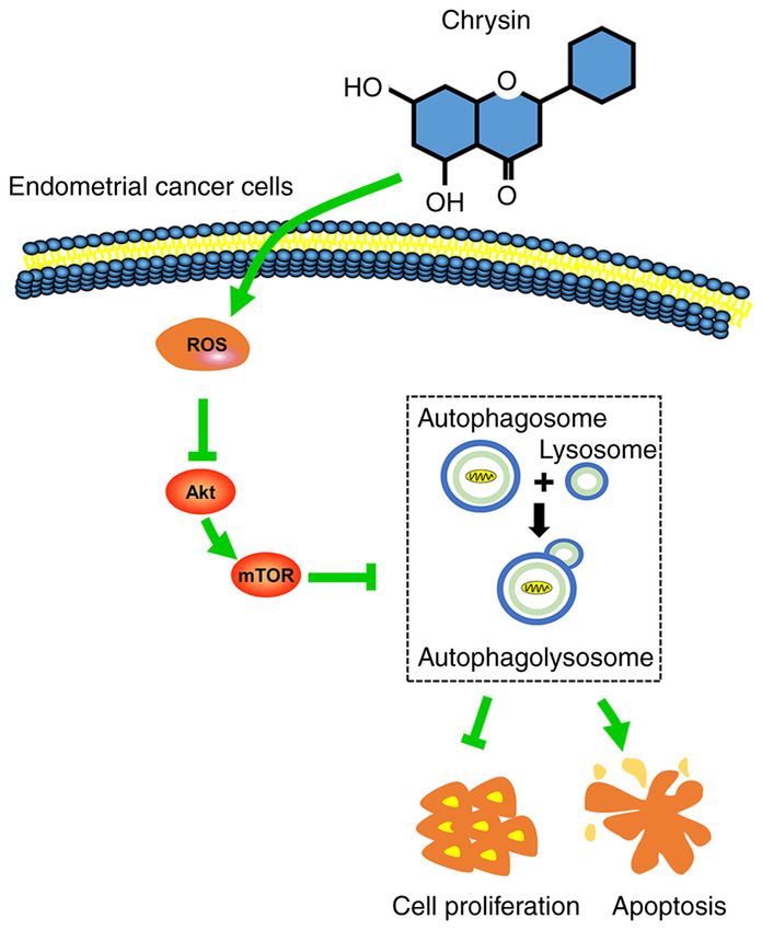
these findings demonstrated that chrysin‑induced autophagy shown in Fig. 1A) is a natural dietary flavonoid, commonly
present in various plant extracts, including honey and propolis.
It has a notable medicinal functions and economic value (8).
Additionally, chrysin has diverse biological properties, specifi‑
cally including anticancer, antioxidant, anti‑inflammatory,
antibacterial, anti‑diabetic and anti‑allergenic effects (9,10).
Correspondence to: Dr Bing Wei, Department of Gynecology Recently, several studies have reported that chrysin exerts
and Obstetrics, The Second Affiliated Hospital of Anhui Medical
its cancer‑suppressive effects on breast, lung, cervical and
University, 678 Furong Road, Hefei, Anhui 230601, P.R. China
E‑mail: m1351565@163.com
bladder cancer cells through regulating multiple cell signaling
pathways selectively (11‑15). However, the anti‑EC possible
*
Contributed equally mechanisms of chrysin have not yet been fully elucidated.
Cancer cell death is often caused by apoptosis, which is
Key words: endometrial cancer, chrysin, autophagy, apoptosis, considered to be the principal anticancer mechanism (16).
reactive oxygen species, Akt/mTOR However, it has been documented that autophagy and apop‑
tosis are intertwined processes (17). Autophagy is the process
of engulfing and degrading cytosolic proteins and damaged
2 HE et al: CHRYSIN INDUCES AUTOPHAGY IN ENDOMETRIAL CANCER
organelles (18). It has been reported that the inhibition of Gibco; Thermo Fisher Scientific, Inc.) at 37˚C in a 95% O2 and
autophagy increases the anticancer efficiency by inducing the 5% CO2 atmosphere in a humidified incubator.
apoptotic process (19). In addition, reactive oxygen species
(ROS) have been found to be the associated factors in the Cell viability assay. The Cell Counting Kit‑8 (CCK‑8;
anticancer effects of chrysin‑induced autophagy. For example, Biosharp Life Sciences) was used to analyze the viability
Lin et al (20) indicated that chrysin induced ROS production of the HEC‑1A and Ishikawa cells following treatment with
and then autophagy, resulting in the attenuation of the viability chrysin. Briefly, 5x10³ cells in 100 µl medium were plated into
of human colorectal cancer cells. This suggests that there each well of a 96‑well plate until they became adherent. In
may be a link among anticancer drugs, such as chrysin, ROS experiments evaluating the effect of chrysin alone, cells were
levels and autophagy pathways in EC. However, to date, at treated with chrysin (0, 10, 20, 40 and 80 µM) for 24, 48 or
least to the best of our knowledge, there are no reports on the 72 h. In the experiments that evaluated the combined effect of
pharmacological mechanisms of chrysin in EC, particularly chrysin and CQ, cells were pretreated with 5 µM CQ for 1 h
concerning its role in the regulation of cell ROS levels and prior to exposure to 40 µM chrysin, and the cells were then
autophagy pathways, which remains unclear. further incubated for 48 h at 37˚C. Subsequently, 10% CCK‑8
The present study thus aimed to investigate the molecular solution (the ratio of volume of medium and CCK‑8 was 9:1)
mechanisms of the anticancer role of chrysin and focused on was added to the culture medium. Following further incuba‑
how the compound regulates the autophagy pathway in EC tion for 1 h at 37˚C in the dark, a Varioskan LUX microplate
cells. reader (Thermo Fisher Scientific, Inc.) was used to measure
the absorbance at 450 nm in each well.
Materials and methods
Colony formation assay. HEC‑1A and Ishikawa cells were
Reagents and antibodies. Chrysin and LY294002 were inoculated into 6‑well plates at a low cell density (1,000 cells
purchased from MedChemExpress. N‑acetylcysteine (NAC) per well) and incubated at 37˚C overnight. The cells were
and chloroquine (CQ) were obtained from Sigma‑Aldrich the exposed to 40 µM chrysin and cultured for ~1 week. The
(Sigma‑Aldrich; Merck KGaA). The cells were pretreated medium was replaced every 3 days, in order to maintain stable
with 5 µM CQ, or 10 µM LY294002, or 10 mM NAC for 1 h chrysin concentration levels. The plates were periodically
and were then treated with chrysin for additional 24 or 48 h. observed until distinct colonies were formed and then fixed
The antibodies used for western blotting included mono‑ with 4% paraformaldehyde. After staining with 0.1% crystal
clonal anti‑LC3 (dilution 1:1,000; cat. no. ab192890; Abcam), violet solution (Beyotime Institute of Biotechnology) for
monoclonal anti‑sequestosome‑1/p62 (dilution 1:1,000; 15 min at room temperature, the colonies (>50 cells per colony)
cat. no. ab207305; Abcam), monoclonal anti‑autophagy‑related were counted with ImageJ software (version 1.38; National
gene 5 (ATG5; dilution 1:1,000, cat. no. ab108327; Abcam), Institutes of Health).
polyclonal anti‑Beclin 1 (dilution 1:1,000; cat. no. AF5128;
Affinity Biosciences), polyclonal anti‑Bcl‑2 (dilution 1:1,000; Flow cytometric analysis. Apoptotic cells were detected by
cat. no. AF6139; Affinity Biosciences), polyclonal anti‑Bax flow cytometry and quantified according to the percentage of
(dilution 1:1,000; cat. no. AF0120; Affinity Biosciences), apoptotic cells in each group. Briefly, HEC‑1A and Ishikawa
monoclonal anti‑Akt (dilution 1:1,000; cat. no. 4691; Cell cells that had been treated with the indicated concentrations of
Signaling Technology, Inc.), monoclonal anti‑phosphorylated chrysin with or without 5 µM CQ were digested with trypsin
(p)‑Akt (Ser473; dilution 1:1,000; cat. no. 4060; Cell Signaling without EDTA and harvested. Subsequently, the cells were
Technology, Inc.), monoclonal anti‑mTOR (dilution 1:1,000; resuspended in 100 µl binding buffer, and incubated with 5 µl
cat. no. 2983; Cell Signaling Technology, Inc.), monoclonal Annexin V‑FITC and 10 µl propidium iodide (PI) for 15 min
anti‑p‑Mtor (Ser2448; dilution 1:1,000; cat. no. 5536; Cell at room temperature in the dark. The apoptotic ratio was
Signaling Technology, Inc.) and polyclonal anti‑GAPDH measured using a Navios flow cytometer (Beckman Coulter,
(dilution 1:10,000; cat. no. AF7021; Affinity Biosciences). Inc.). The upper and lower right quadrants on the dot‑plot
Horseradish peroxidase‑conjugated goat anti‑rabbit IgG graphs represented late and early apoptotic cells, respectively.
(dilution 1:10,000; cat. no. ZB‑2301) and goat anti‑mouse IgG
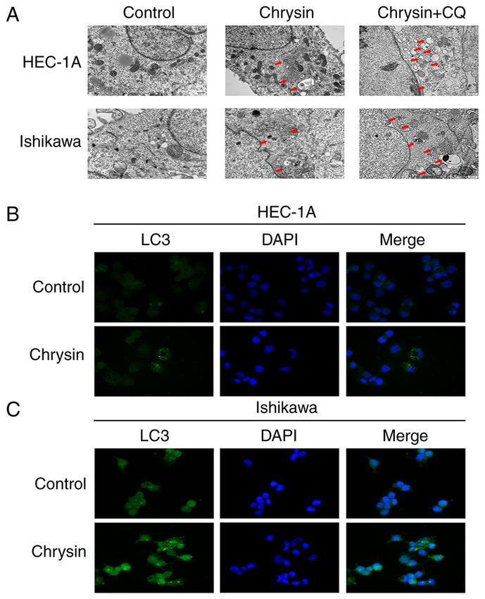
(dilution 1:10,000; cat. no. ZB‑2305) secondary antibodies were Transmission electron microscopy (TEM) examination. To
purchased from Beijing Zhongshan Jinqiao Biotechnology, observe the cell morphological changes of autophagosomes
Co., Ltd. and autolysosomes, TEM was performed. Briefly, HEC‑1A and
Ishikawa cells, following exposure to 40 µM chrysin for 48 h,
Cell lines and cell culture. The human endometrioid adenocar‑ were fixed with 2.5% glutaraldehyde at 4˚C overnight, and then
cinoma cell line, HEC‑1A (cat. no. HTB‑112), was purchased post‑fixed with 1% osmium tetroxide (OsO4) for 1 h followed
from the American Type Culture Collection (ATCC) and by incubation with 2% uranyl acetate at room temperature for
the endometrioid adenocarcinoma cell line, Ishikawa a further 1 h. After washing with phosphate‑buffered saline
(cat. no. 99040201), was purchased from the European (PBS; Gibco; Thermo Fisher Scientific, Inc.), the cells were
Collection of Authenticated Cell Cultures (ECACC). Cells dehydrated in an ethanol series, infiltrated with propylene
were maintained in high‑glucose DMEM (Gibco; Thermo oxide and finally embedded in epoxy resin. Ultrathin sections
Fisher Scientific, Inc.) supplemented with 10% v/v FBS (70 nm) were prepared, stained with uranyl acetate and lead
(Gibco; Thermo Fisher Scientific, Inc.) and penicillin/strep‑ citrate, and then examined in a JEM‑1400 TEM system
tomycin (100 U/ml penicillin and 100 µg/ml streptomycin; (JEOL, Ltd.).
INTERNATIONAL JOURNAL OF MOLECULAR MEDICINE 48: 172, 2021                                            3

                                                          Figure 1. Continued.

Western blotting. The cells were lysed with RIPA lysis buffer         with 0.5% (v/v) Triton X‑100 for 15 min at room tempera‑
(Beyotime Institute of Biotechnology), and the supernatant            ture, the cells were blocked with 5% bovine serum albumin
was collected by centrifugation at 13,000 x g for 30 min at 4˚C       (Beyotime Institute of Biotechnology) for 30 min and incubated
in order to extract total cellular protein. After quantifying the     with anti‑LC3 antibody (dilution 1:200; cat. no. ab192890;
protein concentration in the supernatant using a BCA protein          Abcam) overnight at 4˚C. Finally, the cells were incubated
assay kit (Beyotime Institute of Biotechnology), total protein        with the FITC‑labeled goat anti‑rabbit IgG (H+L) secondary
was mixed by loading buffer and boiled at 100˚C for 5 min. The        antibody (dilution 1:400; cat. no. A0562; Beyotime Institute of
denatured cell proteins (10‑20 µg) were separated on 6‑15%            Biotechnology) for 1 h at room temperature, and the cell nuclei
SDS‑PAGE according to the molecular weight of the target              finally were stained with DAPI for 10 min in the dark. All
proteins, and subsequently transferred onto polyvinylidene            samples were imaged using an Axio Scope A1 fluorescence
difluoride membranes. The membranes were then blocked                 microscope (Zeiss GmbH).
for 2 h at room temperature in 5% skimmed milk, and then
incubated with the aforementioned diluted primary antibodies          Intracellular reactive oxygen species (ROS) analysis.
at 4˚C overnight, following by incubation with the aforemen‑          Intracellular ROS levels were determined using a ROS assay
tioned secondary antibodies for 1 h at room temperature. The          kit (Beyotime Institute of Biotechnology); 2',7'‑dichlorofluores‑
protein bands were visualized with an enhanced chemilumi‑             cein diacetate (DCFH‑DA), a ROS‑sensitive fluorescent dye,
nescent reagent (Thermo Fisher Scientific, Inc.), and analyzed        was used as the ROS detection probe. DCFH‑DA was deacety‑
using ImageJ version 1.38 (National Institutes of Health).            lated to non‑fluorescent DCFH, and ROS then oxidized DCFH
                                                                      to produce fluorescent DCF. The fluorescence intensity was
Immunofluorescence assay. Following treatment with 40 µM              proportional to the oxidant production levels. HEC‑1A and
chrysin for 48 h, HEC‑1A and Ishikawa cells were fixed with           Ishikawa cells were treated with 5 µM CQ or 10 mM NAC for
4% paraformaldehyde for 15 min. Following permeabilization            1 h prior to treatment with 40 µM chrysin for 48 h. Processed
4                                     HE et al: CHRYSIN INDUCES AUTOPHAGY IN ENDOMETRIAL CANCER

Figure 1. Chrysin inhibits the proliferation and induces the apoptosis of endometrial cancer cells. (A) Chemical structure of chrysin. (B and C) HEC‑1A and
Ishikawa cells were treated with chrysin (0, 10, 20, 40 and 80 µM), and Cell Counting Kit‑8 solution was added at 24, 48 and 72 h. (D) Following 40 µM chrysin
treatment for 48 h in HEC‑1A and Ishikawa cells, colony formation assay was performed. (E) Following treatment with chrysin (0, 20, 40 and 80 µM) for 48 h,
cell apoptosis was analyzed by flow cytometry using Annexin V‑fluorescein isothiocyanate and 10 µl propidium iodide staining in HEC‑1A and Ishikawa cells.
(F and G) HEC‑1A and Ishikawa cells were exposed to chrysin (0, 10, 20, 40 and 80 µM) for 48 h, and the Bcl‑2 and Bax levels were confirmed by western blot
analysis. Data are expressed as the mean ± standard deviation (n=3). *P
INTERNATIONAL JOURNAL OF MOLECULAR MEDICINE 48: 172, 2021                                                     5

control values, whereas comparisons between multiple groups
were performed using Tukey's multiple comparison test.
Statistical analysis was performed with GraphPad Prism 8.0
software (GraphPad Software, Inc.). P
6                                    HE et al: CHRYSIN INDUCES AUTOPHAGY IN ENDOMETRIAL CANCER

Figure 3. Concentration‑dependent effect of chrysin affects the expression of the autophagy‑related protein LC3II in endometrial cancer cells.
(A and B) Following treatment of HEC‑1A and Ishikawa cells with various concentrations of chrysin (0, 10, 20, 40 and 80 µM) for 48 h, the levels of LC3II,
Beclin 1 and p62 were examined by western blotting. (C and D) HEC‑1A and Ishikawa cells were transfected with si‑negative control or siATG5 for 24 h,
and then treated with chrysin at 40 µM for 24 h. The expression of ATG5 and LC3 was detected by western blotting. #P
INTERNATIONAL JOURNAL OF MOLECULAR MEDICINE 48: 172, 2021 7
Figure 4. Continued.
Furthermore, HEC‑1A and Ishikawa cells were pre‑treated been reported as a potent inhibitor of breast cancer resistance
with NAC and subsequently treated with chrysin, in order to protein, which is one of the ATP‑binding cassette (ABC)
evaluate the association between Akt/mTOR signal suppres‑ transporters, and is commonly involved in the multidrug resis‑
sion and ROS accumulation. The results of western blotting tance of chemotherapy (25). TNF‑related apoptosis‑inducing
reveaeld that NAC pretreatment upregulated the expression of ligand (TRAIL) has been regarded as an anti‑cancer agent.
p‑Akt and p‑mTOR in chrysin‑treated cells (Fig. 7A and B). However, certain types of cancer, including gliomas, are
Overall, these findings indicated that chrysin inhibited the resistant to TRAIL‑induced cell death (26). Chrysin has been
activity of the Akt/mTOR signaling pathway by inducing the shown to overcome TRAIL resistance in breast, pancreatic,
accumulation of intracellular ROS in EC cells. cervical, colon, prostate and bladder cancer, as well as in
hepatoma and melanoma cells (27). Thus, chrysin represents
Discussion a potential therapeutic target for the development of clinical
applications.
At present, cisplatin and paclitaxel, which are the first‑line In the present study, it was revealed that chrysin induced
chemotherapeutics applied for EC therapy, exert an inhibi‑ protective autophagy through the ROS/Akt/mTOR signaling
tory effect on cancer cell growth. However, chemoresistance pathway in EC cells. More specifically, chrysin induced
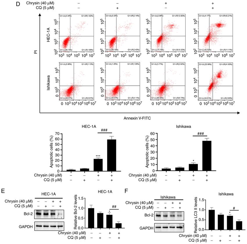
remains a major obstacle in EC therapy (24). Chrysin has autophagy through the upregulation of the intracellular8 HE et al: CHRYSIN INDUCES AUTOPHAGY IN ENDOMETRIAL CANCER Figure 4. Chrysin induces cytoprotective autophagy in endometrial cancer cells. (A and B) HEC‑1A and Ishikawa cells were treated with 40 µM chrysin without or with 5 µM CQ for 48 h, and cell viability was then assessed using a CCK‑8 assay. (C) HEC‑1A and Ishikawa cells were pretreated with CQ (5 µM) for 1 h before being exposed to 40 µM chrysin for colony formation assay. (D) HEC‑1A and Ishikawa cells were pretreated without or with 5 µM CQ for 1 h, followed by treatment with chrysin (40 µM) for 48 h. Cell apoptosis was then analyzed by flow cytometry using Annexin V‑FITC/PI staining. (E and F) HEC‑1A and Ishikawa cells were untreated or treated with 5 µM CQ for 1 h, followed by chrysin treatment at 40 µM for 24 h. Subsequently, the expression levels of Bcl‑2 were examined by western blotting. Values are presented as the mean ± standard deviation (n=3). *P
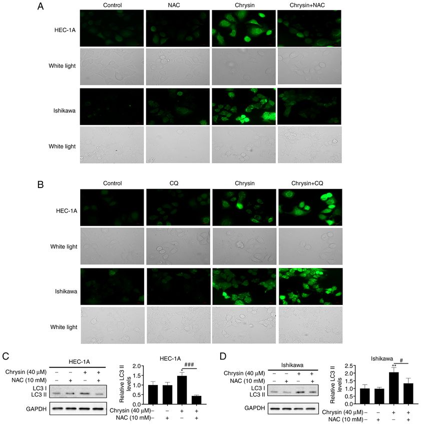
INTERNATIONAL JOURNAL OF MOLECULAR MEDICINE 48: 172, 2021                                                           9

Figure 5. Chrysin induces autophagy via ROS in endometrial cancer cells. (A) Increased levels of intracellular ROS induced by chrysin (40 µM) were detected
by microscopy. NAC (10 mM) suppressed chrysin‑induced ROS increase in HEC‑1A and Ishikawa cells. Scale bar, 200 µm. (B) Increased levels of intracel‑
lular ROS induced by chrysin (40 µM), as detected by microscopy. CQ (5 µM) promoted chrysin‑induced ROS accumulation in HEC‑1A and Ishikawa cells.
Scale bar, 200 µm. (C and D) HEC‑1A and Ishikawa cells were pretreated without or with 10 mM NAC for 1 h, and then incubated with 40 µM chrysin for 24 h.
The expression of LC3 was detected by western blotting. *P
10                                    HE et al: CHRYSIN INDUCES AUTOPHAGY IN ENDOMETRIAL CANCER

Figure 6. The Akt/mTOR pathway is involved in chrysin‑induced autophagy in endometrial cancer cells. (A and B) HEC‑1A and Ishikawa cells were exposed
to chrysin at different concentrations (0, 10, 20, 40 and 80 µM) for 48 h, and the effects of chrysin on the levels of Akt, p‑Akt, mTOR, p‑mTOR were examined
by western blotting. (C‑F) HEC‑1A and Ishikawa cells were either not treated, or treated with 10 µM LY294002 for 1 h, and then incubated with 40 µM chrysin
for 48 h, Next, Akt, p‑Akt, mTOR, p‑mTOR and LC3II protein expression was analyzed by western blotting. Values are presented as the mean ± standard
deviation of three independent experiments. *P
INTERNATIONAL JOURNAL OF MOLECULAR MEDICINE 48: 172, 2021                                                     11

Figure 7. ROS‑mediated inactivation of the Akt/mTOR signaling pathway is involved in autophagy induced by chrysin in endometrial cancer cells.
(A and B) HEC‑1A and Ishikawa cells were pretreated without or with 10 mM NAC for 1 h, and then incubated with 40 µM chrysin for 48 h. Next, Akt, p‑Akt,
mTOR and p‑mTOR protein expression was analyzed by western blotting. The results are presented as the mean ± standard deviation of three independent
tests. *P
12                              HE et al: CHRYSIN INDUCES AUTOPHAGY IN ENDOMETRIAL CANCER

inhibition of cell proliferation and promoted chrysin‑induced        5. Rai R, Essel KG, Benbrook DM, Garland J, Zhao YD and
                                                                        Chandra V: Preclinical efficacy and involvement of AKT, mTOR,
apoptosis, indicating that chrysin‑induced autophagy played             and ERK kinases in the mechanism of sulforaphane against
a pro‑survival role in EC cells. Thus, combination treatment            endometrial cancer. Cancers (Basel) 12: 1273, 2020.
with an autophagy inhibitor may be a promising intervention          6. Ma L, Zhang M, Zhao R, Wang D, Ma Y and Li A: Plant natural
                                                                        products: Promising resources for cancer chemoprevention.
strategy for EC treatment.                                              Molecules 26: 933, 2021.
                                                                     7. Sauter ER: Cancer prevention and treatment using combination
Acknowledgements                                                        therapy with natural compounds. Expert Rev Clin Pharmacol 13:
                                                                        265‑285, 2020.
                                                                     8. Song S, Gao K, Niu R, Wang J, Zhang J, Gao C, Yang B and Liao X:
Not applicable.                                                         Inclusion complexes between chrysin and amino‑appended
                                                                        β ‑cyclodextrins (ACDs): Binding behavior, water solubility,
                                                                        in vitro antioxidant activity and cytotoxicity. Mater Sci Eng C
Funding                                                                 Mater Biol Appl 106: 110161, 2020.
                                                                     9. Mani R and Natesan V: Chrysin: Sources, beneficial phar‑
This study was funded by the National Science Foundation                macological activities, and molecular mechanism of action.
                                                                        Phytochemistry 145: 187‑196, 2018.
for Young Scientists of China (grant. no. 81801511).                10. Kasala ER, Bodduluru LN, Madana RM, Athira KV, Gogoi R
                                                                        and Barua CC: Chemopreventive and therapeutic potential of
Availability of data and materials                                      chrysin in cancer: Mechanistic perspectives. Toxicol Lett 233:
                                                                        214‑225, 2015.
                                                                    11. Moghadam ER, Ang HL, Asnaf SE, Zabolian A, Saleki H,
The datasets used and/or analyzed during the current study              Yavari M, Esmaeili H, Zarrabi A, Ashrafizadeh M and Kumar AP:
are available from the corresponding author on reasonable               Broad‑spectrum preclinical antitumor activity of chrysin: Current
                                                                        trends and future perspectives. Biomolecules 10: 1374, 2020.
request.                                                            12. Roy S, Sil A and Chakraborty T: Potentiating apoptosis and
                                                                        modulation of p53, Bcl2, and Bax by a novel chrysin ruthenium
Authors' contributions                                                  complex for effective chemotherapeutic efficacy against breast
                                                                        cancer. J Cell Physiol 234: 4888‑4909, 2019.
                                                                    13. Brechbuhl HM, Kachadourian R, Min E, Chan D and Day BJ:
The present study was conceived and designed by YH, YY,                 Chrysin enhances doxorubicin‑induced cytotoxicity in human
LZ and BW. The experiments were conducted by YH, YS,                    lung epithelial cancer cell lines: The role of glutathione. Toxicol
                                                                        Appl Pharmacol 258: 1‑9, 2012.
YY, HH, YF and YW. YH and YS wrote the manuscript with              14. Zhang T, Chen X, Qu L, Wu J, Cui R and Zhao Y: Chrysin and its
support from BW and YY, HH, YF, YW LZ and BW critically                 phosphate ester inhibit cell proliferation and induce apoptosis in
revised the manuscript for important intellectual content. All          hela cells. Bioorg Med Chem 12: 6097‑6105, 2004.
                                                                    15. Lima APB, Almeida TC, Barros TMB, Rocha LCM, Garcia CCM
authors agreed to be accountable for all aspects of the work            and da Silva GN: Toxicogenetic and antiproliferative effects of
in ensuring that questions related to the accuracy or integrity         chrysin in urinary bladder cancer cells. Mutagenesis 13: geaa021,
of any part of the work are appropriately investigated and              2020.
                                                                    16. Koff JL, Ramachandiran S and Bernal‑Mizrachi L: A time to
resolved. YH and BW confirm the authenticity of all the raw             kill: Targeting apoptosis in cancer. Int J Mol Sci 16: 2942‑2955,
data. All authors have read and approved the final manuscript.          2015.
                                                                    17. Pang X, Zhang X, Jiang Y, Su Q, Li Q and Li Z: Autophagy:
                                                                        Mechanisms and therapeutic potential of flavonoids in cancer.
Ethics approval and consent to participate                              Biomolecules 11: 135, 2021.
                                                                    18. Crawley O, Opperman KJ, Desbois M, Adrados I, Borgen MA,
Not applicable.                                                         Giles AC, Duckett DR and Grill B: Autophagy is inhibited by
                                                                        ubiquitin ligase activity in the nervous system. Nat Commun 10:
                                                                        5017, 2019.
Patient consent for publication                                     19. Ranieri R, Ciaglia E, Amodio G, Picardi P, Proto MC,
                                                                        Gazzerro P, Laezza C, Remondelli P, Bifulco M and Pisanti S:
                                                                        N6‑isopentenyladenosine dual targeting of AMPK and rab7
Not applicable.                                                         prenylation inhibits melanoma growth through the impairment
                                                                        of autophagic flux. Cell Death Differ 25: 353‑367, 2018.
Competing interests                                                 20. Lin YM, Chen CI, Hsiang YP, Hsu YC, Cheng KC, Chien PH,
                                                                        Pan HL, Lu CC and Chen YJ: Chrysin attenuates cell viability
                                                                        of human colorectal cancer cells through autophagy induction
The authors declare that they have no competing interests.              unlike 5‑fluorouracil/oxaliplatin. Int J Mol Sci 19: 1763, 2018.
                                                                    21. Garcia‑Martinez T, Vendrell‑Flotats M, Martinez‑Rodero I,
                                                                        Ordóñez‑León EA, Álvarez‑Rodríguez M, López‑Béjar M,
References                                                              Yeste M and Mogas T: Glutathione ethyl ester protects
                                                                        in vitro‑maturing bovine oocytes against oxidative stress
 1. Urick ME and Bell DW: Clinical actionability of molecular           induced by subsequent vitrification/warming. Int J Mol Sci 21:
    targets in endometrial cancer. Nat Rev Cancer 19: 510‑521,          7547, 2020.
    2019.                                                           22. An Z, Tassa A, Thomas C, Zhong R, Xiao G, Fotedar R, Tu BP,
 2. Troisi J, Raffone A, Travaglino A, Belli G, Belli C, Anand S,       Klionsky DJ and Levine B: Autophagy is required for G1/G0
    Giugliano L, Cavallo P, Scala G, Symes S, et al: Development        quiescence in response to nitrogen starvation in Saccharomyces
    and validation of a serum metabolomic signature for endome‑         cerevisiae. Autophagy 10: 1702‑1711, 2014.
    trial cancer screening in postmenopausal women. JAMA Netw       23. Nunez‑Olvera SI, Gallardo‑Rincon D, Puente‑Rivera J,
    Open 3: e2018327, 2020.                                             Salinas‑Vera YM, Marchat LA, Morales‑Villegas R and
 3. Clarke MA, Long BJ, Del Mar Morillo A, Arbyn M,                     López‑Camarillo C: Autophagy machinery as a promising thera‑
    Bakkum‑Gamez JN and Wentzensen N: Association of endo‑              peutic target in endometrial cancer. Front Oncol 9: 1326, 2019.
    metrial cancer risk with postmenopausal bleeding in women:      24. Guo F, Zhang H, Jia Z, Cui M and Tian J: Chemoresistance
    A systematic review and meta‑analysis. JAMA Intern Med 178:         and targeting of growth factors/cytokines signalling pathways:
    1210‑1222, 2018.                                                    Towards the development of effective therapeutic strategy for
 4. Korets SB, Czok S, Blank SV, Curtin JP and Schneider RJ:            endometrial cancer. Am J Cancer Res 8: 1317‑1331, 2018.
    Targeting the mTOR/4E‑BP pathway in endometrial cancer. Clin    25. Sharom FJ: ABC multidrug transporters: Structure, function and
    Cancer Res 17: 7518‑7528, 2011.                                     role in chemoresistance. Pharmacogenomics 9: 105‑127, 2008.
INTERNATIONAL JOURNAL OF MOLECULAR MEDICINE 48: 172, 2021 13
26. Crommentuijn MH, Maguire CA, Niers JM, Vandertop WP, 37. Deng L, Gao X, Liu B, He X, Xu J, Qiang J, Wu Q and
Badr CE, Würdinger T and Tannous BA: Intracranial Liu S: NMT1 inhibition modulates breast cancer progression
AAV‑sTRAIL combined with lanatoside C prolongs survival in through stress‑triggered JNK pathway. Cell Death Dis 9: 1143,
an orthotopic xenograft mouse model of invasive glioblastoma. 2018.
Mol Oncol 10: 625‑634, 2016. 38. Yang CC, Tsai MH, Li KY, Hou CH and Lin FH: Carbon‑doped
27. Bronikowska J, Szliszka E, Kostrzewa‑Susłow E, Jaworska D, TiO 2 activated by X‑ray irradiation for the generation of reactive
Czuba ZP, Bednarski P and Król W: Novel structurally related oxygen species to enhance photodynamic therapy in tumor treat‑
flavones augment cell death induced by rhsTRAIL. Int J Mol ment. Int J Mol Sci 20: 2072, 2019.
Sci 18: 1211, 2017. 39. Lim W, Ryu S, Bazer FW, Kim SM and Song G: Chrysin attenu‑
28. Rybstein MD, Bravo‑San Pedro JM, Kroemer G and Galluzzi L: ates progression of ovarian cancer cells by regulating signaling
The autophagic network and cancer. Nat Cell Biol 20: 243‑251, cascades and mitochondrial dysfunction. J Cell Physiol 233:
2018. 3129‑3140, 2018.
29. Dou QH, Chen HN, Wang K, Yuan K, Lei Y, Li K, Lan J, Chen Y, 40. Ru JY and Wang YF: Osteocyte apoptosis: The roles and key
Huang Z, Xie N, et al: Ivermectin induces cytostatic autophagy molecular mechanisms in resorption‑related bone diseases. Cell
by blocking the PAK1/akt axis in breast cancer. Cancer Res 76: Death Dis 11: 846, 2020.
4457‑4469, 2016. 41. Guglielmelli P, Barosi G, Rambaldi A, Marchioli R, Masciulli A,
30. Yao C, Liu BB, Qian XD, Li LQ, Cao HB, Guo QS and Zhou GF: Tozzi L, Biamonte F, Bartalucci N, Gattoni E, Lupo ML, et al:
Crocin induces autophagic apoptosis in hepatocellular carci‑ Safety and efficacy of everolimus, a mTOR inhibitor, as single
noma by inhibiting Akt/mTOR activity. Onco Targets Ther 11: agent in a phase 1/2 study in patients with myelofibrosis.
2017‑2028, 2018. Blood 118: 2069‑2076, 2011.
31. Amaravadi RK, Lippincott‑Schwartz J, Yin XM, Weiss WA, 42. Zhou Y, Chen X, Qu N, Zhang B and Xia C: Chondroprotection
Takebe N, Timmer W, DiPaola RS, Lotze MT and White E: of PPARα activation by WY14643 via autophagy involving akt
Principles and current strategies for targeting autophagy for and ERK in LPS‑treated mouse chondrocytes and osteoarthritis
cancer treatment. Clin Cancer Res 17: 654‑666, 2011. model. J Cell Mol Med 23: 2782‑2793, 2019.
32. Ohnishi K, Yano S, Fujimoto M, Sakai M, Harumoto E, 43. Wang YX, Yang L, Wang HQ, Zhao XQ, Liu T, Li YH, Zeng QX,
Furuichi A, Masuda M, Ohminami H, Yamanaka‑Okumura H, Li YH and Song DQ: Synthesis and evolution of berberine deriv‑
Hara T and Taketani Y: Identification of dietary phytochemicals atives as a new class of antiviral agents against enterovirus 71
capable of enhancing the autophagy flux in HeLa and Caco‑2 through the MEK/ERK pathway and autophagy. Molecules 23:
human cell lines. Antioxidants (Basel) 9: 1193, 2020. 2084, 2018.
33. Lu Y, Xiao L, Liu Y, Wang H, Li H, Zhou Q, Pan J, Lei B, 44. Ives A, Nomura J, Martinon F, Roger T, LeRoy D, Miner JN,
Huang A and Qi S: MIR517C inhibits autophagy and the Simon G, Busso N and So A: Xanthine oxidoreductase regulates
epithelial‑to‑mesenchymal (‑like) transition phenotype in human macrophage IL1β secretion upon NLRP3 inflammasome activa‑
glioblastoma through KPNA2‑dependent disruption of TP53 tion. Nat Commun 6: 6555, 2015.
nuclear translocation. Autophagy 11: 2213‑2232, 2016. 45. Su Z, Burchfield JG, Yang P, Humphrey SJ, Yang G, Francis D,
34. Kim K, Dayem AA, Gil M, Yang GM, Lee SB, Kwon OH, Choi S, Yasmin S, Shin SY, Norris DM, Kearney AL, et al: Global redox
Kang GH, Lim KM, Kim D and Cho SG: 3,2'‑Dihydroxyflavone proteome and phosphoproteome analysis reveals redox switch in
improves the proliferation and survival of human pluripotent akt. Nat Commun 10: 5486, 2019.
stem cells and their differentiation into hematopoietic progenitor 46. Huang CR, Chang TW, Lee CT, Shen CJ, Chang WC and
cells. J Clin Med 9: 669, 2020. Chen BK: ARNT deficiency represses pyruvate dehydrogenase
35. Lewinska A, Adamczyk‑Grochala J, Deregowska A and kinase 1 to trigger ROS production and melanoma metastasis.
Wnuk M: Sulforaphane‑induced cell cycle arrest and senes‑ Oncogenesis 10: 11, 2021.
cence are accompanied by DNA hypomethylation and changes
in microRNA profile in breast cancer cells. Theranostics 7:
This work is licensed under a Creative Commons
3461‑3477, 2017.
36. Ye S, Zhao T, Zhang W, Tang Z, Gao C, Ma Z, Xiong JW, Peng J, Attribution-NonCommercial-NoDerivatives 4.0
Tan WQ and Chen J: p53 isoform Δ113p53 promotes zebrafish International (CC BY-NC-ND 4.0) License.
heart regeneration by maintaining redox homeostasis. Cell Death
Dis 11: 568, 2020.You can also read


