Why Cataract and Refractive Surgeons Need the Pentacam
←
→
Page content transcription
If your browser does not render page correctly, please read the page content below
Why Cataract and
Refractive Surgeons
Need the Pentacam
 Four surgeons discuss the device’s value, including new software additions.
                            Produced under an unrestricted educational grant from Oculus, Inc..

             Extremely accurate anterior and posterior corneal surface measurements
          Keratoconus detection software • Holladay Report for more accurate "Ks" for
    postrefractive surgery patients receiving IOL implants • Very early cataract detection with
            exclusive "Densitometry" feature • Accurate post-LASIK ectasia detection
            Accurate anterior chamber measurements aid in proper sizing of ACIOLs

                                   MICHAEL W. BELIN, MD, FACS, MODERATOR
JACK T. HOLLADAY, MD, MSEE, FACS • H. BURKHARD DICK, MD, PHD • RENATO AMBRÓSIO, JR, MD, PHD

E VA LU AT I N G P O S T - L A S I K E C TA S I A
W I T H T H E O C U LU S P E N TA C A M

Front and back corneal measurements improve the
accuracy of calculations.

BY MICHAEL W. BELIN, MD

   Almost 10 years ago, I published an article1 showing the
extreme variability between different topography systems in
the analysis of abnormal corneal shapes. Since then, the abil-
ity to accurately image the anterior corneal surface has
greatly improved. We are now being told, however, that the
elevation of the posterior surface is important in diagnosing
early keratoconus and post-LASIK ectasia. Currently, two
commercially available systems are able to measure back-
surface elevation: the Pentacam comprehensive eye scanner
(Oculus, Inc., Lynnwood, WA), which is a rotating
Scheimpflug device, and the Orbscan corneal topographer
(Bausch & Lomb, Rochster, NY), a scanning slit device.

                                                                 JANUARY 2006 I INSERT TO CATARACT & REFRACTIVE SURGERY TODAY I 1
WHY CATARACT AND REFRACTIVE SURGEONS NEED THE PENTACAM

   ELEVATION DATA                                                  sound data, with which the Pentacam has a very good
      The most common way to display elevation data is to          correlation. We also know that, with the Orbscan, the
   compare it to a known shape to amplify the data. The            postoperative cornea is 35 to 40µm thinner than corre-
   most appropriate shape is a best-fit sphere. The reason for     sponding ultrasound values.
   examining corneal data against a best-fit sphere is that           Another example between the two systems is one of a
   raw elevation data on any patient, no matter how patho-         patient who was recently referred to me for a buttonhole
   logic, all look the same, like trying to identify Earth’s       flap (Figure 3). The patient’s central cornea was clear and
   mountains from space. A topographic picture of the              had undergone no surgery prior to the LASIK procedure
   Earth’s surface, however, detects the hills and the valleys.    that produced the buttonhole, and it was of normal
   Likewise, a best-fit sphere amplifies the corneal surface for   thickness and topography preoperatively. The patient’s
   the ophthalmologist and is intuitively logical (Figure 1).      other eye was completely healthy. No ablation was per-
   One may also compare elevation data to other shapes,            formed, and because a buttonhole was created, there was
   such as an ellipse or a toric ellipsoid. A distortion map is    no flap in the central cornea. On his postoperative
   effectively a best-fit elevation map using a toric ellipsoid.   Orbscan, positive elevation (or positive ectasia or dis-
   Eliminating the area that can be corrected by a pair of         placement of the posterior surface) is visible. On the other
   spectacles (a sphere and cylinder) produces, in essence, an     hand, the Pentacam image looks completely normal. As
   irregularity map.                                               this example illustrates, it is mandatory that the hardware
      Again, not only is examining the posterior surface           be capable of measuring what the ophthalmologist is try-
   important, but many experts have stated that changes in         ing to examine. The Orbscan does not appear capable of
   the posterior surface are often missed and that most            measuring postoperative corneas, a fact that is fairly well
   post-LASIK patients have significant changes on their pos-      known.
   terior corneal surfaces.
                                                                   ONLY AS GOOD AS THE OPERATOR
   DIFFERENCES BETWEEN SYSTEMS                                        Joseph Ciolino, MD, and I studied a series of 124 patients
       The Orbscan has almost no peer-reviewed literature to       to determine their post-LASIK posterior displacement with
   support its accuracy in nonspherical, nonsymmetric              elevation topography. We examined the eyes preopera-
   shapes (what we see clinically), and no peer-reviewed lit-      tively and at 1 month postoperatively and calculated the
   erature has documented the accuracy of that system’s            difference. We used a two-display difference map, a preop-
   posterior data. There are however, numerous articles to         erative first map and a postoperative second map, and the
   show that the Orbscan’s postoperative pachymetry is             difference was the third map. We examined 104 LASIK and
   wrong and typically underestimates the corneal thickness        20 PRK patients. Because we wanted to use postoperative
   by 35 to 40µm. If the anterior surface is relatively easy to    corneas, we used PRK eyes as our control. The average cor-
   map, but the posterior surface is more problematic, and         rection was -3.70D for the LASIK patients and -2.80D for
   pachymetry is determined by subtracting the posterior           the PRK patients, with a range of -0.90 to -10.10D. Central
   surface from the anterior surface, then the error probably      corneal thickness averaged 546µm for the LASIK patients
   is in the posterior surface. Such an underestimation would      and 521µm for the PRK patients, with a range of 493 to
   likely make the posterior surface too anterior or “ectatic”     617µm. The residual bed averaged 329µm for the LASIK
   in the post-LASIK patient.                                      patients and 472µm for the PRK patients, with no calculat-
       Figure 2 clearly illustrates the differences between        ed residual below 263µm.
   pachymetric maps with the Orbscan and the Pentacam.
   The images are posterior elevations maps of a single
   patient imaged on the same day after his LASIK surgery.
   The physician used the same scale and same colors,
   ±75µm. Positive elevation (ectasia) is visible on the
   Orbscan, but appears completely normal with the
   Pentacam. Although many Orbscan users would say that
   up to 50µm of posterior ectasia is normal, it is not. It is a
   problem with that system. The postoperative picture has
   the same normal values as the preoperative picture.
       When comparing Orbscan and Pentacam images, how
   do we know which one is correct? Although it is very diffi-     Figure 1. The best-fit display shows the variation from a
   cult to validate posterior data, we can compare it to ultra-    known shape.

2 I INSERT TO CATARACT & REFRACTIVE SURGERY TODAY I JANUARY 2006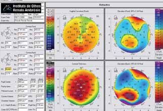
WHY CATARACT AND REFRACTIVE SURGEONS NEED THE PENTACAM

Figure 2. This post-LASIK patient was imaged on the same        Figure 3. This patient was referred after sustaining a button-
day with both the Orbscan and Pentacam. The Orbscan reads       hole. No ablation was performed, and his preoperative
the central cornea 41µm thinner due to a faulty localization    topography was completely normal. Although no tissue was
of the posterior corneal surface. The Orbscan shows a poste-    removed, the Orbscan map shows a dramatic shift in the pos-
rior surface compatible with an ectasia, whereas the            terior elevation, whereas the Pentacam map shows a com-
Pentacam reveals a normal posterior surface.                    pletely normal, unaltered posterior surface.

   When we analyzed the corneas graphically, it was             elevation as abnormal and greater than 15µm on the pos-
apparent that there was no difference in the posterior dis-     terior surface. If there is a gray zone, such as 10µm of ante-
placement between the LASIK and PRK patients. In other          rior elevation and 12µm of posterior, but the pachymetric
words, within our preoperative parameters, routine pos-         map is also displaced toward the cone, then this is much
terior displacement after LASIK did not occur. Central          more suspicious. I rarely look at curvature maps for kera-
corneal thickness, the residual bed’s ablation depth, or the    toconus, because an abnormal curvature map does not
residual bed versus the central corneal thickness ratio did     imply keratoconus. A false-positive may occur with a dis-
not seem to be factors. One has to keep in mind that            placed apex, which will not affect elevation but will signif-
these findings can only be applied to our patient popula-       icantly affect curvature maps.
tion. That is, I neither perform LASIK on corneas with a           An accurate examination of the posterior corneal sur-
preoperative pachymetry of less than 500µm, nor PRK on          face is important. No one would look at half an X-ray;
corneas with less than 475µm. I typically respect a 275-µm      likewise, we should not rely on an analysis that only
post-LASIK residual bed.                                        reports on half of the cornea. In the past, assessing the
   I am not suggesting that post-LASIK ectasia does not         posterior corneal surface was problematic, particularly in
exist. Obviously, we have all seen it. I am proposing, howev-   the postoperative or distorted cornea. The Pentacam’s
er, that its frequency has been greatly exaggerated and that,   unique imaging system, which uses a rotating Scheimpflug
like keratoconus with placido systems, some elevation sys-      camera, appears capable of accurately assessing both the
tems are associated with a very high false-positive rate.       anterior and posterior corneal surfaces, both pre- and
                                                                postoperatively. The Pentacam enables the ophthalmolo-
ASTIGMATISM VERSUS ECTASIA                                      gist to better diagnose ectatic disorders and to better
   I think one issue about which people get very confused       quantify the normal postoperative cornea.
is astigmatism versus ectasia. Ectasia is an island of posi-
tive deviation in the central or paracentral region of the         Michael W. Belin, MD, is Professor of Ophthalmology and
cornea on the elevation map, whereas astigmatism has it         Director of Cornea & Refractive Surgery at the Albany
maximum deviation in the periphery. The higher the              Medical Center Lions Eye Institute and is Adjunct Professor
degree of astigmatism, the greater the peripheral devia-        of Ophthalmology at the University of Ottawa in Canada.
tion, elevated on the flat axis (positive) and depressed on     He has received travel support and honoraria from Alcon
the steep axis (negative deviation). It is also important to    Laboratories, Inc., and Oculus, Inc. Dr. Belin may be reached
look at the pachymetric map, not just in the thinnest           at (518) 475-1515.
region but to evaluate the pachymetric distribution. A
normal cornea is thinnest in the center. Although nothing       1. Belin MW, Ratliff CD. Evaluating data acquisition and smoothing functions of currently avail-
is absolute, I use greater than 12µm of central anterior        able videokeratoscopes. J Cataract Refract Surg. 1996;22:421-426.

                                                                 JANUARY 2006 I INSERT TO CATARACT & REFRACTIVE SURGERY TODAY I 3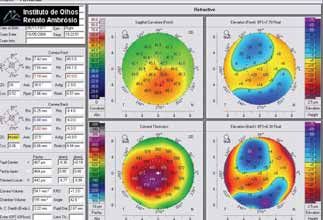
WHY CATARACT AND REFRACTIVE SURGEONS NEED THE PENTACAM

   M E A S U R I N G CO R N E A L P O W E R                        area. Topographers and keratometers have a camera or
                                                                   viewer in their centers that obscures the central area
   A F T E R CO R N E A L R E F R AC T I V E
                                                                   (1.8mm for a topographer and the central 3mm for a ker-
   S U R G E RY                                                    atometer). This first error results in a 15% error of the
                                                                   refractive change from refractive surgery for the ker-
   How the Pentacam improves the accuracy of                       atometer and an error of about 5% for the topographer.
   these calculations.                                                The second error with LASIK and PRK relates to the
                                                                   change in the ratio of the back-to-front radius of the cur-
                                                                   vatures, which is normally 82%. This error accounts for
   BY JACK T. HOLLADAY, MD, MSEE, FACS
                                                                   approximately 10% of the refractive change from refrac-
                                                                   tive surgery. The total is therefore about a 25% (15% +
      I have used the Pentacam comprehensive eye scanner           10%) error in the refractive change for keratometry and
   (Oculus, Inc., Lynnwood, WA) for more than 1 year, ever         approximately 15% (5% + 10%) for topography using the
   since I saw how well it measured the front and back sur-        central refractive power. So, if a patient experienced a
   faces of the cornea. As most surgeons know, the differ-         10.00D refractive change from refractive surgery, then
   ence between topography and tomography is that                  the keratometer would make a 2.50D error, and the
   topography measures the surface of the cornea, whereas          topographer would make a 1.50D error in the measured
   tomography measures its three-dimensional thickness.            corneal power. This is the reason why surgeons do not
      The Pentacam is a rotational Scheimpflug device, and         achieve accurate corneal measurements with topogra-
   the Orbscan corneal topographer (Bausch & Lomb,                 phers or keratometers following refractive surgery.
   Rochester, NY) is a translational device. The major differ-        There is one additional problem with keratometry that
   ence between the two is that the Pentacam takes images          must be addressed. Ophthalmologists have always reported
   of 50 meridional sections through the center of the             keratometric power, not the true net power of the cornea,
   cornea. This approach allows the system to realign the          within IOL calculations. When a 45.00D keratometric read-
   central thinnest point of each section before it recon-         ing is reported, surgeons assume that the standardized ker-
   structs the corneal image, thus eliminating any eye move-       atometric index of refraction of 1.3375 has been used and
   ment that occurs during the examination (Figure 1). The         that the corresponding anterior corneal radius of curvature
   Pentacam is the only device that rotates around a com-          is 7.5mm. All IOL calculation formulas reduce the reported
   mon axis and thus allows the user to toggle down                keratometric power by approximately 2% (approximately
   through each meridional image to see if there is a blink-       0.75D) to achieve the average net power that has been
   ing eyelid or some other type of movement that                  determined for normal corneas in true net power studies.
   degrades the image’s quality for that meridian.                 Therefore, the actual power used in the vergence calculation
                                                                   is approximately 44.25D, when the keratometric power
   REVIEWING THE BASICS
      In addition to providing an image of each section, the
   Pentacam gives densitometry readings related to the
   amount of scattered light (Figure 2). The result is that the
   user may know the density of the crystalline lens and objec-
   tively measure the amount of its cataractous changes. The
   Pentacam can map any structure in the anterior segment
   that is not opaque. For example, the ciliary sulcus cannot be
   imaged in dark brown eyes, because the pigment epitheli-
   um of the iris is opaque.

   PERFECTING CALCULATIONS
      The Pentacam allows users to take direct measure-
   ments of the power of the cornea, thus avoiding the two
   problems that plague keratometers and topographers and
   cause errors, especially with IOL calculations in patients
   who have undergone refractive surgery. The first problem
   is that keratometry and topography cannot sample the            Figure 1. The system allows a three-dimensional display with
   central 2mm of the cornea, which is the most important          the ability to toggle through each meridional section.

4 I INSERT TO CATARACT & REFRACTIVE SURGERY TODAY I JANUARY 2006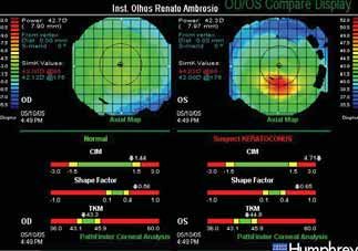
WHY CATARACT AND REFRACTIVE SURGEONS NEED THE PENTACAM

                                                                         PACHYMETRY IN POST-LASIK PATIENTS

                                                                      Surgeons should always regard the pachymetry in post-
                                                                   LASIK patients taken at 3 months with suspicion, because
                                                                   the reading depends on the amount of interface scatter
                                                                   present, which may not be due to scarring. It takes 1 to 2
                                                                   months for the cornea to pump out all of the fluids in the
                                                                   space underneath the LASIK flap. This fluid makes the flap
                                                                   somewhat edematous, which produces forward light scat-
                                                                   ter that causes people to see halos. It is similar to the kind
                                                                   of edema caused by contact lenses.

                                                                 aberrometry, refractometry, and retinoscopy, the highest
Figure 2. The Pentacam’s densitometry readings measure the       correlation is always between 4.0 and 4.5mm. Sampling a
density of the crystalline lens and objectively measure the      zone smaller than 4mm excludes too much of the pupil
amount of its cataractous changes.                               through which the rays are passing. Alternatively, with a
                                                                 zone larger than 5mm, the Stiles Crawford effect weighs the
measures 45.00D. The Pentacam can report the net power           rays beyond 5mm so little that they contribute very slightly
(44.25D), but IOL calculation formulas would reduce the          to the retinal image and are therefore not important.
power by 0.75D to 43.50D and create a new error from dou-
ble compensating the value. The Pentacam therefore               THE PENTACAM’S REPORTS
reports the equivalent K-reading, which would be 45.00D in          In designing the Holladay Report that the Pentacam gen-
the normal patient with no refractive surgery (the same as       erates, Andreas and I wanted all of the patient’s informa-
with keratometry and topography).                                tion available to the physician on one report. Therefore, we
                                                                 included a map for refractive power and one displaying the
TREATMENT RANGE AND FOCUS                                        tangential (which is a poor name; it is really the local radius
    For IOL calculations with the Pentacam, what area            of curvature). The refractive power map uses Snell’s law and
should be used to determine the corneal power? The               shows positive spherical aberration. Any change in a
device has parameters in a zone from 2 to 12mm that can          patient’s refraction will be exactly related to the change in
be used. We must first determine the size zone, or diameter      the topographic map.
(not the radius), for which we want to know the net power.          We also included a tangential curvature map. It does not
When the Pentacam was first developed, I conducted a             use Snell’s law; it only gives the local radius at every point
study with Andreas Steinmueller of Oculus Optikgeraete           on the surface. These kinds of data are also called
GmbH (Wetzler, Germany) to determine the answer to this          instantaneous radius of curvature, because they illustrate
question.
    We first calculated the power of a preoperative cornea
with the Pentacam, a Humphrey Atlas Topography System
(Carl Zeiss Meditec Inc., Dublin, CA), and an Eyesys 2000
Corneal Analysis System (Eyesys Vision, Houston, TX). The
three systems agreed on the same measurement within
approximately 0.03D. Then, we enrolled 50 patients (100
eyes) who had undergone LASIK or PRK and whose refrac-
tions ranged from +3.00 to -8.00D, and we recorded their
refractive changes at 3 months. We vertexed the measured
refractive change to the cornea at 3 months and subtracted
it from the patients’ preoperative corneal power. Next, we
correlated these measurements with the Pentacam over var-
ious treatment zones. The 4-mm zone had the best agree-
ment of the measured corneal power with the calculated
power. In retrospect, this finding is consistent with previous   Figure 3. The Pentacam correlates the measured corneal
studies because, when correlating refraction with wavefront      power and the calculated power.

                                                                 JANUARY 2006 I INSERT TO CATARACT & REFRACTIVE SURGERY TODAY I 5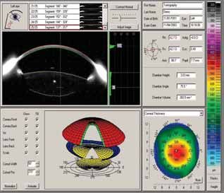
WHY CATARACT AND REFRACTIVE SURGEONS NEED THE PENTACAM

   the radius of curvature of every little bump instanta-          for this kind of lens. Many phakic IOLs, such as toric
   neously. The map does not reflect the change in refrac-         lenses, require precise implantation in terms of the
   tive power, but it will reveal such things as flat spots in     location of enclavation to the iris. As most surgeons
   the periphery, a capability that makes it a valuable tool.      know, the more a lens is displaced from the target axis,
      The lower maps in the Pentacam’s report show eleva-          the greater the induced astigmatism is. For example,
   tion. They become important when discussing terms               even 15º of displacement equals approximately a 50%
   such as best-fit sphere. Using positive and negative refer-     reduction in the quality of the refractive effect. To
   ence points, the map illustrates the height of an area on       optimize our placement of these lenses, our depart-
   the cornea relative to a best-fit sphere. It will also com-     ment uses two Pentacam comprehensive eye scanners
   pare these points to shapes other than a sphere, such as        (Oculus, Inc., Lynnwood, WA).
   an aspheric shape or Q values.
      The back surface float on the Pentacam’s report              PREOPERATIVE CALCULATIONS
   details the back curvature of the cornea and helps the            To illustrate the value of the Pentacam, one patient
   surgeon determine its elevation. The posterior float on         of mine had a preoperative refraction of +7.00D sphere
   the Pentacam is far more accurate than on the Orbscan,          combined with -3.00D of astigmatism, so he had spher-
   but it is still not as accurate as the Pentacam’s front cur-    ical aberration and a spherical equivalent refraction of
   vature. One reason is that the posterior curvature is a         +5.50D. Is he a good candidate for refractive lens ex-
   virtual image seen through the optics of the front of the       change or phakic lens implantation? The latter requires
   cornea and stroma and their thicknesses and curva-              sufficient space in the anterior chamber and would be
   tures. Therefore, to determine the precise back curva-          a good option if the patient could not accommodate.
   ture of the cornea, one needs to know its exact index of        The Pentacam showed that this patient’s eye had a
   refraction, of which there is a gradient in the cornea.         small peripheral anterior chamber. Regarding the depth
   Minute errors ultimately have a minimal effect on back-         of the anterior chamber, the distance from the periph-
   surface pachymetry, but the curvature is usually accu-          eral optic is more important than the central distance
   rate to within 10 to 12µm. On the Orbscan, the float is         (the anterior chamber depth). The ophthalmic litera-
   accurate to within 20µm. On the Pentacam’s posterior            ture and training courses for phakic IOLs refer to the
   float, only changes greater than 10 to 12µm are signifi-        central anterior chamber depth but stress the impor-
   cant. Once we determined that the 4-mm measure-                 tance of the peripheral depth, because these IOLs’
   ment zone was optimal, we again performed the corre-            periphery is closer to the endothelium. The space is
   lation between the measured corneal power and the               important to avoid any intermittent touching of the
   calculated power, and we achieved an R2 of 96%, which
   was ±0.55D of the corneal power (Figure 3).

      Jack T. Holladay, MD, MSEE, FACS, is Clinical Professor
   of Ophthalmology at Baylor College of Medicine in
   Houston and is a consultant for Oculus, Inc. He may be
   reached at (713) 668-7337; docholladay@docholladay.com
   and www.docholladay.com.

   A N T E R I O R S E G M E N T A N A LY S I S F O R
   P H A K I C I O L I M P L A N TAT I O N

   The information that the device supplies allows sur-
   geons to serve patients better.

   BY H. BURKHARD DICK, MD, P H D
                                                                   Figure 1. For this eye with a refractive error of +7.25 comb. -3
                                                                   at 123°, the Pentacam demonstrates that there is insufficient
     Preoperative planning for phakic IOL implantation is          space for a toric phakic IOL in the peripheral anterior cham-
   of the utmost importance in order to exclude poor               ber at the target implantation axis but enough space at the
   candidates who do not have optimal intraocular space            horizontal meridian for phakic lens implantation.

6 I INSERT TO CATARACT & REFRACTIVE SURGERY TODAY I JANUARY 2006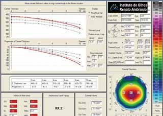
WHY CATARACT AND REFRACTIVE SURGEONS NEED THE PENTACAM

                                                               are always thicker in the periphery. Highly myopic eyes
                                                               have great side height. I do not recommend taking the
                                                               perpendicular measurement between the phakic IOL
                                                               and the endothelium in the periphery, but rather the
                                                               shortest distance. The Pentacam allows the surgeon to
                                                               quickly see the closest distance. Interestingly, when my
                                                               colleagues and I took some measurements with the
                                                               Pentacam, the more important peripheral measure-
                                                               ment was always smaller than the central measure-
                                                               ment.
                                                                  Another particular case illustrates the Pentacam’s
                                                               usefulness well. The patient presented for phakic lens
                                                               implantation. Based on his slit-lamp examination, my
                                                               staff and I were convinced that he was a good candi-
                                                               date for implantation with a Verisyse phakic IOL
                                                               (Advanced Medical Optics, Inc., Santa Ana, CA).
                                                               Although the implantation was successful, during a
Figure 2. Determination of the safety distances after phakic   routine postoperative check, we performed an exami-
IOL implantation in a myopic eye operated elsewhere.           nation with the Pentacam (which was not available in
Obviously, the safety distances are already smaller than       our office prior to the implantation) that revealed
allowed, underlining the necessity to perform Pentacam eval-   synechiae posterior to the iris. The iris bulged slightly
uations in the future follow-up.                               anteriorly, and the space from the endothelium to the
                                                               iris was small. Again, based on the slit-lamp examina-
lens if the patient rubs his eyes, for example, which          tion alone, we were confident of this patient’s candida-
might induce continuous endothelial cell loss. A safe          cy for a phakic implant, but the Scheimpflug image of
amount of space for this kind of lens is a minimum of          the Pentacam provided information that would have
1mm in the periphery. The central anterior chamber             excluded this patient from this procedure. With the
depth should be approximately 3mm, according to                Verisyse IOL, there is a tiny but important distance
phakic IOL manufacturers. However, anterior chamber            between the iris, the pupil, and the internal margin of
depth does not tell the surgeon everything, because            the optic. The central thickness of the anterior cham-
the smallest distance has to be the exclusion criterion,       ber of this patient’s eye was 3.2mm, but the peripheral
and this distance is found in the periphery only. The          distance was just below the safety distance at 900µm.
Pentacam nicely addresses this point. The user simply          Thus, the patient definitely was not a perfect candidate
implements an 8.5-mm line into the Scheimpflug image           for implantation with this -9.00D lens.
that represents the overall diameter of the phakic IOL.
He can then measure the eye’s shortest distance                PRAISE FOR THE PENTACAM
between the IOL’s optic and the endothelium manually.             In conclusion, the Pentacam is more comfortable for
Using this method, the surgeon can easily decide if the        patients than other measurement devices. It operates
patient is an appropriate candidate for toric lens             very quickly, gives immediate results, and is extremely
implantation.                                                  easy to use (it may be operated by anyone in the
                                                               office). It also has the potential for many new applica-
ADDITIONAL APPLICATIONS                                        tions. I feel it is a great addition to my clinical practice;
   Surgeons planning to reduce astigmatism may ana-            it has become an irreplaceable tool, because I no
lyze incisional architecture with the Pentacam, because        longer implant any phakic IOLs without checking the
the device provides a topographic map of both sides of         results that the Pentacam provides. ●
the cornea as well as a pachymetric map. However,
what is most important is the way in which the                   H. Burkhard Dick, MD, PhD, is Clinical Professor and
Pentacam aids in the postoperative evaluation of safety        Head Physician for the Department of Ophthalmology,
issues. With age, the anterior segment grows smaller,          Johannes Gutenberg-University, Mainz, Germany. He
and the crystalline lens thickens. What is much more           acknowledged no financial interest in any product or
important is the closest distance from the optic’s             company mentioned herein. Professor Dick may be
periphery to the endothelium, because myopic lenses            reached at +49 61 31175445; bdick@mail.uni-mainz.de.

                                                               JANUARY 2006 I INSERT TO CATARACT & REFRACTIVE SURGERY TODAY I 7
WHY CATARACT AND REFRACTIVE SURGEONS NEED THE PENTACAM

   E C TA S I A D E T E C T I O N A N D                             would refer to these devices by their technological names
   C L A S S I F I C AT I O N W I T H C O R N E A L                 rather than brand names to help others understand the
   TOMOGRAPHY                                                       different tasks they perform. The Orbscan (Bausch &
                                                                    Lomb, Rochster, NY) was the first tomographer clinically
   How this technology complements topography and                   available, and subsequent devices such as the Pentacam
   thus increases the safety of refractive surgery.                 have improved upon it.

                                                                    T H E P R O G R E S S I O N O F CO R N E A L T H I C K -
   BY RENATO AMBRÓSIO, J R , MD, P H D                              N E S S F O R I D E N T I F Y I N G K E R AT O CO N U S
                                                                       Keratoconus is a noninflammatory pathologic condi-
      In today’s world of refractive surgery, screening             tion characterized by progressive thinning and protru-
   patients preoperatively for a predisposition to developing       sion of the cornea. The thinning process occurs in one
   severe complications such as postoperative ectasia is            particular area so that the surrounding area remains
   vitally important. One of the most high-profile examples         disproportionately thicker. Physiologically, the normal
   of this problem was covered in the October 2005 issue of         cornea is thinner in its center and thicker in the periph-
   Cataract & Refractive Surgery Today, “Anatomy of a               ery. I hypothesize that the gradual increase of the
   Lawsuit II.”1 The Schiffer-Speaker trial was a very sad situa-   corneal thickness from the center toward the periphery
   tion in which a highly skilled surgeon with a very good          in healthy eyes falls within a normal range and that this
   reputation treated a patient who experienced ectasia             characteristic could lead to a criterion for identifying
   after LASIK, and the result led to a disastrous $7.25 mil-       pathology such as ectasia.
   lion lawsuit. The case illustrates the risk of developing           Based on this theory, I conducted studies with certain
   ectasia after LASIK, even when corneal topography and            colleagues that revealed significant differences between
   pachymetry readings—the current standard of care for             keratoconic and normal corneas using different tomo-
   the preoperative evaluation of refractive candidates—are         graphic systems. I performed the first study in 2003 using
   considered normal. Although other similar medicolegal            the Orbscan II at the Hospital de Olhos de Sergipe in
   cases have been reported in the peer-reviewed literature,2       Brazil with Mario Ursulino, MD, and Allan Luz, MD. In
   the exact scope of this problem is unknown, because the          this study, we analyzed 100 normal eyes and 25 eyes with
   majority of cases are not reported. Thus, we must identi-        mild keratoconus. Using the pachymetric numeric map
   fy the need for improving the screening process for              (0.92), we identified and recorded the thinnest point.
   refractive candidates if we are to raise the bar for safety      Also, we manually drew several circles concentric to the
   in refractive surgery. It is critical to identify patients who   thinnest point with radii that increased at 1-mm steps up
   are at risk for ectasia, and any suspicious signs should be      to 7mm. We calculated and recorded the average of the
   considered contraindications for LASIK.                          thickness values of the points located within each circle
      To this end, technologies such as the Pentacam com-           so that we could create a graph for the progression of
   prehensive eye scanner (Oculus, Inc., Lynnwood, WA) are          corneal thickness from the thinnest point toward the
   raising the bar. The Pentacam is a tomographer, which is         periphery for each eye. This approach enabled us to
   different than placido-disc topography. A tomographer            study the rate of increase for each eye proportionally
   enables a mathematical reconstruction of the internal            from the thinnest point. We were very impressed by how
   picture of the element studied, whereas topographers             the lines created were parallel in normal eyes. When we
   study its surface exclusively. Corneal tomography goes           applied statistical tests, we found significant differences
   beyond topography and pachymetry in a preoperative               in the pachymetric progression between normal eyes and
   examination. Corneal tomography enables a three-                 early keratoconic eyes at all positions. Interestingly, there
   dimensional corneal reconstruction, which evaluates its          were some cases with early keratoconus evident on the
   anterior and posterior curvatures and creates a pachy-           anterior corneal maps that had normal posterior eleva-
   metric map. This map gives the thinnest point’s value            tion maps and a peak-to-valley difference of less than
   and location, whereas ultrasound pachymetry evaluates a          0.100µm using the best-fit sphere. Thus, we observed
   single point at the center of the cornea, which might not        that pachymetric progression data could provide infor-
   be the thinnest one. However, the technologies are com-          mation to identify ectasia and add to the surgeon’s arma-
   plementary. For example, the evaluation of the integrity         mentarium in the preoperative screening process. Such
   and stability of the tear film and the corneal surface pro-      information would help indicate the risk of postoperative
   vided by the reflection of the placido rings still gives very    ectasia, which might be much higher than what was pre-
   useful clinical data. I wish that, on the podium, speakers       viously thought.

8 I INSERT TO CATARACT & REFRACTIVE SURGERY TODAY I JANUARY 2006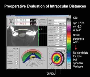
WHY CATARACT AND REFRACTIVE SURGEONS NEED THE PENTACAM

ECTASIA DETECTION: IS THE PACHYMETRIC                               mal BCVA.3 However, the condition first induces structural
PROGRESSION MORE SENSITIVE?                                         changes on the cornea that might be detectable with
   Classically, corneal topography is considered the most           tomography before the condition becomes evident on the
sensitive method for detecting ectasia. More than half of           surface with topography. Some cases considered unilateral
the cases of keratoconus and pellucid marginal degenera-            keratoconus serve as excellent examples for this point
tion identified among refractive candidates by corneal              (Figure 1). Although keratoconus is a bilateral disease, in
topography had normal eye examinations, including nor-              some cases, there is evidence of it in only one eye.
A                                                                   B

C                                                                   D

Figure 1. This 26-year-old male patient with a history of unilateral keratoconus was referred to me for a second opinion. Both
eyes had myopic astigmatism with BSCVAs of 20/20+. Corneal topography detected ectasia OS and a normal pattern OD (A).
Comparing front sagital (axial) and pachymetric maps provided by the Pentacam clearly illustrated the asymmetry between his
eyes (B). Corneal pachymetric progression detected an abrupt increase of the thickness values from the thinnest point (485µm,
located 0.52µm temporally and 0.45µm inferiorly) toward the limbus OD (C). Note the progression of corneal thickness on the
graph with the line very close to the limit.The artificial intelligence indices for an 8-mm zone from the anterior cornea are all nor-
mal for the detection of ectasia. In his left eye, a typical abrupt increase in the pachymetric values is seen from the thinnest point
(449µm, located 0.29µm temporally and 0.93µm inferiorly) toward the limbus.The artificial intelligence indices for an 8-mm zone
from the anterior cornea also detect keratoconus, grade 2 (D). Interestingly, corneal hysteresis and the corneal resistance factor,
measured with the Ocular Response Analyzer (Reichert Ophthalmic Instruments, Depew, NY), were low in both eyes. Corneal
hysteresis was 9.8mmHg OD and 9.1mmHg OS.The corneal resistance factor was 7.74mmHg OD and 7.23mmHg OS. In normal
corneas, corneal hysteresis has a mean of 11.9mmHg (range, 7.63 to 17.9mmHg), and a standard deviation of 1.97.The corneal
resistance factor has a mean of 11.4mmHg (range, 6.19 to 16.81mmHg) with a standard deviation of 2.07.The diagnosis is truly
form fruste keratoconus OD and early keratoconus OS. Because early changes were detected OD, the term unilateral keratoconus
is misleading. Experienced clinicians may note an increase in corneal asphericity, but the changes are very slight or even unde-
tectable by corneal topography (placido) OD. Other such cases in which both eyes show very early forms of ectasia could be
refractive surgery candidates who are at high risk for iatrogenic post-LASIK progressive ectasia.

                                                                     JANUARY 2006 I INSERT TO CATARACT & REFRACTIVE SURGERY TODAY I 9
WHY CATARACT AND REFRACTIVE SURGEONS NEED THE PENTACAM

   Generally, this is not true unilateral keratoconus, but asym-
                                                                            NEW METHOD OF SURFACE ABLATION
   metrical. True unilateral keratoconus is very rare and, in
   most cases, related to trauma to one eye. In most of these           Paolo Vinciguerra, MD, of Milan, Italy, will present this
   cases, the contralateral eye with a normal anterior surface
   might already have developed some changes in the pachy-            year a new, aggressive method of surface ablation in
   metric map that are detectable using the functions we are          which he ablates deeply in the cornea and leaves a very
   developing.
      The evaluation of the posterior corneal elevation might         thin residual bed. He uses a homogenous area of thinning
   also reflect early structural changes as observed in several
                                                                      and a large treatment zone, which leaves the cornea more
   studies using corneal tomography with the Orbscan.4
   However, the need for using a reference plane such as the          biomechanically stable and more receptive to biomechan-
   best-fit sphere makes this approach less than optimal.
   Even by employing a better reference to fit the corneal            ical evaluation. I believe that this technique is another ad-
   contour, such as a toric or an ellipsoid, the map created is       vancement in refractive surgery.
   artificially affected by the reference plane used. This is the
   main reason why the pachymetric map is so important. It
   considers data from the anterior and posterior cornea            ble explanation in eyes that develop iatrogenic ectasia
   and reflects the architecture of the corneal tissue.             after LASIK despite normal corneal topography and
                                                                    pachymetry.
   PACHYMETRY’S FUNCTION                                               This case illustrates why I believe that the standard of
      The Pentacam has implemented several functions to             care today should include the tomographic evaluation
   better describe corneal thickness data. Pachymetric evalu-       of the cornea with a pachymetric map. I believe that the
   ation gives us two important insights. The first is locating     Pentacam is the best tomographer currently available,
   the correct thinnest point of the cornea and its value. The      because one can rely on the data it provides from mul-
   distance and position of the thinnest point relative to the      tiple Scheimpflug images. The examiner can analyze
   apex of the cornea are important characteristics. It is not      each image and evaluate the edge detection to confirm
   uncommon to find cases in which the ultrasound data              accuracy. This step is impossible or else very difficult to
   are higher than the thinnest point detected by the               perform using other tomography systems. Additionally,
   Pentacam. By repeating the ultrasound measurement at             my colleagues and I have conducted studies comparing
   the location detected on the map, we find a lower value          the Pentacam data and central ultrasound measure-
   than the one at the corneal center. For example, one             ments, and we have found a very high similarity with an
   patient of mine who was going to undergo LASIK for               R2 value higher than 0.9.
   approximately -6.00D of myopia appeared to have 510µm               The Pentacam is also useful in differentiating and
   of central corneal thickness with pachymetry by ultra-           classifying various types of ectasia. Pellucid marginal
   sound, but the Pentacam revealed an area that had less           degeneration has different features than keratoconus,
   than 490µm of thickness inferiorly, which was further con-       and this knowledge is important for clinicians as well as
   firmed by careful regional ultrasound pachymetry. Inter-         for academia.5 Treating these conditions with contact
   estingly, the central cornea had 509µm of thickness on           lenses or corneal surgery, including the implantation of
   the map, in agreement with the value first found by the          intrastromal ring segments and keratoplasty, is very dif-
   ultrasound probe. It is critical to note that only because of    ferent. These features are easily recognized by corneal
   the pachymetric map did I find the thinnest spot. This           tomography maps (Figure 2A and B).
   was actually my first day of using the Pentacam. This               Furthermore, one can use the Pentacam’s data to
   patient had been a friend of mine since high school, so          plan a corneal transplant according to the location of
   this information was critical to my decision not to per-         the ectasia on the cornea. It also allows surgeons to
   form LASIK. Based on normal corneal topography, I dis-           plan and prepare for implanting intrastromal ring seg-
   cussed the patient’s options with him and opted for              ments, determine the depth of the incisions, and devel-
   customized wavefront-guided surface ablation. He fared           op customized nomograms for improving outcomes.
   very well in both eyes. I would not have been able to
   explain it if this patient had developed postoperative           R O O M F O R B OT H T E C H N O LO G I E S
   ectasia after LASIK, which, considering the thinnest               It is important to note that corneal placido-based
   value, would have been a significant risk. The mistaken          topography will not be eclipsed by tomography; they
   detection of the thinnest point of the cornea is a possi-        complement each other, and one should not be substi-

10 I INSERT TO CATARACT & REFRACTIVE SURGERY TODAY I JANUARY 2006
WHY CATARACT AND REFRACTIVE SURGEONS NEED THE PENTACAM
A B
Figure 2. These images show keratoconus (A) and pellucid marginal degeneration (B).
tuted for the other. Topography has its purpose; it is still at least 1 year). I also explain that there is a risk of ecta-
crucial for evaluating the corneal surface, and it also has sia progression, with, without, or even despite refractive
advantages for examining the tear film. There are many surgery. ●
circumstances in which topography gives more detail
for surface evaluation. Nevertheless, corneal tomogra- Renato Ambrósio, Jr, MD, PhD, is Director of Cornea and
phy and biomechanical measurements will certainly Refractive Surgery at the Instituto de Olhos Renato
increase the detection of poor candidates for refractive Ambrósio and Refracta-RIO and Clinical Assistant Profes-
surgery and thus avoid postoperative disasters (and law- sor at the Fluminense Federal University in Rio de Janeiro,
suits). Brazil. He is a consultant for Oculus, Inc., but acknowl-
I believe that ectasia is more likely to occur with edged no direct financial interest in any product or tech-
lamellar surgery, and I prefer to perform surface abla- nology discussed herein. Dr. Ambrósio may be reached at:
tion if I suspect any problems related to the architec- 55-21-2234-4233; renatoambrosiojr@terra.com.br.
ture of the cornea. Proceeding with PRK, LASEK, Epi-
LASIK, or other types of surface ablation may suffice, 1. McDermott G, Krafczek AB. Anatomy of a lawsuit II. Cataract & Refractive Surgery Today.
2005;5:10:75-104.
but the bottom line is never perform LASIK on a suspi- 2. Binder PS, Lindstrom RL, Stulting RD, et al. Keratoconus and corneal ectasia after LASIK.
cious cornea, because flap creation by itself disrupts the J Refract Surg. 2005;21:6:749-752.
corneal structure. In my opinion, patients with defini- 3. Ambrosio R Jr, Klyce SD, Wilson SE. Corneal topographic and pachymetric screening of
tive or suspicious signs of ectasia need to be well orient- keratorefractive patients. J Refract Surg. 2003;19:1:24-29.
4. Cairns G, McGhee CN. Orbscan computerized topography: attributes, applications, and
ed. I usually tell patients that customized surface abla- limitations. J Cataract Refract Surg. 2005;31:1:205-220..
tion could be an option, mainly if the cornea and eye’s 5. Ambrosio JR, Klyce SD, Smolek MK, Wilson SE. Pellucid marginal corneal degeneration.
total wavefront measurements are stable over time (for J Refract Surg. 2002;18:1:86-88.
JANUARY 2006 I INSERT TO CATARACT & REFRACTIVE SURGERY TODAY I 11You can also read