Brain Disorders and Chemical Pollutants: A Gap Junction Link?
←
→
Page content transcription
If your browser does not render page correctly, please read the page content below
Review

Brain Disorders and Chemical Pollutants: A Gap
Junction Link? †
Marc Mesnil 1, Norah Defamie 1, Christian Naus 2 and Denis Sarrouilhe 3,*

                                         1 Laboratoire STIM, ERL7003 CNRS-Université de Poitiers, 1 rue G. Bonnet–TSA 51 106, 86073 Poitiers,
                                           CEDEX 09, France; marc.mesnil@univ-poitiers.fr (M.M.); norah.defamie@univ-poitiers.fr (N.D.)
                                         2 Professor Emeritus, Faculty of Medicine, Department of Cellular & Physiological Sciences, Life Sciences

                                           Institute, University of British Columbia, 2350 Health Sciences Mall, Vancouver, BC V6T1Z3, Canada;
                                           ccnaus@gmail.com
                                         3 Laboratoire de Physiologie Humaine, Faculté de Médecine et Pharmacie, 6 rue de La Milétrie, bât D1, TSA

                                           51115, 86073 Poitiers, CEDEX 09, France
                                         * Correspondence: denis.sarrouilhe@univ-poitiers.fr; Tel.: +33-5-49-45-43-58
                                         † This review is dedicated to the memory of Catherine Dejean who passed too early in July 2018.

                                         Abstract: The incidence of brain pathologies has increased during last decades. Better diagnosis
                                         (autism spectrum disorders) and longer life expectancy (Parkinson’s disease, Alzheimer’s disease)
                                         partly explain this increase, while emerging data suggest pollutant exposures as a possible but still
                                         underestimated cause of major brain disorders. Taking into account that the brain parenchyma is
                                         rich in gap junctions and that most pollutants inhibit their function; brain disorders might be the
                                         consequence of gap-junctional alterations due to long-term exposures to pollutants. In this article,
                                         this hypothesis is addressed through three complementary aspects: (1) the gap-junctional organiza-
                                         tion and connexin expression in brain parenchyma and their function; (2) the effect of major pollu-
                                         tants (pesticides, bisphenol A, phthalates, heavy metals, airborne particles, etc.) on gap-junctional
                                         and connexin functions; (3) a description of the major brain disorders categorized as neurodevelop-
                                         mental (autism spectrum disorders, attention deficit hyperactivity disorders, epilepsy), neurobe-
                                         havioral (migraines, major depressive disorders), neurodegenerative (Parkinson’s and Alzheimer’s
Citation: Mesnil, M.; Defamie, N.;       diseases) and cancers (glioma), in which both connexin dysfunction and pollutant involvement have
Naus, C.; Sarrouilhe, D. Brain           been described. Based on these different aspects, the possible involvement of pollutant-inhibited
Disorders and Chemical Pollutants:       gap junctions in brain disorders is discussed for prenatal and postnatal exposures.
A Gap Junction Link? Biomolecules
2021, 11, 51. https://doi.org/           Keywords: Alzheimer’s disease; attention deficit hyperactivity disorders; autism spectrum disor-
10.3390/biom11010051                     ders; blood brain barrier; Brain; connexin; epilepsy; gap junction; glioma; major depressive disor-
                                         ders; migraines; Parkinson’s disease; pesticides; pollutants
Received: 13 October 2020
Accepted: 23 December 2020
Published: 31 December 2020

                                         1. Introduction
Publisher’s Note: MDPI stays neu-
tral with regard to jurisdictional             In the last few decades, brain pathologies have become more prevalent in our socie-
claims in published maps and insti-      ties (Table 1) [1]. For some of these pathologies, the increase in their prevalence is probably
tutional affiliations.                   the consequence of a better diagnosis [autism spectrum disorders, (ASD)] while others,
                                         considered as degenerative, are more commonly associated with aging [Parkinson’s (PD)
                                         and Alzheimer’s diseases (AD)] [1]. For some of these degenerative pathologies, the inci-
                                         dence increase is most prevalent in high-income countries where life expectancy is the
Copyright: © 2020 by the author. Li-     highest. For instance, in the US, the annual incidence rate of AD increases from 1% among
censee MDPI, Basel, Switzerland.         people aged 65 years to 8% for people aged 85 years and older. Based on the projected
This article is an open access article   ageing of the population, these numbers will triple in the next 30 years resulting in an
distributed under the terms and con-
                                         increase of nearly 80% in total societal costs per adult [2]. This situation has reached a
ditions of the Creative Commons At-
                                         point where the proportion of deaths related to AD went up by 146.2% between 2000 and
tribution (CC BY) license (http://cre-
                                         2018, while the overall death rate from stroke, HIV and cardio-vascular diseases de-
ativecommons.org/licenses/by/4.0/).
                                         creased. Globally, direct and indirect costs for healthcare related to AD are estimated at

Biomolecules 2021, 11, 51. https://doi.org/10.3390/biom11010051                                                www.mdpi.com/journal/biomolecules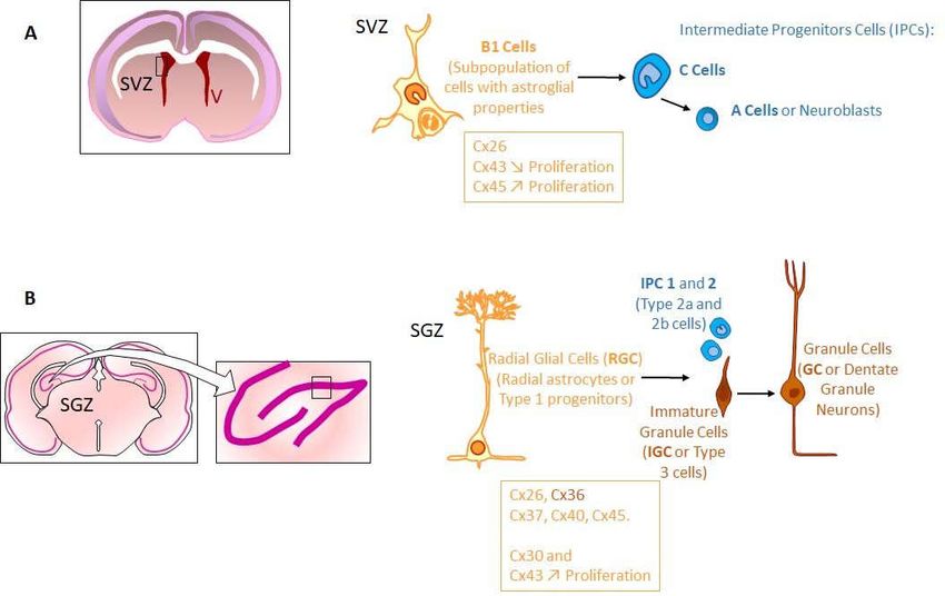
Biomolecules 2021, 11, 51                                                                                                          2 of 64

                                     nearly USD 500 billion annually [2,3]. Taken together, all brain pathologies, therefore, rep-
                                     resent a strong challenge for next few years, in terms of health care and social impact.
                                           Depending on their etiology, diagnosis and evolution, these brain pathologies can be
                                     classified into three major categories of disorders (Table 1), as neurodevelopmental [ASD,
                                     attention deficit hyperactivity disorders (ADHD), epilepsy], neurodegenerative (PD and
                                     AD) and neurobehavioral [migraines, major depressive disorders (MDD)]. Brain cancer
                                     can be included in this list as a 4th category since it cannot be classified as either a neuro-
                                     developmental, neurodegenerative or neurobehavioral disorder (Table 1). Even if still rare
                                     (less than 2% of cancer cases), brain cancer incidence has increased in high income coun-
                                     tries similar to other brain pathologies [4]. The recent rise in incidence of these various
                                     brain pathologies [1,4] leads one to question their possible common etiology.
                                           Part of the answer to this question might be in distinct characteristics of the brain.
                                     Contrary to other organs, human brain development still progresses for almost two dec-
                                     ades after birth, especially during adolescent years. Even later in life, contrary to earlier
                                     held views, the brain is not a “static” organ. This brain plasticity permits humans to adapt
                                     their behavior to their environment throughout their life [5]. Therefore, during intra-uter-
                                     ine and postnatal periods of life (childhood, adolescence), neuronal connections are mod-
                                     ulated under environmental cues and stimulation (education, psychological traumatisms,
                                     etc.) and are maintained during the rest of life. Such connections are the consequences of
                                     cellular plasticity, which depends on synaptic activity and on the presence of stem cells
                                     able to replace damaged brain cells including neurons, astrocytes, and oligodendrocytes.
                                     Due to its multiple functions crucial for survival of the entire organism, the brain is highly
                                     protected against mechanical and chemical trauma by the skull externally and the blood–
                                     brain barrier (BBB) internally. However, despite this high level of protection, the brain can
                                     be reached and exposed to compounds able to cross the BBB. This leads, for instance, to
                                     drug addictions and possibly to the higher prevalence of brain pathologies affecting hu-
                                     mans from birth (ASD, ADHD, epilepsy) to old age (PD and AD).
                                           In order to assure coordinated brain activity, major brain cells (neurons, astrocytes,
                                     oligodendrocytes) are tightly connected through a dense intercellular network. Among
                                     such intercellular interactions, gap junctions (GJs) play important roles necessary to brain
                                     activity by permitting an efficient transfer of information between neurons through elec-
                                     trical synapses, neurotransmitter and ionic buffering of chemical synapses, axonal mye-
                                     lination, neuronal metabolism through astrocytes, etc. Consequently, the brain is one of
                                     the organs abundantly supplied by diversity of GJ-proteins [connexins (Cxs)]. Therefore,
                                     considering the sensitivity of Cxs and GJ functions to a wide range of chemical com-
                                     pounds, it is possible that chronic exposures to BBB-permeable pollutants disturb the gap-
                                     junctional intercellular communication (GJIC) network in brain. Such GJIC disturbances
                                     could then perturb brain physiology (synaptic plasticity, memory storage, behavior, etc.)
                                     and explain the rising incidence of brain pathologies.

            Table 1. Categories, diagnosis, prevalence, symptoms and causes of brain pathologies discussed in this review.

                                                     Prevalence
  Brain Pathologies          Age at Diagnosis
                                                   (% of Indicated            Symptoms                       Probable Causes
    [References]                (Average)
                                                     Population)
Neurodevelopmental
                                                                                                   -Genetics
                            9–10 years old       8–12%               -Attention deficit            -Exposures at pregnancy and young
ADHD 1
                            (may appear as early (of children world- -Hyperactivity                age (cigarette smoking, alcohol and
[6,7]
                            as 3 years old)      wide)               -Impulsivity                  drug uses, lead, …)
                                                                                                   -Brain injuries
                                                1%                   -Lack of social interaction   -Genetics
ASD 2                       From 2 years old or
                                                (of total popula-    -Restricted interest          -Prenatal and postnatal exposures to
[8–10]                      earlier
                                                tion)                -Repetitive behavior          environmental factors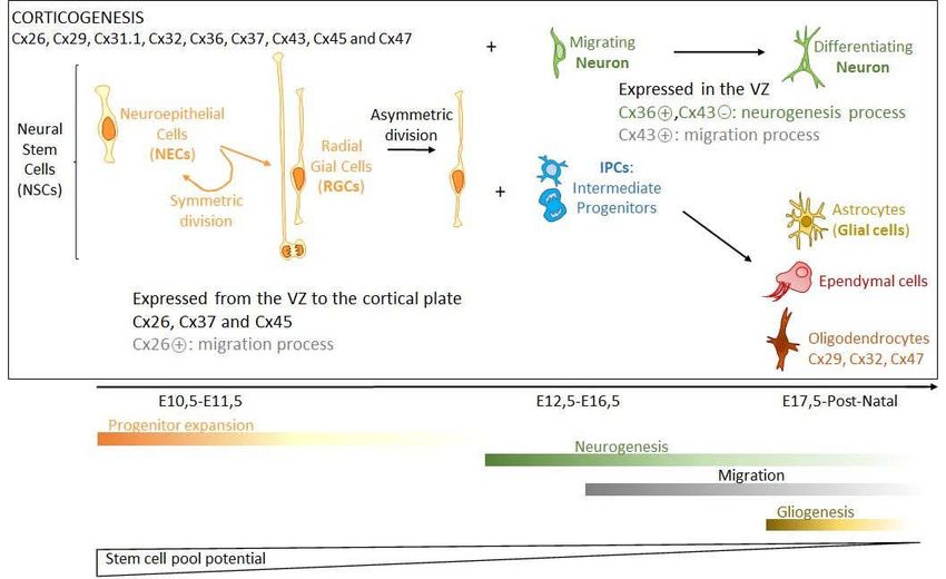
Biomolecules 2021, 11, 51                                                                                                           3 of 64

                                                Prevalence increas-
                                                ing globally over past
                                                50 years
                                                                                                   -Genetics
                                                                                                   -Brain damage from prenatal or peri-
                                                                      -Recurrent and brief epi-
                                                0.4–1%                                             natal causes
Epilepsy                                                              sodes of involuntary move-
                            Any age             (of total popula-                                  -Head injury
[11]                                                                  ments that may be partial or
                                                tion)                                              -Brain infection (meningitis, encepha-
                                                                      generalized
                                                                                                   litis or neurocysticercosis, …)
                                                                                                   -Brain tumour
Neurodegenerative
                                                                      -Decline in memory, lan-
                                                10%
                                                                      guage, problem-
                                                (at age 65 and                                     -Age
                                                                      solving
Alzheimer’s disease                             older in US)                                       -Genetics
                            65 years old                              -Decline in performing eve-
[2,12]                                          146.2% increase in                                 -Environmental factors (cigarette
                                                                      ryday activities through
                                                deaths from 2000 to                                smoking, alcohol use, …)
                                                                      judgment, reasoning, and
                                                2018
                                                                      other cognitive abilities
                                                                      -Movement disorder           -Age
Parkinson’s disease         58 years old        1%                    (tremor, rigidity and brady- -Genetics (5–10% of cases)
[13,14]                     (1 patient/2)       (above 60 years)      kinesia)                     -Environmental factors (pesticide ex-
                                                                      -Depression, anxiety         posure, synthetic heroine use, etc.)
Neurobehavioral
                                                                      -Persistent sad mood
                                                                      -Anxiety                    -Genetics
                                                28.2%
MDD 3                                                                 -Irritability               -Major life change, trauma, stress
                            41.4 years          (life time preva-
[15,16]                                                               -Loss of interest           -Physical illness, medications
                                                lence)
                                                                      -Loss of energy
                                                                      -Fatigue
                                                                                                 -Changes in hormonal levels in
                                                                                                 women
                                                12%                   -Recurrent attacks of mod-
Migraines                                                                                        -Circadian disruption
                            Any age             (of total popula-     erate or severe headache
[17]                                                                                             -Genetics
                                                tion)                 lasting from 4 to 72 h
                                                                                                 -Environmental factors (alcohol use,
                                                                                                 nitrates, carbon monoxide, etc…)
Other
                                                                    -Headache                   -Genetics
                            Children and        0.004–0.008%        -Vertigo                    -Therapeutic irradiation
Cancer 4
                            adults (45–70 years (of total popula-   -Vision trouble             -Electromagnetic fields (?)
[4]
                            old)                tion)               (depends on tumor loca-     -N-nitroso compounds (food)
                                                                    tion)                       -Unknown
            1 Attention deficit hyperactivity disorders. 2 Autism spectrum disorders. 3 Major depressive disorders. 4 Glioma.

                                           The aim of the present review is to evaluate whether chemical pollutants can induce
                                      neuropathologies through their actions on GJIC and Cxs. In order to argue about such a
                                      possibility, this review is made of three major parts reviewing (1) the physiological roles
                                      of GJs and Cxs in the brain, (2) the effect of chemical pollutants on Cx functions and, (3)
                                      the Cx and environmental involvements in major brain pathologies cited above (Table 1).

                                      2. Gap Junction and Connexin Roles in Brain
                                           In the context of animal evolution, the brain was formed through the cephalisation
                                      process. As the anterior part of the central nervous system (CNS), it results from both an
                                      increased number of neurons and a particular cellular organization. The adult human
                                      brain weighs approximately 1.5 kg and, proportional to the body size, its volume is the
                                      highest of all animal species. As a complex organ, the brain is rich in GJs and probably
                                      exhibits the widest range of Cx expression in the body with most of the 21 Cx isoforms
Biomolecules 2021, 11, 51 4 of 64
present (Cx26, Cx29, Cx30, Cx30.2, Cx31.1, Cx31.9, Cx32, Cx36, Cx37, Cx40, Cx43, Cx45,
Cx46, Cx47, Cx57) [18–24]. Paradoxically, while the brain is highly complex in its func-
tional capacities and tissue organization, it only contains a few different cell types express-
ing, for most of them, several Cx isoforms. Before being stabilized in each differentiated
cell type, the Cx expression profile varies during embryogenesis and reveals that Cxs are
involved both in brain formation and in many physiological aspects needed for its func-
tions (Table 2).Biomolecules 2021, 11, 51 5 of 64
Table 2. Gap-junctional organization, connexin expression and functions in human adult brain. Stem cells are not included. Abbreviations : Astro., Astrocytes ;
BBB, Blood-brain barrier ; CMT1X, X-linked Charcot-Marie-Tooth disease type 1 ; Compart., Compartments ; Cx, Connexin ; Ependym., Ependymocytes ; Endo.,
Endothelial ; GJ, Gap junction ; GJIC, Gap-junctional intercellular communication ; HC, Hemichannel ; KO, Knock out mice; Neuroinflam., Neuroinflammation ;
NPC, Neuronal progenitor cells ; ODDD, Oculodentodigital dysplasia ; Oligo., Oligodendrocytes. References are indicated between brackets. In some cases, con-
nexin function as GJIC and/or HC is questioned (?) because not proven yet [25–98].
GJ Com- Cellu- Connexin Expression Brain-Related Physiological Effects of Cx Deficiency in:
part./GJ Net- lar
Works Com-
po-
nents
Cell Major Cell Functions Cx Cx Functions Principal Mice Human
Types Types GJIC HC Physiological Roles (Cx Knock out) (Cx Gene Defect)
(Major of Major Cxs
Cxs)
Neuronal Neu- Action potential transmis- Cx36 + + Electrical synapses Single KO
rons sion [25–27] [27] [28] (no synaptic delay): Cx36: Cx36 :
-Synchronization -Impaired short- and long-term memo- -Juvenile myoclonic
Cx31.1 of interconnected ries [36,37] epilepsy [38,39]
[23,29] neurons [27]
Cx45 -Reduced motor learning [36,37]
[30–32] -Excitability limitation
Cx57 in inhibitory networks [27]
[33–35]
-Role in learning and memory [27]
Panglial syn- Astro- -Component of neuronal Cx43 [40] + [42] + -Glucose/lactate Single KO
cytium cytes synapses (tripartite synap- [44] diffusion
ses) Cx30 [40] + [42] from capillaries Cx43 (astro.): Cx43:
? to neurons -Accelerated hippocampal spreading ODDD patients (ac-
-BBB Cx26 [41] ? [43] and panglial syncytium (oligo., ep- depression and enhanced locomotory cording to muta-
component (astrocytic endym.) [45] activity [57] tions) [61]:
endfeet) -Increased exploratory activity [58] -White matter change
-K+ diffusion -Anxiolytic-like effect in open field [58] -Psychomotor delay
from neuron -Epilepsy
periphery and oligo. -Language disorder
to capillaries [46,47] Cx30:
-Reduced exploratory activity
-Anxiogenic behavior [59]Biomolecules 2021, 11, 51 6 of 64
-Synaptic clearing of neurotrans-
mitters, Double KO
diffusion and recycling towards Cx43 (astro.) / Cx30 [60]:
neurons [45] -Vacuolated oligo.
-Edematous astrocytes
-Modulation of -Myelination abnormalities
neuronal activity [48] -Spatial memory impair
-Sensimotor impair
-BBB maturation [49]
-Gliosis and
modulation
of neuron activity
through Cx43 HC activated
by IL-1β/TNF-α
(neuroinflam.) [50,51]
or Ca2+/K+[52–54].
Such activation leads to ATP re-
lease leading
to paracrine/GJIC-mediated
IP3/Ca2+ wave
propagation
(gliosis) [55] and gliotransmitters
(glutamate) release (synaptic mo-
dulation) [56]
Oligo. -Axon myelination Cx29 [62] ? [64] ? -Myelin integrity [69] Single KO
to facilitate propagation [68] Cx29:
of action potentials Cx32 [63] + -K+ buffering from -No myelin defect [71]
[62,65– neuron periphery
Cx47 [63] 67] to astrocytes [63,70] Cx32 [72]: Cx32:
-Enhanced intrinsic excitability of neu- CMT1X patients:
+ rons -Mostly peripheral
[62,65– -Myelination defects in neocortex neuropathy [75]
67] -Dysfunction of inhibitory synaptic -Encephalopathy
transmission linked to metabolic
stress
Cx47 [66]: (white matter lesions)
-No behavior abnormality [76]
-Vacuolation of nerve fibers Cx47:Biomolecules 2021, 11, 51 7 of 64
-Myelination abnormality Pelizaeus-Merzbacher-
like disease [77,78]:
-Myelination abnor-
mality
Double KO -Impaired psychomo-
Cx32/Cx47 [73]: tor development
-Myelination abnormality
-Astrogliosis
-Microglia activation
Cx30/Cx47 [74]:
-Myelination defects
-Severe motor impairment
-Decreased number of oligodendro-
cytes
-Microglia activation
Epen- -Lining Cx43 [79] + [79]
dym. cerebral
cavities
-Restrictive barrier be-
tween cerebrospinal and
interstitial fluids
-Facilitate circulation of
cerebrospinal fluid by cili-
ated cells
Vascular Endo. -Lining brain capillaries Cx37 [80] + -BBB integrity [81] Cx37 polymorphism:
cells -Ischemic stroke risk?
-Maintenance and control of endo- [82]
Cx40 [80] + thelial
barrier function [81]
Cx43 [80] +
Peri- -Regulation of blood flow Cx37 + -Pericyte contraction? [83–86]
cytes through contraction ca- [80]
pacity + -Maintenance
of vascular
Cx40 [80] + homeostasis [81]
Cx43 [80]Biomolecules 2021, 11, 51 8 of 64
Other Micro- -Inflammation control Cx32 [87] ? + -Neuron death [87]
glia [87]
-Phagocyte micro- Cx36 [88] ? [88] -Control of NPC proliferation, dif-
(With
organisms neurons
ferentiation and migration [93–97]
and damaged cells when
activated)
-Exchange of antigenic
Cx43 [89] + peptides? [98]
(When acti-
vated) [90,91] +
[92]Biomolecules 2021, 11, 51 9 of 64
2.1. Brain Development and Connexin Expression
The brain is the result of an embryonic process starting from the 3rd gestational week
to undergo the four following principal phases of development: (1) dorsal induction (pri-
mary and secondary neurulations), (2) ventral induction (forebrain patterning), (3) neu-
ronal proliferation and migration, and (4) myelination.
Thus, during the 3rd week of embryogenesis, initiation of the CNS evolves with the
development of the notochordal process (neuroectoderm induction). This derivative of
primitive ectoderm rapidly grows in length to be converted, within 20 days, from a hollow
tube to the notochord. Then, the notochord, in relation with the axial mesoderm, induces
the neural plate (primary neurulation). At this stage, the neuroepithelium of the neural
plate begins the formation of the brain and spinal cord by appearing at the cranial end of
the embryo to differentiate craniocaudally. At the beginning of the 4th week, the neural
plate is composed of a broad cranial portion (foetal brain) and a narrow caudal portion
(spinal cord). Then, the foetal brain pursues its own particular and complex evolution,
which corresponds, for the next 5 weeks, to the proencephalic and hemispheric formation.
From the 10th week, neuronal proliferation continues for at least 10 weeks. Soon (12th
week), neuronal migration takes place concomitant with neuronal proliferation up to the
24th week. This neuronal proliferation (neurogenesis) within the cerebral hemispheres
arises from neural precursor cells (NPCs) which are either “type 1 stem-like cells” (nestin+,
GFAP+) or “type 2a progenitors” (nestin+, GFAP-). These self-renewing and multipotential
cells are able to differentiate either into neurons or glial cells. Neurogenesis starts when
those cells differentiate in “type 2b progenitor” cells (nestin+, doublecortin+) to become
“type 3 neuroblasts” (doublecortin+) capable of migrating to form the cortical layers
[24,99–104]. Thereafter, those post-mitotic neurons complete their differentiation by ex-
tending axon and dendrites from their cell body. Neuronal proliferation and migration
continue after birth for several months (postnatal neurogenesis) or even years. Synapto-
genesis accompanies this phenomenon up to puberty, and even probably later. Appar-
ently, NPCs, which are at the origin of neurogenesis, are differently located during em-
bryogenesis. For instance, those from the ventricular zone (VZ) are at the site of origin of
cortical glutamatergic pyramidal neurons, while those from the ganglionic eminence form
GABAergic neurons. After neuronal proliferation and differentiation, programmed neu-
ronal cell death also takes place from the 28th week up to at least the first postnatal week.
During all this neuronal organization, gliogenesis (from the 20–24th weeks) and mye-
lination (from the 26–28th weeks) complete the cellular brain organization up to 2–3 post-
natal years and maybe more. Finally, as brain tissue grows and differentiates, angiogene-
sis forms the particular blood vessel network necessary for food and oxygen supply to
neurons and other brain cells. Importantly, at the time of birth, the BBB is not finalized
due to the incomplete coverage of blood vessels by astrocytic endfeet. Such a process
seems to be completed at postnatal day 10 (P10) and the BBB is considered being mature
at P20 [105].
Cxs and GJIC play important roles in organs during development [106]; this is also
the case for brain formation and cortical development (Figure 1). In rodents, at least 9 Cx
isoforms are highly expressed in the embryonic cerebral cortex (Cx26, Cx29, Cx31.1, Cx32,
Cx36, Cx37, Cx43, Cx45 and Cx47) [24,107,108], and during cortical development, each Cx
isoform has a distinct spatial and temporal pattern of expression [107,109–111], which
might have functional significance as each Cx isoform has distinct permeability and reg-
ulation properties [112]. Cx26, Cx37 and Cx45 are largely distributed from the VZ to the
cortical plate, whereas Cx36 and Cx43 are highly expressed in the VZ and less in the cor-
tical plate [108,113]. Cx43 is widely expressed in embryonic brain before being restricted
mainly to astrocytes as the CNS matures. While Cx43 is a negative modulator of neuronal
differentiation through its carboxyl terminus (C-ter), Cx36 activates this process [114,115].
This is consistent with Cx36 expression in the VZ during the first wave of neurogenesis
with, to a lesser extent, Cx26 [108]. Cx36 expression is maintained during the period ofBiomolecules 2021, 11, 51 10 of 64
neuronal differentiation before being dramatically reduced during postnatal maturation.
It is then restricted to only a few neuron populations (interneurons of hippocampus, thal-
amus, and sparcely in neocortex) where it forms electrical synapses, while most other neu-
rons are connected by chemical synapses [108,109,116–118]. Interestingly, two other Cxs
(Cx26 and Cx43) are involved in neocortical neuronal migration, which seems to be
mainly the consequence of mechanisms driven by the C-ter part of Cx43, independently
from its channel function, but depending on its phosphorylation status [113,119–121]. In
this process, astrocytes participate in the migration of neurons by orientating them [113],
and become involved in synaptogenesis, contributing eventually to the formation of tri-
partite synapses. The differentiation of oligodendrocytes is accompanied by the expres-
sion of specific Cxs (Cx29, Cx32, Cx47), which are involved in the postnatal myelination
process [63,69,122].
During brain development, the presence of Cxs leads to the establishment of GJIC,
which is important to coordinate differentiation of NPC clusters. This is particularly true
in the rodent embryonic CNS where spatio-temporal patterns necessary for formation of
functional domains have to be synchronized in their behaviour [106,123,124]. Moreover,
GJIC probably permits NPCs to exchange signaling messages with adjacent cells to direct
them towards a particular differentiation lineage (neuronal or glial) [24,125]. Such GJIC
compartmentalization is influenced further by changes of Cx expression over time de-
pending on the differentiation process. In parallel to the establishment of GJIC, Cxs are
also involved in brain development via their hemichannel (HC) activity. For instance, dur-
ing corticogenesis, HC-mediated Ca2+ waves increase in number, amplitude, and distance
[126]. Moreover, in the early postnatal neocortex, dendritic GJs mediate the propagation
of IP3 and Ca2+ waves. This process is thought to be at the origin of functional regionaliza-
tion by dividing the immature neocortex into columnar patches of coordinated activity
[123,127–129].
In summary, Cxs are actively involved in brain formation, which largely extends the
classical period of embryogenesis. The numerous Cx isoforms are expressed differently,
in a spatio-temporal pattern modulating cell proliferation, differentiation and migration.
All these processes are mediated differently by Cxs, which establish either GJIC-forming
communicating domains directed towards particular differentiation lineages, para-
crine/autocrine communication through HCs or particular intracellular protein interac-
tions. Activated P2X7 receptors are involved in apoptosis and in the clearance of apoptotic
cells during neurogenesis [130–134]. Even if not completely defined yet, as pannexin
(Panx) channels, Cx HCs could participate in such activation by releasing ATP [24]. Fi-
nally, Cx protein interactions can take place either outside the cells like extracellular parts
of Cx43 interacting with astrocytes during neuronal migration, or inside the cells by con-
trolling particular signaling pathways. Knowing that several Cxs can be expressed in the
same cell type, the events can be controlled in a very subtle way depending on their re-
spective abundance, since different Cxs may have opposite effects in a same cell type (eg.,
Cx43 and Cx36 in neuronal differentiation) [24,119,135].Biomolecules 2021, 11, 51 11 of 64
Figure 1. Corticogenesis. During early cortical development, neural stem cells (NSCs) predominantly undergo symmetric
divisions to expand the NSC pool (Progenitor expansion phase). This pool includes neuroepithelial, radial glial and inter-
mediate progenitor cells. Neuroepithelial cells (NECs) become radial glial cells (RGCs), which by asymmetric divisions
generate RGC and intermediate progenitors (IPCs) or neurons, both migrating and differentiating (Neurogenesis phase).
At later stages of development, NSCs generate the other cell types of the brain including astrocytes, oligodendrocytes, or
ependymal cells (Gliogenesis phase). During cortical development, Cx37 and Cx45 are largely distributed from the VZ to
the cortical plate, whereas Cx36 and Cx43 are highly expressed in the VZ and less in the cortical plate. While Cx43 is a
negative modulator of neuronal differentiation, Cx36 activates this process (in green). Cx26 and Cx43 are involved in
neocortical neuronal migration (in grey). The differentiation of oligodendrocytes is accompanied by the expression of
specific Cxs (Cx29, Cx32, Cx47) that are involved in the postnatal myelination process. The time scale of these phases of
corticogenesis (E: Embryogenesis days; PN: Postnatal) is from mouse [136].
2.2. Adult Brain Organization and Connexin Involvement
At term, adult brain tissue is made of a few cell types contributing to different func-
tional compartments such as neuronal, glial, and vascular compartments. The neuronal
compartment is only made of neurons interacting between each other either through
abundant chemical or less frequent electrical synapses. This compartment is complex with
regard to different types of neurons (projection neurons, interneurons, etc.) presenting a
high variety in spatial and temporal neurotransmitter release and receptor activation. The
glial compartment is the most complex in terms of cell types and organization. Even if
glial cells are smaller than neurons, they are 10 times more numerous and constitute at
least 50% of the total brain cells [137,138]. The glial compartment is composed of macroglia
(astrocytes, oligodendrocytes, ependymocytes) and microglia. The most abundant glial
cells are astrocytes whose ramifications are, either, in contact with neuronal synapses (tri-
partite synapses), oligodendrocytes or cover environing blood capillaries by their endfeet,
which are part of the BBB. Astrocytes control the extracellular interstitial space composi-
tion by buffering K+ ions and clearing neurotransmitters secreted by neuronal activity.
They also secrete gliotransmitters, which can modulate neuronal activity [139]. Oligoden-
drocytes are myelinating cells whose ramifications surround axons of neurons by formingBiomolecules 2021, 11, 51 12 of 64
myelin, which facilitates the propagation of action potentials along axons; myelinated ax-
ons constitute the white matter of the CNS. Ependymocytes line the ventricular cavities
of brain. They constitute a permeable barrier between the cerebrospinal fluid (CSF) and
the interstitial liquid surrounding cells of the CNS. Some of these cells are ciliated and
permit the circulation of the CSF [140–143]. Microglia control inflammation and can dif-
ferentiate into macrophagocytes in the presence of microorganisms or damaged neurons
to phagocytose them. Their role is to protect brain tissue since cells of the immune system
cannot normally access the CNS. The vascular compartment is made of endothelial cells,
which line brain capillaries. The apical face of endothelial cells is in contact with blood
whereas their basal face is in contact, for some of them, with pericytes whose contracting
capacity regulates blood flow. The vascular compartment is separated physically from the
glial compartment by the basal membrane. Therefore, anatomically, the glial compart-
ment is localized between vascular and neuronal compartments.
Due to the limited extracellular space (20%) of CNS, these different cell types are at
high density and extensively interconnected between themselves. Such density and inter-
connection facilitate neuron function by stabilizing their immediate extracellular environ-
ment (astrocytes, oligodendrocytes), bringing nutriments (astrocytes), protecting them
(microglia) and increasing the speed of their communication (oligodendrocytes). Most of
these very different actions have to be highly coordinated and, therefore, all cells are
highly communicating between each other through direct (GJs) or short distance (auto-
crine/paracrine) (HCs) communication. Interestingly, both types of intercellular commu-
nication (direct or autocrine/paracrine), are mediated by the presence of Cxs. This explains
why Cxs are so abundant in the brain parenchyma (Table 2).
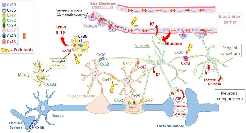
2.2.1. Gap-Junction Compartments in Brain
Interestingly, the functional compartments presented above can be viewed as each
corresponding to a GJ compartment. Indeed, each of these functional compartments can
be defined as a directly communicating network in which GJs permit the free diffusion of
ions and small hydrophilic molecules between cytosols of adjacent cells belonging to the
same functional compartment. Therefore, GJIC defines at least three different GJ compart-
ments (or GJ-connected networks) in a normal brain. One of these compartments (or “net-
works”) is the neuronal compartment in which neurons communicate between them-
selves through GJs (electrical synapses), mostly made of Cx36 [25–27]. A second one, the
vascular compartment is constituted by capillary endothelial cells (endothelium), which
are connected between themselves and with pericytes via Cx37, Cx40, and Cx43. The 3rd
GJ compartment is localized between the two previous ones and constitutes a panglial
compartment (“panglial syncytium”). In this panglial syncytium, the cells expressing Cxs
include astrocytes (Cx43, Cx30 and possibly Cx26) and oligodendrocytes (Cx32, Cx29,
Cx47), resulting in the formation of GJs directly connecting astrocytes, oligodendrocytes
and ependymocytes (Cx43) [41,46,63,144–147]. Other crucial cell types for brain function,
including microglia and stem cells, do not seem to constitute such firmly established GJ
compartments. However, since these cell types express Cxs and are able to make GJs, at
least occasionally, it is possible to consider them as members of “putative” GJ compart-
ments. As presented below, the spatial organization of “well-established” or “putative”
GJ compartments have functional consequences (Figure 2).Biomolecules 2021, 11, 51 13 of 64
Figure 2. Schematic representation of the organization and connexin composition of the gap-junction compartments in the
human adult brain. The different gap-junction compartments are indicated on the right side of the figure (vascular com-
partment, panglial syncytium, neuronal compartment). Principal cell types are presented for each gap-junction compart-
ment (vascular compartment: endothelial cells or endothelium with pericytes; panglial syncytium: astrocytes in green and
oligodendrocytes with myelin surrounding axons of neurons; neuronal compartment: neurons connected by chemical or
electrical synapses). For clarity, pericytes are not enveloped by basement membrane. Microglia are located apart since they
do not seem to establish permanent gap junctions between microglial cells or with cells of the different gap-junction com-
partments. Microglial cells are presented in non-activated and activated states through microglial activation. The connexin
isoforms expressed in the different cell types are indicated according to the code color inserted in the left side of the figure.
For clarity, connexin hemichannels and gap junctions are both presented as cylinders. Cylinders present between cells are
gap junctions made of the indicated connexins, while those located in nonintercellular areas are hemichannels. Cylinders
inside myelin represent reflexive gap junctions between the concentrical layers of myelin around neuronal axons. The red
double arrows in the vascular compartment represent gap junctions made of either Cx43, Cx37 or Cx40 between endothe-
lial cells and between endothelial cells and pericytes. Single red arrows represent ions (K+) and molecules passing through
connexin channels, which are either gap junctions (transfer of K+ and glucose uptaken from extracellular space to be re-
leased in blood or close to neurons, respectively) or hemichannels (release of pro-inflammatory cytokines like TNF-α and
IL-1β by activated microglia; ATP, D-serine or glutamate released by astrocytes). Yellow lightning bolts represents possi-
ble effects of pollutants as either, inhibitors (−) of gap-junctional intercellular communication or indirect activators (+) of
connexin hemichannels (See Discussion part for details). For clarity, ependymocytes and stem cell niches are not shown
and only two astrocytic endfeet are presented as constituents of the blood–brain barrier. At the level of the blood-brain
barrier, the perivascular space delimited by the astrocytic endfeet is part of the so-called glymphatic system involved in
draining interstitial fluid and removing metabolic wastes.
The Neuronal Gap-Junction Compartment
The simplest GJ compartment in the brain is the neuronal compartment, which is
only made of neurons connected by electrical synapses (Figure 2). This neuronal compart-
ment is quite extended in the embryonic CNS where electrical synapses are numerous.
Then, electrical synapses become limited during development [22,24,25,148–152] up to be-
ing restricted, in adults, in particular brain areas like the cerebral cortex (inhibitory GA-
BAergic interneurons) [153–157], hippocampus (synchronous activity among inhibitory
interneurons), reticular thalamic nucleus (synchronization of burst-firing), suprachiasmic
nucleus (circadian behavior) [158,159], hypothalamus (pulsatile oxytocin release) and
brainstem (control of respiratory modulation) [27].Biomolecules 2021, 11, 51 14 of 64
All these electrical synapses are mostly made of Cx36, even if Cx31.1, Cx45, Cx57 and
others have been found in murine neurons of some brain regions such as the retina [30–
35]. Therefore, consistent with the role of neurons in which Cx36 is expressed, Cx36 GJs
mostly promote the synchronization of interconnected neurons [118,160]. If such a syn-
chronization is required, for example, for circadian rhythm, the presence of inhibitory net-
works coupled by Cx36 GJs permits resistance to the development of epileptiform syn-
chronization. In relation to this, it is interesting to note that mutations in the regulatory
promoter of the Cx36 gene (GJD2) are linked to juvenile myoclonic epilepsy (Table 2)
[38,39]. In addition to their function in limiting excitability, neuronal GJs also play a major
role in learning and memory. Indeed, Cx36-KO mice exhibit impaired short- and long-
term memory as well as reduced motor learning (Table 2) [36,37]. Cx31.1, which is ex-
pressed in dopaminergic neurons of the substantia nigra pars compacta [161] and striatal
output neurons [29], seems to play a role in exploration of novel environments and object
recognition since these functions are either elevated or impaired, respectively, in Cx31.1
KO mice [23,162].
A general property of these electrical synapses is their high degree of plasticity,
which makes them capable of modifying their coupling strength under various physio-
logical conditions [27]. Cx36 has also been reported to act not only as a GJ protein but also
as HC components releasing ATP during depolarization in cortical spreading depression
[28]. Further studies could verify this.
The Panglial Syncytium
As a GJ compartment, the panglial syncytium is made of astrocytes, oligodendrocytes
and ependymocytes. Because of their abundance and roles, only the first two cell types
will be considered (Figure 2).
Astrocytes are the most numerous cells in the panglial syncytium. They represent an
intermediate layer between neurons and blood vessels since they project, for some of
them, extensions to synapses (tripartite synapses) as well as to capillaries they surround
by their endfeet [163]. Astrocytes are coupled extensively by GJs mostly made of Cx43 but
also Cx30, which seems to be present more in grey matter [40] where there seem to be
mainly homotypic/homomeric Cx43/Cx43 or Cx30/Cx30 [164]. Contrary to Cx30, whose
expression is delayed during brain maturation, Cx43 is more homogeneously present in
the astrocyte network of white matter, becoming the most abundant Cx in CNS [40,41,165–
168]. Expression of Cx26 has also been reported in rodent astrocytes after birth, where it
was observed in GJs between astrocytes and with oligodendrocytes [41]. However, this
expression of Cx26 in astrocytes is still debated as deletion of both Cx43 and Cx30 in as-
trocytes causes pathological conditions [60], whereas lacZ under the Cx26 promoter is not
expressed in astrocytes [43].
As the other important part of the panglial syncytium, oligodendrocytes form few
GJs with astrocytes, mainly to those they depend on for homeostatic and nutrient support
[169]. In oligodendrocytes, 3 Cx isoforms are expressed (Cx29, Cx32, Cx47) exhibiting dif-
ferent subcellular localizations. Cx47 colocalizes with Cx32 in GJs present in the soma
connected to astrocytes (heterologous GJs) while Cx29 and Cx32 are concentrated in re-
flexive GJs connecting the concentric myelin layers. Cx47 is also present in small GJs link-
ing myelin to astrocytes but not in deeper layers of myelin [62,65,66]. Apparently, oli-
godendrocytes also communicate, but to a much lesser extent, through homotypic Cx47
GJs, with other oligodendrocytes and their precursors in white matter [67]. Therefore, the
only way most oligodendrocytes can communicate between each other is indirectly
through GJs with astrocytic «intermediaries» [170,171].
This particular GJ network, in which at least 5 Cx isoforms are involved, plays fun-
damental roles for neuron survival and activity. Since neurons are distant from blood ca-
pillaries, their energy supply (glucose and lactate) cannot be transmitted directly to them
but has to pass through astrocytes whose endfeet surround the basal membrane of theBiomolecules 2021, 11, 51 15 of 64
vessels [172]. Glucose molecules enter from blood to astrocytic endfeet by specific trans-
porters and then traffic from astrocytes to astrocytes through Cx43/Cx30 GJs to be released
by transporters to the immediate proximity of neurons, where uptake occurs. Apparently,
in astrocytes, glucose is transformed through a glutamate-induced glycolytic degradation
pathway into lactate, which is transferred to neurons via monocarboxylate transporters
[45,173]. Moreover, glucose diffusion passing through astrocytes is not only directed to
supply neurons, but is also distributed, through GJs, to oligodendrocytes and ependymo-
cytes.
This energy supply in neurons is essential for the generation of action potentials. Due
to neuronal activity, these action potentials increase extracellular K+ concentrations, which
could be deleterious for neuronal function. Extracellular K+ ions are taken up by astrocytes
and redistributed though astrocyte network following their gradient before being released
in the interstitial space. Interestingly, GJIC (ionic coupling) of the astrocytic networks
plays a particular role in this phenomenon by equalizing the astrocytic membrane poten-
tial (VM) to levels comparable to its neighbours [174]. This minimizes VM depolarization
due to elevated local [K+]e and thereby maintains a sustained driving force for highly effi-
cient K+ uptake. Thus, GJIC permits to maintain a constant extracellular K+ microenviron-
ment by achieving isopotentiality in astrocytic networks [174]. Astrocyte endfeet contact-
ing capillaries can also release excess K+ ions and associated osmotic water via Kir4.1 po-
tassium channels [175] and AQP4 water channels that are concentrated in the endfoot
plasma membrane [176,177]. Involved in the redistribution of extracellular ions and water,
Cx43-mediated GJIC has been reported to regulate astroglial volume [48,60,178]. Because
astrocytes occupy 50% of the brain volume [179], changes in astrocytic volume classically
lead to opposite variations in extracellular space volume, which can affect neuronal activ-
ity by altering the concentration and diffusion of extracellular ions and neurotransmitters.
Oligodendrocytes also participate in this K+ buffering through their myelin sheath all
along the axon of neurons [70]. After entering the innermost cytoplasmic layer of myelin,
K+ ions pass through successive paranodal loops through Cx32/Cx29-containing reflexive
GJs to the outermost layer of myelin, then into the astrocytes through heterotypic GJs
(Cx47/Cx32: oligodendrocyte side; Cx43/Cx30: astrocyte side). Therefore, through a com-
plex and unique communicating network combining a succession of dense autologous,
heterologous, and then homologous GJs, both astrocytes and oligodendrocytes participate
to K+ siphoning, avoiding the excess of extracellular K+ ions, which do not reenter into
neurons by Na+/K+ ATPase pump activity [63]. Failure to remove sufficient K+ results in
changes in resting membrane potential allowing spontaneous action potentials.
Moreover, if reflexive GJs permit the diffusion of K+ ions through the myelin layers
of oligodendrocytes, they also permit ionic homeostasis necessary for myelin integrity.
Any lack of function due to mutation of Cx47 or Cx32 may cause myelin vacuolation or
dismyelination [70]. This results in loss of velocity of action potentials along neuronal ax-
ons as it is the case in the Pelizaeus–Merzbacher-like disease [Cx47 gene (GJC2) mutation]
[77,78] or Charcot–Mary-Tooth disease [Cx32 gene (GJB1) mutation] [75,76]. Astrocytes
also contribute to this function by relaying to their network this ionic homeostasis. Any
disturbance of astrocytic GJIC may then provoke dismyelination among oligodendrocytes
[60]. Moreover, GJIC-deficient astrocytes display a reduced threshold for the generation
of epileptiform events [47,60]. Therefore, the panglial syncytium entirely contributes to
the efficient propagation of action potentials along the axons by maintaining myelin in-
tegrity.
Neurotransmitter buffering is necessary to maintain efficient synaptic transmission.
Such a buffering is due to astrocytes present in the so-called «tripartite synapses». By ad-
equate membrane transporters, astrocytes uptake neurotransmitters present in the synap-
tic space and enable them to diffuse through the astrocytic network before their eventual
recycling to the neurons. This process has been documented for glutamate [45,173]. This
excitatory amino acid is cleared from the neuronal synapses by astrocytes via glutamate
transporters, and is converted into glutamine, which is released and in turn taken up byBiomolecules 2021, 11, 51 16 of 64
neurons. Metabotropic glutamate receptor activation on astrocytes triggers a variety of
responses via increases in cytosolic Ca2+, such as Ca2+-dependent glutamate release from
the astrocytes, which modulates the activity of both excitatory and inhibitory synapses. In
vivo studies have identified the astrocytic endfoot processes enveloping the vessel walls
as the center for astrocytic Ca2+ signaling and it is possible that Ca2+ signaling events in the
cellular component of the BBB are instrumental in modulation of local blood flow as well
as substrate transport.
By facilitating K+ and neurotransmitter buffering, the maintenance of an efficient
panglial syncytium is therefore necessary to keep the immediate environment of neurons
compatible with their function and survival.
The Vascular Gap-Junction Compartment
Although the human brain accounts for only 2% of total body weight, it demands
20% of the overall energy produced by the body. Since the brain lacks the capacity to store
energy, a vast vascular compartment responds to the continuous demand of oxygen and
glucose (and waste exit) necessary for neuronal survival and activity and for other cells of
the CNS [180]. In an adult brain, it is estimated, by stereological approach, that such en-
ergy supply and waste exit are carried out by a network of 600 km of capillaries (7 µm
diameter) representing an exchange surface of approximately 15–25 m2 [180–183]. Such a
high density of capillaries places each neuron within 20 µm from a capillary. As seen
above, neurons require a tightly regulated extracellular environment to function appro-
priately, and the 20 µm proximity between neurons and capillaries makes them suscepti-
ble to possible chemical fluctuations (ions, hormones, neurotransmitters, amino acids,
etc.). Causes of such fluctuations are numerous and may even include circulating neuro-
toxic substances from external origin (drugs, pollutants, etc.). As a protection, the ex-
change of substances between blood and brain is tightly controlled by the BBB, which,
thus, plays fundamental roles such as regulating ion concentrations, facilitating nutrient
transport, and preventing toxic substances [184].
Anatomically, BBB is therefore constituted by a 600 km syncytium of vascular endo-
thelial cells (endothelium) lining the lumen of the capillaries (Figure 2). A thin layer of
basal membrane separates endothelial cells from less numbered pericytes (ratio 1:3). De-
spite this thin basal membrane, endothelial cells and pericytes are nevertheless in contact
from place to place through GJs [185,186]. Such GJIC and also paracrine communication
are involved in pericyte contraction, which contributes to the local regulation of blood
flow by controlling capillary diameter [83,86]. Endothelial cells and pericytes are envel-
oped by the basal membrane of the capillary, which is almost entirely enclosed by astro-
cyte endfeet (Figure 2). Importantly, the perivascular space delimited by the astrocytic
endfeet is part of the so-called glymphatic system involved in draining interstitial fluid
and removing metabolic wastes [187–189].
Substances can cross the BBB only via paracellular and transcellular pathways re-
stricted by the presence of adherens and tight junctions between endothelial cells. The
transcellular trafficking of endocytic vesicles carrying substances from the apical mem-
brane through the cytoplasm before exiting the basal membrane is more limited than in
the rest of the organism [180,190]. Endothelial cells extensively communicate between
each other and with pericytes through GJs made of Cx37, Cx40 and Cx43 (Figure 2) [80].
This particular GJ compartment is implicated in BBB function since the presence of func-
tional Cxs is necessary for its maintenance [81]. Moreover, expression of Cx30 and Cx43
at the astrocytic endfeet during development coincides with postnatal maturation of BBB.
The absence of these Cxs in the astrocyte endfeet weakens the BBB when hydrostatic vas-
cular pressure is increased [49]. Similarly, the presence of Cx43 in astrocytes helps keep
the endothelial barrier closed for immune cell infiltration [191]. However, Cx43 on the
endothelial side would have an opposite effect on BBB permeability as observed in a mu-
rine model of the familial cerebral cavernous malformations type III (fCCM3). This disease
is caused by loss-of-function mutations in CCM3 that result in dilated capillary beds,Biomolecules 2021, 11, 51 17 of 64
which are susceptible to hemorrhage because of BBB disruption. Interestingly, Cx43 ap-
pears to be up-regulated in developing CCM3 lesions and through its interactions with
ZO-1 would favor BBB breakdown by limiting tight junction formation [192]. Similarly,
the impairment of pericyte-endothelial interaction increases BBB permeability [193].
The situation is probably more complex because GJ channels and Cx HCs of BBB
appear to play opposite roles in the regulation of its permeability. GJIC contributes to
maintaining BBB integrity, while HCs are associated with ATP signaling release and nec-
essary to generate Ca2+ oscillations linked to BBB disruption induced by proinflammatory
signals [194]. Moreover, it is quite possible that Cx HCs then affect the BBB transcellular
pathway by modifying [Ca2+]i, which is involved in the vesicular pathway [194]. Such a
situation would depend then on the neuroinflammation status since the release of gli-
otransmitters or cytokines by activated astroglial and microglial cells affects GJIC and ac-
tivates Cx HCs [195]. In this situation, it would be interesting to determine whether Panxs,
which share many common functions with Cx HCs, might also be involved. This is im-
portant because disruption of the BBB is associated with many diseases of the CNS asso-
ciated with neuroinflammation such as neurodegenerative diseases like AD [196–200].
BBB disruption has been also observed in PD [198,201–203], epilepsy, seizures [204–206],
and brain tumors [207–209]. Moreover, it is also possible that BBB disruption affects the
glymphatic-draining function by disturbing the perivascular space. Disturbance of such a
process seems to be involved in AD by preventing the efficient clearance of amyloid-β
deposits [210,211].
Other Putative Gap-Junction Compartments in Brain
The GJ compartments that are functionally described above are well established after
birth and for the entire life. The integrity of such an organization is maintained and pro-
tected against any cell damage. This protection is assured by cells playing complementary
roles by eliminating pathogens and damaged cells (microglia) and by replacing damaged
cells (stem and precursor cells). Either scattered inside brain parenchyma (microglia) or
concentrated in particular niches (stem cells), these cells do not seem to constitute a well-
established and permanent GJ compartment. However, they express Cxs and are therefore
able to communicate either directly through GJs or through paracrine communication
with other cell types, depending on their activation.
i. Stem Cell Niches
After birth, neurogenesis can occur in limited niches of the mammalian brain (Figure
3). In adults, those niches are located in the subventricular zone (SVZ), the subgranular
zone (SGZ) of the dendrite gyrus of the hippocampus [24,101,104], and in the hypothala-
mus (dorsal α1, α2 regions and in the «hypothalamic proliferative region», adjacent to the
median eminence, in the β region) [212,213]. The restriction of the neurogenesis areas is
the result of a progressive developmental process well described for the SVZ [141,142,214–
219].
Once formed, the SVZ lines the walls of the lateral ventricles (LVs). It contains neural
stem cells (type B cells) [220] located under the ependymal layer of the ventricle [221].
Those cells have projections towards the CSF in the LV, and also contacts with blood ves-
sels (BVs) of the SVZ plexus [217]. This situation allows type B cells receiving signals from
CSF and blood to form transit-amplifying NPCs (type C cells) in asymmetric divisions,
which divide to form neuroblasts (type A cells) [220,222,223]. In humans, during the first
postnatal months, those neuroblasts migrate to the frontal lobe tangentially close to the
walls of LVs and along BVs, through a mechanism probably controlled by astrocyte-re-
leased soluble factors [224–226]. Then, these neuroblasts individually reach cortical tissue
where they differentiate and contribute to inhibitory circuits. These late-arriving inhibi-
tory interneurons could contribute to developmental plasticity since the disruption of
their postnatal migration may underlie neurodevelopmental disorders [227–229]. Inter-
estingly, such a migration coincides with the such a migration happens during the firstBiomolecules 2021, 11, 51 18 of 64
months after birth, a critical period for human brain development, when children begin
to interact with their environment. This is especially true for the human frontal lobe,
which is so important for such interactions and social behavior. Afterwards, neurogenesis
and migration decline, disappearing by two years of age [228–232]. This cessation of post-
natal neurogenesis in the forebrain seems to be particular to human [233,234]. In addition,
SVZ type B cells can also generate oligodendrocyte precursors that contribute to the
maintenance of the oligodendrocyte population in the neighboring corpus callosum, stri-
atum and fimbria-fornix [235,236].
Even if it is not well understood yet, cell-cell interactions seem to play an important
role during the maturation and maintenance of stem cell niches. Indeed, postnatal NPCs
and immature neurons of the SVZ express several types of Cxs (Cx26, Cx43, Cx45)
[107,108,237–244]. GJIC has been demonstrated not only between NPCs (radial glial cells)
but also between them and astrocytes or microglia [237,239–241,245]. Apparently, Cx43
expression increases with postnatal age in SVZ NPCs negatively regulating cell prolifera-
tion, contrary to its promoting role during embryogenesis [242,243,246,247]. In contrast,
in those cells, Cx45 exhibits an opposite role in NPCs by inducing cell cycle reentry via
ATP signaling [243]. Moreover, it has been demonstrated that GJIC mediated by those Cxs
is involved in the migration of NPCs within the SVZ [238]. In the postnatal hippocampus,
almost the same Cxs are expressed (Cx26, Cx30, Cx37, Cx40, Cx43, Cx45) in various NPCs.
However, the expression changes during differentiation (Cx36 appearing in immature
neurons). Interestingly, Cx30 and Cx43 seem to be active regulators of hippocampal NPC
proliferation while Cx43 itself is a negative regulator of proliferation in SVZ [150,248–251].
Although the majority of Cxs are docked to function as GJs, unapposed HCs have also
been documented on the surface of different cell types [252,253].
By the presence of stem cell niches, the postnatal brain exhibits more plasticity than
previously thought and this has implications for memory, learning, and the pathogenesis
of neurodegenerative diseases [254–256]. For instance, in humans, hippocampal neuro-
genesis persists during very late decades of life while it is significantly declined in AD
patients [257]. The fact that NPCs persist in the adult mammalian brain and can integrate
into brain circuity is proof of adult structural plasticity at the cellular level [5]. Interest-
ingly, adult neurogenesis can be regulated by several behavioral factors like running, for
instance, which, contrary to stress, induces such a phenomenon. Other behaviors, like
learning, seem to have more complex effects, suppressing neurogenesis at some stages
(proliferation of progenitor cells), and increasing it at others (differentiation and survival)
[5].
In conclusion, any disruption of proliferation and differentiation of neural stem cells
and migration of their progeny may contribute to neurodevelopmental and neurocogni-
tive deficits such as autism [227–229]. If GJIC is important for such proliferation, differen-
tiation, and survival of neural progenitor cells, any inhibition could indeed have deleteri-
ous effects.Biomolecules 2021, 11, 51 19 of 64
Figure 3. Stem cell niche and neurogenesis in adult. Neural stem cells (NSCs) are mostly retained
in two regions: Sub-ventricular zone (SVZ) and sub-granular zone (SGZ). (A) Frontal cross-section
of the adult brain showing the location of the SVZ, in walls of the lateral ventricles (V). In the SVZ,
NSCs correspond to type B1 cells. B1 cells express Cx26, Cx43 and Cx45. While Cx43 expression
negatively regulates cell proliferation, Cx45 exhibits an opposite role. These B1 cells generate inter-
mediate progenitor cells (IPCs) corresponding to C cells that divide to generate neuroblasts (type
A cells). (B) Frontal cross-section of the adult brain showing the hippocampal formation. The in-
sert indicates the location of the dentate gyrus. The adult dentate gyrus contains radial glial cells,
which are polarized cells with their cell body in the SGZ. Radial glial cells (RGC or RC, type 1
cells) generate IPCs (IPC1 and IPC2 or type 2a and type 2b cells), which differentiate into imma-
ture granule cells (IGCs or type 3 cells) and mature granule cells (GC). In SGZ, Cx26, Cx30, Cx36,
Cx37, Cx40, Cx43, Cx45 are expressed in various NPCs. Interestingly, Cx30 and Cx43 seem to be
active regulators of hippocampal NPC proliferation while Cx43 itself is a negative regulator of
proliferation in SVZ [258].
ii. Microglia
While they belong to the glial cell types (5–20% of glial cells) with macroglia (astro-
cytes and oligodendrocytes), microglia are not part of the panglial syncytium from which
they differ in their embryonic origin [259,260]. So far, despite the fact they do express Cxs,
there is no evidence that microglia establish permanent GJs with astrocytes and/or oli-
godendrocytes. Therefore, microglia can be considered as distinct from macroglia.
As the resident immune cells in the CNS, microglial cells play important roles in
health and disease. In the healthy CNS, they exhibit a ramified morphology with multiple
fine processes that survey their surrounding microenvironment. If a pathological stimulus
appears, as a result of any type of injury or disease, microglial cells acquire an activated
phenotype in which their morphology changes towards a hypertrophic or ameboid-like
appearance and their functional behavior is altered [261–264]. Along with these features,
mounting evidence suggests that microglia continually extend and retract their cell pro-
cesses toward and from synapses, being part of a new spectrum of unexplored capabili-You can also read