Concurrent environmental stressors and jellyfish stings impair caged European sea bass (Dicentrarchus labrax) physiological performances
←
→
Page content transcription
If your browser does not render page correctly, please read the page content below
www.nature.com/scientificreports

              OPEN             Concurrent environmental stressors
                               and jellyfish stings impair caged
                               European sea bass (Dicentrarchus
   received: 15 January 2016
      accepted: 26 May 2016    labrax) physiological performances
     Published: 15 June 2016
                               Mar Bosch-Belmar1,2, Folco Giomi3, Alessandro Rinaldi3,4, Alberta Mandich5,
                               Verónica Fuentes6, Simone Mirto4, Gianluca Sarà3,* & Stefano Piraino1,2,*

                               The increasing frequency of jellyfish outbreaks in coastal areas has led to multiple ecological and
                               socio-economic issues, including mass mortalities of farmed fish. We investigated the sensitivity of
                               the European sea bass (Dicentrarchus labrax), a widely cultured fish in the Mediterranean Sea, to the
                               combined stressors of temperature, hypoxia and stings from the jellyfish Pelagia noctiluca, through
                               measurement of oxygen consumption rates (MO2), critical oxygen levels (PO2crit), and histological
                               analysis of tissue damage. Higher levels of MO2, PO2crit and gill damage in treated fish demonstrated
                               that the synergy of environmental and biotic stressors dramatically impair farmed fish metabolic
                               performances and increase their health vulnerability. As a corollary, in the current scenario of ocean
                               warming, these findings suggest that the combined effects of recurrent hypoxic events and jellyfish
                               blooms in coastal areas might also threaten wild fish populations.

                               Human activities are transforming coastal and marine ecosystems at local, regional, and global scales, exposing
                               both individual organisms and biological communities to dramatic environmental changes by a complex array of
                               interacting stressors1,2. The current trend of induced anthropogenic environmental changes includes increasing
                               sea water temperatures, frequencies of hypoxia episodes, and ocean acidification3,4. Concurrently, zooplankton
                               communities respond to anthropogenic- and climate-induced changes by strong variations in their spatial distri-
                               bution, structure and function5,6. Jellyfish represent one of the components of plankton that seem to be respond-
                               ing positively to the ongoing changes. They are likely to affect the food web structure by their high consumption
                               rates, fast growth and reproduction rates, and wide tolerance to ecosystem changes7–9. Recent analyses of jellyfish
                               population dynamics in Mediterranean coastal zones suggested increasing abundance and frequency of bloom
                               formation10–13. Global changes such as overfishing, eutrophication and ocean warming have been proposed
                               as mechanisms leading to jellyfish increases in many coastal waters worldwide, including the Mediterranean
                               Sea7,11,14–16. These factors are causing severe negative impacts on human economic activities, such as tourism,
                               fisheries, and aquaculture7,17–19.
                                   Aquaculture is an important source of income for local societies and sustains over 40% of global fish pro-
                               duction; mariculture supports nearly 30% (US $23.5 billion) of the total value of farmed finfish species20.
                               Interactions between jellyfish and marine caged fish have been recorded on several occasions in recent years,
                               leading to severe fish mass mortality21. Jellyfish can enter fish cages either intact or fragmented, as tentacles and
                               other body parts (e.g. oral arms), washed by currents and waves against the mesh of cage nets21–23. Overall, more
                               than 400,000 salmon were killed in Irish marine aquaculture facilities in recurrent blooms of the scyphomedusa
                               Pelagia noctiluca in 2007, 2013 and 201424–26. The moon jellyfish Aurelia aurita, and the hydrozoans Muggiaea
                               atlantica and Phialella quadrata were also involved in different farmed fish mortalities, and together with

                               1
                                Dipartimento di Scienze e Tecnologie Biologiche ed Ambientali (DiSTeBA), Università del Salento, Lecce, Italy.
                               2
                                Consorzio Nazionale Interuniversitario per le Scienze del Mare (CoNISMa), Rome, Italy. 3Dipartimento di Scienze
                               della Terra e del Mare (DISTEM), University of Palermo, Italy. 4Istituto per l’Ambiente Marino Costiero, IAMC-
                               CNR, Castellamare del Golfo (TP), Italy. 5Dipartimento di Scienze della Terra, dell’Ambiente e della Vita (DISTAV),
                               University of Genova, Italy. 6Institut de Ciències del Mar, ICM-CSIC, Barcelona, Spain. *These authors jointly
                               supervised this work. Correspondence and requests for materials should be addressed to M.B.-B. (email: maria.
                               boschbelmar@unisalento.it) or S.P. (email: stefano.piraino@unisalento.it)

Scientific Reports | 6:27929 | DOI: 10.1038/srep27929                                                                                             1
www.nature.com/scientificreports/
Figure 1. Gill lesions in fish exposed to Pelagia noctiluca. (a) Control fish gills with unaltered primary
lamellae (pl) with mucous cells (mc) and elongated secondary lamellae (sl) with flat epithelial cells (ep) and
pillar cells (pc), (400×); (b–e). Pathological features in fish gills from the treatment groups after 8 h exposure
to jellyfish (400×): (b) Hyperplasia of primary lamella; (c) Moderate lifting of epithelial cells (*) and cellular
degeneration (arrows); (d) Absence of respiratory epithelium and loss of structure.
P. noctiluca, were identified as potentially harmful species for aquaculture facilities22. In addition, jellyfish can
act as vectors of the bacterium Tenacibaculum maritimum, exacerbating fish gill injuries27. Beyond these few
studies, limited information is available on how jellyfish affect fish health, the biological mechanisms underlying
fish mortalities, or estimates of potential economic losses21. Only a few studies described significant fish injuries
caused by the discharge of cnidocytes (specialized cnidarian stinging cells) in fish tissues (skin, gills) leading to
envenomation and cellular damage23,28.
Temperature and dissolved oxygen concentration in the water column are crucial for the development and
performance of aquatic organisms through direct effects on their metabolic rates29,30. Most fish adapt their phys-
iological responses to sustain their metabolic rates when exposed to temperature changes or decreased dissolved
oxygen levels3,31; however, additional external factors (such as pollutants or different environmental factor) may
impair acclimation processes.
In this framework, we investigated the sensitivity of fish to the co-occurrence of environmental stressors
(water temperature) and jellyfish stings to understand the impact of jellyfish blooms on caged finfish in a global
warming scenario. Experiments were designed to test the combined effects of temperature (“temperature treat-
ment”) and prolonged jellyfish contact (“jellyfish treatment”) on metabolic performances (MO2 and critical
PO2) and tissue damage on fish gills over the time. We used the jellyfish Pelagia noctiluca (Forsskål, 1775), the
strongest stinging and most abundant scyphozoan species in the Mediterranean Sea and Eastern North Atlantic,
and juveniles of Dicentrarchus labrax (Linnaeus, 1758), one of the most common fish species in Mediterranean
marine aquaculture. This study presents important eco-physiological data to the overall fish mariculture sector in
jellyfish-affected coastal areas and also for the scientific community working on the global change susceptibility
of wild fish populations.
Results
Histological analysis. The treatment groups showed obvious gill tissue injuries, most fish displaying lesions
of clinical significance (Fig. 1). The most frequently observed cellular damages were hyperplasia and lamellar
fusion, lamellar oedema and lifting, and cellular hypertrophy and degeneration especially in fish exposed to jel-
lyfish at 27 °C. The gill damage scores in fish exposed to jellyfish were higher than in controls without jellyfish at
both temperatures (21 and 27 °C) (Table 1, Fig. 2a). Wilcoxon pairwise comparisons showed significant interactions
between temperature and jellyfish factors for treated fish but not for control groups (Table 2, Fig. 2a). The number of
goblet cells was significantly higher in fish exposed to jellyfish than in controls; also, the number of chloride cells dif-
fered significantly between control and exposed fish, but not between control fish at different temperatures (Table 1,
Fig. 2b,c).
Scientific Reports | 6:27929 | DOI: 10.1038/srep27929 2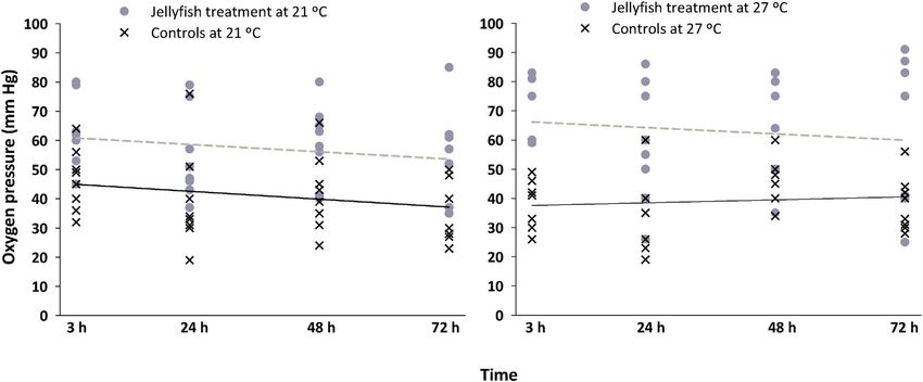
www.nature.com/scientificreports/

                                                     Gill damage                 Goblet cells                Chloride cells
                               Factor           df   H value    P value     df    F value   P value     df     F value P value
                               Jellyfish        1    50.651    1.103 e−12   1     18.869        0.001   1       6.510     0.015
                               Temperature      1     0.010        0.919    1      3.996        0.046   1       3.580     0.063
                               Jelly. x Temp.           –                   1      0.930        0.334   1       3.846     0.062

                              Table 1. Statistical results for histopathological gill damage. Kruskal-Wallis test for temperature and jellyfish
                              factors and one-way ANOVA analyses for goblet cells and chloride cells. p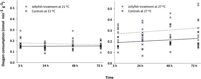
www.nature.com/scientificreports/

                               Gill             Jelly. x       Controls x                                Controls x
                               damage            21 °C           21 °C              Jelly. x 27 °C         27 °C
                               Jelly. x
                                                   –
www.nature.com/scientificreports/
Figure 4. Critical oxygen pressures of Dicentrarchus labrax juveniles exposed to Pelagia noctiluca contact.
Data from fish exposed to jellyfish are represented by grey dots and a dashed regression line; data from control
fish are represented by black “x” and a continuous regression line. Experiments performed at 21 °C and 27 °C
are shown on the left and right panels, respectively. X axes correspond to the time after the fish were exposed to
jellyfish. PO2crit did not change over time: r2 is 0.04 for treated and 0.05 for controls at 21 °C (n.s.) and 0.02 for
treated and 0.01 for controls at 27 °C (n.s.). Regression lines have different intercepts at 21 °C and 27 °C showing
higher PO2crit s for fish exposed to jellyfish.
Figure 5. Unifying model of physiological responses of fish to the interaction of ocean warming and jellyfish
stinging. Dashed lines represent the responses to single factors alone. Briefly, the rise of water temperature is
mirrored by an increase of oxygen consumption rate (MO2), but does not affect the sensitivity of fish to declining
environmental oxygen tension (PO2) (long dashed line); by contrast, jellyfish envenomation causes increased
PO2crit , which enhances the sensitivity to hypoxia (short dashed line). The dotted line represents the physiological
response to the interaction of both factors and shows the enhanced vulnerability of fish.
jellyfish-treated fish, but not between temperatures. No significant changes were observed on fish MO2 and PO2crit
over time following their exposure to P. noctiluca tissues (Table 3).
MO2 values were significantly different at the two temperatures (Table 4, Fig. 3). Oxygen uptake was equiv-
alent in fish exposed to jellyfish stings and control fish at 21 °C, whereas fish exposed to jellyfish at 27 °C had
higher MO2 than controls. PO2crit at 21 °C and 27 °C were significantly different, with higher PO2crit values in
jellyfish-treated fish (averages ranged between 33–55 and 53–70 mm Hg, respectively) than in control fish
(Table 3, Fig. 4). The observed changes of physiological responses of D. labrax juveniles (as MO2 and PO2crit val-
ues) related to temperature and/or exposure to jellyfish tissues were represented in a conceptual model (Fig. 5).
The higher temperature led to increased fish oxygen consumption rate (a), and the jellyfish stings produced an
increased PO2crit (b). The combined effects of temperature and jellyfish stings caused higher oxygen uptake and
PO2crit value (c) than the separate effect of either factor. The synergistic action of envenomation and increased
temperature increased the PO2crit value (i.e., anticipating the switch from the aerobic to anaerobic metabolism),
thereby increasing fish sensitivity to hypoxic conditions.
Scientific Reports | 6:27929 | DOI: 10.1038/srep27929 5www.nature.com/scientificreports/
Discussion
Previous studies on jellyfish impacts on farmed fish hypothesized that respiratory distress may impair the overall
fish metabolism23,25. Here, for the first time, we used an integrated approach to investigate the effects of jelly-
fish blooms on farmed fish by the combined analysis of fish gill integrity and metabolic rates. Significant effects
(increased gill damage, oxygen consumption, and critical oxygen pressure) were observed in fish at higher tem-
perature and exposed to jellyfish.
The increased histological damage in juvenile D. labrax exposed to P. noctiluca jellyfish corroborated previ-
ous observations of adult salmon (Salmo salar) with severe skin and gill injuries induced by jellyfish contacts,
which significantly affected fish health and survival23,25. Severity of gill injuries increased with factors inter-
action (temperature and exposure to jellyfish), which reduced gill plasticity and functioning. The observed
thickening of the gill epithelium due to hyperplasia may increase the diffusion distance for gas exchange, having
profound effects on the efficiency of oxygen transfer across the gill32,33. The increase in goblet cell numbers in
fish contacted by jellyfish was paralleled by [I] increased production of mucus (data not shown), which acts as
a protective barrier against microbial infections34,35 but also forms a barrier to oxygen diffusion and contributes
to hypoxia36, and [II] an increase of chloride cells in the respiratory epithelium, which is a common response to
environmental (chemical or physical) stresses, such as low-calcium and low-magnesium water, or the detection
of toxicants37,38.
With increasing temperature, metabolic rate and oxygen demand of ectothermic fish usually increase, but
oxygen solubility declines, which exacerbates the problem caused by increased respiratory activity2. European
sea bass increases cardiorespiratory and swimming performances in response to increased temperature39,40.
Similarly, higher oxygen consumption rate41,42, growth rate, food intake and feeding efficiency43 also occur in
higher temperature. Several studies indicate that temperature and hypoxia are likely to interact synergistically on
fish metabolism2,44,45. PO2crit values in fish usually increase when temperature rises46,47. Increased temperatures
typically cause a decrease in the affinity of hemoglobin for oxygen, limiting oxygen uptake at the gills and, as a
consequence, reducing fish tolerance to hypoxic conditions2,31. By contrast, other studies suggest that increased
temperature may not affect the tolerance to hypoxia in some fish species due to the intervention of homeostatic
mechanisms (e.g. the recruitment of tissue glycogen or liver lactate clearance capacity)48. An increase in PO2crit
values has been observed during digestion processes49 and may explain why hypoxic conditions might reduce
appetite and growth in many fish species49,50. Increased PO2crit values have also been observed after contamination
by xenobiotics such as heavy metals, pesticides, or nanoparticles in coastal waters51,52.
As suggested by the conceptual model (Fig. 5), our results support the hypothesis that exposure to jellyfish
stings and envenomation may act synergistically with temperature, reducing fish metabolic performance, impair-
ing their ability to withstand hypoxic conditions and, as a consequence, reducing the available energy for critical
processes such as growth and reproduction2. Furthermore, jellyfish venoms may have hemolytic properties53
leading to exacerbation of hypoxia. In conclusion, the interaction of jellyfish envenomations with increasing
temperatures may result in greater vulnerability to hypoxic conditions and in the overall reduction of fish physi-
ological performances.
The reduction of fish homeostatic potentials due to jellyfish outbreaks in coastal waters may co-occur to pro-
duce economic losses to aquaculture facilities. Our study suggests that the interaction of direct climatic stressors
(e.g. warming) with indirect effects of global change (e.g. increasing jellyfish outbreaks) may exacerbate negative
impacts on fish stocks. The consequences of such interactions for human activities are numerous, but mainly
affect fisheries and aquaculture. Due to the continual growth of the aquaculture sector and the increased fre-
quency of jellyfish blooms in coastal areas, the negative interactions of stinging jellyfish on farmed fish is expected
to become a substantial, recurrent issue. More research on the effects of multiple stressors on fish populations in
a global change scenario is needed for a better management of living resources and the development of effective
mitigation plans.
Materials and Methods
Ethical statement. The study was performed in accordance with the EU Directive 2010/63 and Italian DL
2014/26; the experimental protocol was approved by the University of Palermo. Maintenance and handling of ani-
mals during the experiment, as well as the euthanasia procedure, were monitored and carried out by authorized
staff to minimise the animals’ suffering.
Animal collection and maintenance. Two hundred juvenile Dicentrarchus labrax (19.5 ± 5.5 g, means ± S.E.)
were obtained from an aquaculture facility near Licata (Sicily, Italy). The choice of juveniles was related to severe
mortality events caused by jellyfish in different Mediterranean aquaculture facilities where the most affected fish
class ranged 15–60 g in weight28.
The fish were kept in tanks with seawater from a closed recirculated seawater system at controlled salin-
ity, temperature and photoperiod (means ± S.E., 37.8 ± 0.08 salinity, 19.4 ± 0.4 °C, 12 h: 12 h light-dark regime).
Acclimation at the experimental temperatures (21 °C and 27 °C) was gradually achieved during the week before
the start of the experiments. The fish were fed daily with 2.5% of their body mass of commercial fish feed during
the acclimation period. For the duration of the experiments, the fish stock density was maintained between 12.5
and 14 kg m−3, as used in D. labrax aquaculture cages (9–15 kg m−3). Jellyfish were collected by hand net from the
port of Messina (Sicily, Italy) the day before the experiments and were maintained in 25-L buckets with filtered
seawater and at low density (5 jellyfish per bucket) for one day.
Experimental setup. The experiment was carried out at two temperatures, 21 °C and 27 °C. A total of 128
fish (64 treated, 64 controls) were subject to metabolic measurements. Fish were transferred to the treatment
tanks 24 h prior to the start of the experiment and maintained unfed to reduce possible anomalous metabolic
Scientific Reports | 6:27929 | DOI: 10.1038/srep27929 6www.nature.com/scientificreports/
responses due to residual specific dynamic action. Sixteen 7-L treatment tanks were used for the 8-h contact
period, each of them containing five fish to maintain the experimental stock density. The contact duration corre-
sponds to a realistic night time period of high jellyfish concentration in surface waters16,54.
To simulate a realistic encounter between caged fish and jellyfish pressed by currents through aquaculture
cages, jellyfish tissues were manually cut in small pieces (≥1 cm) immediately prior to the start of the jellyfish
exposure. The jellyfish density used was 25 medusae m−3 23. Tanks were supplied with air to keep dissolved oxy-
gen at maximum levels and ensure contact between jellyfish pieces and fish. The treatment started when jellyfish
tissues were randomly placed in 8 of 16 tanks with fish, whereas the other 8 tanks served as controls without jel-
lyfish. Immediately after the exposure period at each temperature, all replicate fish groups were pooled into two
60-L tanks, according to their experimental status (treated or control) to maximise randomisation of subsequent
fish sampling for metabolic measurements. At each of four different sampling times (3, 24, 48 and 72 h after the
end of the contact period), 8 fish (4 control and 4 treated) were individually transferred into the respirometric
chambers for acclimation. We opted to use closed respirometric chambers rather than swim tunnels to allow fish
routine activity and spontaneous movement in a confined environment. We postulate this approach would fairly
reflect the routine metabolic rate of cultured fish at high density and constrained living space conditions, such as
in farming cage systems.
Respirometry and determination of critical oxygen pressure (PO2crit). At each temperature (21
and 27 °C), eight independent 2-L closed respirometers supplied with filtered sea water (Millipore GF/C 0.45 μm )
were used to measure the oxygen consumption rate of individual fish. Chambers were covered with an opaque
plastic material to avoid visual stresses to fish throughout measurements. An agitator and small magnets were
used to maintain homogeneous water mixing inside the experimental chambers. At each sampling time (3, 24, 48
and 72 h after the end of jellyfish exposure), four treated and four control fish were randomly sampled and placed
in individual respirometers. Fish were left undisturbed in the respirometers for 3 h with supplemented air to keep
dissolved oxygen at the saturation level. Then the aerators were removed and chambers were carefully refilled of
water and hermetically closed. Fibre-optic oxygen meters calibrated according to instructions by Pyro Science
(Aachen, Germany) were used to record water oxygen levels. Fish were maintained in the respirometric chambers
until the slope of the oxygen concentration curve changed suddenly. In most cases, that change occurred at 5 to
30% of the initial oxygen concentration. At the lowest oxygen concentration fish status was surely affected but
no mortalities were recorded over the complete duration of the experiment. Fish were then removed from the
respirometers, marked by a small cut in the caudal pin and returned to the original tank in order to maintain the
initial density. All fish recovered well after hypoxic exposure.
Oxygen consumption rates to approximate routine metabolism (MO2) were calculated from the decrease in
oxygen content in the respirometers over time and expressed as mmol min−1 g−1. Those times were standardized
to 45 min at 21 °C and 15 min at 27 °C within the range during which the fish were able to oxyregulate. The critical
oxygen pressure (PO2crit), which represents the transition from oxyregulation to oxyconformation during the
progressive decline of environmental oxygen tension30, was calculated as the break-point of the graph depicting
the PO2 − MO2 relationship. PO2crit was expressed in mm Hg.
Histological analysis of gill tissue. The experiments were performed in full compliance with the national
rules (D.Lgs 116/92 and subsequent amendments) and the European Commission Recommendation guidelines
for the accommodation and care of animals used for experimental and other scientific purposes (2007/526/EC).
After the last sampling time (72 h), 16 experimental fish (4 controls and 4 treated at each temperature) were anaes-
thetised with 0.05% w/v MS222 (3-aminobenzoic acid ethyl ester) and then killed according to the current animal
care rules using a lethal dose of MS-222 (0.1% w/v). Two gill arches were excised from each fish and immediately
preserved in 10% neutral buffered formalin for 48 h and transferred to 70% ethanol for histological analysis.
After dehydration, tissues were embedded in Paraplast (Bio-Optica), cut by microtome into 5 μm sections and
stained using Hematoxylin-Eosin as “routine-staining” to reveal the underlying tissue structures and conditions.
Moreover, the Periodic Acid Schiff (PAS) technique was used to identify the goblet cells. The localisation and
the number of chloride cells was determined by immunocytochemical techniques by using a primary antibody
that recognised sodium, potassium and chloride cotransporters (Na+/K+/Cl− cotransporters NKCC1-T4 1:200)
revealed by a second antibody Donkey Anti-Rabbit IgG (H+L) Alexa Fluor 488 (AF488) Conjugate (Southern
Biotech), 1:200. For each gill arch, 9 randomly selected tissue areas, between 25 and 34 μm2 were screened at 200X
and 400X to count the number of chloride cells.
Gill damage score. Interpretation of the gill damage was based on a recently developed gill histopathology
scoring system (GHS index, Mandich et al. in prep.) that rates the damage on each gill sample by a total score
obtained by summation of partial scores assigned to 12 different criteria. The evaluation of gill damage was per-
formed as follows: for each gill sample a total of 9 sections (photographed fields), each with 10 secondary lamellae
were evaluated for all 12 histopathological criteria. For each criterion, the score ranged from 0 to 6 depending on
the extent and intensity of injuries (0: no significant damage, 1: damage in 1–2 of 10 lamellae; 3: damage in 3–5 of
10 lamellae; 6: 6–10 of 10 lamellae damaged). Gill damage could be of different grades of severity, and advanced gill
damage could mask previous mild injuries. Therefore, the GHS index was supplemented by a secondary classifica-
tion system to separate different stages in the progression of tissue damage (according to Santos et al.55). All histo-
pathological criteria were split into 3 groups. The first group (first-stage lesions) was composed of hyperplasia (cell
number increase) of the lamina and the secondary lamellae, lamellar fusion, reduction of the lamellae, lamellar
oedema (accumulation of an excessive amount of watery fluid in the intercellular spaces), and cellular hypertrophy
(cell size increase). The second-stage lesions included circulatory disturbances of the lamina such as telangiectasia
(dilation of groups of capillaries) and grave cellular anomalies (presence and extension of lamellar lifting); these
Scientific Reports | 6:27929 | DOI: 10.1038/srep27929 7www.nature.com/scientificreports/
more severe injuries lead to effects on tissue functions; the third -stage lesions included the appearance of haemor-
rhage, high granulocyte concentrations, and cellular degeneration of the respiratory epithelium or necrosis, which
represent irreparable damages. The score assigned for each criterion was multiplied according to the severity group
(×1: mild damage group; ×10: moderate injury group; and ×100: the most severe gill damage group).
The goblet and chloride cells were visually counted in each section and analysed separately from the other
histopathological criteria.
Statistical analysis. To obtain critical oxygen pressures (PO2crit) and approximate the break-point in the
respiration curve, a Piecewise linear regression function was used (SigmaPlot v.11).
Normality of respiration data was confirmed with a Shapiro-Wilk test. To test the statistical significance
among treatments for MO2 and for PO2crit, ANCOVA analyses were used, considering MO2 and PO2crit as the
response variables, time after the exposure period as a continuous explanatory variable, and temperatures and
jellyfish treatments as categorical explanatory variables.
The assumptions of normality were not encountered for the histopathological data (Shapiro-Wilk test,
pwww.nature.com/scientificreports/
31. Pörtner, H.-O. Oxygen and capacity-limitation of thermal tolerance: a matrix for integrating climate-related stressor effects in
marine ecosystems. J. Exp. Biol. 213, 881–893 (2010).
32. Malte, H. Effect of pulsatile flow on gas exchange in the fish gill: Theory and experimental data. Respir. Physiol. 88, 51–62 (1992).
33. Skidmore, J. F. & Tovell, P. W. A. Toxic effects of zinc sulphate on the gills of rainbow trout. Water Res. 6, 759–765 (1972).
34. Deplancke, B. & Gaskins, H. R. Microbial modulation of innate defense: Goblet cells and the intestinal mucus layer. Am. J. Clin. Nutr.
73, 1131S–1141S (2001).
35. Rinaldi, L. et al. Oxygen availability causes morphological changes and a different VEGF/FIk-1/HIF-2 expression pattern in sea bass
gills. Ital. J. Zool. 72, 103–111 (2005).
36. Handy, R. D., Eddy, F. B. & Romain, G. In vitro evidence for the ionoregulatory role of rainbow trout mucus in acid, acid/aluminium
and zinc toxicity. J. Fish Biol. 35, 737–747 (1989).
37. Perry, S. F. The chloride cell: structure and function in the gills of freshwater fishes. Annu. Rev. Physiol. 59, 325–347 (1997).
38. Perry, S. F. Relationships between branchial chloride cells and gas transfer in freshwater fish. Comp. Biochem. Physiol. Part A Mol.
Integr. Physiol. 119, 9–16 (1998).
39. Farrell, A. P. Cardiorespiratory performance in salmonids during exercise at high temperature: Insights into cardiovascular design
limitations in fishes. Comp. Biochem. Physiol. - A Mol. Integr. Physiol. 132, 797–810 (2002).
40. Claireaux, G., Couturier, C. & Groison, A.-L. Effect of temperature on maximum swimming speed and cost of transport in juvenile
European sea bass (Dicentrarchus labrax). Fish Physiol. 27, 3420–3428 (2006).
41. Claireaux, G. & Lagardère, J.-P. Influence of temperature, oxygen and salinity on the metabolism of the European sea bass. J. Sea Res.
42, 157–168 (1999).
42. Dalla Via, J. G., Villani, P., Gasteiger, E. & Niederstatter, H. Oxygen consumption in sea bass fingerling Dicentrarchus labrax exposed to
accute salinity and temperature changes: metabolic basis for maximum stocking density estimations. Aquaculture 169, 303–313 (1998).
43. Person-Le Ruyet, J., Mahé, K., Le Bayon, N. & Le Delliou, H. Effects of temperature on growth and metabolism in a Mediterranean
population of European sea bass, Dicentrarchus labrax. Aquaculture 237, 269–280 (2004).
44. Pörtner, H. O., Langenbuch, M. & Michaelidis, B. Synergistic effects of temperature extremes, hypoxia, and increases in CO2 on
marine animals: From Earth history to global change. J. Geophys. Res. Ocean. 110, 1–15 (2005).
45. Pörtner, H. O. & Farrell, A. P. Physiology and Climate Change. Science 322, 690–692 (2008).
46. Sørensen, C., Munday, P. L. & Nilsson, G. E. Aerobic vs. anaerobic scope: sibling species of fish indicate that temperature dependence
of hypoxia tolerance can predict future survival. Glob. Chang. Biol. 20, 724–729 (2014).
47. Collins, G. M., Clark, T. D., Rummer, J. L. & Carton, A. G. Hypoxia tolerance is conserved across genetically distinct sub-populations
of an iconic, tropical Australian teleost (Lates calcarifer). Conserv. Physiol. 1, 1–9 (2013).
48. He, W., Cao, Z.-D. & Fu, S.-J. Effect of temperature on hypoxia tolerance and its underlying biochemical mechanism in two juvenile
cyprinids exhibiting distinct hypoxia sensitivities. Comp. Biochem. Physiol. Part A Mol. Integr. Physiol. 187, 232–341 (2015).
49. Thuy, N. H. et al. Critical oxygen tension increases during digestion in the perch Perca fluviatilis. J. Fish Biol. 76, 1025–1031 (2010).
50. Wang, T., Lefevre, S., Thanh Huong, D. T., Cong, N. van & Bayley, M. The effects of hypoxia on growth and digestion. Fish Physiology
27, 361–396 (2009).
51. Bilberg, K., Malte, H., Wang, T. & Baatrup, E. Silver nanoparticles and silver nitrate cause respiratory stress in Eurasian perch (Perca
fluviatilis). Aquat. Toxicol. 96, 159–165 (2010).
52. Schjolden, J., Sørensen, J., Nilsson, G. E. & Poléo, A. B. S. The toxicity of copper to crucian carp (Carassius carassius) in soft water.
Sci. Total Environ. 384, 239–251 (2007).
53. Mariottini, G. L. & Pane, L. Mediterranean jellyfish venoms: A review on scyphomedusae. Mar. Drugs 8, 1122–1152 (2010).
54. Malej, A. Behaviour and trophic ecology of the jellyfish Pelagia noctiluca (Forsskal, 1775). J. Exp. Mar. Bio. Ecol. 126, 259–270 (1989).
55. Santos, T. C. a et al. Histopathological alterations in gills of juvenile Florida pompano Trachinotus carolinus (Perciformes,
Carangidae) following sublethal acute and chronic exposure to naphthalene. Panam. J. Aquat. Sci. 6, 109–120 (2011).
56. Hothorn, T., Hornik, K., van de Wiel, M. A. & Zeileis, A. A lego system for conditional inference. Am. Stat. 60, 257–263 (2006).
Acknowledgements
We wish to thank Matteo Mercurio for his excellent assistance during the experiment and Marco Bonaldo for
technical support in histological analysis. Furthermore, we are grateful to Dr. Giacomo Milisenda for his help
with jellyfish sampling and statistical advice and Dr. Jennifer E. Purcell for critical revisions. This work has
received funding from the European Union’s projects MED-JELLYRISK (grant n. I-A/1.3/098 - ENPI CBCMED
programme), VECTORS (Vectors of Change in Oceans and Seas Marine Life, Impact on Economic Sectors, grant
n. 266445, FP7th programme), CERES (Climate Change and European Aquatic Resources, grant n. 678193,
Horizon 2020 programme) and from the Italian Ministry of Research and University project PRIN TETRIS 2010
(grant n. 2010PBMAXP_003).
Author Contributions
M.B.B., V.F., S.P. and A.R. conceived and designed the experiments. M.B.B., F.G. and A.R. carried out the
experiments and collected the data. M.B.B. and A.M. performed the histological analysis. M.B.B. and F.G.
performed the data analysis. G.S. and S.M. provided the experimental animals, laboratory space and facilities for
the experiment. M.B.B., F.G. and S.P. wrote the manuscript and all authors reviewed and edited the manuscript.
Additional Information
Competing financial interests: The authors declare no competing financial interests.
How to cite this article: Bosch-Belmar, M. et al. Concurrent environmental stressors and jellyfish stings impair
caged European sea bass (Dicentrarchus labrax) physiological performances. Sci. Rep. 6, 27929; doi: 10.1038/
srep27929 (2016).
This work is licensed under a Creative Commons Attribution 4.0 International License. The images
or other third party material in this article are included in the article’s Creative Commons license,
unless indicated otherwise in the credit line; if the material is not included under the Creative Commons license,
users will need to obtain permission from the license holder to reproduce the material. To view a copy of this
license, visit http://creativecommons.org/licenses/by/4.0/
Scientific Reports | 6:27929 | DOI: 10.1038/srep27929 9You can also read