Data sheet based on syngo MR XA401 - siemens-healthineers.com/freemax
←
→
Page content transcription
If your browser does not render page correctly, please read the page content below

MAGNETOM Free.Max

Data sheet based on
syngo MR XA40  1)

siemens-healthineers.com/freemax

1)
     The product is still under development and not commercially available yet.
     Its future availability cannot be ensured.

                                                         Preliminary Edition – 04/2021
Breaking barriers
MAGNETOM Free.Max

MAGNETOM Free.Max breaks barriers to expand the reach of MRI.
Where patients have felt discomfort, the world's first 80 cm bore sets a
new paradigm in patient comfort. Where infrastructure was an obstacle
to MRI, MAGNETOM Free.Max slots into an existing helium-free infra-
structure. Where access to MRI was not viable, MAGNETOM Free.Max
makes access affordable. And where conventions have limited our think-
ing, MAGNETOM Free.Max breaks out of conventions to explore new
clinical opportunities in MRI.

2   MAGNETOM Free.Max: Data sheet based on syngo MR XA40 · 04.2021

                                             Preliminary Edition – 04/2021
         Our first 80 cm patient bore
         Accessibility for claustrophobic and obese patients

         DryCool technology
         0.7 liters liquid helium
         Sealed-for-life magnet design

         Our most compact whole-body MRI
         Increased flexibility for siting
         Reduced infrastructure costs

         High-V MRI
         0.55 T for daily excellence and new clinical opportunities

         Image processing innovations
         Deep Resolve
         Simultaneous Multi-Slice
         Compressed Sensing

         myExam Companion
         Intuitive operation for any professional

 Providing greater flexibility to offer MR services
 and open up new clinical opportunities

		                                          04.2021 · MAGNETOM Free.Max: Data sheet based on syngo MR XA40   3

                                 Preliminary Edition – 04/2021
Magnet system

Magnet system

DryCool technology

• Short-bore, patient-friendly design, high homogeneity               • Unique design with a minimal thermal mass and
• Easy siting due to AS (Active Shielding) and E.I.S.                   optimized thermal properties and heat transfer paths
  (External Interference Shielding) magnet technology                   for most efficient cool down
                                                                      • Operating mode: Standard operating mode according
                                                                        to IEC 60601-2-33.

Magnet parameters
Operating field strength                                              0.55 T
Magnet type                                                           Superconducting
Field stability over time                                             < 0.1 ppm/h
Weight (with cryogens)                                                1635 kg
Magnet length                                                         1.48 m
System length cover to cover                                          165 cm
             1)
Bore size                                                             80 cm
Type of installation                                                  Fixed
Decay characteristics from full field to 20 mT                        approx. 1.6 s

Guaranteed homogeneity (based on highly accurate 24 plane plot)
10 cm DSV                                                             0.02 ppm
20 cm DSV                                                             0.075 ppm
30 cm DSV                                                             0.3 ppm
40 cm DSV                                                             1.4 ppm
50 × 50 × 45 cm3 DEV                                                  5 ppm

In compliance with the German “Qualifikationsvereinbarung”. Standard deviation Vrms (Volume root-mean square) measured with highly
accurate 24 plane plot method (20 points per plane). Standard active shim with 3 linear channels (1st order). DSV = Diameter spherical
volume (x, y and z direction). DEV = Diameter elliptical volume.

1)
     Incl. shim coils, gradient coil, RF body coil

4          MAGNETOM Free.Max: Data sheet based on syngo MR XA40 · 04.2021

                                                      Preliminary Edition – 04/2021
Magnet system

Shimming
Both: passive and active shimming. Passive shimming during installation.
Standard active shim with 3 linear channels (1st order).
3D Shim                                       Patient-specific automated shim
                                              Time to shim = approx. 6 s

Shielding
Active Shielding (AS)                         7th generation active shielding (AS) technology with counter coils
Fringe field (axial×radial)                   0.5 mT1)                                    4.0 m × 2.5 m
                                              0.1 mT                                      6.0 m × 3.8 m
External Interference Shield                  Patented shielding system integrated into the magnet
(E.I.S.)
                                              Continuous compensation and automatic suppression of external magnetic
                                              field interferences during measurement (caused by moving ferromagnetic
                                              objects or nearby power lines)

DryCool technology

DryCool technology is an innovative cooling technology
that provides a sealed-for-life superconducting magnet
that operates with 0.7 liters of liquid helium. It eliminates
the need for helium refills and a quench pipe.

Cryostat                                                                                  Stainless steel
Helium inventory                                                                          0.7 l liquid helium
Ramp down time                                                                            < 0.5 h
Time to be back in operation after ramp down                                              < 4 h2)
Ramp up time after emergency shut off                                                     < 24 h2)

1)
     Pacemaker safety limit
2)
     Time will extend if refrigerator remains off for a longer period of time. For longer downtimes, the net time off field needs to be added to
     the times stated to estimate the overall ramp-up time.

		                                                                04.2021 · MAGNETOM Free.Max: Data sheet based on syngo MR XA40                   5

                                                          Preliminary Edition – 04/2021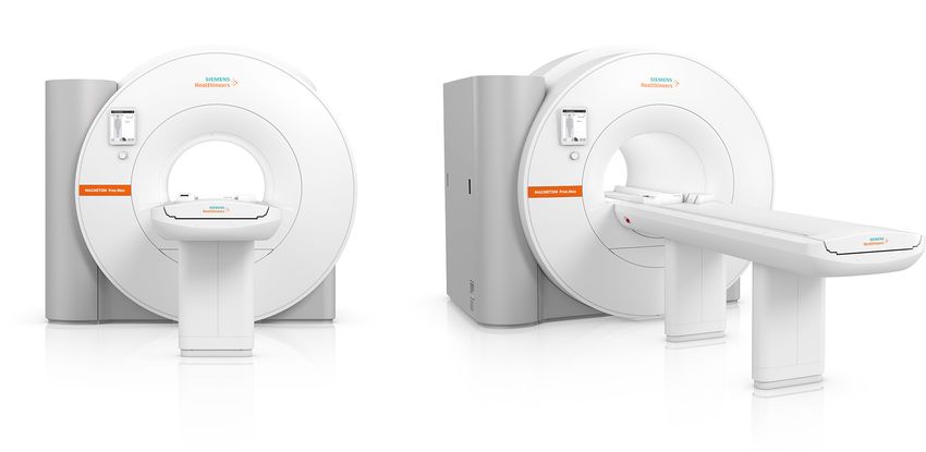
B80 gradients

B80 gradients

General features

• Actively shielded (AS) whole-body gradient coil system               Resolution parameters
• Extremely low eddy currents                                          Min. FoV                     5 mm
• Water-cooled coil and amplifier for maximum
  performance                                                          Max. FoV1)                   500 mm
• All axes force compensated                                           Slice thickness 2D           min. 0.1 mm
                                                                                                    max. 250 mm
                                                                       Partition thickness 3D       min. 0.1 mm
Gradient performance for each axis
                                                                                                    max. 5 mm
Max. amplitude                     26 mT/m
                                                                       Slab thickness 3D            min. 5 mm
Min. rise time                     578 µs                                                           max. 300 mm
Max. slew rate                     45 T/m/s                            Max. matrix                  1024
                                                                       Highest in-plane             20 μm
                                                                       resolution
Vector gradient performance (vector addition of
all 3 gradient axes)
Max. eff. amplitude                45 mT/m                             Gradient amplifier2)
Max. eff. slew rate                78 T/m/s                            • Water-cooled, highly compact, modular design
                                                                       • Ultra-fast solid-state technology with very low
Gradient duty cycle                100 %
                                                                         switching losses
                                                                       Max. output voltage2)        1200 V
                                                                                             2)
                                                                       Max. output current          330 A
                                                                       Max. power                   0.4 MW

1)
     Depending on the application, the maximum FoV in the z-direction can be up to 450 mm
2)
     Values for each of the 3 gradient axes

6         MAGNETOM Free.Max: Data sheet based on syngo MR XA40 · 04.2021

                                                       Preliminary Edition – 04/2021
B80 gradients

Cooling system

Two different customer specific cooling alternatives:
Passive and Active1)

Passive cooling system                       Water consumption                           > 70 l/min2)
                                             Heat dissipation to water                   ≤ 34 kW
                            1)
Active cooling system                        Active cooling system with two Siemens Outdoor Units (SOU) will automatic
                                             adaptation to the required cooling demands (for example, different day /night
                                             mode) to decrease energy cost.

1)
     Depending on the system configuration, this can be an optional feature, actual configuration might deviate.
2)
     Water temperature: 6°C - 14 °C (43°F - 57 °F)

		                                                               04.2021 · MAGNETOM Free.Max: Data sheet based on syngo MR XA40    7

                                                         Preliminary Edition – 04/2021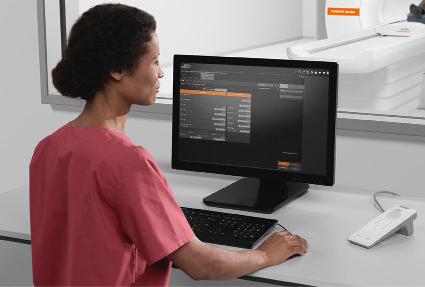
B80 gradients

Sequences
                                                                                 Matrix
                                                                                 64                 128               256
Spin Echo               min. TR [ms]                                             6.4                6.9               7.6
                        min. TE [ms]                                             3.5                4                 4.7
Inversion Recovery      min. TR [ms]                                             31                 32                33
                        min. TE [ms]                                             3.6                4.1               4.9
                        min. TI [ms]                                             24                 24                24
2D GRE                  min. TR [ms]                                             3.1                3.6               4.3
                        min. TE [ms]                                             1.13               1.67              2.4
3D GRE                  min. TR [ms]                                             2.4                2.5               2.9
                        min. TE [ms]                                             0.72               0.89              1.24
TrueFISP                min. TR [ms]                                             2.6                3.2               4
                        min. TE [ms]                                             1.23               1.41              1.79
TSE (HASTE)             min. Echo Spacing [ms]                                   2.28               2.68              3.24
                        min. TR [ms]                                             31                 32                33
                        min. TE [ms]                                             3.6                4.1               4.9
                        max. Turbo Factor = 1024
EPI                     min. Echo Spacing [ms]                                   0.58               0.95              1.18
                        min. TR [ms]                                             42                 43                44
                        min. TE [ms]                                             2.8                3.2               3.6
                        min. Measurement time                                    120                120               160
                        max. EPI Factor = 256
Diffusion Imaging       Max. b-value [s/mm2]                                     10 000             10 000            10 000
                                                               2
                        Min. TE [ms] with b = 1000 s/mm                          67                 70                73

All matrices without interpolation. Combinations of the stated parameters are not always possible; some parameters may require optional
application packages.

8       MAGNETOM Free.Max: Data sheet based on syngo MR XA40 · 04.2021

                                                      Preliminary Edition – 04/2021
DirectRX Technology

DirectRX Technology

General

The unique all digital-out design integrates all RF receive     • Digital-out design – optical links between magnet and
components at the magnet.                                         equipment room to achieve highest RF stability
                                                                • Receive path is integrated in the magnet housing
• Optical RF system improves SNR by reducing electrical         • Receiver with high dynamic range without adjustments
  noise and increasing signal detection

RF transmit technology
Frequency stability (5 min)          ±2×10⁻¹⁰
Frequency control                    32 bits (0.015 Hz)
Phase control                        16 bits (0.006 Degrees)
Body coil                            • Integrated whole body no tune transmit/receive coil with 16 Rungs
                                     • Optimized RF efficiency and signal-to-noise ratio (SNR)
                                     • Real-time Feedback loop for unmatched RF stabilization
Transmitter path                     Transmit amplitude                         16 bit control 25 ns resolution
                                     Gain stability (after first minute)        < 0.05 dB (1 s)
DirectRX Technology

RF receiver technology
Number of coil elements1)                  51
Number of independent receiver             24
channels that can be used
simultaneously in one single scan
in one FOV, each generating an
independent partial image
Quadrature demodulation and                Digital
filtering
Receiver bandwidth                         500 KHz (for each channel)
Receiver signal resolution                 32 bit
ADC sampling rate                          120 MHz
Dynamic range at coil connector            151 dB/Hz instantaneous at coil connector
(referred to 1 Hz resolution
bandwidth)

1)
     Channels that can be connected simultaneously

10        MAGNETOM Free.Max: Data sheet based on syngo MR XA40 · 04.2021

                                                     Preliminary Edition – 04/2021
Local Receive Coils
Local Receive Coils
BioMatrix and Tim 4G technology
BioMatrix technology (integrated into the new BioMatrix technology reduce patient set up time. Lightweight,
Contour L Coil1)) provides seamlessly integrated sensors ergonomically designed coils enable highest patient
to acquire and display the patient’s respiration data comfort.
without need for user interaction.
• No coil changing with multi-exam studies saves patient
All local receive coils of MAGNETOM Free.Max are setup time
designed for highest image quality in combination with • All coils are time-saving “no-tune” coils
easy handling. Tim 4G's DirectConnect and SlideConnect • Low-noise preamplifiers
• AutoCoilSelect for dynamic, automatic, or interactive
selection of the coil elements within the field of view
Standard coils
Head/Neck Coil Application area Head and neck
(DirectConnect)
Dimensions (L × W × H) 495 mm × 350 mm × 334 mm
Weight 6 kg
Spine Coil Application area Spine
(DirectConnect)
Dimensions (L × W × H) 651 mm × 466 mm × 67 mm
Weight 5 kg
BioMatrix Contour L Coil Application area • Thorax
(SlideConnect) • Abdomen
• Pelvis
• Bilateral hip
Dimensions (L × W × H) 416 mm × 632 mm × 65 mm
Weight 700 g
Contour S Coil Application area Multi-purpose
(SlideConnect)
Dimensions (L × W × H) 269 mm × 446 mm × 40 mm
Weight 370 g
Accessories Coil Link
1)
The Respiratory Triggering functionality is still under development and not commercially available yet. Its future availability cannot be
ensured. The Respiratory Triggering functionality is planned to be activated with a future software update.
04.2021 · MAGNETOM Free.Max: Data sheet based on syngo MR XA40 11
Preliminary Edition – 04/2021MAGNETOM Free.Max
Head/Neck Coil
siemens-healthineers.com/freemax
General Applications
The Head/Neck Coil is part of the the Standard Coil • Head examination
Package. • Neck examination
• MR angiography of the head/neck
• 12-channel design with 12 integrated pre-amplifiers, • Combined head/neck examination
three rungs of 4 elements each
• Combined coil for head and neck examination for
optimized workflow
• Tilted with 9 degree angular position Coil specifications
• Upper part removable, designed with 6 elements
• Lower part usable without upper part for highly Max. number of independent channels in one single scan
claustrophobic patients, designed with 6 elements and one single FoV:
• Cable-free lower part with DirectConnect technology
• Lower coil part may stay on the patient table for most • 24 in combination with Spine Coil and BioMatrix
of the examinations Contour L Coil
• Cushioned head stabilizers (removable)
• No coil tuning
• iPAT-compatible in all directions
• Detachable double mirror Possible combinations
• Spine Coil
• BioMatrix Contour L Coil
• Contour S Coil
12 MAGNETOM Free.Max: Data sheet based on syngo MR XA40 · 04.2021
Preliminary Edition – 04/2021Head/Neck Coil
Weight Dimensions
Total 6 kg Length 495 mm
Anterior part 3.5 kg Width 350 mm
Height 334 mm
Manufacturer Siemens Healthcare
Siemens Shenzhen Magnetic Headquarters
Resonance Ltd. Siemens Healthcare GmbH
Siemens MRI Center Henkestr. 127
Gaoxin C. Ave., 2nd 91052 Erlangen
Hi-Tech Industrial Park Germany
518057 Shenzhen Phone: +49 9131 84-0
PEOPLE’S REPUBLIC OF CHINA siemens-healthineers.com
04.2021 · MAGNETOM Free.Max: Data sheet based on syngo MR XA40 13
Preliminary Edition – 04/2021MAGNETOM Free.Max
Spine Coil
siemens-healthineers.com/freemax
General Coil specifications
The Spine Coil is part of the Standard Coil Package. Max. number of independent channels in one single scan
and one single FoV:
• 9-channel coil integrated at the isocenter of the
magnet underneath the patient table • 24 independent channels in combination Head/Neck
• All 9 coil elements available for scanning in one Coil and BioMatrix Contour L Coil
field of view
• Always in position and ready to scan Spine Coil:
• No need to place or remove the spine coil offers maxi-
mum workflow efficiency and hygiene-friendly design • 9 coil elements, available in one FoV
• Cable-free coil with DirectConnect technology • 3 rows of 3 elements each
• Specific coil density: 1.9 channels / 10 cm z-FoV
Applications
Possible combinations
• High-resolution imaging of the whole spine
• Various applications in combination with addi- • Head/Neck Coil
tional coils • BioMatrix Contour L Coil
• Contour S Coil
14 MAGNETOM Free.Max: Data sheet based on syngo MR XA40 · 04.2021
Preliminary Edition – 04/2021Spine Coil
Weight Dimensions
• 5 kg
Length 651 mm
Width 466 mm
Height 67 mm
Manufacturer Siemens Healthcare
Siemens Shenzhen Magnetic Headquarters
Resonance Ltd. Siemens Healthcare GmbH
Siemens MRI Center Henkestr. 127
Gaoxin C. Ave., 2nd 91052 Erlangen
Hi-Tech Industrial Park Germany
518057 Shenzhen Phone: +49 9131 84-0
PEOPLE’S REPUBLIC OF CHINA siemens-healthineers.com
04.2021 · MAGNETOM Free.Max: Data sheet based on syngo MR XA40 15
Preliminary Edition – 04/2021MAGNETOM Free.Max
BioMatrix Contour L Coil
siemens-healthineers.com/freemax
General Coil specifications
The BioMatrix Contour L Coil with Respiratory Sensor1) is Max. number of independent channels in one single scan
part of the Standard Coil Package. and one single FoV:
• Highly flexible blanket coil • 24 in combination with Head/Neck Coil and Spine Coil
• Operates in an integrated fashion with the Spine Coil
for body imaging BioMatrix Contour L Coil:
• The Respiratory Sensor is seamlessly integrated into the
BioMatrix Contour L Coil • 6 coil elements
• Can be combined with further BioMatrix Contour L • 2 rows of 3 elements each
Coils for larger coverage • Specific coil density: 1.4 channels /10 cm z-FoV
• No coil tuning
• iPAT-compatible in all directions
• SlideConnect technology for easy coil set up
• Hygienic, Velcro-free coil design for easy cleaning Possible combinations
• Connection via Coil Link2)
• Head/Neck Coil
• Spine Coil
• 2nd BioMatrix Contour L Coil
Applications
• Thorax
• Abdomen
• Pelvis
• Bilateral hip
1)
The Respiratory Triggering functionality is still under development and not commercially available yet. Its future availability cannot be
ensured. The Respiratory Triggering functionality is planned to be activated with a future software update.
2)
Coil Link not included as part of the coil. Coil Link needs to be purchased separately. One Coil Link is always included in standard system
configuration.
16 MAGNETOM Free.Max: Data sheet based on syngo MR XA40 · 04.2021
Preliminary Edition – 04/2021BioMatrix Contour L Coil
Weight Dimensions (L × W × H)
• 700 g
Length 416 mm
Width 632 mm
Height 65 mm
Manufacturer Siemens Healthineers
Siemens Healthcare GmbH Headquarters
Henkestr. 127 Siemens Healthcare GmbH
91052 Erlangen Henkestr. 127
Germany 91052 Erlangen
Germany
Phone: +49 9131 84-0
siemens-healthineers.com
04.2021 · MAGNETOM Free.Max: Data sheet based on syngo MR XA40 17
Preliminary Edition – 04/2021MAGNETOM Free.Max
Contour S Coil
siemens-healthineers.com/freemax
General Possible combinations
The Contour S Coil is part of the Standard Coil Package. • Head/Neck Coil
• Spine Coil
• Small, versatile and highly flexible blanket coil for • 2nd Contour S Coil
high-resolution imaging of various body regions
• iPAT compatible
• No coil tuning
• Hygienic, Velcro-free coil design for easy cleaning Coil specifications
• Connection via Coil Link1)
• Dedicated positioning aids for knee and foot/ankle Contour S Coil:
allow easy and comfortable patient positioning
• 6-channel design
• 2 rows of 3 elements each
• Specific coil density: 2.2 channels /10 cm z-FoV
Applications
• Shoulder
• Elbow
• Wrist
• Hand
• Unilateral hip
• Knee
• Foot
• Ankle
1)
Coil Link not included as part of the coil. Coil Link needs to be purchased separately. One Coil Link is always included in standard system
configuration.
18 MAGNETOM Free.Max: Data sheet based on syngo MR XA40 · 04.2021
Preliminary Edition – 04/2021Contour S Coil
Weight Dimensions (L × W × H)
• 370 g
Length 269 mm
Width 446 mm
Height 40 mm
Manufacturer Siemens Healthineers
Siemens Healthcare GmbH Headquarters
Henkestr. 127 Siemens Healthcare GmbH
91052 Erlangen Henkestr. 127
Germany 91052 Erlangen
Germany
Phone: +49 9131 84-0
siemens-healthineers.com
04.2021 · MAGNETOM Free.Max: Data sheet based on syngo MR XA40 19
Preliminary Edition – 04/2021MAGNETOM Free.Max
Coil Link
siemens-healthineers.com/freemax
General
One Coil Link is part of the Standard Coil Package. Further
Coil Links available optional.
• Interface connecting Contour L and Contour S Coils to
the system
• 6 integrated low-noise preamplifiers
• Allows flexible coil positioning
• SlideConnect technology for easy coil set up
• Up to two Coil Links can be used simultaneously to
connect two Contour Coils
20 MAGNETOM Free.Max: Data sheet based on syngo MR XA40 · 04.2021
Preliminary Edition – 04/2021Coil Link
Weight Dimensions
• 550 g
Length 1512 mm
Manufacturer Siemens Healthcare
Siemens Shenzhen Magnetic Headquarters
Resonance Ltd. Siemens Healthcare GmbH
Siemens MRI Center Henkestr. 127
Gaoxin C. Ave., 2nd 91052 Erlangen
Hi-Tech Industrial Park Germany
518057 Shenzhen Phone: +49 9131 84-0
PEOPLE’S REPUBLIC OF CHINA siemens-healthineers.com
04.2021 · MAGNETOM Free.Max: Data sheet based on syngo MR XA40 21
Preliminary Edition – 04/2021Computer System
Computer System
syngo Acquisition Workplace
Full multi-tasking for simultaneous functionality, for
example:
• Patient registration and pre-registration
• Scanning
• Reconstruction
• Viewing
• Post-processing
• Filming
• Data storage
• Based on operating system Windows 10
22 MAGNETOM Free.Max: Data sheet based on syngo MR XA40 · 04.2021
Preliminary Edition – 04/2021Computer System
Host computer Processor Intel CFL E-2226GE
Clock rate 3.4 GHz
Main memory (RAM) 64 GB
Hard disk SSD: 480 GB1)
(DICOM Standard, ISO 9660)
CD/DVD driver Not built in, but optionally
connectable by USB
Media drive SDHC card reader
2)
24" Standard Monitor • High resolution widescreen monitor
• Automatic backlight control for long-term brightness stability
Screen size (diagonal) 24”
Horizontal frequency 74 kHz
Vertical frequency 60 Hz
Screen matrix 1920 pixels × 1200 pixels
3)
24" Touch Monitor • High resolution widescreen monitor with touch functionalities
• Automatic backlight control for long-term brightness stability
Screen size (diagonal) 24”
Horizontal frequency 74 kHz
Vertical frequency 60 Hz
Screen matrix 1920 pixels × 1200 pixels
Measurement and reconstruc- Processor Intel CFL E-2124G
tion system
Clock rate 3.4 GHz
Main memory (RAM) 32 GB
Hard disk SSD: 480 GB
Hard disk for raw data SSD:≥240 GB
Reconstruction speed ≥18064 recons per second (2562 PeFT,
full FoV)
≥75675 recons per second (2562 PeFT,
25% FoV)
1)
Using Enhanced DICOM > 2200000 images with a matrix size of 256 × 256 can be stored, when acquiring image stacks with 25 slices
per stack
2)
A standard monitor without calibration is not suitable for diagnostic purposes. Please consider the initial acceptance testing for image
display devices and the follow-up service for constancy testing on a regular base, as offered by Siemens service.
3)
Depending on the system configuration, this can be an optional feature, actual configuration might deviate.
04.2021 · MAGNETOM Free.Max: Data sheet based on syngo MR XA40 23
Preliminary Edition – 04/2021Patient handling
Patient handling
General
BioMatrix Select&GO, Tim 4G and myExam Companion
help increase patient comfort and improve workflow
efficiency.
• BioMatrix Select&GO simplifies how the user interacts
with the MRI scanner. One-touch positioning using the
Select&GO touch display with the underlying BioMatrix
Body Model accelerates patient positioning - powered
by artificial intelligence. Delays due to incorrect
positioning can now be avoided. The user simply
selects the region or organ to be scanned on the touch
display and the patient is automatically and precisely
positioned for the respective scan.
• Set up the patient once, no repositioning, no changing
of coils needed
• Tim 4G's DirectConnect and SlideConnect technology
allow easy and fast coil set up to accelerate workflows
and reduce patient set up times.
• Imaging with optimized high element ultra lightweight
surface coils
• Remote table movement
• Feet-first examinations for many applications (e.g.
lumbar spine, pelvis, colonography) reduces the
level of anxiety experienced by highly claustrophobic
patients
• myExam Companion gives the user advice during the
positioning process, on the Select&GO displays
Patient positioning aids
Set of cushions and positioning aids for comfortable,
safe, and stable patient and local coil positioning.
Optimized for use with multi-purpose flexible local coils.
24 MAGNETOM Free.Max: Data sheet based on syngo MR XA40 · 04.2021
Preliminary Edition – 04/2021Patient handling
Table
Comfortable patient table solution which fits the needs Users can adjust the table speed with predefined speed
for patients with weight capacity up to 220 kg in verti- mode or accelerate continuously with the jog-wheel on
cal1) and horizontal movement. Integrated coils for fast the Select&GO Control Centers.
patient preparation and enhanced user comfort. Scan
range up to 140 cm.
Patient Table2)
Max. weight capacity for vertical1) and horizontal table movement 220 kg (485 lbs)
Max. scan range 1400 mm
Vertical table movement (Only Range 480 mm–898 mm;
applied for the Patient Table with ±10 mm3)
vertical drive)1)
Speed 40 mm/s
Horizontal table movement Max. range 2150 mm
Max. speed 200 mm/s
4)
Repositioning accuracy ± 1 mm
1)
Optional: vertical table drive to lower patient to 48 cm for easy access
2)
Including Standard Patient Table and Patient Table with vertical drive.
3)
Depending on the floor conditions
4)
Accuracy for repositioning from one direction
04.2021 · MAGNETOM Free.Max: Data sheet based on syngo MR XA40 25
Preliminary Edition – 04/2021Patient handling
External triggering
• Interface for trigger input from external sources
(e.g. patient monitoring system) inside the examina-
tion room
Patient communication
Ergonomically designed patient communication
unit – may be placed at any convenient location on the
workplace table or on the wall mounted workspace1).
• Assistance call via squeeze-bulb for the patient
• Response to the patient’s activation of the squeeze-
bulb via communication unit
• Table stop
• Sequence stop
• Volume of speaker in control room
• Volume of speaker and headphones in examination
room for voice commands
• Connection to external audio system
• Independent volume control of voice and music
• Pneumatic system of ergonomically designed
headphones
• Loudspeaker
• Microphone
• Automatic and freely programmable voice commands
for breath-hold examinations
• Automatic mute of music to ensure a proper patient
communication
• Two-way intercom communication, i.e. the patient and
user in the control room can speak and be heard by the
other party
1)
Depending on the system configuration, this can be an optional feature, actual configuration might deviate.
26 MAGNETOM Free.Max: Data sheet based on syngo MR XA40 · 04.2021
Preliminary Edition – 04/2021myExam Companion
myExam Companion
Intelligence that works with you
MAGNETOM Free.Max with myExam Companion enables myExam Autopilot
intuitive operation for any professional. Using the new
possibilities of digitalization and AI, data is turned into Automate intelligently - myExam Autopilot offers users
integrated expertise and tailored assistance to benefit the most advanced and intelligent automation. It enables
user and address the clinical question. This helps users users to scan at high quality with virtually a simple
efficiently achieve high-quality results – regardless of click and has the potential to remove burdensome
their experience level, the patient, or throughput. routine tasks.
myExam Companion is available in three different modes:
myExam Cockpit
myExam Assist Customize intuitively - myExam Cockpit allows users to
customize intuitively. It provides a central workspace for
Flexible and guided - myExam Assist provides guided protocol management. Users can set up and maintain
workflows. Users can select exam strategies or flexibly protocols, build knowledge into standardized exams and
adapt them based on the patient’s condition. It allows make those continuously available for every user in the
for high quality, efficient exams even when condi- MRI department.
tions change.
04.2021 · MAGNETOM Free.Max: Data sheet based on syngo MR XA40 27
Preliminary Edition – 04/2021myExam Assist
myExam Assist
myExam Assist provides guided and flexible workflows. • Decision logic for consistent adaptations
• Useful automation with e.g. automated slice/volume
• Standardized exam strategies for all supported positioning
body regions
myExam Brain Assist
Guidance View
Step-by-step user guidance is seamlessly integrated.
Example images and guidance text are displayed for
each individual step of the scanning workflow to ensure
perfect scanning even by non-expert operators. Both
images and text are easily configurable by the user.
Parameter View
The new streamlined Parameter View displays a user-
defined subset of parameters which are available for
manual pulse sequence optimization. If desired, the user
The myExam Brain Assist is intended to simplify general can switch to the conventional – fully loaded – parameter
brain examinations. It allows the user to attach guidance view at any time.
information to measurement steps (Guidance View),
to arrange most important pulse sequence parameters
on one dialog (Parameter View), and to enter patient-
specific information in the Patient View dialog. By select- AutoPosition
ing a predefined exam strategy, the appropriate pulse
sequences will be loaded into the measurement queue. Accurate positioning of the anatomy in the isocenter
without need for manual positioning.
Patient View
Within the Patient View the user can easily tailor exami-
nations to an individual patient. Exam strategies allow
you to choose the most appropriate strategy with one
mouse click; the complete scan setup is then automati-
cally prepared.
28 MAGNETOM Free.Max: Data sheet based on syngo MR XA40 · 04.2021
Preliminary Edition – 04/2021myExam Assist
AutoAlign Head LS Rerun
Automated positioning and alignment of slice groups to A sequence inside the examination queue can be selected
the anatomy, relying on multiple anatomical landmarks. and a rerun of the corresponding series can be triggered
Provides fast, easy, and reproducible patient scanning with identical sequences or parameters.
and facilitates the reading by consistently delivering high
image quality with a standardized slice orientation, both
for follow-ups and across patients. AutoAlign Head LS
computes the central positioning for many routine brain Inline MPRs
structures such as AC-PC, midbrain & temporal lobes. The
inner ear, the orbits and the optic nerve are also standard Automatic multiplanar reconstruction for 3D datasets.
positioning orientations with the AutoAlign Head LS. It The Multi Planar Reconstruction (MPR) tool can be easily
delivers robust and consistent results independently of configured to automatically generate any required 2D
patient age, head position, disease or existing lesions. images from high resolution 3D acquisitions by using the
position information from the AutoAlign algorithm.
AutoCoverage
Inline Diffusion
Maximizes the speed of the examination by automatically
setting the number of slices and the field of view to fully Automatic calculation of trace-weighted images and ADC
cover the brain. This is performed based on the informa- maps with inline technology.
tion delivered by AutoAlign, eliminating manual setting
and the scanning of unnecessary slices.
Customization
Exam strategies myExam Brain Assist can be easily modified by the user to
their individual standard of care:
Examinations can be easily personalized to the individual
patient condition and clinical need. myExam Brain Assist • Add / remove protocol steps
comes with the predefined examination strategies, which • Change guidance content (images and text)
the user can select according to patient conditions or • Change or add exam strategies
change at any time during the workflow, when condi- • Add clinical decision points
tions change. • Add / remove parameters in the parameter
viewing card
• User-defined offsets to the standard positions delivered
by AutoAlign
BLADE • Customize within myExam add-in functionalities such
as AutoCoverage, AutoFOV, InlineMPR reconstructions.
Motion insensitive turbo spin echo sequence. Improves
image quality by correcting for the effects of motion
during an MR acquisition. BLADE can be used in head,
spine, and other body regions.
04.2021 · MAGNETOM Free.Max: Data sheet based on syngo MR XA40 29
Preliminary Edition – 04/2021myExam Assist
myExam Spine Assist
AutoAlign Spine LS
Automated and highly reliable positioning and align-
ment of slice groups to the spine anatomy, based on
multiple anatomical landmarks. Provides fast, easy, and
reproducible patient scanning and facilitates the reading
by consistently delivering high image quality with a
standardized slice orientation, both for follow-ups and
across patients. AutoAlign Spine LS automatically detects
and labels vertebra and body disks as well as suggests
and provides guided positioning for sagittal, coronal and
double oblique axial slices in the spine. The anterior satu-
ration band is automatically positioned to reduce imaging
artifacts. All settings are open to user modifications.
MyExam Spine Assist simplifies examinations of the
spine (morphology only). It allows users to attach own
guidance information to measurement steps (Guidance AutoLabeling
View), to arrange their most important pulse sequence
parameters on one dialog (Parameter View) and to label Automatic labeling of vertebra for easier examination
spinal structures. planning and faster reading
Patient View Interactive Snapping
Within the Patient View the user can easily tailor Just drag the slide group over the sagittal plane.
examinations to an individual patient. Exam strategies AutoAlign Spine LS delivers automatic double oblique
allow you to choose the most appropriate strategy with positioning of axial slice groups to intervertebral
one mouse click, enabling automatic preparation of the disk layers.
complete MR examination.
AutoCoverage
Parameter View
Maximizes the speed of the examination by automatically
The new streamlined Parameter View displays a user- setting the number of slices and the field of view to fully
defined subset of parameters which are available for cover the C, T or L-spine. This is performed based on the
manual pulse sequence optimization. If desired, the user information delivered by AutoAlign Spine LS, eliminating
can switch to the conventional – fully loaded – parameter manual setting and the scanning of unnecessary slices.
view at any time.
30 MAGNETOM Free.Max: Data sheet based on syngo MR XA40 · 04.2021
Preliminary Edition – 04/2021myExam Assist
Exam strategies Inline curved reconstructions
Examinations can be easily personalized to the individual Automatic curved reconstruction from 3D acquisitions by
patient condition and clinical need. myExam Spine using the position information from the AutoAlign Spine
Assist comes with the following predefined examination LS algorithm.
strategies, which the user can select according to patient
conditions or change at any time during the workflow,
when conditions change:
Customization
• Standard: for fast routine spine examinations
• Post surgery: for detailed evaluation of spine including myExam Spine Assist can be easily modified by the user
fat saturation and Dixon techniques. to their individual standard of care:
• Add/remove protocol steps
• Add guidance content (images and text)
WARP • Change or add exam strategies
• Add clinical decision points
Susceptibility artifact reduction techniques. 2D TSE • Add/remove parameters in the parameter viewing card
sequences combining highbandwidth pulse sequences • User-defined offsets to the standard positions delivered
and the VAT (View Angle Tilting)-technique, tailored to by AutoAlign Spine LS (also for the saturation region)
reduce susceptibility artifacts (e.g. from MR Conditional1) • Inline curved and MPR reconstructions
implants). Available pulse sequences include
T1-weighted, T2-weighted, and STIR contrast.
Rerun
An image inside the examination UI can be selected and
a rerun of the corresponding series can be triggered with
identical sequences or parameters.
1)
MR imaging of patients with metallic implants brings specific risks. However, certain implants are approved by the governing regulatory
bodies to be MR conditionally safe. For such implants, the previously mentioned warning may not be applicable. Please contact the im-
plant manufacturer for the specific conditional information. The conditions for MR safety are the responsibility of the implant manufac-
turer, not of Siemens.
04.2021 · MAGNETOM Free.Max: Data sheet based on syngo MR XA40 31
Preliminary Edition – 04/2021myExam Assist
myExam Large Joint Assist
the most appropriate scan strategy, and then the queue
is automatically loaded and filled with the complete
scan setup.
Guidance View
Step-by-step user guidance is seamlessly integrated.
Example images and guidance text are displayed for each
individual step of the scanning workflow. Both images
and text are easily configurable by the user.
The myExam Large Joint Assist is intended to simplify
orthopedic knee examinations and was extended to also
support hip and shoulder examinations and collectively Parameter View
are called the myExam Large Joint Assist. The myExam
Large Joint Assist includes workflows for the knee, The new streamlined Parameter View displays a user-
hip, and shoulder for 2D and 3D imaging techniques. defined subset of parameters which are available for
Additionally, workflows for single and bilateral hip manual pulse sequence optimization. If desired, the user
examinations are included. The AutoAlign functionality can switch to the conventional – fully loaded – parameter
which provides pre-positioning and pre-alignment of the view at any time.
slice groups by the system, is also available for hip and
shoulder workflows.
Exam Strategies
AutoPosition The workflow can be personalized to the individual
patient condition and clinical need. myExam Large Joint
Accurate positioning of the anatomy in the isocenter Assist comes with the following predefined strategies,
without need for laser light positioning. which the user can select according to patient condi-
tions or change at any time during the workflow, when
conditions change:
Patient View • Standard: Achieve highest image quality in a reason-
able scan time with 2D and 3D pulse sequences
Within the Patient View the user can easily tailor exami-
nations to an individual patient. exam strategies can
be integrated. With one mouse-click you simply choose
32 MAGNETOM Free.Max: Data sheet based on syngo MR XA40 · 04.2021
Preliminary Edition – 04/2021myExam Assist
• Motion Insensitive (BLADE): Compensate for the WARP Susceptibility Artifact Reduction
effects of motion with motion insensitive BLADE pulse
sequences. WARP and advanced WARP (SEMAC1) ) integrates different
• WARP: Optimized strategy for the reduction of suscepti- techniques tailored to reduce susceptibility artifacts
bility artifacts.1) caused by orthopedic MR Conditional1) implants. 2D TSE
sequence combining optimized high-bandwidth pulse
sequences and View Angle Tilting (VAT) technique, helps
in evaluation of soft tissue in proximity of the implant.
AutoAlign SEMAC1) (Slice Encoding for Metal Artifact Correction)
is a technique to correct through-plane distortions by
Automated, localizer based positioning and alignment means of additional phase encoding in slice direction.
of slice groups to the anatomy, relying on anatomical It is especially useful in the case of hip and knee joint
landmarks. Providing fast, easy, and reproducible patient replacements. Available pulse sequences can be found in
scanning and supporting the reading by consistently the library.
delivering high image quality with a standardized slice
orientation.
Customization
AutoCoverage myExam Large Joint Assist can be easily modified by the
user to their individual standard of care:
Maximizes the speed of the examination by automatically
setting the number of slices and the FoV to fully cover • Add/remove protocol steps
knee, hip or shoulder anatomy. This is performed based • Change guidance content (images and text)
on the information delivered by AutoAlign, eliminating • Change or add exam strategies
manual setting and the scanning of unnecessary slices. • Add clinical decision points
This feature is configurable. • Add/remove parameters in the parameter viewing card
Inline MPRs
Automatic multiplanar reconstruction for 3D datasets.
The Multi Planar Reconstruction (MPR) tool uses the
position information from the AutoAlign algorithm and
can be easily configured to automatically generate any
required 2D images from high resolution 3D acquisitions.
1)
MR imaging of patients with metallic implants brings specific risks. However, certain implants are approved by the governing regulatory
bodies to be MR conditionally safe. For such implants, the previously mentioned warning may not be applicable. Please contact the im-
plant manufacturer for the specific conditional information. The conditions for MR safety are the responsibility of the implant manufac-
turer, not of Siemens.
04.2021 · MAGNETOM Free.Max: Data sheet based on syngo MR XA40 33
Preliminary Edition – 04/2021myExam Assist
myExam Abdomen Assist1)
Guidance View
Step-by-step user guidance is seamlessly integrated.
Sample images and guidance text are displayed for each
individual step of the scanning workflow. Both images
and text are easily configurable by the user.
Parameter View
The new streamlined Parameter View displays the
parameters that are really needed for the scan set-up. If
desired, the user can switch to the conventional – fully
loaded – parameter view at any time.
The myExam Abdomen Assist is intended to simplify
examinations of the liver, biliary, and pancreatic system
and kidneys (morphology only). It allows the user to
attach his guidance information to measurement steps AutoPosition
(Guidance View), to arrange his most important measure-
ment parameters on one dialog (Parameter View), and Accurate positioning of the anatomy in the isocenter
to enter patient-specific information in the Patient View without need for laser light positioning.
dialog. By selecting his predefined exam strategy, the
appropriate pulse sequences will be loaded into the
measurement queue.
AutoAlign and AutoCoverage
Automated adaptation of scanning parameters according
Patient View to anatomical and physiological characteristics (including
breath-hold adaptations)
Within the Patient View the user can easily tailor the
exam to each individual patient. Several pre-defined
exam strategies can be integrated. The user just selects
the appropriate strategy with one click, and the queue AutoNavigator
and the complete scan set-up are automatically updated.
Furthermore protocols tailored for use of contrast media Automatic breathing pattern detection and scaling of
can be integrated. triggered scans
1)
Depending on the system configuration, this can be an optional feature, actual configuration might deviate.
34 MAGNETOM Free.Max: Data sheet based on syngo MR XA40 · 04.2021
Preliminary Edition – 04/2021myExam Assist
AutoFoV Decisions
Based on the localizer images the optimal field of view Decisions can be seamlessly integrated into the scan-
(FoV) is automatically estimated. In case the patient ning workflow. The user just selects the queue, and the
moves during the examination, this step can be repeated appropriate pulse sequence or set of pulse sequences are
at any time. added automatically. For the abdomen, pre-configured
decision points are offered for MRCP and Diffusion.
Abdomen Library Assist
MRCP decision point
A storage folder for individual sequences optimized with
myExam Assist functionality. StarVIBE1) and TWIST-VIBE1) myExam Assist provides comprehensive guidance, includ-
pulse sequences are integrated into the Abdomen ing positioning help. MRCP is measured and Inline Radial
Library Assist. Ranges are generated in-line.
4D Movie toolbar Timeline monitoring
With the 4D Movie toolbar the user can navigate in For best overview of multi-phase breath-hold
an optimized way through space and time of multi- examinations, the contrast media enhancement curve is
phase data. visualized.
Exam strategies Automatic timing
The workflow can be personalized to the individual Liver dynamics is done using the Care Bolus approach.
patient’s condition and clinical need. The following Auto Bolus Detection enables the system to monitor the
predefined strategies are included. They can be changed arrival of contrast agent in a user defined ROI. When
at any time during the workflow: “Auto Bolus Detection” is enabled, Auto ROI can be
enabled in the patient view, which allows the system to
• Breath-hold (fast with robust image quality) perform an automatic ROI positioning on the descending
• Respiratory synchronized (using PACE triggering, high aorta at the level of the diaphragm. The ROI positioning
image resolution) can be confirmed and adjusted by the user.
• Motion-insensitive (fast, using BLADE and PACE
triggering)
1)
Depending on the system configuration, this can be an optional feature, actual configuration might deviate.
04.2021 · MAGNETOM Free.Max: Data sheet based on syngo MR XA40 35
Preliminary Edition – 04/2021myExam Assist
Automatic Voice Commands Customization
Seamlessly integrated into the scanning workflow. The Taking full advantage of the new configuration platform.
system plays them automatically at the desired time Providing various guidance and customization options,
point. This assists the user in providing the optimal tim- featuring “AutoTiming”, “Auto Coverage”, “Local Voice
ing of scanning, breathing and contrast media. The user Command”, etc.
can monitor which breath-hold or pauses are actually
played, and could add pauses between the automatic myExam Assist programs can be adapted by the user to
breath-hold commands if necessary. their individual standard of care:
• Add/remove protocol steps
• Change guidance content (images and text)
Inline subtraction • Change or add exam strategies and decision points
• Modify the Parameter View
Within the contrast-enhanced abdomen exam, multiple • myExamAssist Library – alternative pulse
phases are acquired: native, arterial phase, portal-venous sequences with preconfigured add-ins. Only simple
phase and late-phase. The scanner automatically drag&drop needed.
subtracts the native measurement from the arterial,
portal-venous and late phase.
Inline registration
For best visualization of lesions the system can be set
to automatically perform a registration / alignment
of the anatomy for the different dynamic phases. The
importance of registration / correction can be seen when
examining nodular enhancing pathologies.
36 MAGNETOM Free.Max: Data sheet based on syngo MR XA40 · 04.2021
Preliminary Edition – 04/2021myExam Autopilot
myExam Autopilot
myExam Autopilot enables users to automate their
routine MRI intelligently.
• MRI operation drastically simplified
• Automated protocol without the need for any manual
adjustments.
• Clear design with a focus on what users need – and
without any distractions
• Novel usability with touch1) or click interaction
myExam Brain Autopilot
Same as myExam Assist, myExam Brain Autopilot pro-
vides the automations such as AutoPosition, AutoAlign
Head LS, AutoCoverage, inline MRPs, inline Diffusion, and
the flexibility of customization for site-specific standards
of care.
Predefined protocols allow users to scan with minimal
manual interactions. Users can switch to myExam Assist
at any time to further personalized scan settings on the
fly for individuals.
Intuitive user interface
A new and intuitive user interface simplifies scanning
so that exams can be performed, or strategies can be
changed at virtually one click of a button. This new
myExam Brain Autopilot helps users to automate intel- philosophy to operate MRI helps any user to generate
ligently. It enables less trained staff to scan brain MRI at consistent, comprehensive results.
high quality. By using automation and AI, it takes away
burdensome routine tasks for all technologists. The exam workflow of myExam Brain Autopilot is stream-
lined and intuitive. The user interface consists of the
following main areas:
1)
Prerequisite: 24” Touch Monitor (Depending on the system configuration, this can be an optional feature, actual configuration might
deviate.)
04.2021 · MAGNETOM Free.Max: Data sheet based on syngo MR XA40 37
Preliminary Edition – 04/2021myExam Autopilot
Workline Information and control stage
The Workline is the central element to lead and propa- Information and control stage is next to the image stage.
gate the workflow including general information, patient Step specific information as well as situation specific
safety information, exam settings, workflow steps, and controls such as delete or repeat the step are displayed in
control area. Users can start, continue, stop, insert or this area.
remove pause for the examinations from Workline. Users
can activate the step in Workline to view scanned images
and information, preview of pending steps, and access
step specific controls such as repeat or delete via the info Summary
and control stage.
It is the last step of myExam Autopilot workflow. All
acquired image series are summarized as thumbnail
overview here.
Image stage
Image distribution and basic filming tasks can be done in
The image stage is the image viewing area. Users can this workflow step as well.
preview scan regions of pending steps and judge scanned
image quality.
Exam settings
The Exam settings is a dedicated dialog offers the pos-
sibility to change settings of the workflow such as patient
language, exam strategies, decisions and patient comfort
settings. The dialog can be accessed at any time during
the workflow.
38 MAGNETOM Free.Max: Data sheet based on syngo MR XA40 · 04.2021
Preliminary Edition – 04/2021myExam Autopilot
myExam Spine Autopilot
Intuitive user interface
A new and intuitive user interface simplifies scanning
so that exams can be performed, or strategies can be
changed at virtually one click of a button. This new
philosophy to operate MRI helps any user to generate
consistent, comprehensive results.
The exam workflow of myExam Spine Autopilot is
streamlined and intuitive. The user interface consists of
the following main areas:
Workline
myExam Spine Autopilot helps users to automate intel-
ligently. It enables less trained staff to scan general The Workline is the central element to lead and propa-
cervical, thoracic and lumbar spine MRI at high quality. gate the workflow including general information, patient
By using automation and AI, it takes away burdensome safety information, exam settings, workflow steps, and
routine tasks for all technologists. control area. Users can start, continue, stop, insert or
remove pause for the examinations from Workline. Users
Same as myExam Assist, myExam Spine Autopilot can activate the step in Workline to view scanned images
provides the automations such as AutoAlign Spine LS, and information, preview of pending steps, and access
AutoLabeling, interactive Snapping, AutoCoverage, inline step specific controls such as repeat or delete via the info
curved reconstructions, and the flexibility of customiza- and control stage.
tion for site-specific standards of care.
Predefined protocols allow users to scan with minimal
manual interactions. Users can switch to myExam Assist Spine planning
at any time to further personalized scan settings on the
fly for individuals. A dedicated spine planning step within myExam Spine
Autopilot for users to plan the center of scan regions
for the following transversal protocols steps. Automatic
labeling of vertebra is provided for easier examination
planning.
04.2021 · MAGNETOM Free.Max: Data sheet based on syngo MR XA40 39
Preliminary Edition – 04/2021myExam Autopilot
Image stage Information and control stage
The image stage is the image viewing area. Users can Information and control stage is next to the image stage.
preview scan regions of pending steps and judge scanned Step specific information as well as situation specific
image quality. controls such as delete or repeat the step are displayed in
this area.
Exam settings
Summary
The Exam settings is a dedicated dialog offers the pos-
sibility to change settings of the workflow such as patient It is the last step of myExam Autopilot workflow. All
language, exam strategies, decisions and patient comfort acquired image series are summarized as thumbnail
settings. The dialog can be accessed at any time during overview here.
the workflow.
Image distribution and basic filming tasks can be done in
this workflow step as well.
40 MAGNETOM Free.Max: Data sheet based on syngo MR XA40 · 04.2021
Preliminary Edition – 04/2021myExam Autopilot
myExam Knee Autopilot
Intuitive user interface
A new and intuitive user interface simplifies scanning
so that exams can be performed, or strategies can be
changed at virtually one click of a button. This new
philosophy to operate MRI helps any user to generate
consistent, comprehensive results.
The exam workflow of myExam Knee Autopilot is stream-
lined and intuitive. The user interface consists of the
following main areas:
Workline
myExam Knee Autopilot helps users to automate intel-
ligently. It enables less trained staff to scan knee MRI at The Workline is the central element to lead and propa-
high quality. By using automation and AI, it takes away gate the workflow including general information, patient
burdensome routine tasks for all technologists. safety information, exam settings, workflow steps, and
control area. Users can start, continue, stop, insert or
Same as myExam Assist, myExam Knee Autopilot pro- remove pause for the examinations from Workline. Users
vides the automations such as AutoPosition, AutoAlign, can activate the step in Workline to view scanned images
AutoCoverage, Inline MPRs, and the flexibility of custom- and information, preview of pending steps, and access
ization for site specific standards of care. step specific controls such as repeat or delete via the info
and control stage.
Predefined protocols allow users to scan with minimal
manual interactions. Users can switch to myExam Assist
at any time to further personalized scan settings on the
fly for individuals. Image stage
The image stage is the image viewing area. Users can
preview scan regions of pending steps and judge scanned
image quality.
04.2021 · MAGNETOM Free.Max: Data sheet based on syngo MR XA40 41
Preliminary Edition – 04/2021myExam Autopilot
Exam settings Summary
The Exam settings is a dedicated dialog offers the pos- It is the last step of myExam Autopilot workflow. All
sibility to change settings of the workflow such as patient acquired image series are summarized as thumbnail
language, exam strategies, decisions and patient comfort overview here.
settings. The dialog can be accessed at any time during
the workflow. Image distribution and basic filming tasks can be done in
this workflow step as well.
Information and control stage
Information and control stage is next to the image stage.
Step specific information as well as situation specific
controls such as delete or repeat the step are displayed in
this area.
42 MAGNETOM Free.Max: Data sheet based on syngo MR XA40 · 04.2021
Preliminary Edition – 04/2021myExam Cockpit
myExam Cockpit
myExam Cockpit allows users to customize intuitively.
• Configure all your protocols to your individual needs
• Configure and set-up myExam Autopilot or Assist
programs
• Efficient protocol management with explorer and
editor in one
• Instantly edit, save, and run your protocols – for
maximum flexibility even during an examination
myExam Cockpit
Explorer and Program Editor on one page
myExam Cockpit offers two tasks: Explorer and Program
Editor. In the Explorer you browse through exams and
organize your exams. In the Program Editor, you modify
them and you can find protocol histories and compare
your exams.
A program overview
With myExam Cockpit, you can see the whole exam
workflow, the different User Trees, Exam, Strategies,
Decisions, pulse sequences and add-ins visualized
together on one page.
Configure all exams from one central
interface Dynamic search delivers highlighted results
In the Explorer, searching for pulse sequences is very
Designed to realize the full potential of myExam quick. Just type in your search query, and results are
Autopilot and Assist programs. myExam Cockpit is your highlighted instantly.
central interface for all exam management tasks. This
includes flexible configuration of all myExam programs,
according to your standards of care. In the following,
we introduce the most important features of the new
myExam Cockpit.
04.2021 · MAGNETOM Free.Max: Data sheet based on syngo MR XA40 43
Preliminary Edition – 04/2021myExam Cockpit
Editing exams instantly Drag & drop from the sidebar
In order to modify an exam opened in the Explorer, In the Program Editor, you can add pulse sequences to a
you can immediately switch to the Program Editor with strategy by drag & drop from the sidebar.
one click.
User-friendly toolbar
Adding a new exam strategy
Use the toolbar for opening and saving of programs, for
In the Program Editor, just drag & drop or click on the copy, paste, undo, redo – in the same way as you are
strategy button in the sidebar, and a new exam strategy used to in Office programs.
is added to your exam workflow. This step automatically
creates a new myExam Assist or Autopilot program.
44 MAGNETOM Free.Max: Data sheet based on syngo MR XA40 · 04.2021
Preliminary Edition – 04/2021You can also read