Development and application of computational methods for cancer subtype detection from -omics data
←
→
Page content transcription
If your browser does not render page correctly, please read the page content below
Development and application of computational
methods for cancer subtype detection from -omics
                      data

           Cumulative inaugural dissertation
        in partial fulfillment of the requirments for the degree of
                Doctor rerum naturalium (Dr. rer. nat.)

                                   by
                         Jens Preussner

                           submitted to the
                   Faculty of Biology and Chemistry
                       Justus-Liebig-University
                          Giessen, Germany

                           prepared in the
                Department of Cardiac Remodelling
          Max Planck Insititute for Heart and Lung Research
                      Bad Nauheim, Germany

                           Bad Nauheim, 2020
Preface
Thesis reviewers
First reviewer Prof. Dr. Alexander Goesmann, Bioinformatics and Systems Biology,
Justus-Liebig-University, Giessen, Germany

Second reviewer Prof. Dr. Dr. Thomas Braun, Max Planck Institute for Heart and
Lung Research, Bad Nauheim, Germany

Examiner Prof. Dr. Reinhard Dammann, Institute for Genetics, Justus-Liebig-University,
Giessen, Germany

Examiner Prof. Dr. Stefan Janssen, Algorithmic Bioinformatics, Justus-Liebig-University,
Giessen, Germany

Date of defense
July 8th, 2020

Declaration
I declare that I have completed this dissertation single-handedly without the unautho-
rized help of a second party and only with the assistance acknowledged therein. I have
appropriately acknowledged and cited all text passages that are derived verbatim from
or are based on the content of published work of others, and all information relating
to verbal communications. I consent to the use of an anti-plagiarism software to check
my thesis. I have abided by the principles of good scientific conduct laid down in the
charter of the Justus Liebig University Giessen “Satzung der Justus-Liebig-Universität
Giessen zur Sicherung guter wissenschaftlicher Praxis” in carrying out the investigations
described in the dissertation.

                                            i
Abstract
Cancer is a complex and dynamic disease manifesting in ~100 distinct cancer
types that arise in multiple cell types and organs due to different but related mech-
anisms. Research from the last decade has revealed vast heterogeneity within and
between cancer types, hampering effective treatment and calling for more person-
alized treatment strategies. This thesis develops methodology for detection and
molecular characterization of cancer subtypes by focusing on the analysis of ex-
periments generating large datasets. The first objective was to provide algorithms
for rapid detection and quantification of microRNAs and analysis and visualiza-
tion of DNA methylation. The second objective was to investigate the cellular and
molecular origin of embryonal rhabdomyosarcoma (ERMS), a rare and aggressive
childhood cancer.
Two new computational methods were implemented and evaluated by compari-
son to previously published findings. DNA copy number alterations and gene expres-
sion estimates were obtained from a novel model system for ERMS and integrated
with molecular data from cancer patients. Cell tracing experiments unambiguously
demonstrated that ERMS is derived from tissue-resident muscle stem cells, at least
in the model system used. In-depth data analysis revealed a diverse molecular basis
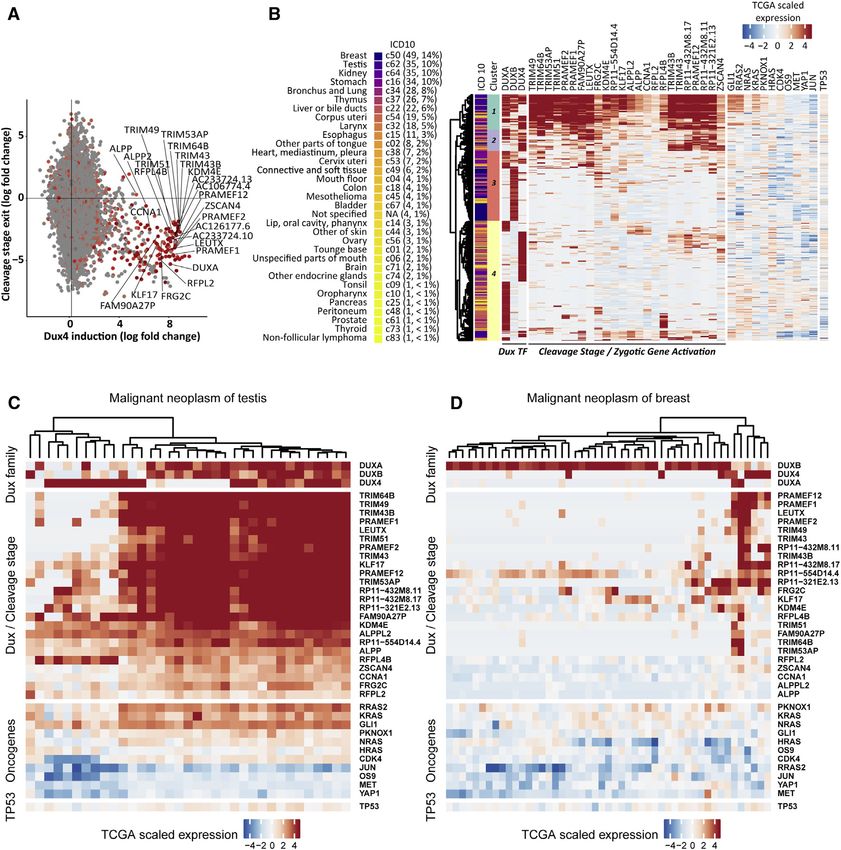
of ERMS, confirming cancer heterogeneity. Surprisingly, activation of zygotic Dux
factors identified a novel cancer subtype that is not limited to ERMS, but occurs
in a broad range of different human cancer.
Based on the results, it can be concluded that computational methods and in-
tegrative data analysis are useful to delineate the origin of cancer subtypes and
provide a valuable starting point for selection of relevant therapeutic targets. How-
ever, future research is needed to establish more holistic analysis approaches and
transfer findings into existing clinical routines.
iii
Contents

Preface                                                                                    i

Abstract                                                                                 iii

List of Figures                                                                          vii

1 Introduction                                                                            1
  1.1 Rationale . . . . . . . . . . . . . . . . . . . . . . . . . . . . . . . . . . .     1
  1.2 Background . . . . . . . . . . . . . . . . . . . . . . . . . . . . . . . . . .      2
       1.2.1 Genomic alterations transform cells into cancer cells . . . . . . .          2
       1.2.2 Disrupted context: Cancer as a tissue-based disease . . . . . . .            4
       1.2.3 The stem cell as a cancer cell of origin . . . . . . . . . . . . . . .       6
       1.2.4 Skeletal muscle regeneration as a model for stem cell tumours? .             8
       1.2.5 Small RNA mediated carcinogenesis . . . . . . . . . . . . . . . .           10
       1.2.6 Epigenetic mechanisms in cancer initiation and progression . . .            11
       1.2.7 Cancer subtype detection using integrated molecular analysis . .            14
  1.3 Lack of knowledge and objectives . . . . . . . . . . . . . . . . . . . . . .       15
       1.3.1 Knowledge gaps . . . . . . . . . . . . . . . . . . . . . . . . . . .        15
       1.3.2 Objectives . . . . . . . . . . . . . . . . . . . . . . . . . . . . . . .    17
  1.4 Thesis contributions . . . . . . . . . . . . . . . . . . . . . . . . . . . . .     19
  1.5 Results and discussion . . . . . . . . . . . . . . . . . . . . . . . . . . . .     20
       1.5.1 Sensitive computational quantification of miRNA sequences from
             NGS sequencing . . . . . . . . . . . . . . . . . . . . . . . . . . .        20
       1.5.2 Computational analysis of DNA methylation in arbitrary genomic
             regions . . . . . . . . . . . . . . . . . . . . . . . . . . . . . . . . .   21
       1.5.3 Lineage-tracing reveals cellular origin of ERMS and enables in-
             depth analysis of cancer stem cells . . . . . . . . . . . . . . . . .       22
       1.5.4 Computational analysis of copy number variation reveals molecu-
             lar origin of ERMS . . . . . . . . . . . . . . . . . . . . . . . . . .      22
       1.5.5 Integrative analysis of zygotic Dux factors defines a new cancer
             subtype . . . . . . . . . . . . . . . . . . . . . . . . . . . . . . . .     23
       1.5.6 Gene expression analysis reveals epigenetic plasticity conferred by
             tumourigenic Duxbl . . . . . . . . . . . . . . . . . . . . . . . . .        24
  1.6 Conclusion . . . . . . . . . . . . . . . . . . . . . . . . . . . . . . . . . .     24

2 Publications                                                                           27
  2.1 MIRPIPE: quantification of microRNAs in niche model organisms . . .                 27

                                             v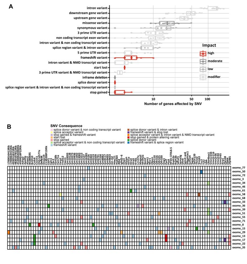
Contents

           2.2   ADMIRE: analysis and visualization of differential methylation in ge-
                 nomic regions using the Infinium HumanMethylation450 Assay . . . . .           30
           2.3   A molecular subtype of cancer originating from adult stem cells during
                 regeneration is driven by Dux transcription factors . . . . . . . . . . . .   41

      Gratitude                                                                                73

      3 References                                                                             75

vi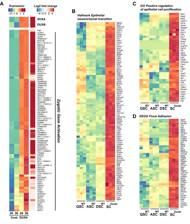
List of Figures

1   Heterogeneity in the process of cancer formation. . . . . . .     . . . . . .   .   .   .    3
2   Cancer as a tissue-based disease. . . . . . . . . . . . . . . .   . . . . . .   .   .   .    5
3   Cell lineages in tumour initiation and heterogeneity. . . . .     . . . . . .   .   .   .    7
4   Stem-cell dependent regeneration of skeletal muscle fibres. .      . . . . . .   .   .   .    9
5   The epigenetic landscape in lineage development and cancer        initiation.   .   .   .   13

                                          vii
1      Introduction

1.1     Rationale
Cancer is one of the leading causes of death around the world, being responsible for
nearly 10 million deaths in 2018 (Bray et al., 2018). Cancer is a complex disease that
can arise in multiple tissues, originating in numerous cell types and by different albeit
related mechanisms. Currently, ~100 distinct cancer types are known to emerge from
interactions of hundreds to thousands of macromolecules. Recent decades of research
have generated detailed insights into variations of cancer initiation, progression, severity
and treatment resistance. However, a clear vision and path for the cure of cancer is still
missing (Koutsogiannouli, Papavassiliou, & Papanikolaou, 2013; Nurse, 1997).
    Efficiency of tumour treatment depends on and is affected by cellular and molecular
tumour profiles. However, individual tumours from different patients exhibit different
molecular profiles and properties like cellular morphology, gene expression, metabolism,
proliferation or metastatic potential. Such intratumoural heterogeneity is caused by
cancer subtypes and hampers effective design of treatment strategies. It is one of the
biggest challenges for successful cancer treatment. A promising approach to overcome
intratumoural heterogeneity aims to identify individual patients with similar cancer
subtypes and to tailor specific treatments for those patients (Senft, Leiserson, Ruppin,
& Ronai, 2017; Vincent, 2017). Termed precision medicine, it requires integration of
patient data from multiple sources (e.g. genomics, epigenomics, clinical data, lifestyle
and environment) to identify therapeutic targets that are essential for subtype-specific
tumour initiation and progression. To fulfil those requirements and to support clinical
decision making, appropriate computational methods for managing, integrating and
analysing large and complex data sets are needed (Singer et al., 2017).
    The initial sequencing of the human genome (International Human Genome Se-
quencing Consortium, 2001) has marked the beginning of cancer genomics and initiated
a new era of modern biomedical research. Disruptive advances in DNA sequencing
technology revolutionised not only cancer research, but also the way how genome-wide
questions can be addressed (MacConaill, 2013). With the advent of next generation se-
quencing (NGS) technology, it became possible to profile cancer genomes (Pugh et al.,
2012; Stephens et al., 2009), which significantly enhanced the ability to study neoplastic
transformation based on changes in the genome sequence. Notwithstanding, enourmous
amounts of data generated by NGS introduced new challenges in computational data
analysis (Mardis, 2011; Wu, Rice, & Wang, 2012). Transformation of this data to gain
a holistic understanding of the complex and dynamic systems of cancer is challenging
(Grizzi et al., 2006; Sigston & Williams, 2017).

                                             1
1. Introduction
This thesis provides methodological development and application of software within
the scope of computational cancer biology, focusing on the analysis of data from NGS
experiments to support the characterization of the molecular basis of cancer subtypes
and the investigation of mechanisms underlying cancer formation. The objectives are:
to provide an algorithm for rapid detection and quantification of small, regulatory RNA;
to develop a software pipeline for analysis and visualization of CpG-site methylation in
a case-controlled setting; to investigate the cellular and molecular origin of a childhood
cancer, embryonal rhabdomyosarcoma, by an integrative analysis of data generated
from NGS experiments and to delineate potential cancer subtypes and the mechanisms
of tumour formation.
1.2 Background
The origin of cancer and its development has been a subject of debate, covered by
several theories. In the early nineteenth century, a professor of anatomy and pathology
at the Royal Anatomical Museum in Berlin, Johannes Müller, recognised for the first
time the cellular structure of cancer. Using microscopic pathology, he observed how
morbid growth resembles the tissue from which the cancerous growth springs. Since
then, modern oncology seeks the origin of cancer in a transformation of a healthy cell
into a disease state characterized by uncoordinated and excessive cell growth.
The upcoming sections highlight several theories that provide explanation for dif-
ferent cancer-causing mechanisms with implications for cancer classification, diagnosis,
therapy and research. Additionally, a brief introduction into integrated molecular data
analysis is provided.
1.2.1 Genomic alterations transform cells into cancer cells
While normal cells retain their ability to control the production and release of growth-
promoting signals and thereby provide tissue architecture and homeostasis of cell num-
ber, cancer cells are characterised by chronic proliferation and constant re-entry into the
growth-and-division cycle (Hanahan & Weinberg, 2011). The somatic mutation theory
postulates that molecular events such as genomic mutations precede cell transforma-
tion (Fig. 1a), black lightning and red cells) allowing cells to overcome cell control
mechanisms. By such, transformed cancer cells are able to e.g. escape the control of
growth suppressors, bypass mechanism of induced cell death, delay or avoid entry into
cell senescence, induce angiogenesis and/or alter cell-to-cell contacts to activate inva-
sion of surrounding tissue (Hanahan & Weinberg, 2011). At the core of this theory,
mutations in so-called master genes, i.e. genes that have the potential to cause cancer
(oncogenes) or genes that protect the cell from cancer progression (tumour suppressor
genes), determine the onset of cancer.
The search for transformation-causing genome alterations accelerated through recent
advances in NGS technologies which led to an impressive accumulation of data from
large-scale genome sequencing projects like the Cancer Genome Project or The Cancer
Genome Atlas. Collectively, both projects list 81 million simple somatic mutations across
21.2. Background
a b
Somatic cells Natural selection process Environment Lifestyle Treatment
Figure 1: Heterogeneity in the process of cancer formation. (a) Molecular events
(straight arrows, exemplified for one cell) increase genome context heterogeneity (col-
ors) and confer differences in fitness between somatic cells. Oncogenic transformation
(black lightning) enables positive natural selection and the evolution of cancer. (b)
Cell population heterogeneity is additionally increased by external factors acting on the
individuals genome context.
cancer genomes from nearly 25,000 individuals in the International Cancer Genome
Consortium Data Portal Release 28. However, the search did not reveal a single pattern
of genetic alterations that is universal to most cancers. Instead, a tremendous genetic
heterogeneity in underlying mechanisms of cancer formation emerged, despite common
features of cancer cells.
The observed genetic heterogeneity among cancer and even within similar cancer
types suggests that most individual tumours exhibit altered genome contexts (genes,
regulatory elements and genomic topology), with different genomic mutations. The
Genome Theory is an extension of the somatic mutation theory and seeks to explain
cancer heterogeneity by additionally including cell population heterogeneity and the
process of natural selection (Heng et al. (2010), Fig. 1). Cancer formation is seen as
an evolutionary process initiated through internal (e.g. somatic mutations) or external
(e.g. environment, lifestlye, treatment) stress that results in genome context instability.
Additional genetic or epigenetic mutations may occur in individual somatic cells with
instable genomic contexts, increase the cell population heterogeneity and confer differ-
ences in fitness between somatic cells. Importantly, as genetic or epigenetic mutations
are heritable and can be passed to a cells progeny, all requirements for natural selec-
tion are met and evolution of somatic cells within individual patients towards cancer is
enabled.
The occurence of genome-level alterations in an instable genome context is a stochas-
tic process and therefore the probability of successful progression towards cancer is high-
est through alterations significantly impacting the phenotype of a cell. However, the
stochastic nature complicates the prediction of which pathway will become dominant
prior to tumour formation and renders the characterization of individual cancer path-
31. Introduction
ways less meaningful. Ultimately, it impedes the establishment of a general model of
cancer origin and obscures understanding of how cancer can be managed in classical
clinical treatment.
1.2.2 Disrupted context: Cancer as a tissue-based disease
Organs and higher-level structures are comprised of tissue with functional (parenchyma)
and structural (stroma) parts. Information exchange between cells of a tissue via cell-to-
cell contacts, cytokine signaling and the extracellular matrix, a macromolecular scaffold
to support surrounding cells, enables maintenance of cell differentiation and tissue struc-
ture. Tissue-level interactions are important for embryonic development, regeneration
and morphogenesis, e.g. to provide necessary mechanical forces for proper tissue for-
mation (Hernández-Hernández, Rueda, Caballero, Alvarez-Buylla, & Benítez, 2014).
Therefore, disruption of tissue organisation is thought to be carcinogenic through en-
tailed disruption of tissue-level interactions.
The physiomitotic theory sees carcinogenesis as a problem of tissue organisation and
relates the acquisition of mitotic activities to development of cancer by non-regulated
cell turnover among normal tissue (Hirata & Hirata, 2002; Paduch, 2015). Two types
of mitosis maintain tissue histology and continuity: duplicating mitosis regenerates a
basal pool of undifferentiated cells in a space-restricted duplication area by creating two
identical daughter cells. Those cells are constantly consumed in surrounding areas by
maturating mitosis, which creates two daughter cells that are more mature than the
parent cell and contribute to tissue diversity and functionality via cell differentiation
(Fig. 2a). Disrupted tissue organisation and regulation may evoke duplicating mitosis
at ectopic sites among normal tissue, thereby creating aberrant and undefined tissue
identity. This might lead to non-regulated cell turnover and maturation into cancerous
tissue (Fig. 2b).
Similarly, the tissue organisation field theory argues that interactions among differ-
ent tissue components cannot be explained on a cellular level and that carcinogenesis
cannot be reduced to cellular events (Paduch, 2015). Cancer initiation is preceded by a
carcinogenic event and chronically disrupts reciprocal interactions between stroma and
parenchyma of a tissue, but cannot be observed in individual, isolated cells.
A well-known example and important feature of interactions of stroma with sur-
rounding cells is the maintenance of polarized epithelial sheets, a basic tissue type that
lines outer surfaces of organs and surfaces of inner cavities. Cell polarity is established
by interaction with the basement membrane and cell-to-cell contacts, like adherens junc-
tions, gap junctions, tight junctions and desmosomes (Fig. 2c). Alterations of epithelial
sheets, e.g. through wounding and subsequent activation of stromal fibroblasts, can
lead to epithelial cell movement and proliferation. Similarly, sustained inflammation
and continuous exposition to factors produced by invading immune cells and enzymes
degrading the extracellular matrix (ECM, e.g. matrix metalloproteinases) stimulate pro-
liferative and apoptotic mechanisms, which can lead to selection of apoptosis-resistant,
premalignant cells and enhance formation of carcinoma (Fig. 2c, Bissell & Radisky
(2001)). Loss or downregulation of E-cadherin, an important component of adherens
41.2. Background
a
Undifferentiated cell
Differentiating cell(s)
Non-regulated, duplicating cell
b c
Epithelial cells
Basement membrane
Extracellular matrix
Stroma
Figure 2: Cancer as a tissue-based disease. (a) Two types of mitosis maintain normal
tissue histology and continuity under the physiomitotic theory: Duplicating mitosis
(circular arrow) in a space-restricted duplication area (light blue shading) regenerates
a basal pool of undifferentiated cells (blue), which are consumed by maturating mitosis
(straight arrows) and contribute to more differentiated cells (green) to establish diverse
and functional tissue (green shading). Duplicating mitosis at ectopic sites (red shading)
among normal tissue might create cancerous tissue (red cells) and lead to non-regulated
cell turnover (circular and straight arrows). (b) Schematic depiction of basic epithelial
sheets. Epithelial cells are polarized by interaction with the basement membrane and
underlying stroma. (c) Alteration of epithelial sheets through activation of stromal
fibroblasts (jagged cells), degraded ECM (brown) or invading immune cells (blue) can
stimulate epithelial cell movement and proliferation.
junctions, leads to a premalignant cell type that is prone to invasion and metastasis
by passing through an epithelial-to-mesenchymal transition (Christofori & Semb, 1999).
Accordingly, restoration of E-cadherin expression in such cells can suppress cellular
transformation.
Experiments have shown that restoration of the cellular micro-environment can also
lead to healthy phenotypes, e.g. normal differentiation is observed when teracarcinoma
cells are injected into blastocysts, even after long passaging (Illmensee & Mintz, 1976).
Similarly, experiments from 3D culturing with reconstituted basement membranes or
co-culturing of malignant cells with normal stroma reverted their carcinogenic proper-
ties (Weaver et al., 1997). In summary, these experiments suggest that normal stroma
provides contextual cues that promotes normal tissue identity and restricts prolifera-
tion of existing pre-malignant cells. In contrast, non-functional stroma releases this
suppression and permits neoplastic transformation (Bissell & Radisky, 2001). These
findings lead to the hypothesis that carcinogenesis might be reversed when neoplastic
51. Introduction
tissue comes into contact with functional tissue or components thereof.
1.2.3 The stem cell as a cancer cell of origin
Similar to maintenance of tissue identity, the development of normal tissue requires
complex crosstalk between cells, their local environment and the whole organ. Migra-
tion and proper localization of precursor cells is a prerequisite for formation of mature
descendants that can carry out their tissue-specific function. The specific pattern of a
cells tissue-forming division(s) has been termed its lineage (Chisholm, 2001). Further-
more, dissection of such cell lineages revealed a hierarchical organisation and helped
to identify interactions and molecular signalling pathways that are important in tissue
development and diseases. However, unidirectional division alongside the cell lineage
would quickly lead to exhaustion of a cells tissue-generative potential and therefor calls
for a mechanism such as duplicating mitosis as proposed by the physiomitotic theory:
Stem cells are tissue-specific multipotent precursor cells residing at the apex of a lin-
eage, and are capable of both (i), generation of common progenitor cells with increasing
lineage commitment and (ii) self-renewal to regenerate and sustain the pool of stem
cells. The inherent proliferative capacity and the ability to give rise to different, mature
cell types renders stem cells particularly fascinating for the study of tissue development,
regeneration and in the search for the cellular origin of cancer.
An important distinction has to be made between the origin of cancer cells (i.e. the
normal cell that acquires the first cancer-promoting alteration (Creton et al., 2012)) and
cancer stem cells, i.e. a cellular subset within a tumour that exclusively sustains malig-
nant growth (Visvader, 2011). Intertumoural heterogeneity, i.e. the variability among
discrete tumour types arising from the same tissue, has put forward two hypothesis
how cancer stem cells are formed: (i) All tumours originate from common progenitor
cells that accumulate different genetic or epigenetic mutations through their extended
longevity and therefore result in different tumour types or (ii) different cells along the
lineage hierarchy that still possess or can re-instigate proliferative capacity or prevent
terminal differentiation (e.g. more restricted progenitor cells) constitute different can-
cer cell types including cancer stem cells upon oncogenic transformation (Perez-Losada
& Balmain (2003), Visvader (2011) and Fig. 3a). Cells with self-renewal capacity are
of paramount importance for tumour growth as they ensure long-term clonal growth.
However, not all cancer cells possess self-renewal capacity and not all cells from which
cancer origins are bona fide stem cells. So, how do cancer cells acquire their stemness,
if not from normal tissue stem cells?
The lineage-dependency hypothesis suggests that many tumours might be depen-
dent on (or addicted to) lineage-survival programmes that also operate during normal
lineage development (Garraway & Sellers, 2006). In this view, cancer cells can aquire
their stemness from lineage precursor cells, but rely on persistence and deregulation
of lineage-specific proliferation and differentiation pathways (Fig. 3b). The lineage-
dependency hypothesis inextricably associates lineage descendance and differentiation
state of progenitor cells to cancer biology and complements oncogene addiction, in which
61.2. Background
a b
Cell-of-origin model
Lineage survival Tumor initiation
Subtype A
lineage-associated “Conditioned”
Stem cell transcription factors genetic alterations
Survival
Subtype B Tumor survival
lineage dependency
and deregulation
Subtype C
Survival
Precursor cells
Lineage differentiation
Growth arrest
differentiation-associated
transcription factors
Growth arrest
Differentiated Tumor metastasis
descendants adapted from Visvader (2011), Garraway and Sellers (2006)
Figure 3: Cell lineages in tumour initiation and heterogeneity. (a) Cells along a lineage
hierarchy that still possess proliferative capacity or can prevent terminal differentiation
constitute cancer subtypes upon oncogenic transformation (black lightning). (b) Lineage
survival and normal development are often dependent on lineage-associated transcrip-
tion factors. Genetic alterations might be conditioned by lineage and subsequent tumour
initiation crucially depends on persistence or deregulation of survival mechanisms pro-
grammed into precursor cell development: A mechanism termed lineage dependency.
tumour-specific gain-of-function events elicit a dependency on growth signalling that is
absent in normal lineage development.
Activation of the same oncogenic pathway in tumours originating from different cell
lineages may also profoundly influence tumour phenotype and degree of malignancy. For
example, primary human melanocytes transformed with a set of genes form melanomas
that frequently metastasize, while human fibroblasts or epithelial cells transformed with
the identical set of genes rarely do (Gupta et al., 2005). Ultimately, therapeutic ap-
proaches might exploit lineage dependency for context-specific treatment, for example
when synthetic lethality exists between two genetic factors (Kaelin, 2005).
A straightforward approach to evaluate the oncogenic capacity of different lineage
stem and progenitor cell populations relies on reproducible separation of functionally
defined subpopulations using e.g. cell sorting techniques. Relevant oncogenic lesions
are introduced together into different precursor cell populations ex vivo with a fluores-
cent reporter, followed by orthotopical transplatation into immunocompromised mice.
Emergence of pre-neoplastic or neoplastic tissue from transduced subpopulations serves
as readout for evaluation of oncogenic potential for each subpopulation. Complimentary,
and with sufficient knowledge about cell-specific promoters, in vivo conditional target-
ing of cell populations is also conceivable. This approach makes use of genetic mouse
models to conditionally activate either an oncogene or inactivate a tumour suppressor
71. Introduction
gene in different lineage subpopulations, e.g. by Cre-mediated deletion. Depending on
the activated cell-specific promoter, different cancer subtypes might arise and reveal the
cellular origin of the specific cancer subtype from within the cell lineage (Hayashi &
McMahon, 2002; Visvader, 2011). However, established lineages and knowledge of cell
specific promoters are missing for many tissues and organs and therefore hamper the
approach described above.
1.2.4 Skeletal muscle regeneration as a model for stem cell tumours?
The ability of movement is an evolutionary advantage of all animals, and is powered
by muscles. Vertebrate locomotion receives its power from striated, skeletal muscle,
one of the three major muscle types in the body, that is composed of multiple bundled
muscle fibres (fascicles). Each fibre is a multinucleated muscle cell formed by fusion of
differentiated mononuclear muscle cells (myoblasts) and exhibits force and movement
by coordinated activity of myosin II motor proteins within an actin filament scaffold.
Skeletal muscle retains a remarkable ability to regenerate and adapt to changes in re-
quirements, mediated by and dependent on muscle stem cells that reside in a niche
between the muscle sarcolemma and the basal lamina of individual muscle fibres. Adult
muscle stem cells are bona fide stem cells, being capable of both, self-renewal and myo-
genic differentiation, which ultimately leads to differentiated muscle cells (Almada &
Wagers, 2016; Günther et al., 2013).
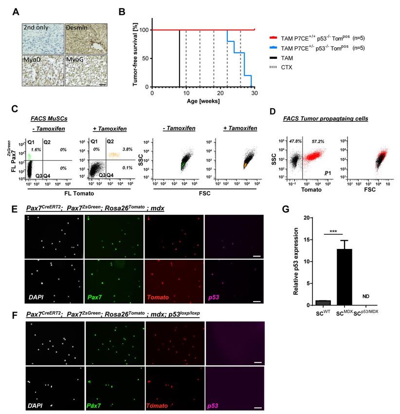
Muscle stem cells that are characterised by expression of Paired box protein 7 (Pax7),
a transcriptional regulator, are mainly quiescent under homeostatic conditions. Upon
muscle trauma, otherwise quiescent muscle stem cells become activated through ex-
posure to extrinsic stimuli and switch to a highly proliferative state. Activation and
proliferation of muscle stem cells depends on expression of two transcriptional regu-
lators, myogenic factor 5 (Myf5) and myogenic determintation protein (Myod1), and
precedes commitment to differentiation (Braun & Gautel, 2011). Downregulation of
Pax7 and expression of myogenin (Myog) in a subset of activated muscle stem cells in-
duces differentiation and ultimately leads to cell-cycle exit and formation of myoblasts
that fuse with other myoblasts or existing muscle fibres to repair the muscle (Almada
& Wagers, 2016; Braun & Gautel, 2011). Activated stem cells may also inhibit Myod1
expression and re-instating quiescence, thereby replenishing the pool of muscle stem
cells for future rounds of muscle repair (Fig. 4).
Duchenne muscular dystrophy (DMD) is a genetic disorder leading to muscle weak-
ness and decrease in the muscle mass (muscle wasting, atrophy). Dystrophin, the gene
product causing DMD in affected individuals, is part of a larger complex that stabilizes
the membrane of striated muscle cells. Dystrophic fibres are prone to get damaged
by mechanical stress and die after repeated muscle contraction. Such fibres are often
replaced by fibrotic, adipose or connective tissue that is not able to transmit sufficient
muscular force (Almada & Wagers, 2016). Muscle degeneration elicits repair by ex-
pansion and differentiation of stem cells, but regenerated muscle fibres will also lack
a functional dystrophin such that chronic cycles of degeneration and regeneration are
passed through. Until now, the role of muscle stem cells in DMD remains elusive, with
81.2. Background
Muscle fibers wt mdx
Myf5
Pax7 Myod1
Pax7
Myog
Myf5
Myod1
Quiescent Activated
satellite cell satellite cells Differentiating myoblasts
Figure 4: Stem-cell dependent regeneration of skeletal muscle fibres. In uninjured
muscle (wt), quiescent muscle stem cells (left) reside between the muscle sarcolemma
and the basal lamina of individual muscle fibres. Pro-myogenic stimuli from muscle
trauma or under genetic disorders like DMD (mdx, right) activate muscle stem cells
and lead to proliferation (middle). Differentiating myoblasts (right) arise through cell-
cycle exit of a subset of activated muscle stem cells and constitute a basis for formation
of novel fibres or repair of existing fibres.
only some evidence for a specialized role of dystrophin during stem cell division, but
an important role of dystrophin for the pathological environment in disease progression
(Almada & Wagers, 2016).
A genetic and experimental model of DMD is the mdx mouse, whose muscles retain
a lifelong capacity to regenerate fibres and exhibits loss of muscle fibres and exten-
sive fibrosis (Boldrin, Zammit, & Morgan (2015), Fig. 4). Recently, it was shown
that germline inactivation of the tumour suppressor p53 in chronically regenerating
mdx mice develop rhabdomyosarcoma (RMS) (Camboni, Hammond, Martin, & Martin,
2012; Chamberlain, Metzger, Reyes, Townsend, & Faulkner, 2007), a rare and aggressive
childhood cancer and the most common soft-tissue sarcoma in children and adolescents.
Histologically, RMS resembles developing skeletal muscle and is marked by expression
of actin and myosin as well as myogenic factors (Drummond et al., 2018; El Demellawy,
McGowan-Jordan, de Nanassy, Chernetsova, & Nasr, 2017). RMS is subdivided into
four subgroups, with alveolar RMS (ARMS) and embryonal RMS (ERMS) being two
major subgroups accounting for nearly all childhood cases of RMS, while spindle cell
RMS and pleomorphic RMS occurring mostly in adolescents. A broad molecular basis
has been identified in RMS, with interference of myogenic differentiation and emergence
of chromosomal aberrations being main drivers of cancer progression. For example,
aberrant expression of Notch2, Yap1, members of the Wnt gene family and Tgf-1 sig-
nalling have been implicated in disruption of myogenic differentiation (Chen et al., 2014;
Judson et al., 2012; Schaaf et al., 2005; Wang et al., 2010). On the other hand, ex-
91. Introduction
pression of Egr1, Met and signalling by the Fgf family seem to maintain proliferation of
RMS cells (Sarver, Li, & Subramanian, 2010; Taulli et al., 2006; Wachtel et al., 2014).
Genomic amplifications and translocations, as well as loss of heterozygosity from spe-
cific whole chromosomes have been reported for ERMS and ARMS subtypes. Prominent
examples include interference of Pax3 and Pax7 expression levels in ERMS, promoting
migration and invasiveness (Bridge et al., 2002; Chiappalupi, Riuzzi, Fulle, Donato, &
Sorci, 2014), and Pax3-Foxo1 or Pax7-Foxo1 gene fusions in ARMS, leading to commit-
ment of mesenchymal stem cells to the myogenic lineage by transactivation of Myod1 or
Myog (Ren et al., 2008). In addition to genetic mechanisms, epigenetic and small RNA-
mediated mechanisms have also been described to deregulate myogenic differentiation
enabling escape of RMS cells from suppressive mechanisms (see also chapters 1.2.5 and
1.2.6).
RMS is marked by large heterogeneity that not only manifests in distinct subtypes,
but also by different underlying genetic and epigenetic mechanisms. However, the cellu-
lar origin of RMS has remained elusive, despite large efforts to characterize the molecular
basis of many RMS specimen in recent years. As introduced in chapter 1.2.3, high tu-
mour heterogeneity can emerge from a cellular origin with stem cell-like properties.
Subsequently, the cellular origin of RMS was claimed to reside in tissue-resident stem
cells, e.g. muscle stem cells or mesenchymal stem cells (Hettmer & Wagers, 2010). As
such and for RMS subtypes showing features of myogenic differentiation, the mdx mouse
model with its constant regeneration of skeletal muscle could be used as a model for
stem cell-dependent carcinogenesis and serve the discovery of the cellular origin of RMS.
1.2.5 Small RNA mediated carcinogenesis
Micro RNAs (miRNAs) are small, non-coding RNAs of ~22 nucleotides and serve numer-
ous roles in negative gene regulation. In animals, most miRNAs exhibit their regulatory
role through imperfect binding of a sequence in the 3’ untranslated region (3’-UTR) of
messenger RNA from target genes. Complementary binding can either repress transla-
tion of target gene(s) or mediate mRNA degradation, through a mechanism similar to
RNA interference in plants (Jones-Rhoades, Bartel, & Bartel, 2006).
MiRNA biogenesis begins with the transcription of either independent miRNA genes
or intronic regions from protein-coding genes into large precursor molecules (pri-miRNAs).
Imperfect base-paring of folding pri-miRNAs results in hairpin structures that are fur-
ther cleaved by an RNase III type endonuclease (Drosha, RN3) together with a double-
stranded RNA binding domain (dsRBD) protein (Han, 2004) into ~70 nucleotide hair-
pins called pre-miRNAs, leaving a short characteristic single-stranded overhang at the 3’-
end of pre-miRNAs. Exportin 5 recognizes such an overhang and arranges the transport
of pre-miRNAs to the cytoplasm (Yi, 2003), where a second complex consisting of Dicer,
a RNase III type enzyme, and TRBP, a dsRBD protein, cleave the pre-miRNA twice
into a miRNA duplex (Chendrimada et al., 2005). One strand (the mature miRNA)
preferentially enters the miRNA-induced silencing complex (miRISC), while the other is
degraded, although the complementary miRNA is also competent for miRNA-mediated
silencing (Schwarz et al., 2003). Imperfect double-strand pairing of pre-miRNAs as well
101.2. Background
as imperfect digestion by Drosha and Dicer result in miRNAs with varying 3’- (silent
modification, isomiR) or 5’-ends, that might affect complementary binding, representing
a challenge for computational miRNA quantification following RNASeq.
Forward genetic experiments have revealed great importance of the miRNA machin-
ery, exemplified by muscle-specific knockout of Dicer leading to complete embryonic
development but perinatal death (Bernstein et al., 2003). Other experiments identi-
fied numerous individual miRNAs with roles in processes, like timing of development
(Abrahante et al., 2003), differentiation (Chen, 2004) and growth control (Brennecke,
Hipfner, Stark, Russell, & Cohen, 2003). Since phenotypic consequence remains elusive
for the vast majority of miRNA, also computational approaches can and have been used
to elucidate miRNA function (Liu & Wang, 2019; Ulitsky, Laurent, & Shamir, 2010).
Progression of cancer growth can also be altered by expression of certain miRNAs.
The dedifferentiated phenotype of ERMS, for example, can result from downregula-
tion of muscle-specific miRNAs (myomiRs, i.e. miR-1, miR-206 and miR-133a/b), that
promote myogenic differentiation under normal conditions. Transfection of miR-206,
a skeletal muscle-specific miRNA, induces cell differentiation in C2C12 myoblast cells
(Kim, Lee, Sivaprasad, Malhotra, & Dutta, 2006). Additionally, expression of miR-1
and miR-206 are highly induced during muscle stem cell differentiation (Chen et al.,
2010) and transfection of miR-206 into a RMS cell line notably decreased tumour cell
migration and proliferation even more than switching to a differentiation medium (Taulli
et al., 2009). Contrarily to those findings Boettger, Wüst, Nolte, & Braun (2014) report
on miR-206/miR-133b dispensability for muscle stem cell differentiation, highlighting
complex modulatory effects and overlapping functions of myomiRs. Non-myomiRs can
as well promote myogenic differentiation, e.g. miR-26a mediates downregulation of cell-
cycle progression by targeting Ezh2. Vice versa, downregulation of miR-26a in RMS
results in upregulation of Ezh2 and therefore prevents myogenic differentiation (Ciara-
pica et al., 2009). Amplification of 13q31 in 25% of ARMS cases results in enhanced
expression of the miR-17-92 cluster (oncomiR-1 ), a bona fide oncogene (Jin et al., 2013;
Reichek et al., 2011; Sandhu et al., 2013), potentially targeting tumour suppressors like
PTEN. Deregulation of another oncogene in RMS, miR-183, is reported to promote
tumour cell migration, by targeting the transcription factor Egr1, a direct regulator up-
stream of other tumour suppressor genes (Mohamad, Kazim, Adhikari, & Davie, 2018;
Sarver et al., 2010).
1.2.6 Epigenetic mechanisms in cancer initiation and progression
Development of normal tissue, as discussed in chapter 1.2.4, requires distinct cell types
to arise during lineage-specification. Although equipped with identical genomic infor-
mation, different cell types exhibit different gene expression programs and are able to
pass such information on to their progeny (Margueron & Reinberg, 2010). How are
such expression patterns specified and maintained? It is now accepted that not only
information encoded as DNA in a cells genome, but also epigenetic information (i.e.
the stable and heritable non-genetic counterpart to DNA) provides an important layer
of regulation and plays pivotal roles in cell lineage specification and cell identity main-
111. Introduction
tenance (Margueron & Reinberg, 2010). In eukaryotes, DNA is organized in a massive
macromolecular complex called chromatin. Chromatin is formed by wrapping 147 base
pairs of DNA around a histone octamer (nucleosomes), then compacting those further
into topologically associated domains (TADs) separated by insulator proteins to allow
independent and specific regulation. The activity of a genomic locus is controlled by
its chromatin organisation. Accessible chromatin structures expose DNA elements, like
proximal gene promoter sequences or distal enhancer sequences, to regulatory tran-
scription factors and the transcriptional machinery to drive gene expression. Compact
and inaccessible chromatin structures prevent such activity and render a locus inactive
(Flavahan, Gaskell, & Bernstein, 2017).
Dynamic changes needed during tissue development call for mechanisms able to al-
ter chromatin organisation in response to changed conditions (John & Rougeulle, 2018).
Chromatin remodelling resulting in transcriptional repression is, for example, enforced
by the Polycomb protein family, which can post-transcriptionally modify specific his-
tone residues (e.g. trimethylation of histone H3, lysine 27 (H3K27me3)). Repressive
chromatin states can be propagated through cell division by retention of catalytic en-
zymes on replicating DNA (Simon & Kingston, 2013) and functional interaction with
DNA methylation and other regulatory proteins (Flavahan et al., 2017). Conversely,
regulatory activity by e.g. binding of transcription factors and chromatin modifiers,
seems to block repressive chromatin compaction (Zaret & Mango, 2016). Further, ac-
tive loci that are marked by trimethylation of histone H3, lysine 4 (H3K4me3), in turn
inhibit recruitment of DNA methyltransferases for de novo DNA methylation (Ooi et
al., 2007), which is another potent epigenetic mechanism for stabilization of transcrip-
tional repression (Jones, 2012). Methylated DNA functions as a silencing mark and is
involved in processes like X-chromosome inactivation (Venolia & Gartler, 1983), repres-
sion of transposable and repetitive DNA elements (Yoder, Walsh, & Bestor, 1997) and
might influence genome function when present at regulatory elements, like enhancers or
chromatin insulators (Bell & Felsenfeld, 2000).
A compelling conceptualization of epigenetic regulation during cell lineage develop-
ment has been postulated by developmental biologist Conrad H. Waddington, outlined
in his assay entitled The strategy of genes more than 60 years ago (reprinted in Wadding-
ton (2014)). In his hypothesis, differentiating cells proceed downhill along branching
valleys in an energetic landscape (Fig. 5a). The valleys correspond to discrete cellu-
lar states and their topological layout is defined by underlying gene regulatory networks
(GRN) that actively shape and maintain cellular identity (Zaret & Mango, 2016). Walls
between valleys restrict cell lineage capacity, by preventing cells to randomly “switch
states” (i.e. hopping over to another valley), and epigenetic mechanisms effectively
modulate the height of walls. Compacted and repressing chromatin, for example, pre-
vents spurious activation of non-lineage gene regulatory factors, restricting changes in
gene activity and increasing the height of energy walls between cell states, which blocks
changes in cell state and cell type identity (Flavahan et al., 2017).
Initiation and progression of cancer can result from various mechanisms disrupting
normal epigenetic regulation. Overly restrictive chromatin (i.e. high energy walls be-
tween valleys of Waddingtons landscape, Fig. 5b) can be achieved by gain-of-function
121.2. Background
a b
Stem cell
Cancer stem cell
Lineage capacity
Progenitor cells Overly restrictive chromatin,
no terminal differentiation
c
Cancer cells
Overly permissive chromatin,
enhanced plasticity
Normal differentiated cell types
Figure 5: The epigenetic landscape in lineage development and cancer initiation. (a)
Depiction of normal lineage development in the conceptualized epigenetic landscape
from C. Waddington. Stem cells (blue) reside at the apex of a lineage and exhibit
high lineage capacity. Progenitor cells proceed downhill along branching valleys into
differentiated cell types. Oncogenetic events (black lightning) in lineage progeny with
high lineage capacity can create cancer stem cells by switching cell states and might
lead to development of cancer (red arrow). (b) Restrictive chromatin might arrest
progenitor cells in their proliferative state, prevent their terminal differentiation and
lead to cancer initiation. (c) Deteriorated and permissive chromatin confers enhanced
cellular plasticity and might lead to spurious gene activation or cellular reprogramming
and predisposes for cancer initation.
mutations of Ezh2, the catalytic subunit of Polycomb repressive complex 2 (Prc2). A
hyperactive methyltransferase activity of Ezh2 leads to expansive, genome-wide H3K27
methylation (Sneeringer et al., 2010) and the loss of active chromatin marks. Overly
restrictive chromatin arrests developing B-cells in a proliferative state and prevents
terminal differentiation, leading to B-cell lymphoma (Béguelin et al., 2013). Epigenetic
restriction can also arise from aberrations in DNA methylation: The CpG island methy-
lator phenotype (CIMP) results from DNA hypermethylation and is characterized by
silencing of tumour suppressor genes and DNA mismatch repair genes (Hitchins et al.,
2007). Deterioration of overall chromatin topology (the layout of Waddingtons land-
scape, Fig. 5c) can be achieved by disruption of CTCF binding (Flavahan et al., 2016), a
methylation-sensitive DNA binding protein that accomplishes partitioning of chromoso-
mal loops into functional units by insulating TADs. Regulatory TAD boundaries protect
against gene activation from overly promiscuous enhancers from neighboring TADs and
loss thereof can lead to the activation of oncogenes (Hnisz et al., 2016). Upregulation of
members of the Histone Lysine Demethylase (Kdm) protein family has been implicated
in formation of overly permissive chromatin (i.e. low energy walls between valleys).
Enhanced epigenetic plasticity allows for rapid cell reprogramming or adaptation and
131. Introduction
drives diverse cancer types (Black et al., 2015; Liau et al., 2017; Roesch et al., 2013).
Finally, loss of imprinting through DNA hypomethylation may permit reactivation of
oncogenes, e.g. the insulin growth factor signalling pathway in some sarcomas (Rikhof,
de Jong, Suurmeijer, Meijer, & van der Graaf, 2009). In such cases, both, the maternal
and paternal copy of the Igf2 gene are transcribed, leading to elevated mRNA levels and
predispose for cancer through autocrine signalling.
1.2.7 Cancer subtype detection using integrated molecular analysis
Identification and assignment of a tumour’s molecular subtype an important step to-
wards precision medicine and a prerequisite for tailoring therapy for individual patients
(Senft et al., 2017; Vincent, 2017). Traditional clinical practice from the last decades
is largely based on identification and assignment of tumour subtype on histopathology,
cytology and expression or mutational status of known tumour markers. The clinical
outcome is often determined by the individual expertise of clinicians and the classifi-
cation scheme used (Ellis, 2006). With the advent of NGS technology and the estab-
lishment of novel clinical routines for sample processing and data analysis, molecular
characterization of tumours became feasible (Noushmehr et al., 2010; Prat & Perou,
2011), promising to fulfil two goals of precision medicine: First, the discovery of molec-
ular biomarkers that are predictive of disease outcome or effective drug treatment and
second, a better mechanistic understanding of the molecular basis of tumour initiation
and progression (Senft et al., 2017).
Increasing scale of NGS-based assays has so far been very useful in dissection of
tumour heterogeneity. Although genome-wide screening of mutational status (Kuijjer,
Paulson, Salzman, Ding, & Quackenbush, 2018), miRNA expression (Blenkiron et al.,
2007), DNA methylation (Zhang et al., 2018) and RNA expression have led to the
characterization of many cancer subtypes, the characterization of cancer using isolated
assays suffers from certain limitations. For example, genomic profiling alone detects the
presence or absence of genetic drivers, but fails to predict the activity of correspond-
ing proteins and pathways. Simultaneous characterization using two or more assays
might overcome such limitations and enhance clinical decision making towards targeted
therapy, but requires effective integration strategies.
Data integration combines data from different sources, thereby enhancing accessibil-
ity and possibly enriching results from queries. Data are typically integrated across two
axes: vertically, i.e. between different data types (e.g. genomic data, expression data
or clinical data) and/or horizontally, i.e. within the same datatype, data from different
providers or batches. The Cancer Genome Atlas Research Network provides a large col-
lection of tumour samples that have been characterized using different assays (Hoadley
et al., 2018) from numerous data generation centers. An early approach for vertical data
integration used results from separate clustering of data types and performs clustering
of cluster assignments (CoCA, Hoadley et al. (2014)). However, such an approach
does not benefit from synergistic effects of combining evidence levels. It was succeeded
by methods using simultaneous interrogation of subtype clustering from different data
types, like iCluster (Shen, Olshen, & Ladanyi, 2009), which jointly models cancer sub-
141.3. Lack of knowledge and objectives
types as latent variables from different data types, or tumorMap (Newton et al., 2017),
which uses similarity of molecular tumour profiles to embed samples into a standardized
similarity space. A natural approach to encode sample similarity is graph-based: Wang
et al. (2014) constructed sample similarity networks for each data type individually and
subsequently fused those networks into a common similarity space, thereby performing
joint vertical and horizontal data integration. In their setup, cancer subtypes emerge
as connected components in the graph and edges between samples provide information
about the data type from which the evidence comes from. Interpretability is also a
feature of Multi-Omics Factor Analysis (MOFA, Argelaguet et al. (2018)), a versatile
statistical framework that infers a low-dimensional representation and captures major
sources of variation across data types. Latent factors underlying the representation can
be linked to most relevant features revealing shared variation between different omics
layers. Recent technological advancement in the field of deep neural networks led to
the development of methods for cancer subtype classification (Gao et al., 2019; Tabibu,
Vinod, & Jawahar, 2019), using molecular or histological features for classifier training.
However and in contrast to other applications, classification by neural networks is a
supervised task and can only be used to classify new samples, but does not detect novel
subtypes once more data are available.
1.3 Lack of knowledge and objectives
The question of how and why cancer develops already has led to numerous studies to
characterise molecular tumour profiles and the cellular origin of different cancer types.
Methods from molecular biology and assays based on high-density micro arrays or NGS
are often used in conjunction to draw conclusions and validate results. A plethora of
computational methods have been implemented for analysis of data from those assays
and form the basis for approaches integrating across different data types. The upcoming
sections will line out major knowledge gaps in computational approaches to handle
data from such assays and in the search of cancer origin and cancer subtype detection,
exemplified using ERMS. Further, objectives of this thesis were developed from identified
knowledge gaps.
1.3.1 Knowledge gaps
In an extensive genomic analysis of tumours from 9 ERMS patients, Chen et al. (2013)
set out to define biological signatures to predict patient outcome and assign targeted
therapy for a high-risk subtype. They identified a subtype with defects in oxidative
metabolism, but otherwise report a multiplicity of mutations in known cancer consen-
sus pathways such as in Ras family genes, SHH/Wnt signalling or cell-cycle checkpoints.
Analysis of therapy-resistant tumour subclones highlighted complex genetic changes and
clonal evolution. The observations from Chen et. al., can answer two questions: (i)
Which genomic mutations occur in ERMS and (ii) are mutations reoccurring? The
ERMS subtype accumulated both, single nucleotide polymorphisms and larger copy
number variations. Genomic alterations were not reoccurring among cancer samples
151. Introduction
and indicate a large tumour heterogeneity - a result that complicates further subtype
detection and follows observations from large genome sequencing projects. Extending
the somatic mutation theory (see chapter 1.2.1), the genome theory explains cancer het-
erogeneity by introducing karyotype heterogeneity. Further, the genome theory assumes
that initial genome context instability ignites a series of genome alterations from which
the fittest (in terms of cell proliferation and expansion) is selected by an evolutionary
process. Open question thus are
• (i) Which mechanisms induce genomic instability in ERMS?
• (ii) How do accumulated genome alterations confer to cellular fitness?
• (iii) Which hallmarks do accumulated genome alterations operate?
• (iv) Do accumulated genome alterations further specify ERMS subtypes?
Following the stem cell theory of cancer (see chapter 1.2.3), the cancer cell of origin of
ERMS could possibly be either the muscle stem cell itself, or any of its potent progenitor
cells, e.g. activated stem cells or pro-myogenic precursor cells. Elevated expression of
Pax7, which occurs exclusively in fusion-negative RMS, led Tiffin, Williams, Shipley,
& Pritchard-Jones (2003) to propose the origin of RMS within the myogenic lineage.
Transformation of precursor cell populations from the myogenic lineage with either
expression of oncogenic Kras (Blum et al., 2013; Hettmer et al., 2011), or lineage-
specific deletion of tumour suppressor genes (Rubin et al., 2011), led to tumours that
phenotypically resembled their presumable myogenic origin. However, it was not possible
to discriminate the cellular origin of Kras-expressing tumours by transcriptional analysis
(Hettmer et al., 2011). The studies presented above lack direct experimental evidence,
leaving the possibility that other cell types in the muscle compartment might act as
tumour initiator by e.g. cell migration mechanisms. Therefore, the cellular origin of
RMS ultimately remained to be disclosed.
MiRNAs play a role in cancer initiation, progression and maintenance (see chapter
1.2.5) and discovery of the entire regulatory repertoire of these small molecules is crucial
for understanding their function in a given biological system (Gomes et al., 2013). With
the application of next generation sequencing in miRNA research (Tam, de Borja, Tsao,
& McPherson, 2014), the numbers of identified miRNAs increased rapidly, as well as
computational approaches to predict or detect them (Gomes et al., 2013). However,
until recently, isomiR variation due to imperfect digestion of pre-miRs by Drosha and
Dicer was dismissed as sequencing artefacts and led to underestimation of the miRNome
complexity (Neilsen, Goodall, & Bracken, 2012), which constitute a challenge for proper
computational detection and quantification of miRNAs. Further, miRNA detection
often relies on the presence of a preferably complete genomic reference to align miRNA
reads to a genomic locus or reference database, or extensive homology searches to exploit
evolutionary conservation of a nearby species. Thus, two open questions in analyses of
data from miRNA-seq are: (i) How can isomiR variation be detected and properly
quantified and (ii) does a reference-free approach to miRNA detection exist and is it as
161.3. Lack of knowledge and objectives
sensitive as the conventional approach described above? Can it additionally be used to
incorporate knowledge from other organisms?
To functionally characterise miRNAs, the delineation of miRNA target genes, i.e.
those genes that could be silence by complimentary miRNA binding, is required and of-
ten accomplished using in silico target prediction tools (reviewed in Oulas et al. (2015)).
Although prior knowledge from databases harbouring validated miRNA target inter-
actions (MTIs) exists, target prediction and integration of other experimental data,
e.g. from expression studies, remains challenging (Bayer, Kuenne, Preussner, & Looso,
2016).
Epigenetic regulation plays an important role in cell lineage development and disrup-
tion of such regulation might serve tumour initiation as well as tumour maintenance (see
chapter 1.2.6). However, mechanistic insight into how epigenetic lesions take effect (also
in cooperation with or followed by ordinary genetic stimuli) is missing. Open questions
with diagnostic and therapeutic implications include whether or not an initiating ge-
netic hit (e.g. gain or loss of function) becomes secondary, once a downstream epigenetic
lesion has occurred and altered the cellular state towards permanent tumourigenicity.
Advances in microarray and next generation sequencing technology enable assaying dif-
ferent mechanisms of epigenetic regulation at high resolution and in large numbers of
samples (Lister & Ecker, 2009; The ENCODE Project Consortium et al., 2007) but re-
quire specialised computational analysis (reviewed in Bock & Lengauer (2008)). Finally,
DNA methylation as an important mechanism of epigenetic regulation is included, due
to it’s important contribution to cancer development and diagnosis (Kulis & Esteller,
2010; Seki et al., 2015; Sun et al., 2019). Profiling DNA methylation using microarray
technology (Bibikova et al., 2011) allows researchers to assay large number of samples
across the whole genome. Computational analysis of data from such technologies aims
to identify differentially methylated regions between two or more groups and search
for functional enrichment in those regions (Bock, 2012; Laird, 2010). However, such
approaches are complicated by the spatial interdependencies of individual CpG sites.
Open questions include how data from single CpG sites can be aggregated into regions
in order to capture higher-order methylation patterns across broader genomic regions.
It is unclear whether such data can be analysed without the need of prior knowledge,
e.g. the definition of genomic regions, location of CpG islands, promoters or other gene
regulatory regions. Annotation of differentially methylated regions to nearby genes or
known genomic features for further downstream interpretation of results is not straight-
forward and requires flexible and fast software solutions (Kondili et al., 2017).
1.3.2 Objectives
Based on the previously described shortcomings of currently available methods for anal-
ysis of data obtained from high-density arrays or NGS experiments, two of the three
main objectives of this thesis focus on methodological improvements in miRNA quan-
tification and the analysis of DNA methylation pattern in arbitrary genomic regions.
Another objective aims at the disclosure of cellular and molecular origins of ERMS,
using -omics datasets, applied bioinformatics and advanced methods from cell biology.
17You can also read