Diamond Light Source Ltd Review 2018/19 - TU ...

Page created by Jose Mejia
 
CONTINUE READING
Diamond Light Source Ltd Review 2018/19 - TU ...
Diamond Light Source Ltd Review 2018/19

                                                                     1

                        DIAMOND LIGHT SOURCE ANNUAL REVIEW 2018/19
Diamond Light Source Ltd Review 2018/19 - TU ...
Contents

                                                                    Foreword                                        2
                                                                    CEO Welcome                                     5
                                                                    Beamline Development and Technical Summary      6
                                                                    Macromolecular Crystallography Group            8
                                                                    Biological Cryo-Imaging Group                   22
                                                                    Structures and Surfaces Group                   28
                                                                    Magnetic Materials Group                        36
                                                                    Imaging and Microscopy Group                    46
                                                                    Crystallography Group                           56
                                                                    Spectroscopy Group                              66
                                                                    Soft Condensed Matter Group                     74
                                                                    Phase III Overview                              84
                                                                    Integrated Facilities and Collaborations        86
                                                                    Machine Operation and Development               90
                                                                    Optics and Metrology Group                      92
                                                                    Detector Group                                  94    1

                                                                    Scientific Software, Controls and Computation   96
                                                                    Key Facts and Figures                           100
                                                                    Industrial Liaison                              102
                                                                    Engaging with Diamond Light Source              104
                                                                    Governance and Management                       106
                                                                    Staffing and Financial Information              107
                                                                    Committee Membership                            108

DIAMOND LIGHT SOURCE ANNUAL REVIEW 2018/19   DIAMOND LIGHT SOURCE ANNUAL REVIEW 2018/19
Diamond Light Source Ltd Review 2018/19 - TU ...
Foreword

    T                                                                                                                             An increase in Diamond’s
          he past 12 months have seen an increase in the
          complementary capabilities and facilities that Diamond is
          able to offer its over 10,000 users. With the operations of
    the electron Bio-Imaging Centre (eBIC) now fully integrated into
                                                                                                                              scientific output naturally leads
                                     the user programme, researchers
                                     can access a one-stop shop for
                                                                                                                              to a demand to further develop
                                     structural biology right here                                                            our data analysis capabilities.
                                     at Diamond. The unique offer
                                     enabled by further integrated                                                            Progress in this area is extensive,
                                     facilities such as the electron
                                     Physical Science Imaging Centre                                                          with efficient data analysis
                                     (ePSIC), the MPL (Membrane
                                     Protein Lab), the XChem
                                                                                                                              pipelines delivering results in
                                     Fragment Screening service and
                                     the XFEL Hub, put Diamond at the
                                                                                                                              close-to-real time.
                                     leading edge of transformative
                                     science on the world stage.
    An increase in Diamond’s scientific output naturally leads to a demand to
    further develop our data analysis capabilities. Progress in this area is extensive,
    with efficient data analysis pipelines delivering results in close-to-real time.
    One particular project is focusing on citizen science as a means to help train
2   Artificial Intelligence systems, which is a key focus of the UK’s industrial                                                                                    3
    strategy.

    Located on a world-leading site for scientific discovery, Diamond aims to
    be a cornerstone of the Harwell Campus. We are pleased to have seen key
    investments in our new neighbours, the Rosalind Franklin Institute (RFI) and
    Faraday Institution (FI), further increasing the impact of the Harwell Campus
    on UK science and innovation. We will continue to strengthen our relationships
    with these institutes and act as a catalyst for the activities they are undertaking.

    Professor Sir Adrian Smith
    Chairman of the Board of Directors

                                                        DIAMOND LIGHT SOURCE ANNUAL REVIEW 2018/19   DIAMOND LIGHT SOURCE ANNUAL REVIEW 2018/19
Diamond Light Source Ltd Review 2018/19 - TU ...
CEO Welcome

                                                 A
                                                       s we close the financial year 2018-19, it is good to reflect upon the achievements of this period. 2018 was the Year of Engineering and
                                                       this saw an increase in news and features from the engineering team resulting in some striking headlines. For example, the Daily
                                                       Express showcased the work done with Martian Meridianiites within the facility. These minerals were named after the place they
                                                 were discovered on Mars and provide direct evidence that there used to be surface water on this planet. This wonderful and engaging science
                                                 was made possible by the design of a special cold cell built by our talented engineers.
                                                 The Year of Engineering has left a legacy of enhanced engagement, which we can now build on ensuring that career opportunities are more
                                                 widely promoted, and staff continue to be good role models helping to attract the best talents into our organisation.
                                                 With the investment made in Diamond over the past 18 years, funded by the              to take advantage of such advances. We have already started a very substantial
                                                 taxpayers through the Science and Technology Facilities Council (STFC) - UK            rolling programme of upgrades to beamlines supported by a considerable uplift
                                                 Research & Innovation (86%) and the Wellcome Trust (14%), our institute now            in capital from our shareholders. We aim to complement this programme with
                                                 offers access to 32 beamlines and five complementary facilities (eBIC, ePSIC, MPL,     the Diamond-II upgrade of the machine and instruments, which with a factor
                                                 XFEL Hub and XChem). The final beamline of the current phase of construction           of 20 times reduction in emittance coupled with an increase in electron beam
                                                 – DIAD – saw first light in the past year and will take first users in the next        energy from 3.0 to 3.5 GeV will provide up to a factor of 70 increase in brightness
                                                 twelve months. All are pushing the boundaries of visualisation of molecules and                                                              and coherence of Diamond’s
                                                 materials at the atomic level, and enabling high quality science for both industry                                                           photon beams at the higher
                                                 and academia. An indication of the role Diamond plays in establishing the UK at                                                              energies. A new lattice
                                                 the forefront of science worldwide is the high impact of its work embodied in                                                                geometry will allow us to
                                                 almost 8,000 peer review scientific articles published since the organisation was                                                            not only keep and enhance
                                                 created, with citation rates significantly higher than the national average across                                                           all current beamlines, but
                                                 the spectrum of science it serves.                                                                                                           offers the opportunity for
                                                                                                                                                                                              up to five new additional
                                                 A great illustration of the impact we are having is the work by an international                                                             beamlines to be created.
                                                 team led by the University of Portsmouth and the US National Energy Research                                                                 The science case has been
4                                                                                                                                                                                                                              5
                                                 Laboratory that will enable a natural plastic digesting enzyme to be exploited                                                               well received and approved
                                                 more effectively in waste treatment. The unique visualisation tools used to                                                                  by our Science Advisory
                                                 elucidate the way the enzyme works were provided by several of our beamlines.                                                                Committee (SAC) and
                                                 The research made the headlines all over the world, generating the equivalent of                                                             Diamond Industrial Science
                                                 over £23M of advertising spend in press and broadcast coverage.                                                                              Committee (DISCo). The
                                                                                                                                        Conceptual Design Report (CDR) has also been approved by an international
                                                 This review contains many more examples of high impact science drawn from              expert panel, clearing the way to work on the Technical Design Report (TDR) for
                                                 across the broad range of areas of fundamental science that Diamond serves,            which we will need an increase in staff and resources for much more detailed
                                                 and the societal challenges it helps address. These include health and wellbeing,      planning.
                                                 the environment, transport and energy challenges of the future, as well as
                                                 new functional materials, including electronic devices and high-performance            To remain at the cutting-edge of science, we need to ensure we continue to attract
                                                 engineering alloys, and new processing technology for innovative manufacturing.        people from all over the world because they bring their different perspectives and
                                                                                                                                        skills. We also need to engage more closely with other facilities that provide
          To remain at the cutting               All of these achievements reflect the skills, hard work and commitment of the
                                                 dedicated staff that we have within our walls. They also reflect the strong
                                                                                                                                        complementary technical expertise. These are global endeavours, but the highest
                                                                                                                                        density of world-class light source facilities and suitably trained scientists and
      edge of science, we need                   engagement of a vibrant and growing user community in academia and
                                                 industry. In the last financial year, we received 1,788 proposals for experiments
                                                                                                                                        engineers are in Europe, so it is more critical than ever that we maintain close and
                                                                                                                                        effective ties with our closest and strongest allies. To this end, in 2017 we became
      to ensure we continue to                   on our instruments via peer reviewed access routes, requesting a total of 22,117
                                                 shifts. After peer review, 1,191 proposals were awarded beamtime. This resulted
                                                                                                                                        Members of LEAPS – the League of European Accelerator-based Photon Sources
                                                                                                                                        – an organisation that brings together every synchrotron and Free Electron Laser
      attract people from all                    in 12,497 experimental shifts being awarded across 30 beamlines and eight
                                                 electron microscopes. We welcomed 6,332 onsite user visits from academia
                                                                                                                                        (FEL) facility in Europe. Over the past year, the collaboration has developed a
                                                                                                                                        collection of projects for enabling technology, from new sources and optics to
      over the world because                     across all instruments, with an additional 4,459 remote user visits. The machine
                                                 continues to perform to the highest standard with 98.4% uptime and 90.3 hours
                                                                                                                                        detectors and data analysis tools, that we will work on together to ensure that
                                                                                                                                        we are all able to support the most brilliant science and innovation well into the

      they bring their different                 mean time between failures (MTBF).                                                     future.

                                                 However, we cannot afford to rest on our laurels. The pace of technical
      perspectives and skills.                   development is rapid and relentless so if we are to continue to offer the scientific
                                                                                                                                        Prof Andrew Harrison
                                                                                                                                        CEO Diamond Light Source
                                                 community the best opportunities for world-leading research, we need to plan

    DIAMOND LIGHT SOURCE ANNUAL REVIEW 2018/19                                                       DIAMOND LIGHT SOURCE ANNUAL REVIEW 2018/19
Diamond Light Source Ltd Review 2018/19 - TU ...
Beamline Development and Technical Summary                                                                                                                                         Diamond’s beamlines: current operational status April 2019
                                                                                                                                                                                        Beamline Name and Number              Main Techniques                                                                     Energy / Wavelength Range                              Status
                                                                                                                                                                                        I02-1 - Versatile MX micro (VMXm)     Micro- and nano-focus in vacuum cryo-macromolecular crystallography (VMXm)          7 - 28 keV                                             Commissioning
                                                                                                                                                                                                                              In situ microfocus macromolecular crystallography, Serial Synchrotron
                                                                                                                                                                                        I02-2 - Versatile MX in situ (VMXi)                                                                                       10 - 25 keV                                            Commissioning

     I
                                                                                                                                                                                                                              Crystallography
         n its twelfth year of experiments, Diamond is now operating with 32 beamlines and 11 electron microscopes. The next year will see                                              I03 - MX                              Macromolecular crystallography (MX), Multiwavelength Anomalous                      5 - 25 keV                                             Operational
                                                                                                                                                                                                                              Diffraction (MAD)
         the completion of the latest phase of construction when the DIAD beamline welcomes first users. Of the electron microscopes, nine are
                                                                                                                                                                                        I04 - Microfocus MX                   MX, MAD                                                                             6 - 18 keV                                             Operational
         cryo-electron microscopes specialising in life sciences, with two provided for industry use in partnership with Thermo Fisher Scientific,
                                                                                                                                                                                        I04-1 - Monochromatic MX              MX, XChem fragment screening                                                        13.53 keV (fixed wavelength)                           Operational
     and make up eBIC (electron Bio-Imaging Centre). The two remaining microscopes dedicated to advanced materials research are supplied by
                                                                                                                                                                                        I05 - ARPES                           Angle-Resolved PhotoEmission Spectroscopy (ARPES) and nano-ARPES                    18 - 240 eV; 500 eV                                    Operational
     Johnson Matthey and the University of Oxford. These microscopes form ePSIC (electron Physical Science Imaging Centre) and are operated
                                                                                                                                                                                                                              X-ray Absorption Spectroscopy (XAS), X-ray photoemission microscopy and X-ray
     under strategic collaboration agreements to provide for substantial dedicated peer reviewed user access. Both the eBIC and ePSIC centres                                           I06 - Nanoscience                     magnetic circular and linear dichroism                                              80eV - 2200eV                                          Operational
     are next to the Hard X-ray Nanoprobe beamline (I14). For academic research, Diamond instruments (beamlines and microscopes) are free at                                            I07 - Surface and Interface           Surface X-ray diffraction, Grazing Incidence X-ray Diffraction (GIXD), Grazing      6 - 30 keV                                             Operational
                                                                                                                                                                                        Diffraction                           Incidence Small Angle X-ray Scattering (GISAXS), X-ray Reflectivity (XRR)
     the point of access through peer review. For proprietary research, access can be secured through Diamond’s industry team.
                                                                                                                                                                                                                              Ambient Pressure XPS and NEXAFS                                                     250 - 2800 eV                                          Operational
     Following a restructure in 2018, the instruments are organised into eight science groups as described below.                                                                       B07 - VERSOX: Versatile Soft X-ray
                                                                                                                                                                                                                              NEXAFS and High-Throughput XPS                                                      50 - 2200 eV                                           Commissioning
                                                                                                                                                                                                                                                                                                                  I08 branch: 250 eV - 4.4 keV                           Operational
                                                                                                                                                                                        I08 - Scanning X-ray Microscopy       Scanning X-ray microscopy, NEXAFS/ XANES, X-ray fluorescence                        J08 - Soft and Tender X-ray Ptychography branch:
                                                               Cryo-TXM B24                               I02-2 VMXi                                                                                                                                                                                                                                                     Construction
                                                                                                                                                                                                                                                                                                                  250 - 2000 eV
                                              Microfocus and Serial MX I24                                   I02-1 VMXm
                                                                                                                    I03 MX                                                              I09 - Atomic and Electronic
                                              Circular Dichroism B23                                                                                                                                                          XPS (including HAXPES), X-ray Standing Waves (XSW), Near Edge X-ray                 Hard X-rays: 2.1 - 18+ keV
                                                                                                                         I04-1 MX and XChem                                             Structure of Surfaces and                                                                                                                                                        Operational
                                                                                                                                                                                                                              Absorption Fine Structure (NEXAFS), energy-scanned photoelectron diffraction        Soft X-rays: 0.1 - 2.1 keV (currently 0.1 - 1.9 keV)
                                     Long Wavelength MX I23                                                                  I04 Microfocus MX                                          Interfaces
                                                                                                                                 I05 ARPES                                              I10 - BLADE: Beamline for             Soft X-ray resonant scattering, XAS and X-ray magnetic circular and linear          Circular: 400-1600eV; Linear Horizontal: 250-
                   MIRIAM: IR Microspectroscopy B22                                                                                                                                                                                                                                                                                                                      Operational
                                                                                                                                                                                        Advanced Dichroism Experiments        dichroism                                                                           1600eV; Linear Vertical: 480-1600eV
                                                                                                                                     I06 Nanoscience                                    I11 - High Resolution Powder          X-ray powder diffraction                                                            6 - 25(30) keV (0.5 - 2.1 Å)                           Operational
      Small Angle Scattering and Diffraction I22                                                                                                                                        Diffraction
                                                                                                                                       I07 Surface and Interface Diffraction            DIAD: Dual Imaging and
                       High Throughput SAXS B21                                                                                                                                                                               Simultaneous imaging and diffraction                                                8 - 38 keV                                             Construction
                                                                                                                                                                                        Diffraction
               Inelastic X-ray Scattering I21                                                                                           B07 VERSOX: Versatile Soft X-ray
                                                                                                                                                                                                                              Time-resolved imaging and tomography (phase- and attenuation-contrast),
                                                                                                                                        I08 Scanning X-ray Microscopy                   I12 - JEEP: Joint Engineering,        time-resolved powder diffraction, single crystal diffraction, diffuse scattering,   53 keV - 150 keV monochromatic or continuous           Operational
         LOLA: Versatile X-ray Spectroscopy I20                                                                                                                                         Environmental and Processing          energy dispersive X-ray diffraction (EDXD), high-energy small angle X-ray           white beam
                                                                                                                                        I09 Atomic and Electronic Structure of                                                scattering (under development)
    Small-Molecule Single-Crystal Diffraction I19                                                                                       Surfaces and Interfaces
6                                                          Krios
                                                             I                                                                                                                                                                Phase contrast imaging, tomography, full-field microscopy (under                    Imaging branch: 8 - 30keV                                              7
                                                                                                                                                                                                                              commissioning), coherent diffraction and imaging (CXRD,CDI), ptychography
                                        Core XAS B18                                                                                                                                    I13 - X-ray Imaging and Coherence                                                                                                                                                Operational
                                                                                                                                     I10 BLADE: X-ray Dichroism and Scattering                                                and photocorrelation spectroscopy (XPCS) (under commissioning), innovative
                                                                                                                                                                                                                              microscopy and imaging                                                              Coherence branch: 7 - 20keV
                            Microfocus Spectroscopy I18
                                                                                                                                   I11 High Resolution Powder Diffraction
                                        Test beamline B16                                                                                                                                                                     Scanning X-ray fluorescence, X-ray spectroscopy, ptychography and transmission
                                                                                                                                   Long Duration Experiments (LDE)                      I14 - Hard X-ray Nanoprobe                                                                                                5 - 23 keV                                             Optimisation
                                                                                                                                                                                                                              diffraction
                                  Materials and Magnetism I16                                                                DIAD: Dual Imaging and Diffraction
                                                                                                                                                                                                                                                                                                                  Monochromatic and focused 20 - 80 keV
                                                         XPDF I15-1                                                                                                                     I15 - Extreme Conditions              Powder diffraction, single crystal diffraction                                                                                             Operational
                                                                                                                                                                                                                                                                                                                  White beam
                                                     Extreme Conditions I15                                            I12 JEEP: Joint Engineering, Environmental and Processing
                                                                                                                                                                                        I15-1 - XPDF                          X-ray Pair Distribution Function (XPDF)                                             40, 65, and 76 keV                                     Operational
                                                     Hard X-ray Nanoprobe I14
                                                                                                                  I13 X-ray Imaging and Coherence                                       I16 - Materials and Magnetism         Resonant and magnetic single crystal diffraction, fundamental X-ray physics         2.5 - 15 keV                                           Operational
                                                                         Krios                          ARM
                                                                           II                           300F                                                                                                                                                                                                      4 - 20 keV monochromatic focused
                                                                    Talos      Krios                                                                                                    B16 - Test beamline                   Diffraction, imaging and tomography, topography, reflectometry                      4 - 45 keV monochromatic unfocused                     Operational
                                                                   Arctica      III                       ARM
                                                                                       Krios
                                                                                        IV      Krios
                                                                                                          200F                                                                                                                                                                                                    White beam
                                                                         Scios                    V
                                                                                 Glacios                                                                                                                                      Micro XAS, micro Extended X-ray Absorption Fine Structure (EXAFS), micro
                                                                                           Aquilos                                                                                      I18 - Microfocus Spectroscopy                                                                                             2.05 - 20.5 keV                                        Operational
                                                                                                                                                                                                                              fluorescence tomography, micro XRD
            Macromolecular Crystallography                    Structures and Surfaces                                    Magnetic Materials                    Imaging and Microscopy   B18 - Core XAS                        X-ray Absorption Spectroscopy (XAS)                                                 2.05 - 35 keV                                          Operational
                                                                                                                                                                                        I19 - Small-Molecule Single-          Small-molecule single-crystal diffraction                                           5 to 25 keV / 0.5 to 2.5 Å                             Operational
           Crystallography                                    Biological Cryo-Imaging                                    Spectroscopy                          Soft Condensed Matter    Crystal Diffraction
                                                                                                                                                                                        I20 - LOLA: Versatile X-ray           X-ray Absorption Spectroscopy (XAS), X-ray Emission Spectroscopy (XES) and          Dispersive branch: 6 - 26 keV                          Optimisation
                                                                                                                                                                                        Spectroscopy                          Energy Dispersive EXAFS (EDE)
     Electron Microscopes                                                                                                                                                                                                                                                                                         Scanning branch: 4 - 20 keV                            Operational
                                                                                                                                                                                                                                                                                                                  Currently 250 - 1500 eV (to be upgraded to 250
                                                                                                                                                                                        I21 - Inelastic X-ray Scattering      Resonant Inelastic X-ray Scattering (RIXS), X-ray Absorption Spectroscopy (XAS)                                                            Optimisation
          Microscope             Main Capabilities                                                                          Accelerating Voltages          Operational Status                                                                                                                                     - 3000 eV)

          Titan Krios I          Cryo-EM, Cryo-ET                                                                           80, 120, 200, 300 kV           Operational since 2015       B21 - High Throughput SAXS            BioSAXS, solution state small angle X-ray scattering                                8 - 15 keV (set to 13.1 keV by default)                Operational
          Titan Krios II         Cryo-EM, Cryo-ET                                                                           80, 120, 200, 300 kV           Operational since 2016       I22 - Small Angle Scattering and      Small angle X-ray scattering and diffraction: SAXS, WAXS, USAXS, GISAXS.            7 - 20 keV                                             Operational
                                                                                                                                                                                        Diffraction                           Micro-focus.
          Titan Krios III        Cryo-EM, Cryo-ET                                                                           80, 120, 200, 300 kV           Operational since 2017
                                                                                                                                                                                                                                                                                                                  nanoFTIR : 4000-900 cm-1 (2.5-11 µm)
          Titan Krios IV         Cryo-EM, Cryo-ET                                                                           80, 120, 200, 300 kV           Operational since 2017       B22 - MIRIAM: Multimode                                                                                                   microFTIR: 10,000-100 cm-1 (1-100 µm)
                                                                                                                                                                                        InfraRed Imaging And                  IR micro- & nano-spectroscopy, IR imaging, THz spectroscopy                                                                                Operational
          Titan Krios V          Cryo-EM, Cryo-ET                                                                           80, 120, 200, 300 kV           Operational since 2018                                                                                                                                 Spectroscopy (FTIR): 10,000-10 cm-1 (1-1000 µm)
                                                                                                                                                                                        Mircrospectroscopy                                                                                                        Imaging (FPA): 10,000-900 cm-1 (1-11 µm)
          Talos Arctica          Cryo-EM, Cryo-ET                                                                           200 kV                         Operational since 2016
                                                                                                                                                                                        I23 - Long Wavelength MX              Long wavelength macromolecular crystallography                                      3 - 8 keV (1.5 - 4.1 Å)                                Optimisation
          Glacios                Cryo-EM, Cryo-ET                                                                           200 kV                         Installed, March 2019
          Scios                  Cryo-SEM, Cryo-FIB                                                                         3 to 30 kV                     Operational since 2017                                                                                                                                 125-500 nm & 165-650 nm for CD Imaging at
                                                                                                                                                                                        B23 - Circular Dichroism              Circular Dichroism (CD)                                                             50 µm resolution, 96-cell High-Throughput CD           Operational
          Aquilos                Cryo-SEM, Cryo-FIB                                                                         3 to 30 kV                     Operational since 2019                                                                                                                                 (HTCD) and High-Pressure CD up to 200 MPa
          JEOL ARM200F           Atomic scale STEM imaging, EELS, EDX, electron diffraction                                 80, 200 kV                     Operational since 2017       I24 - Microfocus and Serial MX        Macromolecular crystallography, MAD, Serial Crystallography                         6.5 - 25.0 keV                                         Operational
          JEOL ARM300F           Atomic scale TEM and STEM imaging, electron diffraction, 4D-STEM, EDX                      30, 60, 80, 160, 200, 300 kV   Operational since 2017       B24 - Cryo Transmission X-ray         Full field X-ray imaging                                                            200eV - 2600eV                                         Optimisation
                                                                                                                                                                                        Microscopy (TXM)

                                                      DIAMOND LIGHT SOURCE ANNUAL REVIEW 2018/19                                                                                                                                                     DIAMOND LIGHT SOURCE ANNUAL REVIEW 2018/19
Diamond Light Source Ltd Review 2018/19 - TU ...
Macromolecular Crystallography Group
    Dave Hall, Science Group Leader

    M
             acromolecular crystallography (MX) is a key technique in the structural biologist’s toolkit for understanding the function of
             biologically relevant molecules by revealing their shape and interactions at atomic resolution. The information derived from MX
             experiments can be complemented by many other techniques at Diamond for life science research (see the Soft Condensed Matter
    and Imaging and Microscopy sections of this review), coupled with experiments in the researcher’s lab, to giver deeper insight adopting an
    integrated structural biology approach.
    At Diamond, seven beamlines (I03, I04, I04-1, I23, I24, VMXi, and VMXm), alongside the XChem fragment screening facility, the UK XFEL Hub,
    and the Membrane Protein Facility, are dedicated to exploiting the technique of MX for the benefit of the UK structural biology community,
    alongside researchers from Europe and further afield.
                                                                                               been developed by beamline I24, and are already rolled out to the beamline as
                                                                                               well as I03 and I04. Building on the approach used on I24 for fixed-target serial
                                                                                               crystallography, samples can now be moved continuously during grid scans
                                                                                               allowing data collection at the maximum frame-rate of the detector, with no
                                                                                               compromise on positional accuracy. With grid scans now running at >100 Hz,
                                                                                               many more samples can be rastered over during a shift, and the throughput of
                                                                                               automated X-ray centering will significantly increase. Data collection using this
                                                                                               approach on the hard X-ray Imaging and Coherence beamline I13 has reached
                                                                                               rates of 600 Hz, with a positional accuracy of 50 nm, illustrating that detectors       In the push to increase
                                                                                                                                                                                   throughput across the beamlines,
                                                                                               are now the limiting step in raster scanning.

                                                                                                   Building on the work for Eiger detectors on VMXi, the first of the Eiger2
    First users and beamline team on VMXm, from left to right: Gwyndaf Evans (PBS), Jose
                                                                                               X 16M series detector model has been installed on beamline I04 recently. It
                                                                                               replaces the first generation Pilatus2 detector, and provides a significant
                                                                                                                                                                                   upgrades and methods are
    Trincao, Anna Warren, Emma Beale, Diamond, and Ivo Tews and Rachel Bolton, University of

8
    Southampton.                                                                               number of advantages and improvements that are benefitting the user
                                                                                               community and science output. Data acquisition rates have increased five-fold
                                                                                                                                                                                   continuously being rolled out                                                                                                                                                                      9
        It has been an exciting year for the VMXm micro/nanofocus MX beamline.
    After performing the first protein diffraction experiments at room temperature
                                                                                               over the previous detector, and a typical data set can now be collected in less
                                                                                               than 15 seconds. The Eiger2 detector’s much smaller pixel size, coupled with
                                                                                                                                                                                   across the facilities.
    and in vacuum back in May 2018, first user experiments followed in October                 no read-out time and vastly increased count rate capability, is leading to high
    2018. The group of Dr Ivo Tews from University of Southampton measured data                dynamic range data with reduced background noise, improving data quality,
    from their sample crystals mounted on cryoelectron microscopy grids held at                and is beneficial in resolving large unit cells. Recently we installed the second
                                                                                                                                                                                   The I04-1 beamline team, clockwise from left: Jose Brandao-Neto, Richard Gillams, Frank von Delft (PBS), Alex Dias, Romain Talon, Ailsa Powell, Alice Douangamath, Anthony Aimon, Rachel Skyner.
    cryogenic temperatures in the evacuated sample environment. The compact                    Eiger2 X 16M detector on beamline I03 to complement the MX beamline suite’s
    sample space incorporates an on-axis video microscope and a Scanning                       capabilities.                                                                       way, every experiment is tracked, and the responsible Diamond scientist is                           however, one recent highlight has been the study of potassium binding in
    Electron Microscope (SEM) for sample visualisation. The SEM has already                                                                                                        automatically informed which dewars are needed and where they are. The                               the selectivity filter for potassium channels (Nature Communications, DOI
    proved invaluable in identifying the location, size, and shape of micron sized                                                                                                 puck barcodes are scanned as they are loaded into the robot storage dewar                            10.1038/s41467-018-06957-w), where access to the potassium K-edge at
    crystals. The smallest X-ray beam size measured to date on VMXm is currently                                                                                                   and, consequently, the user is presented with a list of their samples when they                      I23 unequivocally identified the biologically relevant state without recourse
    0.4 x 1.2 µm, and further commissioning and optimisation will take place                                                                                                       start the data acquisition software. All these tools will be essential over the                      to complex substitution experiments. I23 can access the absorption edges of
    throughout 2019.                                                                                                                                                               next few months as sample throughput is likely to increase further following                         calcium, potassium and chlorine allowing unambiguous identification of the
                                                                                                                                                                                   the installation of new, faster detectors and, when automated, queued data                           nature of these atoms even at low resolution. Over the next few years we will
        Beamline VMXi provides a highly intense X-ray beam for the study of                                                                                                        collections become more widely used.                                                                 be able to give further insight into binding of these important elements in
    crystals at room temperature, particularly in the media in which the crystals                                                                                                                                                                                                       biology, information which remains elusive in cryo-EM. In the coming months,
    are grown. Through 2018, significant upgrades have been made that enable                                                                                                            Automated collection of data on beamline I04-1 has underlined the                               significant improvements to the beamline will be made to facilitate user mode
    VMXi to progress to a full user programme, with the beamline being the test                                                                                                    success of the crystal-based fragment screening facility, XChem, which to                            and, to optimise best use of time on I23, adaptors for the I23 sample holders
    bed for the first 2nd generation Eiger2 X detector (4M), which is capable of                                                                                                   date has supported more than 130 new screens from academia and industry                              have been developed that enable fast sample screening on beamline I03.
    collecting data at very fast rates with extremely high count rates - perfectly                                                                                                 for fragment-based drug discovery. In October 2018, the I04-1/XChem team
    matched to room temperature data collection with a very intense X-ray source.                                                                                                  hit the milestone of the 100,000th XChem crystal collection – just three                                 Serial crystallography experiments at MX beamlines are complemented by
    A new sample viewing system, and improved alignment configuration, now                     Katherine McAuley (PBS), Neil Paterson and Mark Williams on I03.                    and half years since the XChem programme started. To meet the MX user’s                              Diamond hosting the UK XFEL Hub, a Wellcome funded initiative. It has a remit
    enable accurate automatic collection of data sets from many 10s, if not 100s                                                                                                   increasing demands for XChem, the platform has been expanded, and a                                  to develop hardware and software for Serial Femtosecond Crystallography
    of crystals per hour. Early experiments with serial crystallography delivery                   Remote data collections are routine on the cryo-MX beamlines at Diamond         dedicated XChem support team has been put in place. More information on                              (SFX), alongside supporting user training and access to SPB/SFX (single
    methods have been trialled and will add a further strength to the beamline                 and, over the last few years, there has been a steady increase in the numbers of    the XChem facility can be found in the Integrated Facilities section.                                particles, clusters and biomolecules SFX) at the European XFEL and other hard
    capabilities in the coming years.                                                          samples shipped to Diamond for remote sessions, with more recently up to 90                                                                                                              X-ray facilities worldwide. Recently the XFEL Hub was internationally reviewed,
                                                                                               dewars (or 10,000 sample mounts) on-site at any one time. Consequently, the             The team of beamline I23 has been working to establish MX in a                                   with strong backing of the work and research the Hub is doing, and its support
        In the push to increase throughput across the beamlines, upgrades and                  logistics of managing users’ dewars and pucks has become more complicated,          wavelength range which hasn’t been accessible before, and has required                               of the UK life science community who are awarded XFEL beamtime around the
    methods are continuously being rolled out across the facilities. One such                  requiring new systems for tracking and streamlining the process of setting          building the first MX beamline operating in vacuum. This has necessitated a                          world. More details on the XFEL Hub can be found in the Integrated Facilities
    development in the last year is a significant improvement in grid scans – the              up for remote experiments. Tracking has been resolved by barcoding pucks            completely new approach to sample delivery being developed. A variety of                             section.
    ability to raster a sample in the X-ray beam and collect data. Fast grid scans have        and shipping dewars, and utilising the experiment database ISPyB. In this           studies have already been published, mainly for experimental native phasing,

                                                            DIAMOND LIGHT SOURCE ANNUAL REVIEW 2018/19                                                                                                                                       DIAMOND LIGHT SOURCE ANNUAL REVIEW 2018/19
Diamond Light Source Ltd Review 2018/19 - TU ...
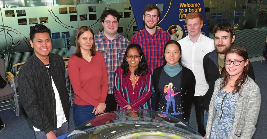
Macromolecular Crystallography Group Beamlines I02 and I04-1

     Using structure-based design to engineer chimeric protein vaccines                                                                                                                                         a.
                                                                                                                                                                                                                                             Antisera                                                          N. meningitidis strain used in SBA                                        PorA dependent
     Related publication: Hollingshead S., Jongerius I., Exley R. M., Johnson S., Lea S. M. & Tang C. M. Structure-based design of chimeric                                                                             fHbp                    PorA                 position                   Isolate                    fHbp peptide                      PorA VR2                       SBA titre
     antigens for multivalent protein vaccines. Nat. Commun. 9, 1051 (2018). DOI: 10.1038/s41467-018-03146-7                                                                                                            V1. 4                 P1.10_1                   151                 M11. 240189                        V3.84                         P1.10_1                            1/20
                                                                                                                                                                                                                        V1. 4                  P1.14                    151                 M15.240853                         V3.45                           P1.14                           1/640
     Publication keywords: Meningococcal; Vaccine; Serogroup B; fHbp; PorA                                                                                                                                             V3.45                    P1.4                    151                 M11. 240123                        V1.92                            P1.4                           1/160

     A
                                                                                                                                                                                                                       V3.45                    P1.9                    151                 M11. 240431                        V2.19                            P1.9                          1/1280
           major World Health Organisation objective is to develop vaccines against pathogenic bacteria. However, vaccine development is often
                                                                                                                                                                                                                b.
           hindered by variation in the molecules at the surface of the bacteria, and the manufacturing challenges inherent to working with
                                                                                                                                                                                                                                             Antisera                                                          N. meningitidis strain used in SBA                                        fHbp dependent
           such molecules. Researchers conducted a proof-of-principle study to assess the feasibility of taking portions of intractable molecules
                                                                                                                                                                                                                        fHbp                    PorA                 position                   Isolate                    fHbp peptide                      PorA VR2                       SBA titre
     and creating chimeras by splicing them into well-behaved molecules, with the ultimate goal of producing vaccines that raised immune
     responses against both types of molecule.                                                                                                                                                                                                                          151                                                                                                                   1/1024
                                                                                                                                                                                                                        V1.1                   P1.16                    294                H44/76 Δ PorA                        V1.1                            N/A                           1/4096
     They used Macromolecular Crystallography beamlines I02 and I04-1 to carry out structural investigations to confirm that this chimera-
                                                                                                                                                                                                                                                                        309                                                                                                                   1/2048
     generating approach doesn’t compromise key structural features of the molecules. This is especially important when it comes to molecules
     with the potential for use in therapeutic drugs. The study demonstrated that the approach is valid. Structures of the chimeric molecules                                                                  Figure 2: Serum bactericidal assay titres. (a) α-PorA SBA titres generated using ChA/alum antisera and serogroup B N. meningitidis isolates with mismatched fHbp variants. (b) α-fHbp SBA titres
                                                                                                                                                                                                               generated using ChA/alum antisera and serogroup B N. meningitidis isolate H44/76 ∆porA.
     revealed that both molecules retained the structure observed in their native context, or in immune-relevant complexes. Immunisation
     studies revealed that the chimeric molecules were able to elicit immune responses against each component.                                                                                                 ChAs, fHbp is exploited as a molecular scaffold to display an immunogenic                              initiate complement-mediated lysis of N. meningitidis. This assay demonstrated
                                                                                                                                                                                                               surface exposed loop from PorA, known as the variable region 2 (VR2, Fig.                              PorA specific SBA titres ranging between ≥20 and ≥1280 (Fig. 2a) (above
                                                                                                                                                                                                               1a). Both fHbp and the VR2 loop exhibit antigenic diversity, with fHbp                                 the ≥8 threshold for an accepted correlate of protective immunity against
                                                                                                                                                                                                               peptides falling into three variant groups or two subfamilies depending on                             N. meningitidis). Furthermore, fHbp specific SBA titres, obtained from ChAs
                                                                                                                                                                                                               the classification system: V1 (subfamily B), V2, and V3 (both subfamily A). In                         composed of fHbp V1.1 and VR2 P1.16, range between ≥1024 and ≥4096 (Fig.
                                                                                                                                                                                                               general, immunisation with a particular fHbp induces cross-protection against                          2b).
                                                                                                                                                                                                               strains that express an fHbp belonging to the same, but not a different, variant
                                                                                                                                                                                                               group, although there can be cross-protection between fHbp variant groups                                  Previous studies had found that linear PorA VR2 peptides failed to elicit
                                                                                                                                                                                                               V2 and V3 (subfamily A). Unlike fHbp, the PorA VR2 offers limited or no cross-                         antibodies that recognised native PorA, while cyclic VR2 peptides, with identical
                                                                                                                                                                                                               protection against strains expressing a PorA peptide with a different VR2.                             residues fixed in a β-turn, elicited PorA specific bactericidal antibodies. In the
10                                                                                                                                                                                                                                                                                                                    ChAs generated in this study, the VR2 N- and C- termini are effectively locked              11
                                                                                                                                                                                                               Therefore to optimise coverage of the vaccine, the fHbp and PorA VR2 peptides
                                                                                                                                                                                                               used must be carefully selected. This was achieved using the comprehensive                             into a “biological” β-turn peptide mimetic by neighbouring fHbp β-strands, as
                                                                                                                                                                                                               epidemiology data available in the meningococcal genome library3.                                      confirmed by our atomic structures.

                                                                                                                                                                                                                   A proof of principle study was designed to ascertain: i) the introduction                              This study provides proof of principle that ChAs can be used to display
                                                                                                                                                                                                               of a PorA VR2 loop did not alter the overall architecture of fHbp, ii) functional                      selected antigens from a soluble protein, which forms a scaffold, and an
                                                                                                                                                                                                               immune responses could be elicited against both fHbp and PorA antigens, iii)                           epitope from an integral membrane protein. The structural data conclusively
                                                                                                                                                                                                               ChA composition can be manipulated to adapt to circulating fHbp and PorA                               demonstrates that fHbp is not perturbed by insertion of a PorA VR2, and
                                                                                                                                                                                                               antigens.                                                                                              that the VR2 loop P1.16 adopts a conformation known to induce bactericidal
                                                                                                                                                                                                                                                                                                                      antibody responses. The immune response data demonstrates that correlates of
                                                                                                                                                                                                                   Structures were determined of ChAs with PorA VR2 P1.16 inserted                                    protection are elicited by both antigens in the ChA, and that ChA composition
                                                                                                                                                                                                               into position 151, 294, or 309 of a V1 fHbp (Fig. 1b). These structures were                           can be adapted to provide protection against relevant circulating N. meningitidis
                                                                                                                                                                                                               solved using molecular replacement to resolutions of 2.9 Å, 3.7 Å, and 2.6 Å,                          strains.
                                                                                                                                                                                                               respectively. All ChA scaffolds align with good agreement to native fHbp (Fig.
                                                                                                                                                                                                               1b), demonstrating that the structure of fHbp is not perturbed by insertion of                         References:
                                                                                                                                                                                                               VR2 loop P1.16. In each structure, the VR2 loop adopts a β-turn conformation                           1. Pizza M. et al. Identification of vaccine candidates against serogroup
                                                                                                                                                                                                               that extends away from the fHbp scaffold, and mimics an identical free VR2                                B meningococcus by whole-genome sequencing. Science (80-. ). 287,
                                                                                                                                                                                                               peptide in a complex with a bactericidal Fab fragment (Fig. 1c).                                          1816–1820 (2000). DOI: 10.1126/science.287.5459.1816
     Figure 1: (a) Schematic of N. meningitidis cell surface, shown are the major antigens fHbp (in a complex with human complement factor H domains 6-7) and PorA. (b) Structural alignment of fHbp
     scaffolds from V1 fHbp peptides with a PorA loop inserted at residue 151 (pink, PDB: 5NQP), 294 (green, PDB: 5NQX) or 309 (orange, PDB: 5NQY). (c) Structural alignment of the PorA VR2 epitopes in the                                                                                                          2. van der Ley P. et al. Topology of outer membrane porins in pathogenic
     fHbp scaffold (151, pink; 294, green; 309, orange) with a linear peptide of PorA VR2 P1.16 (black) in a complex with a bactericidal Fab fragment (grey) from monoclonal antibody MN12H2 (PDB: 2MPA).          The adaptability of the ChA approach was tested by generating
                                                                                                                                                                                                               ChAs composed from different combinations of fHbp and PorA VR2. The                                       Neisseria spp. Infect. Immun. 59, 2963 LP-2971 (1991)
                                                                                                                                                                                                               comprehensive meningococcal genome data available for strains isolated                                 3. Meningitis Research Foundation Meningococcus Genome Library (https://
         To combat the rise in multi-drug resistant bacteria, the World Health                                immune responses against epitopes that are masked in the whole organism. To                      in the UK enable design of ChAs that have exact sequence matches to the                                   pubmlst.org/bigsdb?db=pubmlst_neisseria_mrfgenomes)
     Organisation is advocating the development of vaccines against bacterial                                 circumvent these issues, we used structure-based design to engineer chimeric                     most common antigens in a given region3. Prevalent PorA VR2s in circulating
     pathogens. Historically, vaccines were based on protein toxoids, polysaccharide                          antigens (ChAs), and developed a next generation vaccine for prevention of                       serogroup B N. meningitidis isolates are P1.4, P1.9, P1.14, P1.16 and P1.10_1,                         Funding acknowledgement:
     capsules, or outer membrane vesicles. However, these approaches are not                                  meningococcal disease.                                                                           and prevalent fHbp peptides are V1.4 and V3.45. Five different ChAs were                               Action Medical Research (Award number GN2205).
     feasible for many pathogens, and immune evasion mechanisms, such as                                                                                                                                       constructed, in which a VR2 peptide was inserted into position 151 in V1.4
                                                                                                                  The meningococcal ChAs produced for our study are composed of two major                                                                                                                             Corresponding author:
     antigenic diversity, further limit the strategies available to develop successful                                                                                                                         or V3.45. The ability of these ChAs to elicit immune responses was examined
                                                                                                              N. meningitidis antigens, factor H binding protein (fHbp), a surface exposed                                                                                                                            Prof Susan Lea, University of Oxford, susan.lea@path.ox.ac.uk
     vaccines. In addition, many vaccine candidates are integral membrane proteins                                                                                                                             by immunising groups of CD1 mice with each ChA, followed by the use of a
     which can present manufacturing problems, and issues such as generating                                  lipoprotein1, and PorA, an integral outer membrane porin2 (Fig. 1a). In the                      serum bactericidal assay (SBA), to assess the ability of the immune sera to

                                                                DIAMOND LIGHT SOURCE ANNUAL REVIEW 2018/19                                                                                                                                                                DIAMOND LIGHT SOURCE ANNUAL REVIEW 2018/19
Diamond Light Source Ltd Review 2018/19 - TU ...
Macromolecular Crystallography Group Beamlines I03 and I04-1

     Understanding human sleeping sickness                                                                                                                                    References:
                                                                                                                                                                              1. Pays E. et al. The molecular arms race between African trypanosomes and
                                                                                                                                                                                                                                                                                     4. Lane-Serff H. et al. Evolutionary diversification of the trypanosome
                                                                                                                                                                                                                                                                                        haptoglobin-haemoglobin receptor from an ancestral haemoglobin
                                                                                                                                                                                 humans. Nat. Rev. Microbiol. 12, 575 (2014). DOI: 10.1038/nrmicro3298.                                 receptor. Elife 5, e13044 (2016). DOI: 10.7554/eLife.13044
     Related publication: Zoll S., Lane-Serff H., Mehmood S., Schneider J., Robinson C. V, Carrington M. & Higgins M. K. The structure of serum
                                                                                                                                                                              2. Higgins M. K. et al. Structure of the trypanosome haptoglobin{\                                     5. Zoll S. et al. The structure of serum resistance-associated protein and
     resistance-associated protein and its implications for human African trypanosomiasis. Nat. Microbiol. 3, 295–301 (2018). DOI: 10.1038/
                                                                                                                                                                                 textendash}hemoglobin receptor and implications for nutrient uptake                                    its implications for human African trypanosomiasis. Nat. Microbiol. 3,
     s41564-017-0085-3
                                                                                                                                                                                 and innate immunity. Proc. Natl. Acad. Sci. 110, 1905–1910 (2013). DOI:                                295–301 (2018). DOI: 10.1038/s41564-017-0085-3
                                                                                                                                                                                 10.1073/pnas.1214943110
                                                                                                                                                                                                                                                                                     Funding acknowledgement:
     Publication keywords: African trypanosomes; Innate immunity; Apolipoprotein L1; SRA                                                                                      3. Lane-Serff H. et al. Structural basis for ligand and innate immunity factor                         MRC (MR/L008246/1 and MR/P001424/1); Wellcome Trust.
                                                                                                                                                                                 uptake by the trypanosome haptoglobin-haemoglobin receptor. Elife 3,

     T
           he worst forms of human sleeping sickness (African trypanosomiasis) are caused by a species of trypanosome (single-celled parasite)                                   e05553 (2014). DOI: 10.7554/eLife.05553                                                             Corresponding authors:
                                                                                                                                                                                                                                                                                     Prof Matt Higgins, University of Oxford, matthew.higgins@bioch.ox.ac.uk and
           known as T. b. rhodesiense. Most African trypanosomes are unable to infect humans as they are killed by a component of human blood,
                                                                                                                                                                                                                                                                                     Prof Mark Carrington, University of Cambridge, mc115@cam.ac.uk
           known as trypanolytic factor, and only two trypanosome subspecies are able to infect humans. T. b. rhodesiense is able to do so due
     to the presence of a single molecule called SRA. Although it was clear that SRA is the sole molecule responsible for allowing this parasite to
     develop human resistance, we did not know how it worked, or what it looked like. We also did not know how it binds to the active component
     of the trypanolytic factor, a molecule called ApoLI.
     A team of researchers from the Universities of Oxford and Cambridge investigated how SRA stops the trypanolytic factor from working, using
     the Macromolecular Crystallography beamlines I03 and I04-1 to collect diffraction data from crystals of SRA bound to an antibody, and to
     determine the structure of the molecule. They were able to work out what SRA looks like, and to map onto the structure the regions of SRA
     that bind to the trypanolytic factor. This provides a first view of how SRA gives a trypanosome the ability to infect humans. However, it still
     leaves many questions, and understanding exactly how ApoLI interacts with SRA remains an exciting future research challenge.
          African trypanosomes are single-celled eukaryotic pathogens that are             factor uptake. Our structures revealed how this mutation decreases the affinity
     transmitted by tsetse flies, and live within the blood streams and tissue spaces      of the receptor for lytic factor, and how it helps to protect this trypanosome
     of infected mammals. Infection causes disease, including the debilitating             subspecies from destruction2,3.
     wasting illness of cattle, nagana, which restricts the productivity of livestock in
     sub-Saharan Africa. While there are numerous species of African trypanosome,              We more recently turned our attention to T. b. rhodesiense, which
     only two subspecies, T. b. gambiense and T. b. rhodesiense, can proliferate in        counteracts the action of lytic factor by expressing an anti-toxin that binds
12   humans. This is due to lytic factors, present only in humans and some other           to, and inactivates, ApoL1. The anti-toxin, known as the serum resistance                                                                                                                                                                                                                      13
     primates, which are taken up by trypanosomes and causes their death1.                 associated protein (SRA), is found primarily within the lysosome of
     Human-infective species are able to negate the action of this lytic factor. This      trypanosomes, and is sufficient to allow a trypanosome to become human
     confers the ability to grow within infected humans, and to cause the disease          infective. Our goal was to determine the structure of SRA, and to investigate
     Human African trypanosomiasis, also known as sleeping sickness. The aim of            how it binds to, and inactivates, ApoLI.
     our work is to understand how human-infective trypanosomes inactivate the
     lytic factor, and then to determine whether we can make them susceptible to               Determining the structure of SRA proved challenging. As we later
     lytic factor mediated killing.                                                        discovered, a large loop at one end of SRA is flexible, and prevented the
                                                                                           formation of well-ordered crystals. We were able to solve this problem by
         Our first contribution towards these goals used beamline facilities               producing a panel of monoclonal antibodies that bind to SRA, and discovered
     at Diamond Light Source to understand how lytic factor is taken up by                 that the complex of SRA with one of these antibodies formed crystals of
     trypanosomes. The lytic factor is a complex of several components, including          sufficient quality5. Armed with data collected from beamline I03 at Diamond,       Figure: The centre of the Figure shows a schematic of a blood stream form T. b. brucei. The left-hand side of the figure shows the T. b. rhodesiense haptoglobin-haemoglobin receptor (HpHbR, blue) bound
     the pore-forming protein, apolipoprotein LI (ApoLI), which acts as the toxin          we were then able to determine the structure of SRA. Together with data            to a complex of the SP domain of haptoglobin (Hp, yellow), the a-subunit of haemoglobin (Hba, orange) and the b-subunit of haemoglobin (Hbb, red). This is found on the cell surface, and within the
                                                                                                                                                                              flagella pocket of trypanosomes. The right-hand panel shows the surface resistance associated protein (SRA, rainbow) of T. b. rhodesiense, which is found primarily in the lysosome of trypanosomes.
     and a complex of haptoglobin related-protein bound to haemoglobin (HprHb).            from hydrogen-deuterium exchange mass spectrometry, and validated by
     Trypanosomes have a receptor on their cell surface that binds to haptoglobin-         mutagenesis, this allowed us to map onto SRA the residues that interact with
     haemoglobin (HpHb), allowing them to scavenge this valuable nutrient from             ApoLI.
     the blood. However, the presence of HprHb in lytic factor allows this toxin
     to hitch a ride on the receptor, taking it into the trypanosome cell, where it            This provided the first detailed structural insight into SRA and its mode of
     mediates its toxic effect. We used beamlines I03 and I04-1 to solve structures of     interaction with ApoLI5. It also revealed that prevailing models for how SRA
     HpHb receptors from human-infective and non-infective trypanosomes, alone             prevents ApoLI function were not compatible with our structural findings.
     and also bound to HpHb2-4. This revealed how the lytic factor is recognised by        However, it still leaves many questions, and understanding exactly how ApoLI
     trypanosomes. It also helped us to understand how T. b. gambiense becomes             forms pores, and how the binding of SRA prevents these pores from forming,
     human-infective, as it has a mutation of the HpHb receptor which reduces lytic        remains an exciting future research challenge.

                                                        DIAMOND LIGHT SOURCE ANNUAL REVIEW 2018/19                                                                                                                                       DIAMOND LIGHT SOURCE ANNUAL REVIEW 2018/19
Diamond Light Source Ltd Review 2018/19 - TU ...
Macromolecular Crystallography Group Beamlines I03, I04 and I23

     Turning the plastic tide – engineering enzymes to tackle plastic pollution
     Related publication: Austin H. P., Allen M. D., Donohoe B. S., Rorrer N. A., Kearns F. L., Silveira R. L., Pollard B. C., Dominick G., Duman
     R., El Omari K., Mykhaylyk V., Wagner A., Michener W. E., Amore A., Skaf M. S., Crowley M. F., Thorne A. W., Johnson C. W., Woodcock H. L.,
     McGeehan J. E. & Beckham G. T. Characterization and engineering of a plastic-degrading aromatic polyesterase. Proc. Natl. Acad. Sci. 115,
     E4350--E4357 (2018). DOI: 10.1073/pnas.1718804115

     Publication keywords: Biodegradation; Polyethylene terephthalate (PET); Plastics recycling; Cutinase

     W
                hile climate change has rightly received significant global attention in recent years, we have only just awoken to the sheer
                magnitude of plastic pollution. Eight million tons of plastic waste enters our oceans annually, impacting the environment,
                wildlife, and human health. Programmes like Blue Planet II have changed our perceptions about the effects of plastic, but we
     currently lack effective solutions to deal with the scale of the problem.
     We need to find a way to recycle plastic in a circular fashion. 93% of plastic bottles are never made into new plastic bottles, but are instead
     ‘downcycled’ into lower grade plastic, incinerated, or buried in landfill. Polyethylene terephthalate (PET) is one of the most common polluting
     plastics, and is used for single-use plastic bottles, textiles and carpets.
                                                                                                                                                                                            Figure 2: Some of the team that solved the PETase crystal structure on I23 – Armin Wagner, John McGeehan, Mark Allen, Ramona Duman and Kamel El-Omari.
     Biological enzymes have the potential to break down solid PET into its original building block monomers, and an international team of                                                  shown that these bacteria secreted two enzymes that can break apart the ester                     broccoli under the tap. This waxy layer is also made of monomers that are linked
     researchers used the state-of-the-art Long-Wavelength Macromolecular Crystallography (MX) beamline (I23) to investigate the 3D structure                                               bonds holding the PET monomers together, providing a food and energy source                       together by ester bonds, thus bacteria have been evolving polyester-degrading
     of a newly-discovered PETase enzyme to see how it works, and how to make it more efficient.                                                                                            for the growing organisms. This study highlights the diversity of bacteria and                    enzymes to invade plant cells for millions of years. We were interested to see
                                                                                                                                                                                            shows how they can adapt to new substrates, even man-made materials. Given                        if we could turn PETase back to an ancestral cutinase by changing the DNA
     They discovered that the PETase enzyme is closely related to a cutinase enzyme produced by bacteria to digest the protective natural                                                   that PET has only been in the environment for around 60 years, it is incredible                   sequence. To our surprise, rather than reduce PET-degrading activity, we actually
     polyester coating on plant leaves. The 3D structure allowed them to design an improved enzyme that can digest PET faster, which offers                                                 that these bacteria have evolved to tackle this difficult material in such a short                improved it. While only a modest increase, this does indicate that PETase has the
     great potential for future engineering of this enzyme for industrial recycling applications.                                                                                           timeframe. The big questions for us were: how did these enzymes evolve, how                       potential to be further engineered to be faster and more efficient at digesting
                                                                                                                                                                                            do they work, and can we utilise them as part of a recycling solution?                            PET. This opens up the exciting possibility of turning polyester plastics back to
          Plastic pollution has reached alarming levels in the environment,                            and can be moulded into highly complex solids. From films to the ubiquitous
                                                                                                                                                                                                                                                                                              monomers so that they can be directly reused for new plastic bottles or even
     particularly in our oceans. From documentary programmes such as Blue Planet                       fizzy drink bottle, they protect and extend the lifetime of many products. So            In addition to researchers at the Centre for Enzyme Innovation at the
14                                                                                                                                                                                                                                                                                            upcycled to improved materials.                                                     15
     II, through to media from around the globe, the sheer scale of the problem is                     why then have they created such a global scourge? It is the very properties          University of Portsmouth, we worked across multiple scientific disciplines and
     now receiving the attention that it deserves. Many plastics were only patented                    that the chemists were asked to create, low cost and durability, that have had       countries with teams led by Dr Gregg Beckham at the National Renewable                                We now need to engineer an even faster enzyme and produce it at large
     in the 1940s, but have gained such traction in our everyday lives that their                      unforeseen consequences. To understand why, we need to look at the nature            Energy Laboratory, Colorado, Dr Lee Woodcock at the University of South                           scale. Fortunately, the technology exists, and there are many large industrial
     production has reached truly staggering rates. The plastics industry is worth                     of common plastics.                                                                  Florida, Tampa, Dr Munir Scaf at the University of Campinas, Sao Paulo, Brazil                    companies that have the capacity to generate large quantities of enzymes.
     $1 trillion, and in the UK it represents our second largest manufacturing                                                                                                              and Dr Armin Wagner at Diamond Light Source, UK (Fig. 2). We were able to                         Examples include those currently used in biological washing powders,
     sector. Few could have predicted that in only a few decades huge plastic waste                         Plastic polymers such as polyethylene terephthalate, or PET, are made           produce and isolate the enzyme in our laboratories, follow the digestion of PET                   biofuels production, and food and drink manufacturing. We are now working
     patches would be found floating in our oceans, and once pristine beaches                          from simple monomer building blocks that are linked together via ester bonds,        plastic directly from a single-use drinks bottle over several days, and measure                   in partnership between academia and industry to provide us with the
     would be contaminated all over the world (Fig. 1). Recent studies indicate the                    hence the name polyesters. Most monomers are extracted from petroleum,               the production of the monomer building blocks being produced.                                     best chance of reaching a real-world solution. We must be mindful though
     pervasive nature of these materials, and plastics have now been discovered in                     although more recent developments have seen building blocks produced                                                                                                                   that any real solution will involve a reduction in our reliance on plastics by
     our polar sea ice and in the deepest parts of our oceans. The flow of plastic                     directly from plants. Petroleum based monomers are very cheap because of                  Utilising the exceptional I23 beamline at Diamond, not only could we solve                   innovative alternatives and implementation of a global recycling infrastructure.
     into the environment continues to grow, and it is now with some urgency that                      low oil prices, and it is therefore often cheaper to manufacture virgin-PET than     the structure of the PETase enzyme from native crystals, but we could collect                     Responsibilities lie with both industry and consumers, and now that we are
     we need to find sustainable alternatives and reduce our reliance on plastics for                  to use recycled material. PET also suffers from down-cycling, and in the case        a very high-resolution structure extending to 0.9 Å (Fig. 3)2. This provided                      acutely aware of the problem, we can all play a positive role in reducing our
     single-use applications.                                                                          of single-use plastic bottles, only a small percentage will be turned back into      initial structural insights, and we employed computer modelling to further                        environmental impact and developing solutions.
                                                                                                       new plastic bottles. A reduction in chemical properties results in most recycled     understand how this enzyme binds to PET and breaks the ester-bonds. We
        Plastics have incredible properties and have revolutionised many industries.                   PET being used for clothing fibres, eventually reaching an end-of-cycle use          found that the enzyme has a similar shape to a family of naturally occurring                      References:
     They are cheap to manufacture, strong, light, transparent, waterproof, inert,                     as fillings or carpet. From these low value products, there is little financial      enzymes called cutinases. Cutin is a natural polyester found in plants and forms                  1. Yoshida S. et al; A bacterium that degrades and assimilates poly(ethylene
                                                                                                       incentive to recycle further and much ends up in landfill or is incinerated. While   a protective and waterproof barrier that can be observed by simply running                           terephthalate). Science (80-. ). 351, 1196–1199 (2016). DOI: 10.1126/
                                                                                                       burning plastic can be used to generate electricity, this practice contributes to                                                                                                         science.aad6359
                                                                                                       greenhouse gases with the release of carbon dioxide. Of course, any recycling                                                                                                          2. Austin H. P. et al. Characterization and engineering of a plastic-degrading
                                                                                                       process can only take place when there is a proper infrastructure for the                                                                                                                 aromatic polyesterase. Proc. Natl. Acad. Sci. 115, E4350--E4357 (2018).
                                                                                                       collection and treatment of waste; infrastructure that is severely lacking in                                                                                                             DOI: 10.1073/pnas.1718804115
                                                                                                       many parts of the world. The consequence is that much of the plastic that is not
                                                                                                       dumped into landfill or incinerated will eventually find its way into rivers and                                                                                                       Funding acknowledgment:
                                                                                                       oceans, feeding the already giant plastic-waste islands from the gyres created                                                                                                         BBSRC (BB/P011918/1); National Renewable Energy Laboratory (NREL); US
                                                                                                       by global ocean currents. We must reduce our reliance on single-use plastic,                                                                                                           DOE Office of Energy Efficiency and Renewable Energy. US National Science
                                                                                                       and develop better collection and recycling methods that are truly circular and                                                                                                        Foundation; University of Portsmouth.
                                                                                                       sustainable.
                                                                                                                                                                                                                                                                                              Corresponding author:
                                                                                                           In 2016, a team of scientists in Japan published an exciting research                                                                                                              Prof John McGeehan, University of Portsmouth, john.mcgeehan@port.ac.uk
     Figure 1: Single-use materials such as PET plastic bottles can persist for hundreds of years in
     the oceans, concentrating in huge ocean gyres such as the Great Pacific Garbage Patch. Photo
                                                                                                       paper describing the discovery and isolation of a strain of bacteria, Ideonalla      Figure 3: The PETase enzyme structure is shown bound to a polymer of PET plastic. Image
     credit: DAVID JONES.                                                                              sakaiensis, which has the remarkable ability to survive on a diet of PET1. It was    credit: H.L. Woodcock.

                                                                 DIAMOND LIGHT SOURCE ANNUAL REVIEW 2018/19                                                                                                                                           DIAMOND LIGHT SOURCE ANNUAL REVIEW 2018/19
Diamond Light Source Ltd Review 2018/19 - TU ...
Macromolecular Crystallography Group Beamlines I04 and I04-1

                                                                                                                                                                                                              a                                                                                                                              b
     Insc:LGN tetramers revert breast cancer stem cell expansion by promoting                                                                                                                                                   Superdex-200
                                                                                                                                                                                                                                                                                                        LGNTPR: Insc
                                                                                                                                                                                                                                                                                                        PINSTPR: InscASYM                      InscASYM-ΔαB             -         -   +   -    -     +
                                                                                                                                                                                                                                                                                                                                                                                                                    Figure 2: Human LGN:Insc complex oligomerisation.

     asymmetric cell divisions                                                                                                                                                                                                                                                                                                                                                                                      (a) SEC elution profiles of Drosophila LGNTPR:InscASYM

                                                                                                                                                                                                                  Absorbance at 280 nm (mAu)
                                                                                                                                                                                                                                           200                                     158 KDa
                                                                                                                                                                                                                                                                                                                                                         InscASYM -               +   -   -    +     -              (blue trace) and human LGNTPR:Insc (green trace) with
                                                                                                                                                                                                                                                                                                                        17 KDa                                  KDa                                      IB:
                                                                                                                                                                                                                                                                                                      44 KDa
                                                                                                                                                                                                                                                                                                                                                                75 -                                     GFP
                                                                                                                                                                                                                                                                                                                                                                                                                    associated Coomassie-stained SDS–PAGE separations of
                                                                                                                                                                                                                                           150
                                                                                                                                                                                                                                                 670 KDa
                                                                                                                                                                                                                                                                                                                                                                                                         (LGNTPR)   peak fractions. The elution profile of globular markers is
     Related publication: Culurgioni S., Mari S., Bonetti P., Gallini S., Bonetto G., Brennich M., Round A., Nicassio F. & Mapelli M. Insc:LGN                                                                                                                                                                                                                   37 -                                    FLAG       reported as a dashed gray line. (b) Immunoprecipitation
                                                                                                                                                                                                                                           100
                                                                                                                                                                                                                                                                                                                                                                                                         (LGNTPR)   experiments (IPs) from HEK293T cells transfected
     tetramers promote asymmetric divisions of mammary stem cells. Nat. Commun. 9, 1025 (2018). DOI: 10.1038/s41467-018-03343-4                                                                                                                                                                                                                                  50 -
                                                                                                                                                                                                                                                                                                                                                                                                         Insc
                                                                                                                                                                                                                                                                                                                                                                                                                    with human GFP-LGNTPR and FLAG-LGNTPR alone or in
                                                                                                                                                                                                                                           50                                                                                                                    37 -
                                                                                                                                                                                                                                                                                                                                                                                                                    combination with human InscASYM or InscASYM-ΔαB (lacking
                                                                                                                                                                                                                                                                                                                                                                                                                    residues 62–191). IPs with anti-FLAG antibodies were
     Publication keywords: Mammary stem cells; Cell divisions; Asymmetric fate                                                                                                                                                                                               1.3                1.6                          1.9                                100 -                                    vinculin
                                                                                                                                                                                                                                                                                                                                                                                                                    immunoblotted (IB) with the indicated antibodies. FLAG-
                                                                                                                                                                                                                                                                Elution volume (ml)                                                                                      Lysates          IP FLAG                   LGNTPR co-immunoprecipitates with GFP-LGNTPR only when

     I
                                                                                                                                                                                                                                       Insc                                                                                                                                                                         bound to InscASYM, but not to InscASYM-ΔαB.
         n stem cells, asymmetric cell division (ACD) generates two distinct cells: a stem cell, and a cell committed to differentiate. Defects in this                                                           LGNTPR

         delicate and extremely coordinated process can cause cell over-proliferation and cancer. How stem cell ACDs are executed remains largely                                                                 PinsTPR
         unclear. The knowledge of the architecture of key ACD players constitutes a remarkable advance in the understanding of the operational                                                                   InscASYM

     principles of asymmetric divisions. An international research group determined the three-dimensional structure of the Insc:LGN complex,
     which revealed that oligomerisation and clustering of LGN (spindle orientation protein) and Insc are essential for ACDs.                                                                                open on one side; the cylinder is closed by the Insc helix bundle, increasing the                                                                                               Figure
                                                                                                                                                                                                                                                                                                                                                                                      decreases   the number 2 of mammospheres formed in sphere-forming assays
                                                                                                                                                                                                             steadiness of the complex.                                                                                                                                               (Fig. 3b). Notably, the Insc region encompassing the ASYM domain is sufficient
     To obtain an optimal structural characterisation of this complex, several tens of crystals obtained in different conditions were tested on the
                                                                                                                                                                                                                                                                                                                                                                                      to reduce the sphere-forming efficiency, while the the InscASYM domain
     Macromolecular Crystallography (MX) beamlines I04 and I04-1, but only two diffracted to 3.4 Å and 4.0 Å resolution. The final combination                                                                   The LGNTPR:InscASYM tetramers are present in the crystals in two slightly
                                                                                                                                                                                                                                                                                                                                                                                      lacking the four-helix-bundle, and forming a 1:1 complex with LGN, is not (Fig.
                                                                                                                                                                                                             different conformations: one is extended and symmetric, and the other is
     of the two datasets allowed the structural determination. The group showed that LGN forms intertwined tetramers with Insc, which are                                                                                                                                                                                                                                             3b). These results clearly demonstrate that the oligomerisation of InscASYM with
                                                                                                                                                                                                             slightly more compact and asymmetric. Small Angle X-ray Scattering (SAXS)
     markedly stable. Most importantly, in mammary stem cells, the tetrameric molecular assembly suffices to promote asymmetric cell divisions,                                                                                                                                                                                                                                       LGN is essential for the asymmetric functions of Insc in stem cell divisions.
                                                                                                                                                                                                             analyses indicated that the LGNTPR:InscASYM complex in solution is flexible and
     and reverts the over-proliferation caused by loss of the tumour suppressor gene p53.                                                                                                                    can acquire both conformations (Fig. 1c, the extended and compact tetramers                                                                                                  Conclusively, the crystallographic structure of the LGNTPR: InscASYM complex
          Stem cells are characterised by two unique properties: (I) the ability of                          Initial structural characterisation of the LGN TPR-domain (LGNTPR) in                           are shown in yellow and green ribbons respectively, and fitted into the SAXS                                                                                             revealed a stable hetero-tetrameric assembly with intertwined architecture,
     engendering all differentiated cell types, and (II) the capacity of self-renewal                    complex with a 35-residue Insc peptide (InscPEPT), showed that the association                      envelope). We speculate that this intrinsic structural plasticity of LGNTPR:InscASYM                                                                                     which is conserved throughout evolution. The steadiness of the LGN:Insc
     through several rounds of cell divisions keeping an undifferentiated state.                         is mutually exclusive with NuMA, and revealed a nanomolar binding affinity1.                        might be functional to the scaffolding role of the complex at the plasma                                                                                                 structural organisation suggests that the association of LGN with Insc is
     Specifically, stem cells divide asymmetrically, generating a daughter stem                          However, no information was available for the entire operational domain of                          membrane, which is intrinsically flexible.                                                                                                                               constitutive, and that the LGN:Insc interaction promotes asymmetric fate
     cell (identical to the mother) attached to the maternal niche, and a daughter                       Insc, which is able to recapitulate all its functions in stem cell divisions (the                                                                                                                                                                                            specification without exchanging with the NuMA-bound pool of LGN that
                                                                                                                                                                                                                  The human LGNTPR and InscASYM counterparts were coexpressed in insect
     cell inclined to relocate and differentiate. This asymmetric cell division (ACD)                    so-called asymmetric domain, InscASYM). Therefore, we set out to determine                                                                                                                                                                                                   associates with the microtubule motor dynein. Most importantly, the LGN:Insc
                                                                                                                                                                                                             cells and in mammalian cells, but the poor yield did not enable any structural
     requires the spatial coordination between the division plane and cell polarity                      the structure of the LGNTPR:InscASYM complex, which would constitute a valuable                                                                                                                                                                                              structure revealed that the tetrameric organisation of the complex is essential
16                                                                                                                                                                                                           characterisation. Size-exclusion chromatography analyses of the human                                                                                                                                                                                               17
     cues. The current view is that cell polarity is established by Par proteins                         advance in the understanding of the molecular principles of asymmetric cell                                                                                                                                                                                                  to promote asymmetric divisions of mammary stem cells, and suffices to
                                                                                                                                                                                                             purified LGNTPR:Insc complex agreed with a 2:2 stoichiometry (Fig. 2a). In
     (Par3:Par6:aPKC) at the apical site, while the division plane is defined by                         division and stem cell replication, with substantial implications for cancer.                                                                                                                                                                                                revert aberrant stem cell over-proliferation caused by p53 loss, with important
                                                                                                                                                                                                             addition, the ability of human LGN to pull down another copy of LGN only
     the position of the mitotic spindle regulated by the Ga:LGN:NuMA spindle                                                                                                                                                                                                                                                                                                         therapeutic implications.
                                                                                                                     The Drosophila melanogaster LGNTPR and InscASYM polypeptides were                       in presence of InscASYM demonstrated that the hetero-tetrameric assembly
     orientation complex. The protein Inscuteable (Insc) links these two cellular                                                                                                                            observed in flies is conserved in humans (Fig. 2b). The 2:2 stoichiometry of the
                                                                                                               coexpressed in bacteria and purified as a 2:2 stoichiometry complex with an                                                                                                                                                                                            References:
     processes as a result of its concomitant binding to Par3 and LGN.                                                                                                                                       LGN:Insc assembly is lost when the region encompassing the four-helix bundle
                                                                                                                                                           experimentally measured mass of 180                                                                                                                                                                                        1. Culurgioni S. et al. Inscuteable and NuMA proteins bind competitively
      a                                                                      b                 α-bundle                                                    kDa (Fig. 1a). After the identification of        of human Insc is deleted (Fig. 1b), confirming that this region is essential for                                                                                            to Leu-Gly-Asn repeat-enriched protein (LGN) during asymmetric cell
                   60                             6                                                                                                                                                          oligomerisation (and not for LGN association).
                                                                                                                                                           the first crystal hits, extensive efforts                                                                                                                                                                                     divisions. Proc. Natl. Acad. Sci. 108, 20998–21003 (2011). DOI: 10.1073/
            Absorbance 280 nm (mAU)

                                                  5.5                               INSC-2                                                                 dedicated to the optimisation of the                                                                                                                                                                                          pnas.1113077108
                                                   Log Molecular Weight

                  45 180 kDa                                                                                                                                                                                     To determine the functional relevance of the InscASYM-dependent LGN
                                                                                                                                   LGN-B
                                                                                                                                                           crystallisation condition yielded plate-          oligomerisation for stem cell asymmetric divisions, we analysed the ectopic
                  30
                                                  5                                                                                                                                                                                                                                                                                                                                   2. Cicalese A. et al. The Tumor Suppressor p53 Regulates Polarity of Self-
                                                                               C N                                                                         shaped crystals diffracting to 3.3-4.0 Å.         expression of different constructs of Insc in murine mammary stem cells
                                                  4.5                                                                                       N                                                                                                                                                                                                                                            Renewing Divisions in Mammary Stem Cells. Cell 138, 1083–1095 (2009).
                                                                                                                                                   C       Despite the low resolution, the good data         (MaSCs) depleted of the tumour suppressor gene p53 (p53-KO). Notably, p53-
                  15                                                                                                                                                                                                                                                                                                                                                                     DOI: 10.1016/j.cell.2009.06.048
                                                  4                                   LGN-A                                                                quality and the consistency across the            KO MaSCs show low Insc expression, and replicate abnormally through cycles
                   0                                                                                                                α3                     whole data collection resulted in high            of symmetric divisions, causing over-proliferation2. Restoring Insc expression                                                                                           Funding acknowledgement:
                                                  3.5
                        12        13        14
                                                                                                                                   INSC-1                  redundancy datasets, and permitted                in p53-KO MaSCs increases the number of ACDs (Fig. 3a), and consistently                                                                                                 Italian Foundation for Cancer Research (AIRC) (IG18692, IG14085); Italian
                        Elution volume (ml)                                                                o
                                                                                                               90
                                                                                                                                                           the generation of electron density                                                                                                                                                                                                                        Ministry of Health (RF-2013-02357254);
      c                                                                      d                                                                             maps that were suitable for structure         a                                                                                              b                                                                                                            BioStruct-X (FP7/2007-2013, grant agreement
                                                                                                         C           C                                                                                                                                                                                                       Hs Insc
             100                                                                     Armadillo                                                             determination. The overall architecture                                                                                                                                                 61 192 316
                                                                                                                                                                                                                                                                                               ACD                              FL
                                                                                                                                                                                                                                                                                                                                         1                            532                                            N°283570).
                                                                                                                                                           of the LGNTPR:InscASYM complex consists                         CD49f
                                                                                                                                                                                                                                                                  100
                                                                                                                                                                                                                                                                                               SCD                              ASYM
                                                                                                                                                                                                                                                                                                                             ASYM-ΔαB
                                                                                   INSC-2

                                                                                                                                                                                                                                                 Type of division (%)
                                                                                                                                 INSC-1
                                                                                                                                                           of an intertwined hetero-tetramer,                                                                                                                      200                                                                                               Corresponding authors:

                                                                                                                                                                                                                                                                                                        Sphere number
               10
                                                                                                                                                                                                             EV                                                         75                                                     ***                              ***
                                                                                                                                                           where each Insc chain contacts one                                                                                                                      150
                                                                                                                                                                                                                                                                                                                                             ***
                                                                                                                                                                                                                                                                                                                                                                                               ***                   Dr Marina Mapelli, European Institute of
                                                                               o
                                                                                                                                                                                                                                                                        50                                                                                                  ***                           ***
                                                                                                                                                                                                                                                                                                                                       n.s.
                                                                                                                                                           TPR domain (LGN-A) through its InscPEPT                                                                                                                 100
                                                                                                                                                                                                                                                                                                                                                                        n.s.
                                                                                                                                                                                                                                                                                                                                                                                                                     Oncology,
                1                                                                                                                                                                                                                                                       25                                                                                                                           n.s.
                          Extended                                                                                                     N                   region, and the other (LGN-B) with its
                                                                                                                                                                                                           INSC                                                                                                         50                                                                                           marina.mapelli@ieo.it and Dr Simone
                                                                                   N                                                                                                                                                                                                                                          M1                            M2                                M3
                                                                                       α1                                                                                                                                                                                0                                               0                                                                                           Culurgioni, Exscientia,
                          Compact                                                                                                                          four-helix bundle. The final portion of                                                                                 EV        INSC                            EV FL YM αB                    EV FL YM αB                       EV FL YM αB
                                                                              LGN-A                                                 LGN-B                                                                                                                                                                                        AS M-                          AS M-Δ                            AS M-              sculurgioni@exscientia.co.uk
              0.1                                                                                                                                                                                                                                                                                                                      Δ                                                                Δ
                 0.0         0.1         0.2         0.3                                                                                                   each InscASYM is organised in Armadillo                                                                                                                                  Y                              Y                                 Y
                                                                                                                                                                                                                                                                                                                                 AS                             AS                                AS
                                      -1
                                  q (Å )
                                                                                                                                                           repeats that contact one another,
                                                                                                                                                                                                        Figure 3: Insc promotes asymmetric cell divisions of murine mammary stem cells. (a) Quantification of the division mode of mammary epithelial
      Figure 1: Structure of Drosophila LGNTPR:InscASYM complex. (a) Static-Light-Scattering profile of the LGNTPR:InscASYM tetramer. (b-d) Cartoon        further stabilising the assembly (Fig.       cells isolated from p53-KO mice evaluated by pair-cell assay. Cells were infected with empty lentivirus (EV) or virus expressing full-length Insc
      representation of the LGNTPR:InscASYM tetramer in two orthogonal orientations. The two protomers of LGNTPR are coloured gold and orange, while 1b and 1d). The two TPR domains are
      the two InscASYM copies are in cyan and purple. The two-fold symmetry axis of the tetramer is marked as a red oval in b. (c) SAXS data of the
                                                                                                                                                                                                                                                                                                                                       Figure
                                                                                                                                                                                                        (INSC) and imaged after the first division by staining with the CD49f surface marker of the mammary basal lineage. (b) Sphere-forming           3
                                                                                                                                                                                                                                                                                                                                                    assays
      Drosophila LGNTPR:InscASYM complex, and ab initio model of the SAXS-derived envelope superposed to the crystallographic   Figure
                                                                                                                                    structures of 1
                                                                                                                                                  extended complementarily juxtaposed in a head-
                                                                                                                                                                                                        performed with p53-KO mammospheres infected with empty lentivirus (EV) or viruses expressing Insc (FL), InscASYM (ASYM), or InscASYM-ΔαB. The
                                                                                                                                                                                                        sphere-forming efficiency (SFE) was quantified over three subsequent passages M1-M2-M3.
      and compact tetramers.                                                                                                                               to-head interaction, forming a cylinder

                                                                          DIAMOND LIGHT SOURCE ANNUAL REVIEW 2018/19                                                                                                                                                                                                         DIAMOND LIGHT SOURCE ANNUAL REVIEW 2018/19
You can also read