Diet-Microbiota Interplay: An Emerging Player in Macrophage Plasticity and Intestinal Health
←
→
Page content transcription
If your browser does not render page correctly, please read the page content below
International Journal of
               Molecular Sciences

Review
Diet–Microbiota Interplay: An Emerging Player in Macrophage
Plasticity and Intestinal Health
Cian O’Mahony * , Asma Amamou and Subrata Ghosh

                                            APC Microbiome Ireland, College of Medicine and Health, University College Cork, T12 YT20 Cork, Ireland;
                                            aamamou@ucc.ie (A.A.); subrataghosh@ucc.ie (S.G.)
                                            * Correspondence: 121126891@umail.ucc.ie

                                            Abstract: Inflammatory bowel diseases (IBD) are chronic disorders of the gastrointestinal tract with
                                            an increasing prevalence worldwide. Targeted therapies for IBD are limited by several factors, includ-
                                            ing the therapeutic ceiling and the high incidence of non-responders or loss-of-response. In order
                                            to improve therapeutic efficacy, there is critical need to decipher disease pathogenesis, currently
                                            not well understood. Macrophages, innate immune cells that exhibit high plasticity, perpetuate
                                            inflammatory signalling in IBD through excessive release of inflammatory mediators. In recent years,
                                            pioneering research has revealed the importance of the interplay between macrophages and gut mi-
                                            crobiota in maintaining intestinal homeostasis. Particular attention is focusing on microbiota-derived
                                            metabolites, believed to possess immunomodulatory properties capable of manipulating macrophage
                                            plasticity. Microbiota-derived short-chain fatty acids (SCFAs) and indole compounds, along with
                                            dietary sourced omega-3 (ω-3) polyunsaturated fatty acids (PUFA), exert anti-inflammatory effects,
                                            attributable to interactions with macrophages. Before we can effectively incorporate these metabolites
                                            into IBD therapies, a deeper understanding of microbiota–macrophage interactions at a molecular
                                            level is necessary. Therefore, the aim of this review is firstly to detail current knowledge regarding

                                     how diet and microbiota-derived metabolites modify macrophage plasticity. Later, we discuss the
Citation: O’Mahony, C.; Amamou,
                                            concept of therapeutic strategies directed at microbiota–macrophage interactions, which could be
A.; Ghosh, S. Diet–Microbiota               highly valuable for IBD therapies in the future.
Interplay: An Emerging Player in
Macrophage Plasticity and Intestinal        Keywords: intestinal inflammation; macrophage plasticity; gut microbiota; diet; short-chain fatty
Health. Int. J. Mol. Sci. 2022, 23, 3901.   acid; indole; polyunsaturated fat
https://doi.org/10.3390/
ijms23073901

Academic Editor: Silvia Melgar
                                            1. Introduction
Received: 31 January 2022
                                                  Inflammatory bowel disease (IBD), including Crohn’s disease (CD) and ulcerative
Accepted: 30 March 2022
                                            colitis (UC), are chronic inflammatory disorders with considerable morbidity posing major
Published: 31 March 2022
                                            burdens on healthcare systems [1]. Despite extensive research, the aetiology of IBD still re-
Publisher’s Note: MDPI stays neutral        mains elusive, which complicates efforts for developing effective treatments. Macrophages
with regard to jurisdictional claims in     are indispensable components of the innate immune system that form a front-line barrier
published maps and institutional affil-     against pathogens [2]. Under normal circumstances, macrophages phagocytose pathogens,
iations.                                    dead cells, and cell debris, which primes the cell for orchestrating responses to tissue dam-
                                            age and infection [3,4]. However, impaired macrophage function perpetuates inflammation
                                            in IBD through excessive release of pro-inflammatory mediators, leading to the recruit-
                                            ment of T lymphocytes to the site of injury, eventuating as extensive tissue damage [5].
Copyright: © 2022 by the authors.
                                            Under these circumstances, tissue repair mechanisms become overwhelmed, leading to the
Licensee MDPI, Basel, Switzerland.
                                            build-up of fibrotic tissue depositions, which further impairs organ function. These mani-
This article is an open access article
                                            festations are partly driven by macrophage-mediated release of pro-fibrotic factors, leading
distributed under the terms and
conditions of the Creative Commons
                                            to excessive fibroblast and myofibroblast activation. As intestinal fibrosis currently has no
Attribution (CC BY) license (https://
                                            specific treatment, therapeutic approaches to restore normal macrophage activity may have
creativecommons.org/licenses/by/            a knock-on effect by halting progression of tissue fibrosis. Accordingly, macrophages are
4.0/).

Int. J. Mol. Sci. 2022, 23, 3901. https://doi.org/10.3390/ijms23073901                                           https://www.mdpi.com/journal/ijms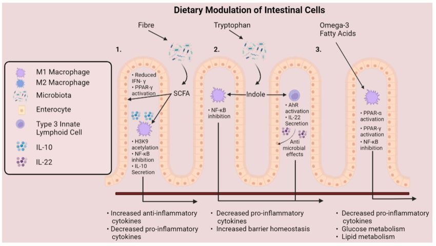
Int. J. Mol. Sci. 2022, 23, 3901                                                                                            2 of 17

                                   an attractive target for therapies aiming to alleviate intestinal inflammation and promote
                                   healing for individuals with IBD.
                                         Macrophages can be categorised as having originated from blood monocytes, or
                                   otherwise, as being tissue-resident macrophages [6]. Macrophages in the first category—
                                   monocyte-derived macrophages—develop from monocytes circulating throughout the
                                   blood. These monocytes quickly migrate to the site of infection to undergo differentiation,
                                   where they provide replacements for depleted monocytes. In recent years, fate-mapping
                                   studies have characterised tissue-resident macrophages, which are derived from the foetal
                                   yolk sac during embryonic development and are embedded in their niche tissue before
                                   birth [7,8]. Uniquely, tissue-resident macrophages attain the somewhat limited capacity to
                                   replenish independently of blood monocytes [9]. Moreover, tissue-resident macrophages
                                   exhibit vast heterogeneity that varies according to ontogeny, tissue locality, and func-
                                   tional programming [10]. These characteristics permit macrophages to display differing
                                   phenotypes depending on the microenvironment that they inhabit.
                                         Of particular interest to IBD research are intestinal-resident macrophages, which sense
                                   antigens derived from food and bacteria that breach the mucosal barrier of the alimentary
                                   canal [11]. Overall, intestinal macrophages constitute the largest pool of macrophages
                                   throughout the body and are described as being highly phagocytic, whilst remaining
                                   resistant to Toll-like receptor (TLR) stimulation [12,13].
                                         Besides forming a first line of defence against pathogens, tissue-resident macrophages
                                   promote tissue healing and resolution following injury [12]. Therefore, in the mammalian
                                   gut, tissue-resident macrophages are imperative for maintaining intestinal homeostasis
                                   and function. In IBD, disturbances to epithelial barrier integrity lead to tissue-resident
                                   macrophages becoming overwhelmed by large infiltrations of the phenotypically different
                                   circulating monocytes, thus promoting a hyper-inflammatory intestinal microenviron-
                                   ment [13]. Under normal circumstances, it is believed that circulating monocytes soon
                                   transition toward a pro-healing phenotype to engage in tissue resolution activities. How-
                                   ever, this phenotypic transformation is likely disrupted in IBD, leading to the continuance
                                   of inflammatory signalling and tissue destruction. Accordingly, targeting interplay between
                                   macrophages and the microbiota—an interaction believed to dampen gut inflammation—is
                                   a novel approach that possesses enormous potential for restoring intestinal function in IBD.
                                         Over 1000 species of bacteria colonise the intestinal microenvironment, and many
                                   are pivotal in maintaining a homeostatic equilibrium. In the gut, microbiota reciprocate
                                   with the host for providing energy by helping to maintain intestinal barrier integrity
                                   and by defending against invading pathogens. Accordingly, dysbiosis of the intestinal
                                   microbiota has been implicated in a multitude of diseases [14]. Recent advancements
                                   in our understanding of these microbiota have propelled host–microbiota interactions
                                   to the forefront of medical research [15]. The most prevalent microbiota species include
                                   Actinobacteria, Firmicutes, and Proteobacteria, while populations of Fusobacteria, Spirochetes,
                                   and Verrucomicrobia are also present in lesser abundance [16].
                                         These microbial species interact intimately with the food products that are consumed,
                                   and microbiota composition varies according to dietary intake. Of particular significance
                                   is the breakdown of food by the microbiota to release an abundance of metabolites into
                                   the gut lumen. These processes give rise to two primary outcomes that are critical for
                                   intestinal homeostasis: altering gut microbial diversity and transforming the inflamma-
                                   tory signature of intestinal cells. Firstly, dietary patterns can reshape the microbiota by
                                   encouraging or inhibiting the growth of certain species. Alternatively, several foods and
                                   microbiota-derived metabolites exert immunomodulatory effects on intestinal cells, such as
                                   inducing changes to macrophage phenotype—an event commonly described as modifying
                                   macrophage plasticity (Figure 1). Considering the importance of microbiota–macrophage
                                   crosstalk, disruptions to this communication can have devastating consequences for the
                                   host. Notably, interactions leading to excessive activation of pro-inflammatory cytokine
                                   release can inflict damage to epithelial cells and induce necrosis, manifesting as IBD [15].
                                   Therefore, several dietary approaches aiming to enhance microbiota–macrophage inter-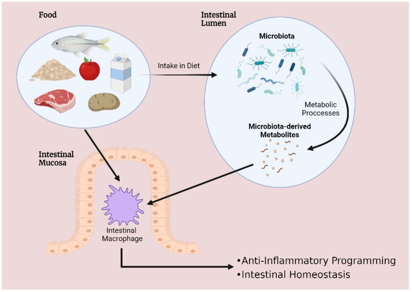
Int. J. Mol. Sci. 2022, 23, 3901                                                                                               3 of 17

                                   actions are being considered as interventions to complement targeted therapies for IBD.
                                   Moreover, given that evidence points toward macrophages constituting major players in
                                   mediating intestinal immunity, studies concerning the interplay amongst microbiota and
                                   macrophages may provide novel insights into factors underpinning IBD pathogenesis. A
                                   deeper understanding of this topic could enlighten us toward the development of novel
                                   medicines for IBD.

                                   Figure 1. Interplay between diet, microbiota and intestinal macrophages encourages gastrointestinal
                                   homeostasis. Disruptions to this delicate balance are implicated in a wide range of disorders—
                                   including inflammatory bowel disease.

                                   2. The Role of Macrophages in the Development and Progression of Intestinal
                                   Inflammation
                                   2.1. Macrophage Plasticity in Inflammatory Bowel Disease
                                        Macrophages are multifaceted, highly dynamic cells that exhibit vast heterogeneity,
                                   reflecting both the tissue microenvironment that they occupy, along with the lineage that
                                   they are derived from [6]. In order to conduct specialised duties, macrophages exhibit a
                                   high degree of plasticity and display specific phenotypes, permitting the macrophage to
                                   adapt to the surrounding microenvironment. Intestinal-resident macrophages for instance,
                                   reside in the lamina propria, enabling the macrophage to sample extracellular surroundings
                                   and process antigens that they encounter. These surveillance duties facilitate a myriad of
                                   functions and defence mechanisms. Therefore, interactions of intestinal macrophages with
                                   microbes or foods could be viewed as critical events in shaping macrophage plasticity. For
                                   example, the tissue microenvironment could be expected to modulate cellular plasticity by
                                   virtue of a combination of a diverse cytokine milieu and environmental cues. This notion
                                   has been highlighted in murine studies, where pro-inflammatory macrophages that display
                                   high expression of Ly6C (LyC6hi ) are reported to undergo in situ functional switches to
                                   pro-restorative LyC6low macrophages in response to the surrounding environment [17].
                                        Broadly speaking, in vitro macrophage plasticity is commonly classified according to
                                   the broad categorisation of being inflammatory M1 macrophages or the anti-inflammatory
                                   M2 macrophages. While transitions between macrophage phenotypes have been well-
                                   defined in vitro, labelling of macrophage populations is convoluted in vivo due to the
                                   complexity of macrophage heterogenicity. Specifically, evidence suggests that macrophage
                                   plasticity exists as part of a spectrum, where M1 and M2 represent polar ends [12]. M1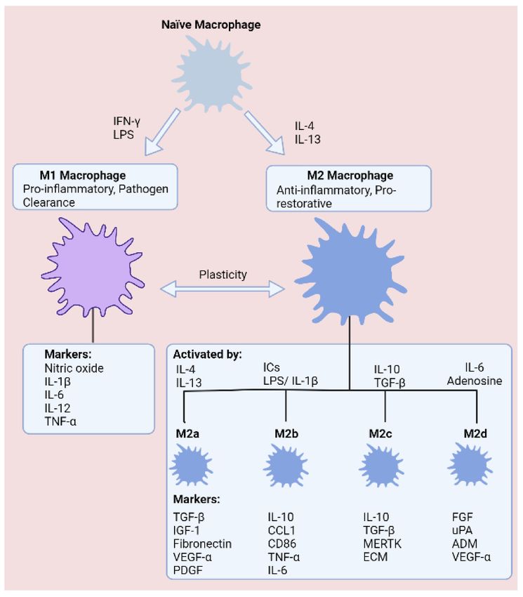
Int. J. Mol. Sci. 2022, 23, 3901                                                                                           4 of 17

                                   macrophages exhibit several distinguishing features that help underpin a pro-inflammatory
                                   phenotype. First of all, pro-inflammatory M1 macrophages are induced through stimulation
                                   with lipopolysaccharide (LPS) and interferon gamma (IFN-γ), leading to secretion of
                                   tumour necrosis factor alpha (TNF-α), interleukin 1 beta (IL-1β), interleukin 6 (IL-6), and
                                   interleukin (IL-12) to encourage microbicidal activity and phagocytosis [3]. These robust
                                   immune responses are energy demanding, and in recent years, it has become evident that
                                   macrophages undergo metabolic reprogramming in response to infection, which dictates
                                   the ability of the macrophage to conduct rapid responses to microbial invasion [18].
                                         Given the necessity for almost-instantaneous responses to prevent microbial dis-
                                   semination within the body, M1 macrophages are understood to upregulate glycolysis
                                   during polarisation as a means of supporting vigorous immune responses and structural
                                   changes. In respect of recruiting immune cell populations and enhancing microbial killing,
                                   M1 macrophages accumulate succinate during pro-inflammatory conditions, which sta-
                                   bilises HIF-1α and promotes secretion of IL-1β [19]. At the other end of the spectrum,
                                   M2 macrophages are instrumental in suppressing immune responses, minimising tissue
                                   damage, and resolving tissue healing following injury. In an alternative manner to M1
                                   macrophages, the anti-inflammatory programming of M2 macrophages is fuelled through
                                   oxidative phosphorylation (OXPHOS) and fatty acid oxidation (FAO). OXPHOS is preferred
                                   on account of the abundance of ATP molecules produced relative to glycolysis, providing
                                   additional energy for tissue resolution. Understandably, FAO is another crucial metabolic
                                   pathway, as FAO generates acetyl-coA, thus helping to ensure maximum ATP generation
                                   from the tricarboxylic acid (TCA) cycle and OXPHOS. M2 macrophages have been further
                                   branched into the M2a, M2b, M2c, and M2d subtypes, which are classified according to
                                   cytokine secretion, expression of cell surface markers, and transcriptome and biological
                                   activities (Figure 2).
                                         While M1/M2 nomenclature is a simplification, the fact of the matter is that equi-
                                   librium between these opposing phenotypes is critical for intestinal tissue homeostasis.
                                   Therefore, it is of high value that inflammatory signalling is subject to stringent regulatory
                                   mechanisms. Disruptions to this status quo may tip the balance toward autoimmunity, or
                                   conversely, may be deleterious by promoting immunodeficiency and an inability to clear
                                   harmful microbes.

                                   2.2. The cGAS-STING Signalling Pathway: An Emerging Regulator of Macrophage Plasticity
                                        A driving force behind perpetuated inflammation and tissue injury in IBD involves
                                   an intricate link between the innate and adaptive immune systems—namely, recruitment
                                   of T lymphocytes to the site of injury through the release of chemokines by macrophages.
                                   Therefore, pathways involved in mediating these responses are of high importance to un-
                                   ravel. Activation of the cyclic GMP-AMP synthase (cGAS)—stimulator of interferon genes
                                   (STING) signalling pathway, which promotes macrophage cross-priming—is reported to
                                   be crucial for bridging this link [20].
                                        cGAS is a cytosolic dsDNA sensor that detects intracellular DNA derived from
                                   pathogens or damaged host cells. Upon recognising dsDNA, cGAS generates the sec-
                                   ondary messenger 20 30 -cGAMP from ATP and GTP, which in turn activates the endoplasmic
                                   reticulum-localised STING protein. These interactions initiate a downstream signalling
                                   cascade culminating in activation of transcription factors nuclear factor kappa B (NF-kB)
                                   and interferon regulatory factor 3 (IRF3), stimulating the release of interferon-beta (IFN-β)
                                   and pro-inflammatory cytokines. Activation of the cGAS-STING axis in macrophages is an
                                   important initial step in the recruitment and activation of T lymphocytes, setting in motion
                                   clonal expansion of these cell populations. Later, these T lymphocytes become embodied
                                   as indispensable arms of the adaptive immune system. Indeed, heightened activation of
                                   macrophage cGAS-STING signalling has previously been associated with a high-fat diet,
                                   demonstrating an intricate link between diet and systemic inflammation. Moreover, a func-
                                   tional role of the cGAS-STING signalling axis extends to intestinal epithelial cells, where
                                   the pathway mediates protective effects by promoting intestinal barrier integrity [21,22].
Int. J. Mol. Sci. 2022, 23, 3901 5 of 17
Figure 2. States of macrophage polarisation. Macrophages are commonly defined as classically acti-
vated (LPS and IFN-γ) M1 macrophages or alternatively activated (IL-4 and IL-13) M2 macrophages,
which comprise four subgroups (M2a–M2d). Macrophages exhibit high plasticity, enabling transitions
between phenotypes according to the surrounding tissue microenvironment.
Interestingly, recent studies have highlighted a potentially novel role for cGAS-STING
in modulating macrophage plasticity (Figure 3). Specifically, in a DSS-induced model of
murine colitis, elevated expression of STING protein was documented, leading to height-
ened sensitivity to the STING agonist: cyclic dinucleotide (CDN) [23]. As expected, the
authors demonstrated that STING expression did indeed worsen colitis, whilst directing
naïve and M2 macrophages in the direction of an M1 phenotype. Moreover, reactive
oxygen species (ROS)-induced oxidation of DNA confers resistance to degradation and
enhances activation of cGAS [24]. Despite previous studies denoting that cGAS-STING
signalling may be detrimental in inflammatory disease, little is known regarding the extent
to which the pathway controls macrophage plasticity in IBD. Indeed, it is plausible that
dsDNA derived from host cells or microbiota may potentiate activation of cGAS-STING
signalling in IBD. Moreover, whether excessive pathway activation promotes abnormal T
lymphocyte migration to the intestinal microenvironment has not been studied. Nonethe-
less, pioneering discoveries in recent years emphasise the multi-functional basis of this
DNA sensor. Importantly, these findings reveal another potential means of modifying
macrophage plasticity in the gastrointestinal tract and encouraging homeostasis in the
intestinal microenvironment.Int. J. Mol. Sci. 2022, 23, 3901 6 of 17
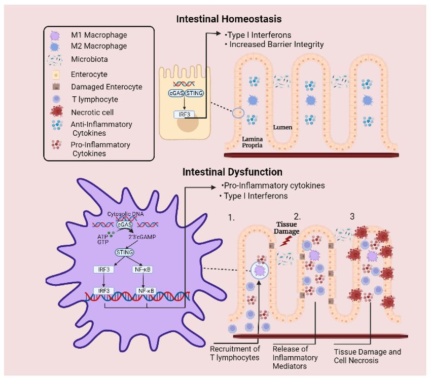
Figure 3. Involvement of cGAS-STING signalling pathway in intestinal inflammation. Under normal
circumstances, cytosolic DNA is sensed by cGAS-STING signalling axis, which is involved in promot-
ing intestinal barrier integrity. Meanwhile, excessive inflammation is prevented by cytosolic DNAse
enzymes that degrade free DNA. However, under inflammatory conditions, the regulatory capacity of
these enzymes is overcome when a DNA threshold is reached, leading to abnormal levels of cytosolic
DNA. Upregulated expression of STING protein and elevated pathway activation culminates in
sustained release of pro-inflammatory cytokines and type I interferons. (1) This mechanism activates
the adaptive arm of the immune system and leads to the recruitment of T lymphocytes to the site of
injury. (2) Perpetual release of inflammatory mediators by T lymphocytes leads to aggregations of
immune cells and extensive release of inflammatory mediators. (3) Surrounding epithelial cells are
damaged by the sustained inflammatory response, resulting in cell necrosis and tissue destruction.
3. Dietary Approaches for Targeting Macrophage Plasticity
While several studies have detailed associations between microbiota imbalances and
IBD [25], others have defined the effects that microbiota-derived metabolites exert on im-
mune cells. The microbiota generates an abundance of metabolites, and many of these
enter systemic circulation, where they mediate modulatory effects upon reaching target
tissues [26]. A better understanding of how these metabolites influence gut homeosta-
sis could enlighten us toward ‘immunonutrition’ approaches, which encompass dietary
interventions for circumventing excessive inflammation in IBD. Various foods are in the
spotlight as candidates for immunonutrition by virtue of favourable interactions with the
microbiota. Because of this important research, several metabolites have been highlighted
that modify immune cell function, leading to suppression of intestinal inflammation and
enhanced tissue healing. Notably, foods that give rise to high levels of short chain fatty
acids (SCFAs), indole-compounds, and omega-3 fatty acids (ω3FAs) have been underlinedInt. J. Mol. Sci. 2022, 23, 3901 7 of 17
as possessing immunomodulatory capacity. Whether these effects can be harnessed to
manipulate macrophage plasticity, and thus control the inflammatory state of the cells, is a
highly important question in medical research. Essentially, the microbiota could be viewed
as a critical connection between food intake and downstream modulation of immune cell
function. Accordingly, targeting the microbiota to regulate metabolite production could
serve as a novel platform for the development of adjuvants for IBD therapies. In the
following section we have highlighted three dietary-derived metabolites: SCFAs, indole-
compounds, and ω3FA, which display distinct therapeutic promise as adjuvant therapies
for treating IBD.
3.1. Short-Chain Fatty Acids
Co-evolution has given rise to the emergence of symbiotic relationships between the
host and commensals. The microbiota in the gut are chief examples that acquire energy
from the food that passes along the intestinal track of the host. Microbiota reciprocate
to this gesture by promoting intestinal barrier homeostasis, dampening inflammation,
strengthening the epithelial barrier, and preventing colonisation by pathogens [27]. A
predominant mechanism of conducting protective duties is via production of SCFAs,
which are fatty acids containing fewer than six carbon atoms, the most common in the
mammalian gut being acetate (C2), propionate (C3), and butyrate (C4) [28]. Intriguingly,
SCFAs ameliorate disease activity in IBD and promote healing of the colon [29,30].
In the mammalian gut, microbiota generate SCFAs through fermentation of indi-
gestible fibres. Accordingly, several high-fibre foods have been reported to induce favourable
outcomes for reducing risk of and disease state of IBD. Remarkably, fibre exhibits several
clinical benefits in IBD, such as prolonging remission and reducing lesions in the intestinal
mucosa [31]. Imbalances in consumption of fruit and vegetables, both abundant in fibre,
were reported to be a risk factor for the emergence of IBD [32,33]. Oats are another rich
source of dietary fibre and have been shown to prevent deterioration of gastrointestinal
symptoms in UC [34]. Likewise, high-fibre legumes, such as navy and black beans, are ca-
pable of attenuating intestinal inflammation in murine models of IBD [35]. Taken together,
it is evident that intake of high-fibre foods is advantageous for maintaining intestinal health.
Indeed, interplay between fibre and microbiota, leading to generation of SCFAs, is a critical
event in this process.
SCFA concentration varies along the gastrointestinal tract, with mean concentrations
reaching 13 mmol/kg in the terminal ileum, 80 mmol/kg in the terminal colon, and
131 mmol/kg in the caecum [36]. This disparity in SCFA concentrations is likely dependent
on the diversity of microbial populations along different portions of the tract. Butyrate is
primarily produced by the microbial phylum Firmicutes and shows highest concentration
in the colon and caecum [37]. In particular, the bacteria Faecalibacterium prausnitzii and
Eubacterium rectale, from the families Ruminococcaceae and Lachnospiraceae, respectively, are
exceptionally robust producers of butyrate [38]. Alternatively, propionate and acetate are
predominantly generated by Bacteroidetes in the small and large intestines [37]. Considering
that production of SCFAs is predominantly conducted by certain bacterial populations,
it no surprise that altered microbiota composition is commonly seen in individuals with
IBD [39]. SCFA-producing Butyricicoccus, for example, are depleted in patients with active
IBD [40].
Over the past decades, it has been well established that foods that generate high levels
of SCFAs are protective against IBD. Indeed, several important pieces of evidence now
support the hypothesis that these effects are at least partly resulting from the effects of
SCFA on immune cells. Of particular significance are studies proposing that favourable
outcomes are largely stemming from modulation of intestinal macrophages. A deeper
understanding of these interactions, especially regarding the mechanisms that SCFAs can
reprogram macrophages toward anti-inflammatory behaviour, would be an invaluable tool
for IBD therapy.Int. J. Mol. Sci. 2022, 23, 3901 8 of 17
Following production by the microbiota, SCFAs are available to interact with macrophages
stationed along the intestinal mucosa. Luminal SCFAs are taken up by the cell through various
means, including passive diffusion and carrier-mediated transportation or otherwise through
binding to G-protein coupled receptors (GPR41, GPR43, and GPR109a) [41]. Following uptake
by the cell, SCFAs mediate immunomodulatory changes to immune cells through several
mechanisms [42–45]. The most studied SCFA—butyrate—exerts anti-inflammatory effects in
IBD through inhibition of NF-kB activation, suppressing release of pro-inflammatory cytokines
(Figure 4) [46]. In accordance, acetate and propionate exhibit comparable inhibitory properties
with butyrate at suppressing NF-κB activation [47]. Indeed, while SCFAs also activate GPCR
signalling, immunomodulatory effects are mediated independently of these receptors [42].
Figure 4. Mechanisms that dietary metabolites modulate cells of the gastrointestinal tract. Fibre
and tryptophan are metabolised by the microbiota to release short-chain fatty acids and indole
compounds into the gut lumen. (1). Short-chain fatty acids enhance PPAR-γ activation and reduce
IFN-γ production of intestinal epithelial cells, while inducing immunomodulatory effects on intestinal
macrophages. (2). Indole suppresses activation of NF-kB and stimulates production of IL-22 by group
3 innate lymphoid cells, promoting epithelial barrier homeostasis. (3) Omega-3 polyunsaturated fats
promote an anti-inflammatory macrophage phenotype through activation of PPAR-α and PPAR-γ,
along with inhibition of NF-κB, culminating as decreased production of pro-inflammatory cytokines.
Anti-inflammatory effects mediated by butyrate have encouraged various laboratories
to delineate molecular interactions underpinning these effects. Traditionally, inhibition of
histone deacetylases (HDAC) has been believed to underlie the anti-inflammatory signature
of butyrate [48]. Investigations by Chang et al., for example, determined that butyrate
acts as an inhibitor of HDAC, which is an enzyme that regulates gene transcription by
removing acetyl groups from histones. In the study, butyrate downregulated NO, IL-6,
and IL-12 in intestinal macrophages, likely through enhanced histone acetylation [42].
These findings build on prior data showing that butyrate attenuates LPS-induced secretion
of TNF-α, IL-1β, IL-6, and NO, whilst prompting the release of anti-inflammatory IL-10
in RAW 264.7 monocytes [45]. On the basis that butyrate is an HDAC inhibitor, studies
have speculated that the SCFA induces epigenetic reprogramming [43]. In line with this
concept, bone marrow-derived macrophage (BMDM) polarisation toward an M2 phenotype
was induced by butyrate through acetylation of H3K9, which showed to be a histone
modification that enhances STAT6 activation [49]. The question remains whether butyrate
can modulate macrophage plasticity. Production of ROS, which are key regulators ofInt. J. Mol. Sci. 2022, 23, 3901 9 of 17
an M1 phenotype, can be attenuated in neutrophils following butyrate treatment [50,51].
Therefore, it is plausible that butyrate exerts similar effects on macrophages. Collectively,
these observations represent a molecular basis to explain why a high-fibre diet has a
protective effect against intestinal disorders [31].
Ultimately, although SCFAs appear to be central in mitigating against intestinal injury,
we have still not mapped the entire signalling landscape underpinning these effects. While
evidence points toward butyrate activating G-protein coupled receptors (GPR41, GPR43,
and GP109a) and regulating several immune and inflammatory processes [41], whether
these receptors are involved in ameliorating inflammation in IBD has yet to be elucidated.
A greater knowledge of these interactions could inform us of more appropriate targets
for the treatment of IBD in the future. Moreover, while transcriptions factors are central
in modulating macrophage plasticity [52], the true nature of interplay amongst SCFAs
and transcription factors, and the ensuing impact on macrophage function, have not yet
been clarified.
3.2. Tryptophan-Derived Metabolites
Tryptophan is an essential amino acid (humans cannot produce tryptophan endoge-
nously), meaning instead that tryptophan must be supplemented in the diet. Many foods
are good sources of tryptophan, including oats, milk, cheese, tuna fish, chicken, and
turkey [53]. Amongst these tryptophan-rich foods, oats, lean meats, milk, and cheese are
all associated with ameliorated intestinal inflammation in IBD [54].
Indeed, consumption of tryptophan in the diet is associated with intestinal homeosta-
sis. Serum tryptophan levels, for example, are lower in individuals with IBD, denoting that
tryptophan deficiency or degradation may exacerbate gut dysbiosis [55]. This notion ex-
tends to animal models of colitis, such as in mice, where mice deficient in tryptophan were
recorded to display exacerbated colitis [56] (p. 2). From a medical standpoint, it is apparent
that dietary tryptophan supplementation could represent a means of attenuating intestinal
inflammation. This is supported by animal models, where dietary supplementation of
tryptophan reduced the severity of DSS-induced colitis in pigs [57].
Again, intestinal microbiota play a fundamental role in the beneficial outcomes of
dietary tryptophan. In the body, tryptophan is subject to biosynthetic manipulation by the
host and microbes to generate various metabolites [58]. Specifically, ingested tryptophan is
metabolised in the intestines by three distinct pathways; the first two of these pathways—
the serotonin and kynurenine pathways—are conducted by host enzymes, whilst the third
pathway, which produces the immunomodulatory indole compounds, is performed by
microbiota [59].
A broad-spectrum of indole compounds is generated by the intestinal flora—the struc-
ture of each metabolite differing according to the biochemical transformations they are
subjected to. Clostridium sporogenes, from the phylum Firmicutes for example, generates
indole-propionic acid from tryptophan through induction of the enzyme phenyllactate de-
hydratase [60]. Moreover, Lactobacilli reuteri generates indole-3-aldehyde from tryptophan—
a reaction catalysed by an aromatic amino acid aminotransferase [61]. Alternatively, indole
may also be sourced directly from the diet from produce such as broccoli, which similarly
has been shown to attenuate murine colitis [62].
Encouragingly, indole compounds have also been reported to promote mucosal home-
ostasis in the intestines [63]. The mechanistic basis for indole alleviating intestinal inflam-
mation can be partly attributed to interactions with the Aryl hydrocarbon receptor (AHR),
which various indole compounds display affinity for [64]. The AHR is a transcription
factor that upon activation is responsible for several anti-inflammatory mechanisms, in-
cluding regulating intestinal homeostasis. In macrophages, AHR dampens LPS-induced
inflammation—as mice lacking macrophage-specific expression of AHR are more sus-
ceptible to LPS-induced septic shock [65]. Given that macrophages propel inflammatory
signalling in IBD, whether interactions between indole and macrophages can modulate
cellular plasticity is an avenue of interest.Int. J. Mol. Sci. 2022, 23, 3901 10 of 17
Interestingly, indole compounds have previously been shown to suppress macrophage
inflammatory signalling in the context of treating liver disease. Notably, Krishnan et al. re-
ported that pre-treating macrophages with microbial-derived tryptophan catabolite indole-
3 acetate (I3A) attenuates production of TNF-α, IL-1β, and monocyte-chemoattractant
protein-1 (MCP-1) in response to LPS and palmitic acid [66]. Similarly, Zhao et al. detailed
that pre-treating murine J774A. 1 macrophages with indole-3-propionic acid lessened secre-
tion of IL-1β, TNF-α, and IL-6 via repression of NF-kB [67]. Taken together, these results
suggest that indole compounds act to promote a more tolerogenic macrophage phenotype
by decreasing responsiveness to inflammatory stimuli. Therefore, it is not beyond the
realm of possibility that similar outcomes may arise to attenuate inflammatory signalling
in the intestines.
Several bodies of evidence give the impression that indole–AHR interactions may be
crucial for maintaining intestinal mucosal immunity. Firstly, AHR activation negatively
regulates production of pro-inflammatory TNF-α and IL-6 [68]. Next, expression of AHR
has been documented to be diminished in patients with IBD [69]. Furthermore, indole com-
pounds have been shown to act as agonists of AHR [64] Collectively, these studies illustrate
that a microbiota-targeted approach aiming to optimise binding of indole compounds to
AHR may be an attractive target for future IBD therapies.
More importantly, this work has evinced the potential of tryptophan metabolites as
another addition to the armoury for modulating disease course in IBD. Considering the
diverse signalling landscape in IBD, these metabolites could be highly beneficial by virtue
of an ability to target immune cell populations and induce downstream modulation of
intestinal epithelial cell function. Although it is apparent that indole compounds mediate
immune altering effects, the question remains whether these findings can be translated to a
meaningful therapy for IBD. For this to occur, we firstly need to identify means to prolong
bioavailability of indole compounds in the body, such as by increasing production, or by
reducing consumption by the microbiota. Accordingly, future studies should validate the
predominant tryptophan-metabolising microbiota, along with identifying the most potent
indole compounds. Importantly, further studies should be attentive to whether indole
compounds can alter tissue-resident macrophage plasticity, which has not been studied.
3.3. Omega-3 Polyunsaturated Fatty Acids
Dietary fats are generally viewed as negative elements of the Western diet—central
to the pathogenesis of a wide range of diseases. However, in recent years, studies are
beginning to portray fatty acids in a more favourable light, as the beneficial impact of certain
fatty acids on intestinal health become apparent. The ω3FAs, such as eicosapentaenoic
(EPA), docosahexaenoic acid (DHA), and α-linolenic acid, are abundant in oily fish, nuts,
seeds, and plant oils.
Adherence to the Mediterranean diet, which is rich in foods with high levels of ω3FAs
(seafood, seeds, nuts, and olive oil), has been closely interlinked by epidemiological studies
to lessened risk of many inflammatory diseases, including IBD [70]. Notably, meta-analysis
of twelve studies that measured fish consumption revealed an inverse relationship between
dietary intake of fish and risk of IBD [71]. In a similar manner, dietary supplementa-
tion of extra-virgin olive oil and canola oil, both containing α-linolenic acid, alleviated
gastrointestinal symptoms and reduced inflammatory markers in patients with UC [72].
When ingested, ω3FAs serve as precursors for synthesis of anti-inflammatory medi-
ators such as resolvins, protectins, lipoxins, and moresins, which dampen inflammation
and protect against organ damage [73]. Accordingly, clinical studies have studied these
ω3FAs with the aim of elucidating involvement in intestinal disease. In a study of pa-
tients with UC, Pearl et al. showed lower levels of ω3FAs were present in colonic mucosa
biopsy samples, suggesting dysfunctional fatty acid metabolism may be involved in exac-
erbating disease [74]. Considering the enormous therapeutic potential of harnessing the
immunomodulatory properties of ω3FAs, various laboratories have attempted to charac-
terise the precise mechanisms underpinning their anti-inflammatory signature.Int. J. Mol. Sci. 2022, 23, 3901 11 of 17
Interactions with the microbiota is one such means that ω3FAs engage in anti-inflammatory
activities and facilitate intestinal homeostasis. Studies have uncovered that ω3FAs mediate
positive effects by altering microbiota diversity. Indeed, this effect encompasses both increases
in beneficial bacteria, along with reductions in populations of harmful bacteria. Notably, ω3FA
encourages growth of SCFA-producing microbes, including the Lachnospiraceae family of bacte-
ria [75]. On the other hand, ω3FA supplementation in infants led to lessened abundance of
pathogenic microbes, such as Enterobacteriaceae [76].
The anti-inflammatory profile of dietary ω3FA may also be altered by biosynthetic
manipulation by the microbiota. For example, α-linolenic acid is metabolised by Lacto-
bacillus plantarum AKU 1009a to generate the bioactive metabolite 10-oxo-cis-12-cis-15-
octadecadienoic acid (α-Keto acid) [77]. Interestingly, pioneering research has shown that
anti-inflammatory properties are retained by α-Keto acid [78]. The authors revealed that
α-Keto acid was capable of inducing M2 macrophage polarisation in mice administered a
high-fat diet.
Omega-3 fatty acids are also postulated to modulate macrophage function through in-
teractions with the peroxisome proliferator-activated receptors (PPARs), including PPAR-α,
PPAR-β/δ, and PPAR-γ. PPARs are a family of transcription factors involved in cellular
differentiation and metabolism [79]. PPARα interacts with both EPA and DHA, leading to
modulation of genes central to glucose and lipid homeostasis [80]. Activation of PPAR-α
promotes FAO, one of the preferred metabolic pathways for shaping M2 macrophage
responses [80]. The relationship between ω3FAs and PPAR-γ, which is master of lipid
homeostasis, is also of heightened interest, as ligation of PPAR-γ skews macrophages
toward an M2 phenotype and inhibits NF-kB [81]. Consequently, pro-inflammatory media-
tors are reduced, characterised by suppression of prostaglandin E2 (PGE2), TNF-α, IL-6,
and IL-1β release [82,83].
Several studies have focused on the modifying effects ω3FAs exert on macrophages,
which sense and respond to other dietary components. It appears that these responses
extend to ω3FAs, as macrophages cultured in the presence of EPA exhibited lessened
production of TNFα and IL-6 induced by LPS challenge, whilst displaying upregulated
secretion of IL-10 [84]. Moreover, in a study involving krill oil, which contains high lev-
els of EPA and DHA, application of krill oil attenuated LPS-induced secretion of TNF-α,
IL-6, and IL-1β through regulation of NF-kB and nucleotide-binding oligomerization do-
main (NOD)-like receptor signalling [85]. While LPS downregulated PPAR-γ, a marker
of M2 polarisation, this was reversed by krill oil treatment, which is favourable for direct-
ing macrophage plasticity toward an M2 phenotype. However, despite ω3FA inducing
macrophage responses consistent with anti-inflammatory reprogramming, neither study
carried out macrophage phenotyping or metabolomics following ω3FA treatment, which
could be the basis of further studies.
Other studies conducted investigations using in vivo models of inflammation, with
rodent and porcine models generally being the preferred approaches. In a DSS-induced
model of colitis in rats, the ω3FA α-linoleic acid, found in various seeds and oils, was
capable of abrogating disease markers of intestinal inflammation, as illustrated by down-
regulated expression of IL-1β and TNF-α [86]. Similar conclusions were drawn by Arisue
et al., who showed that fish oil ameliorates intestinal injury and proposed that this may
occur through downregulation of oxidative stress [87]. Furthermore, in a porcine model,
where animals were challenged with LPS, piglets administered a diet supplemented with
fish oil showed reduced intestinal NF-kB protein and TLR4 expression, along with di-
minished TNF-α secretion [88]. Future research could broaden the scope of this study
by mapping gene expression back to individual intestinal cell populations using novel
transcriptomic technologies. Single-cell RNA sequencing could permit us to determine
inflammatory signatures of the intestinal cell repertoire and delineate the effects of ω3FA
on macrophage plasticity.
Collectively, data obtained in these studies highlight that ω3FAs are an encouraging
prospect as an adjuvant for IBD therapy. Nonetheless, interplay between tissue-residentInt. J. Mol. Sci. 2022, 23, 3901 12 of 17
macrophages and ω3FAs remains relatively unstudied. Questions remain regarding
whether these metabolites can modulate responses and transcriptional activity of intestinal
macrophages populations, which warrants further research. Moreover, considering the im-
portance of metabolic programming in governing cellular responses, interactions between
ω3FA and PPAR-γ, which are central in lipid metabolism, are worthy of further research.
4. Future Therapeutic Perspectives
4.1. Novel Therapeutic Approaches
While it is well established that macrophages are crucial drivers of tissue destruction
and fibrinogenesis in IBD, therapies specifically targeted at macrophages are limited. Ideally,
future therapies would be directed to dampen macrophage inflammatory signalling, whilst
halting tissue fibrosis that progressively impairs organ function.
While several studies have documented several microbiota-derived metabolites pos-
sess immunomodulatory properties, little is known regarding the extent to which these
alter macrophage plasticity and metabolism. Targeting macrophage plasticity is a novel
approach that has gained traction in recent years, and there is a strong rationale for deter-
mining whether dietary inventions can control these molecular switches. Encouragingly,
therapies involving dietary supplementation of SCFAs, indole-compounds, or ω3FAs could
provide a safe approach for attenuating intestinal injury.
However, questions remain regarding the clinical efficacy and gut bioavailability of
these molecules. To address these potential limitations, we should consider means of
optimising cellular-specific delivery and limiting degradation along the gastrointestinal
tract. Firstly, development of a framework for identifying patient subtypes that respond
best to each intervention, along with clarifying targeted therapies that metabolites are most
effective in combination with, would be highly beneficial. Together these could serve as a
basis for minimising non-responders to therapy and optimising clinical efficacy. While these
metabolites may not represent an approach for completely halting inflammatory signalling
and intestinal injury, they offer a means of raising the therapeutic ceiling of more established
interventions—a notable example being SCFA supplementation enhancing targeted therapy
against TNF [89]. Moreover, we could benefit greatly from studying molecular interactions
of these molecules with the epigenome and intracellular signalling pathways, as this could
enlighten us toward novel targets for pharmacological interventions.
Another potential benefit of dietary-based approaches for IBD is that there may be
scope for achieving synergistic effects when administering interventions concurrently.
Synergistic effects can be defined as when multiple treatments interact to reach an overall
effect greater than each treatment on an individual level. While previous studies have
primarily focused on individual metabolites, combined dietary-based approaches could
offer a means of targeting multiple anti-inflammatory pathways simultaneously, which
could accentuate treatment efficacy. SCFAs for example, heighten responsiveness of the
AHR in colonocytes [90], indicating that co-application of SCFA and indole compounds
could represent a promising approach for IBD therapy. Further studies are warranted to
establish synergism that may arise from therapies targeting the microbiota.
4.2. Novel Technologies
Specific delivery of these molecules toward macrophages would be crucial for opti-
mising therapeutic efficacy. Recent advances in nanomedicine have led to development
of novel drug delivery systems. Nanoparticles, for example, may provide an approach
for macrophage-specific drug delivery for treatment of inflammatory disease [91]. There-
fore, encapsulation of metabolites within nanoparticles could be a strategy for tailoring
macrophage-specific therapies. Moreover, modifying the microbiome to optimise metabo-
lite bioavailability is another approach that should be considered. In this context, treat-
ments could involve supplemented tryptophan/SCFA/ω3FA metabolites with precise
microbiome modulating interventions.Int. J. Mol. Sci. 2022, 23, 3901 13 of 17
Ultimately, previous findings support the concept of applying dietary-based therapies
for promoting the activation of anti-inflammatory mechanisms in the gut. Interest in
this area has been rapidly growing as we comprehend how far macrophage involvement
extends within the pathogenesis of inflammatory disease. Although this is a relatively
new area of study, the field is gaining great momentum by virtue of the discovery of novel
approaches for studying immune cell interactions in greater detail. Several developments
in the area of mass cytometry and transcriptomics have revolutionised our understanding
of complex cellular heterogenicity. Spatial transcriptomic technologies, for example, have
been employed to uncover tissue-wide heterogenicity in cancer research. Evidently, there
is a large scope for discovery through application of this technology to characterise the
inflammatory signature of macrophages in diseased tissue. Future studies should prioritise
applying these technologies to IBD research, ensuring that such breakthroughs could reveal
a clearer picture of how macrophages propel the dysregulated tissue architecture prevalent
in IBD [92].
5. Conclusions
Collectively, there is compelling evidence for employing microbiota-derived metabo-
lites to target macrophage plasticity for IBD therapy. While the entire repertoire of molecular
signalling pathways that co-ordinate macrophage responses to microbiota-derived metabo-
lites has not yet been determined, we can be assured that our knowledge of this fascinating
interplay is rapidly growing. All things considered, macrophage-specific targeted therapies
are a novel approach that could revolutionise the treatment of inflammatory disease in
the future.
Author Contributions: C.O.: writing of the original draft, reviewing, and editing. A.A. and S.G.:
revised the manuscript. All authors have read and agreed to the published version of the manuscript.
Funding: C.O. is funded by a PhD studentship from University College Cork. A.A. is supported
by an international mobility grant from the Société Française de Nutrition Clinique et Métabolisme
(SFNCM) and support from APC Microbiome. S.G. is funded by the award of an SFI Research
Professorship grant.
Institutional Review Board Statement: Not applicable.
Informed Consent Statement: Not applicable.
Data Availability Statement: Not applicable.
Conflicts of Interest: The authors declare no conflict of interest.
References
1. Alatab, S.; Sepanlou, S.G.; Ikuta, K.; Vahedi, H.; Bisignano, C.; Safiri, S.; Sadeghi, A.; Nixon, M.R.; Abdoli, A.; Abolhassani, H.; et al.
The global, regional, and national burden of inflammatory bowel disease in 195 countries and territories, 1990–2017: A systematic
analysis for the Global Burden of Disease Study 2017. Lancet Gastroenterol. Hepatol. 2020, 5, 17–30. [CrossRef]
2. Viola, M.F.; Boeckxstaens, G. Niche-specific functional heterogeneity of intestinal resident macrophages. Gut 2021, 70, 1383–1395.
[CrossRef] [PubMed]
3. Verdeguer, F.; Aouadi, M. Macrophage heterogeneity and energy metabolism. Exp. Cell Res. 2017, 360, 35–40. [CrossRef]
[PubMed]
4. Yoshida, N.; Frickel, E.-M.; Mostowy, S. Macrophage–microbe interactions: Lessons from the zebrafish model. Front. Immunol.
2017, 8, 1703. [CrossRef] [PubMed]
5. Mahida, Y.R. The key role of macrophages in the immunopathogenesis of inflammatory bowel disease. Inflamm. Bowel Dis. 2000,
6, 21–33. [CrossRef] [PubMed]
6. Italiani, P.; Boraschi, D. Development and functional differentiation of tissue-resident versus monocyte-derived macrophages in
inflammatory reactions. In Macrophages; Springer: Berlin/Heidelberg, Germany, 2017; pp. 23–43.
7. Honold, L.; Nahrendorf, M. Resident and monocyte-derived macrophages in cardiovascular disease. Circ. Res. 2018, 122, 113–127.
[CrossRef] [PubMed]
8. Gomez Perdiguero, E.; Klapproth, K.; Schulz, C.; Busch, K.; Azzoni, E.; Crozet, L.; Garner, H.; Trouillet, C.; De Bruijn, M.F.;
Geissmann, F.; et al. Tissue-resident macrophages originate from yolk-sac-derived erythro-myeloid progenitors. Nature 2015, 518,
547–551. [CrossRef] [PubMed]Int. J. Mol. Sci. 2022, 23, 3901 14 of 17
9. Yona, S.; Kim, K.W.; Wolf, Y.; Mildner, A.; Varol, D.; Breker, M.; Strauss-Ayali, D.; Viukov, S.; Guilliams, M.; Misharin, A.; et al.
Fate mapping reveals origins and dynamics of monocytes and tissue macrophages under homeostasis. Immunity 2013, 38, 79–91.
[CrossRef] [PubMed]
10. Bain, C.C.; Scott, C.L.; Uronen-Hansson, H.; Gudjonsson, S.; Jansson, O.; Grip, O.; Guilliams, M.; Malissen, B.; Agace, W.W.;
Mowat, A. Resident and pro-inflammatory macrophages in the colon represent alternative context-dependent fates of the same
Ly6C hi monocyte precursors. Mucosal Immunol. 2013, 6, 498–510. [CrossRef] [PubMed]
11. Wang, S.; Ye, Q.; Zeng, X.; Qiao, S. Functions of macrophages in the maintenance of intestinal homeostasis. J. Immunol. Res. 2019,
2019, 1512969. [CrossRef] [PubMed]
12. Das, A.; Sinha, M.; Datta, S.; Abas, M.; Chaffee, S.; Sen, C.K.; Roy, S. Monocyte and macrophage plasticity in tissue repair and
regeneration. Am. J. Pathol. 2015, 185, 2596–2606. [CrossRef]
13. Shaw, T.N.; Houston, S.A.; Wemyss, K.; Bridgeman, H.M.; Barbera, T.A.; Zangerle-Murray, T.; Strangward, P.; Ridley, A.J.;
Wang, P.; Tamoutounour, S.; et al. Tissue-resident macrophages in the intestine are long lived and defined by Tim-4 and CD4
expression. J. Exp. Med. 2018, 215, 1507–1518. [CrossRef]
14. Giuffrè, M.; Campigotto, M.; Campisciano, G.; Comar, M.; Crocè, L.S. A story of liver and gut microbes: How does the intestinal
flora affect liver disease? A review of the literature. Am. J. Physiol. Gastrointest. Liver Physiol. 2020, 318, G889–G906. [CrossRef]
15. Han, X.; Ding, S.; Jiang, H.; Liu, G. Roles of macrophages in the development and treatment of gut inflammation. Front. Cell Dev.
Biol. 2021, 9, 385. [CrossRef]
16. Fassarella, M.; Blaak, E.E.; Penders, J.; Nauta, A.; Smidt, H.; Zoetendal, E.G. Gut microbiome stability and resilience: Elucidating
the response to perturbations in order to modulate gut health. Gut 2021, 70, 595–605. [CrossRef]
17. Ramachandran, P.; Pellicoro, A.; Vernon, M.A.; Boulter, L.; Aucott, R.L.; Ali, A.; Hartland, S.N.; Snowdon, V.K.; Cappon, A.;
Gordon-Walker, T.T.; et al. Differential Ly-6C expression identifies the recruited macrophage phenotype, which orchestrates the
regression of murine liver fibrosis. Proc. Natl. Acad. Sci. USA 2012, 109, E3186–E3195. [CrossRef]
18. Galván-Peña, S.; O’Neill, L.A. Metabolic reprograming in macrophage polarization. Front. Immunol. 2014, 5, 420.
19. Tannahill, G.M.; Curtis, A.M.; Adamik, J.; Palsson-McDermott, E.M.; McGettrick, A.F.; Goel, G.; Frezza, C.; Bernard, N.J.; Kelly, B.;
Foley, N.H.; et al. Succinate is an inflammatory signal that induces IL-1β through HIF-1α. Nature 2013, 96, 238–242. [CrossRef]
20. Ou, L.; Zhang, A.; Cheng, Y.; Chen, Y. The cGAS-STING pathway: A promising immunotherapy target. Front. Immunol. 2021,
12, 795048. [CrossRef]
21. Fischer, J.C.; Bscheider, M.; Eisenkolb, G.; Lin, C.C.; Wintges, A.; Otten, V.; Lindemans, C.A.; Heidegger, S.; Rudelius, M.;
Monette, S.; et al. RIG-I/MAVS and STING signaling promote gut integrity during irradiation-and immune-mediated tissue
injury. Sci. Transl. Med. 2017, 9, eaag2513. [CrossRef]
22. Hu, S.; Fang, Y.; Chen, X.; Cheng, T.; Zhao, M.; Du, M.; Li, T.; Li, M.; Zeng, Z.; Wei, Y.; et al. cGAS restricts colon cancer
development by protecting intestinal barrier integrity. Proc. Natl. Acad. Sci. USA 2021, 118, e2105747118. [CrossRef]
23. Martin, G.R.; Blomquist, C.M.; Henare, K.L.; Jirik, F.R. Stimulator of interferon genes (STING) activation exacerbates experimental
colitis in mice. Sci. Rep. 2019, 9, 14281. [CrossRef]
24. Gehrke, N.; Mertens, C.; Zillinger, T.; Wenzel, J.; Bald, T.; Zahn, S.; Tüting, T.; Hartmann, G.; Barchet, W. Oxidative damage of
DNA confers resistance to cytosolic nuclease TREX1 degradation and potentiates STING-dependent immune sensing. Immunity
2013, 39, 482–495. [CrossRef] [PubMed]
25. Alam, M.T.; Amos, G.C.; Murphy, A.R.; Murch, S.; Wellington, E.M.; Arasaradnam, R.P. Microbial imbalance in inflammatory
bowel disease patients at different taxonomic levels. Gut Pathog. 2020, 12, 1. [CrossRef] [PubMed]
26. Dvořák, Z.; Poulíková, K.; Mani, S. Indole scaffolds as a promising class of the aryl hydrocarbon receptor ligands. Eur. J. Med.
Chem. 2021, 215, 113231. [CrossRef]
27. Hiippala, K.; Jouhten, H.; Ronkainen, A.; Hartikainen, A.; Kainulainen, V.; Jalanka, J.; Satokari, R. The potential of gut commensals
in reinforcing intestinal barrier function and alleviating inflammation. Nutrients 2018, 10, 988. [CrossRef] [PubMed]
28. Ríos-Covián, D.; Ruas-Madiedo, P.; Margolles, A.; Gueimonde, M.; de los Reyes-gavilán, C.G.; Salazar, N. Intestinal short chain
fatty acids and their link with diet and human health. Front. Microbiol. 2016, 7, 185. [CrossRef] [PubMed]
29. Lührs, H.; Gerke, T.; Müller, J.G.; Melcher, R.; Schauber, J.; Boxberger, F.; Scheppach, W.; Menzel, T. Butyrate inhibits NF-κB
activation in lamina propria macrophages of patients with ulcerative colitis. Scand. J. Gastroenterol. 2002, 37, 458–466. [CrossRef]
30. Van der Beek, C.M.; Dejong, C.H.; Troost, F.J.; Masclee, A.A.; Lenaerts, K. Role of short-chain fatty acids in colonic inflammation,
carcinogenesis, and mucosal protection and healing. Nutr. Rev. 2017, 5, 286–305. [CrossRef] [PubMed]
31. Pituch-Zdanowska, A.; Banaszkiewicz, A.; Albrecht, P. The role of dietary fibre in inflammatory bowel disease. Przeglad
Gastroenterologiczny 2015, 10, 135. [CrossRef]
32. Gilat, T.; Hacohen, D.; Lilos, P.; Langman, M. Childhood factors in ulcerative colitis and Crohn’s disease: An international
cooperative study. Scand. J. Gastroenterol. 1987, 22, 1009–1024. [CrossRef]
33. Amre, D.K.; D’souza, S.; Morgan, K.; Seidman, G.; Lambrette, P.; Grimard, G.; Israel, D.; Mack, D.; Ghadirian, P.;
Deslandres, C.; et al. Imbalances in dietary consumption of fatty acids, vegetables, and fruits are associated with risk
for Crohn’s disease in children. Off. J. Am. Coll. Gastroenterol. ACG 2007, 102, 2016–2025. [CrossRef]
34. Nyman, M.; Nguyen, T.D.; Wikman, O.; Hjortswang, H.; Hallert, C. Oat bran increased fecal butyrate and prevented gastroin-
testinal symptoms in patients with quiescent ulcerative colitis—Randomized controlled trial. Crohns Colitis 2020, 2, otaa005.
[CrossRef]Int. J. Mol. Sci. 2022, 23, 3901 15 of 17
35. Zhang, C.; Monk, J.M.; Lu, J.T.; Zarepoor, L.; Wu, W.; Liu, R.; Pauls, K.P.; Wood, G.A.; Robinson, L.; Tsao, R.; et al. Cooked navy
and black bean diets improve biomarkers of colon health and reduce inflammation during colitis. Br. J. Nutr. 2014, 111, 1549–1563.
[CrossRef]
36. Cummings, J.; Pomare, E.; Branch, W.; Naylor, C.; MacFarlane, G. ‘Short chain fatty acids in human large intestine, portal, hepatic
and venous blood. Gut 1987, 28, 1221–1227. [CrossRef]
37. Russo, E.; Giudici, F.; Fiorindi, C.; Ficari, F.; Scaringi, S.; Amedei, A. Immunomodulating activity and therapeutic effects of short
chain fatty acids and tryptophan post-biotics in inflammatory bowel disease. Front. Immunol. 2019, 10, 2754. [CrossRef]
38. Louis, P.; Flint, H.J. Diversity, metabolism and microbial ecology of butyrate-producing bacteria from the human large intestine.
FEMS Microbiol. Lett. 2009, 294, 1–8. [CrossRef]
39. Loh, G.; Blaut, M. Role of commensal gut bacteria in inflammatory bowel diseases. Gut Microbes 2012, 3, 544–555. [CrossRef]
40. Eeckhaut, V.; Machiels, K.; Perrier, C.; Romero, C.; Maes, S.; Flahou, B.; Steppe, M.; Haesebrouck, F.; Sas, B.; Ducatelle, R.; et al.
Butyricicoccus pullicaecorum in inflammatory bowel disease. Gut 2013, 62, 1745–1752. [CrossRef]
41. Sun, M.; Wu, W.; Liu, Z.; Cong, Y. Microbiota metabolite short chain fatty acids, GPCR, and inflammatory bowel diseases.
J. Gastroenterol. 2017, 52, 1–8. [CrossRef]
42. Chang, P.V.; Hao, L.; Offermanns, S.; Medzhitov, R. The microbial metabolite butyrate regulates intestinal macrophage function
via histone deacetylase inhibition. Proc. Natl. Acad. Sci. USA 2014, 111, 2247–2252. [CrossRef] [PubMed]
43. Flemming, A. Butyrate boosts microbicidal macrophages. Nat. Rev. Immunol. 2019, 19, 135. [CrossRef] [PubMed]
44. Ji, J.; Shu, D.; Zheng, M.; Wang, J.; Luo, C.; Wang, Y.; Guo, F.; Zou, X.; Lv, X.; Li, Y.; et al. Microbial metabolite butyrate facilitates
M2 macrophage polarization and function. Sci. Rep. 2016, 6, 24838. [CrossRef] [PubMed]
45. Liu, T.; Li, J.; Liu, Y.; Xiao, N.; Suo, H.; Xie, K.; Yang, C.; Wu, C. Short-chain fatty acids suppress lipopolysaccharide-induced
production of nitric oxide and proinflammatory cytokines through inhibition of NF-κB pathway in RAW264. 7 cells. Inflammation
2012, 35, 1676–1684. [CrossRef]
46. Segain, J.P.; De La Blétiere, D.R.; Bourreille, A.; Leray, V.; Gervois, N.; Rosales, C.; Ferrier, L.; Bonnet, C.; Blottiere, H.M.;
Galmiche, J.P. Butyrate inhibits inflammatory responses through NFκB inhibition: Implications for Crohn’s disease. Gut 2000, 47,
397–403. [CrossRef]
47. Tedelind, S.; Westberg, F.; Kjerrulf, M.; Vidal, A. Anti-inflammatory properties of the short-chain fatty acids acetate and propionate:
A study with relevance to inflammatory bowel disease. World J. Gastroenterol. WJG 2007, 13, 2826. [CrossRef]
48. Candido, E.P.M.; Reeves, R.; Davie, J.R. Sodium butyrate inhibits histone deacetylation in cultured cells. Cell 1978, 14, 105–113.
[CrossRef]
49. Gujral, P.; Mahajan, V.; Lissaman, A.C.; Ponnampalam, A.P. Histone acetylation and the role of histone deacetylases in normal
cyclic endometrium. Reprod. Biol. Endocrinol. 2020, 18, 84. [CrossRef]
50. Li, G.; Lin, J.; Zhang, C.; Gao, H.; Lu, H.; Gao, X.; Zhu, R.; Li, Z.; Li, M.; Liu, Z. Microbiota metabolite butyrate constrains
neutrophil functions and ameliorates mucosal inflammation in inflammatory bowel disease. Gut Microbes 2021, 3, 1968257.
[CrossRef]
51. Rendra, E.; Riabov, V.; Mossel, D.M.; Sevastyanova, T.; Harmsen, M.C.; Kzhyshkowska, J. Reactive oxygen species (ROS) in
macrophage activation and function in diabetes. Immunobiology 2019, 224, 242–253. [CrossRef]
52. Tugal, D.; Liao, X.; Jain, M.K. Transcriptional control of macrophage polarization. Arterioscler. Thromb. Vasc. Biol. 2013, 33,
1135–1144. [CrossRef]
53. Richard, D.M.; Dawes, M.A.; Mathias, C.W.; Acheson, A.; Hill-Kapturczak, N.; Dougherty, D.M. L-tryptophan: Basic metabolic
functions, behavioral research and therapeutic indications. Int. J. Tryptophan Res. 2009, 2, IJTR-S2129. [CrossRef]
54. Campmans-Kuijpers, M.J.; Dijkstra, G. Food and food groups in inflammatory bowel disease (Ibd): The design of the groningen
anti-inflammatory diet (graid). Nutrients 2021, 13, 1067. [CrossRef]
55. Nikolaus, S.; Schulte, B.; Al-Massad, N.; Thieme, F.; Schulte, D.M.; Bethge, J.; Rehman, A.; Tran, F.; Aden, K.; Häsler, R.; et al.
Increased tryptophan metabolism is associated with activity of inflammatory bowel diseases. Gastroenterology 2017, 153, 1504–1516.
[CrossRef]
56. Hashimoto, T.; Perlot, T.; Rehman, A.; Trichereau, J.; Ishiguro, H.; Paolino, M.; Sigl, V.; Hanada, T.; Hanada, R.; Lipinski, S.; et al.
ACE2 links amino acid malnutrition to microbial ecology and intestinal inflammation. Nature 2012, 487, 477–481. [CrossRef]
57. Kim, C.J.; Kovacs-Nolan, J.A.; Yang, C.; Archbold, T.; Fan, M.Z.; Mine, Y. l-Tryptophan exhibits therapeutic function in a porcine
model of dextran sodium sulfate (DSS)-induced colitis. J. Nutr. Biochem. 2010, 21, 468–475. [CrossRef]
58. Alkhalaf, L.M.; Ryan, K.S. Biosynthetic manipulation of tryptophan in bacteria: Pathways and mechanisms. Chem. Biol. 2015, 22,
317–328. [CrossRef]
59. Agus, A.; Planchais, J.; Sokol, H. Gut microbiota regulation of tryptophan metabolism in health and disease. Cell Host Microbe
2018, 23, 716–724. [CrossRef]
60. Dodd, D.; Spitzer, M.H.; Van Treuren, W.; Merrill, B.D.; Hryckowian, A.J.; Higginbottom, S.K.; Le, A.; Cowan, T.M.; Nolan, G.P.;
Fischbach, M.A.; et al. A gut bacterial pathway metabolizes aromatic amino acids into nine circulating metabolites. Nature 2017,
51, 648–652. [CrossRef]
61. Zelante, T.; Iannitti, R.G.; Cunha, C.; De Luca, A.; Giovannini, G.; Pieraccini, G.; Zecchi, R.; D’Angelo, C.; Massi-Benedetti, C.;
Fallarino, F.; et al. Tryptophan catabolites from microbiota engage aryl hydrocarbon receptor and balance mucosal reactivity via
interleukin-22. Immunity 2013, 39, 372–385. [CrossRef]You can also read