Draft genome of a commonly misdiagnosed multidrug resistant pathogen Candida auris
←
→
Page content transcription
If your browser does not render page correctly, please read the page content below
Chatterjee et al. BMC Genomics (2015) 16:686
DOI 10.1186/s12864-015-1863-z

 RESEARCH ARTICLE                                                                                                                                  Open Access

Draft genome of a commonly misdiagnosed
multidrug resistant pathogen Candida auris
Sharanya Chatterjee1†, Shuba Varshini Alampalli1†, Rishi Kumar Nageshan1, Sivarajan T. Chettiar1,
Sangeeta Joshi2 and Utpal S. Tatu1*

  Abstract
  Background: Candida auris is a multidrug resistant, emerging agent of fungemia in humans. Its actual global
  distribution remains obscure as the current commercial methods of clinical diagnosis misidentify it as C. haemulonii.
  Here we report the first draft genome of C. auris to explore the genomic basis of virulence and unique differences
  that could be employed for differential diagnosis.
  Results: More than 99.5 % of the C. auris genomic reads did not align to the current whole (or draft) genome
  sequences of Candida albicans, Candida lusitaniae, Candida glabrata and Saccharomyces cerevisiae; thereby
  indicating its divergence from the active Candida clade. The genome spans around 12.49 Mb with 8527 predicted
  genes. Functional annotation revealed that among the sequenced Candida species, it is closest to the
  hemiascomycete species Clavispora lusitaniae. Comparison with the well-studied species Candida albicans showed
  that it shares significant virulence attributes with other pathogenic Candida species such as oligopeptide
  transporters, mannosyl transfersases, secreted proteases and genes involved in biofilm formation. We also identified
  a plethora of transporters belonging to the ABC and major facilitator superfamily along with known MDR
  transcription factors which explained its high tolerance to antifungal drugs.
  Conclusions: Our study emphasizes an urgent need for accurate fungal screening methods such as PCR and
  electrophoretic karyotyping to ensure proper management of fungemia. Our work highlights the potential genetic
  mechanisms involved in virulence and pathogenicity of an important emerging human pathogen namely C. auris.
  Owing to its diversity at the genomic scale; we expect the genome sequence to be a useful resource to map
  species specific differences that will help develop accurate diagnostic markers and better drug targets.
  Keywords: Nosocomial infections, Drug resistance, Virulence, Fungemia, Misdiagnosis, Candida haemulonii, Next
  Generation Sequencing (NGS)

Background                                                                              common cause of bloodstream infection is Candida, ac-
Hospital acquired infection (HAI) also known as noso-                                   counting for more than 85 % of all fungemias in USA
comial infections are gaining momentum and Centre for                                   and Europe [3, 4]. However the rising number of im-
Disease control USA, estimates about 99,000 deaths a                                    munocompromised people, unwarranted use of multiple
year due to infections acquired from hospital [1]. HAI                                  broad spectrum antibiotics and the advent of implanted
are caused by organisms that include bacteria and fungi                                 medical devices [5] has paved the way for rare non albi-
entering via surgical sites, urinary and other catheters.                               cans Candida species [6] as agents of invasive mycoses
Nosocomial associated invasive fungal diseases are of                                   and nosocomial bloodstream infections [7]. Recently in-
public health concern and candidemia is becoming very                                   vasive non albicans candidiasis cases have been reported
prevalent in European countries [2]. The fourth most                                    from many parts of the world [8–11]. Among the non
                                                                                        albicans Candida species, C. tropicalis and C. glabrata
* Correspondence: tatu@biochem.iisc.ernet.in                                            have emerged as important opportunistic pathogens [12].
†
 Equal contributors                                                                       Most recently, species belonging to the Candida hae-
1
 Department of Biochemistry, Indian Institute of Science, Bengaluru,
Karnataka, India, 560012
                                                                                        mulonii complex [13] has been described as important
Full list of author information is available at the end of the article                  agent of fungemia with a significant global distribution
                                          © 2015 Chatterjee et al. Open Access This article is distributed under the terms of the Creative Commons Attribution 4.0
                                          International License (http://creativecommons.org/licenses/by/4.0/), which permits unrestricted use, distribution, and
                                          reproduction in any medium, provided you give appropriate credit to the original author(s) and the source, provide a link to
                                          the Creative Commons license, and indicate if changes were made. The Creative Commons Public Domain Dedication waiver
                                          (http://creativecommons.org/publicdomain/zero/1.0/) applies to the data made available in this article, unless otherwise stated.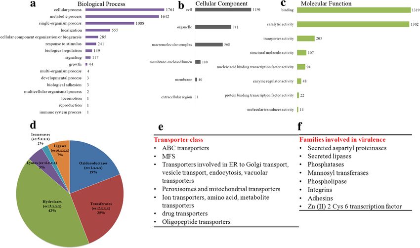
Chatterjee et al. BMC Genomics (2015) 16:686                                                                     Page 2 of 16

[14, 15] and Lehman et al. [16] categorised these species        causative Candida species from clinical cases of inva-
belonging to C. haemulonii into two genetically distinct         sive non-albicans candidiasis. In collaboration with
groups. Furthermore infections caused by two phenotyp-           Manipal Hospital, Bengaluru we have screened clinical
ically related species – C. pseudohaemulonii [17] and C.         samples from invasive cases of Candidiasis (Additional
auris are on the rise [18]. First described in 2009 by           file 2: Table S1). Identification of the isolates was done
Satoh et al. [19] in a Japanese patient, it is striking to see   by Vitek2 (bioMerieux, Marcy, I’Etoile, France) per-
the aggressive pace at which C. auris has expanded its           formed at Manipal Hospital. We saw a significant in-
clinical spectrum worldwide from minor cases of superfi-         crease in the frequency of Candidiasis from 2012 to
cial infections such as ear canal infections to highly inva-     2014 and we also found that non albicans Candida spe-
sive cases of bloodstream infections [20]. Previous studies      cies are occupying the centre stage in such infections.
[21] as well as our study demonstrate that all these clinical    Case reports from bloodstream infections revealed that
isolates have a precociously high tolerance to Amphoteri-        in 2012, 24.3 % of infections were caused by C. albicans
cinB (AmB) [22] and Fluconozole (Fcz) [14, 15], the first        and C. tropicalis. However in 2014, 38.3 % of the cases
line treatment antifungals. Even more concerning is the          were reported to be caused by C. haemulonii. Both C.
rapid emergence of resistance to echinocandins [23], the         albicans and C. tropicalis were susceptible to the com-
newest class of antifungals which may leave no treatment         monly used antifungals AmB and Fcz (Table 1). How-
option available leading to clinical failure.                    ever, all the clinical isolates identified as C. haemulonii
   Many pathogenic species within the Candida clade              showed increased tolerance to both Fcz and AmB.
such as Candida albicans and Candida glabrata have been          These isolates are referred as Candida isolates (Ci)
extensively studied at the genome level, while emerging          henceforth. As shown in Fig. 1, the isolates had MIC50
fungal pathogens Candida auris and Candida haemulonii            value of >32 μg/mL and >7 μg/mL for Fcz and AmB
remains unexplored. The basic characteristics of the gen-        (Fig. 1a,b) respectively. Since the patients were never
ome of C. auris was recently made available [24]. However        administered AmB previously, it is difficult to comment
detailed information regarding the genome architecture,          that AmB resistance in these set of clinical isolates was
virulence and mechanisms of multidrug resistance of these        inherent or acquired. However all these isolates were
emerging novel complexes of pathogenic yeasts are lacking.       susceptible to caspofungin, the newer class of antifun-
Furthermore, the commercial automated systems routinely          gal drugs- echinocandins (data not shown). The isolate
fail to identify C. auris correctly; thereby its actual occur-   Ci 6684 which showed resistance to both AmB and FcZ
rence is underreported. Even more alarming is the fact that      with highest MIC50 values was used for further analysis.
misdiagnosis may lead to incorrect treatment or delay of         The antifungal susceptibility profile of Ci 6684 is pre-
proper treatment, thereby increasing the chances of fatal-       sented in Table 2.
ities. As expected, C. auris fungemia is associated with a
high mortality rate (66 %) and therapeutic failure [25]. It      Complete genome sequence of the clinical isolate Ci 6684
also does not exhibit the known attributes responsible for       We sequenced the genome of Ci 6684 using Illumina se-
virulence in Candida species such as hyphae formation            quencing technology. A high-quality reference genome
and the cells are much smaller in size than that of C. albi-     using Illumina reads was assembled de novo as described
cans (Additional file 1: Figure S2). Towards understanding       in Methods (Additional file 3: Figure S1). The assembled
the basic biology of the multidrug resistant pathogen, we        draft genome of Ci 6684 comprises 99 scaffolds with an
have carried out whole genome sequencing of a multidrug          estimated genome size of 12,498,766 bp, 44.53 % GC
resistant clinical isolate of C. auris using Illumina sequen-    and 1.327 % Ns. The average base is found in the scaf-
cing technology and report that C. auris has a highly diver-     fold with a scaffold N50 of 279 Kb. A total of 8358 pro-
gent genome. Analysis using C. albicans as a reference           tein coding genes, 7 rRNAs and 189 tRNAs were
genome revealed a set of orthologs such as drug trans-           predicting using different tools (Description in Methods
porters, oligopeptide transporters, secreted proteinases and     and Additional file 3: Figure S1).
mannosyl transferases which may play a role in virulence           The basic annotation of 8358 predicted protein coding
and drug resistance. However most of the genome is               genes were done using blastp against current RefSeq
uncharacterized and we speculate that some of these hypo-        fungal protein database and protein NR database. 5175
thetical proteins may be involved in species specific charac-    proteins found orthologs with a mean query coverage of
teristics which promote its aggressiveness as a pathogen.        94.68 % (40–100 %), mean identity of 60.73 % (21.72–
                                                                 100 %) and E-value > e−10. 42.38 % of Ci 6684 proteins
Results and discussion                                           were orthologous to C. lusitaniae ATCC 42720. However
Clinical isolates of Candida show multi drug resistance          majority of the proteins were assigned as hypothetical,
With the background of the growing incidences of can-            since the closely related Candida species C. lusitaniae
didiasis we have determined the hierarchy of the                 protein database have also annotated those similar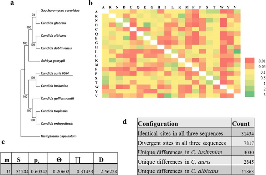
Chatterjee et al. BMC Genomics (2015) 16:686                                                                                                            Page 3 of 16

Table 1 In vitro antifungal susceptibility pattern of pathogenic Candida clinical isolates (from bloodstream) to the most commonly
used drugs belonging to the four different classes of antifungals
Antifungal              Species (n)
drugs                   MIC50 range (μg/ml)
                        Clinical isolates_ Cha (34)      C. albicans (20)        C. tropicalis (34)     C. glabrata (9)        C. lusitaniae (2)   C. parapsilosis (23)
Fluconazole             16–64                            1                       1                      0.5–1                  0.5–1               ≤1
Amphotericin B          4–16                             0.25                    0.25                   0.25–0.5               0.12–0.25           0.25–0.5
Flucytosine             0.25                             ≤1                      ≤1                     ≤1                     ≤1                  ≤1
Caspofungin             0.25                             0.25                    0.25                    0.12                  0.12                0.25–1
a
All these isolates were identified as C. haemulonii by Vitek2, which routinely fails to identify closely related species such as C. auris

proteins to be hypothetical/ functionally uncharacterized.                              able to grow at 40 °C and 42 °C as reported for C. auris
Ci 6684 was found to be diploid with a similar FACS pro-                                but not for C. haemulonii [20]. Electrophoretic karyotyp-
file to that of C. albicans SC-5314 by flow cytometric ana-                             ing by PFGE of Ci6684 yielded 5 bands and the pattern
lysis as shown in Additional file 4: Figure S3. Table 3                                 was similar to that reported previously for C. auris [26]
summarizes the general features of the genome of Ci 6684                                (Fig. 2b). Because diagnostic laboratories rely only on
along with known pathogenic Candida species. The aver-                                  automated systems like Vitek 2 or APIC20C which rou-
age size (bp) of coding sequence domain (CDS) of Ci 6684                                tinely identifies C. auris as C. haemulonii or C. famata,
seems to be least, whilst the intergenic distance (bp) is                               the actual occurrence of C. auris fungemia is under re-
similar to that of other species.                                                       ported [25, 27, 28]. Our results emphasize the need to
                                                                                        develop accurate species identification system based on
Phylogenetic analysis reveals Ci 6684 is closely related to                             molecular typing methods to ensure proper management
Candida auris                                                                           of fungemia. Another recently developed method for
Phylogenetic tree based on the partial sequence of 18 s                                 identifying C. auris by MALDI-TOF has also been re-
rRNA, ITS1, 5.8 s rRNA complete sequence, ITS2 and                                      ported [29]. Henceforth Ci 6684 will be referred to C.
28 s rRNA partial sequence revealed that Ci 6684 be-                                    auris 6684 in the remaining study.
longs to Candida auris clade of Korean and Indian iso-                                    In order to determine the evolutionary position of C.
lates with 99 % bootstrapped confidence (Fig. 2a). To                                   auris 6684 in the fungal genus tree, a concatenated
further confirm its origin, we performed multiple se-                                   phylogenetic tree was constructed based on orthologs of
quence alignment with the Indian C. auris isolates and                                  95 conserved proteins (Additional file 2: Table S2) from
found complete conservation of rRNA and ITS se-                                         11 pathogenic species under the phylum Ascomycota
quences (Additional file 5). The same isolate was also                                  (Fig. 3a). Our analysis shows bifurcation of C. albicans

    Fig. 1 In vitro antifungal susceptibility testing of clinical isolates of Candida: All the isolates were identified as C. haemulonii by Vitek2. Susceptibility
    testing was done by broth microdilution method at 37 °C for 48 h as mentioned in materials and methods. a Comparison of MIC50 values of all
    isolates for Fcz indicates all clinical isolates have MIC range of 32–64 μg/ml. b Comparison of MIC50 values of all isolates for AmB shows that all clinical
    isolates are resistant to AmB. Candida isolate Ci 6684 has the highest MIC50 value of > = 16 μg/ml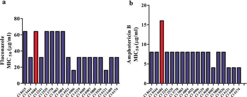
Chatterjee et al. BMC Genomics (2015) 16:686 Page 4 of 16
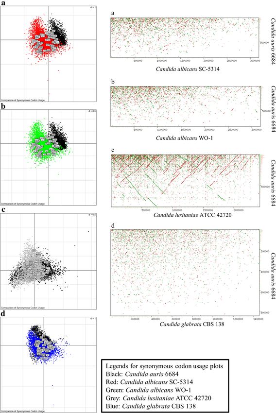
Table 2 In vitro antifungal susceptibility profile of Candida the genome level. To further investigate we compared
clinical isolate Ci 6684 synonymous codon usage between C. auris 6684, C.
Drug MIC50 (μg/ml) Susceptibility albicans SC-5314, C. albicans WO-1, C. lusitaniae
Fluconazole 64 R ATCC 42720 and C. glabrata CBS-138 (Fig. 4a - d). The
Amphotericin B 16 R codon usage in C. auris 6684 shows very less overlaps to
codon usage in C. albicans (SC-5314 and WO-1) as
Flucytosine 1 S
shown in Fig. 4a and b. The synonymous codon usage
Caspofungin 0.25 S
appears to be significantly overlapping for C. auris 6684
and C. lusitaniae which correlates and supports the re-
and C. auris 6684 in two distinct clades. However, we latedness found in the results of phylogenetic analyses
can see that C. auris 6684 and C. lusitaniae falls in the (Figs. 3a and 4c). In addition, the codon usage in C.
same clade, indicating convergence at the protein level. auris 6684 also shows fair overlap with C. glabrata
This is further confirmed by the amino acid substitution where there was no similarity found at the genomic scale
matrix of the house keeping machinery by maximum between the two (Fig. 4d). The difference in codon usage
likelihood estimation wherein the number of amino acid can be to enhance optimal protein structure and func-
substitutions per site between sequences is low (Fig. 3b). tion from the already prevailing behaviours in C. albi-
Tajima’s neutrality test indicates a positive D value which cans. This observation suggests the codon usage bias;
reflects low levels of polymorphism in the core house- which is required for understanding the selective pres-
keeping machinery of all these species including C. auris sures involved in evolution of these fungal species. In
6684 (Fig. 3c). Tajima’s relative rate test was performed the same light, the dot plots of whole (or draft) genome
to determine the heterogeneity of evolutionary rates be- comparison of C. auris 6684 with respect to C. albicans
tween C. lusitaniae and C. auris 6684 with C. albicans (SC-5314 and WO-1) and C. glabrata CBS-138 showed
used as an out group (Fig. 3d). The χ2 test statistic was no linearity at the genome scale (Fig. 4a and d) which
5.83 (P = 0.01580 with 1 degree of freedom). P-value was supports the observations seen in synonymous codon
less than 0.05; hence null hypothesis was rejected, usage plots. C. auris 6684 genome seemed to have linear
thereby indicating different rates of evolution for these genomic synteny with C. lusitaniae genome which was
species. very evident with the blastp results (Fig. 5) as well as
synonymous codon usage.
Candida auris has a highly divergent genome In this study, genomic relatedness was carried out
To gain deeper insights into the genome conservation using GGD calculator (Genomic-to-Genomic Distance
and evolution of C. auris with other pathogenic Candida calculator), formula 2, performed at http://ggdc.dsmz.de
species, we performed whole genome alignment of se- (Meier-Kolthoff et al., 2012). The GGD was calculated
quencing reads against C. albicans SC-5314, C. glabrata between C. auris 6684 and C. albicans (SC-5314 and
CBS-138, C. lusitaniae ATCC 42720 and Saccharomyces WO-1), C. lusitaniae ATCC 42720, C. glabrata CBS-138
cerevisiae S288c. More than 99.5 % of the C. auris 6684 and S. cerevisiae S288c (Table 4). The genomic distances
reads did not align to the current whole (or draft) gen- based on HSP/MUM (high-scoring segment pair/ max-
ome sequences of these four species mentioned above. imal matches that are unique in both sequences) found
This indicates that C. auris 6684 is highly divergent at out using BLAT [30] were on an average 0.20952,
Table 3 General features of Candida species and clinical isolate Ci 6684 genome
Organism Size (Mb) Number of chromosomes GC content (%) Number of Average CDS Average intergenic
(or scaffolds) genes size (bp) distance (bp)
C. albicans SC5314 28.6* 17 33.43 12869 1456.39 859.23
C. dubliniensis CD36 14.6 8 33.25 5992 1522.16 8357.8
C. orthopsilosis Co 90–125 12.66 8 36.93 5766 1491.86 4945.93
C. tropicalis MYA-3404 14.6 23 33.01 6258 1453.47 894.8
C. guilliermondii ATCC 6260 10.61 9 43.62 5920 1401.41 427.87
C. lusitaniae ATCC 42720 12.1 9 44.37 5941 1387.45 774.18
C. glabrata CBS 138 12.3 14 38.62 5235 1526.93 773.63
S. cerevisiae S288C 12.2 17 38.15 5916 1485.5 435.79
Ci 6684 12.5 99 44.53 8358 1024.55 828.8395
*Candida albicans SC-5314 used is assembly number 22 and is shown with a diploid set of chromosomes and genes. Rest of the Candida species are from the
current genomic data available at CGD and Broad Institute. ND - not determined. All decimals are rounded off at the second digit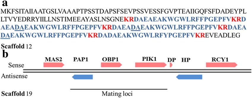
Chatterjee et al. BMC Genomics (2015) 16:686                                                                                             Page 5 of 16

 Fig. 2 ITS phylogeny and electrophoretic karyotyping reveals that Ci 6684 belongs to C. auris clade. a Phylogenetic tree based on the partial
 sequence of 18 s rRNA, ITS1, 5.8 s rRNA complete sequence, ITS2 and 28 s rRNA partial sequences of species belonging to the C. haemulonii
 complex and related Candida species. Our isolate Ci 6684 is related to C. auris as it falls in the same clade. b Electrophoretic karyotyping shows 5
 bands similar to those reported for C. auris by Oh J B et al., where they reported genotypic relatedness between C. haemulonii and closely related
 species. C. albicans (lane 1) shows four distinct bands. Corresponding C. auris lane (lane 2) shows five bands

indicating the number of identical bases between the ge-                     GO terms for all the three domains. As evident from
nomes is inversely proportional to the HSP length. The                       Fig. 6a, a major proportion of the genome is devoted to
probability that these species belong to the same species                    cellular and metabolic processes. A significant number of
or same subspecies is 0 as indicated by logistic regres-                     proteins were annotated to have transporter activity apart
sion of DNA-DNA hybridization (GGDC transform the                            from binding and catalytic activity.
genomic distances analogues to DNA-DNA hybridization).                          We also performed enzyme classification analysis based
                                                                             on Enzyme Commission (EC) numbers predictions for
Functional annotation of the C. auris 6684 genome                            each sequence. We found that hydrolases are the largest
Functional annotation was done in Blast2GO that com-                         group of C. auris 6684 enzymes (42 %), followed by trans-
bined the blastp annotation results (against NR database)                    ferases (25 %) and oxidoreductases (19 %). Blast2GO iden-
with the predicted InterProScan results. The assigned GO                     tified 466 enzyme (Fig. 6d) out of which 329 enzymes got
descriptions to each protein were considered at an E-value                   mapped to KEGG pathways. BlastKOALA was used to re-
greater than e−10. Out of 8358 predicted proteins 10958                      construct KEGG pathways for C. auris 6684. 2775 pro-
GO terms were annotated to 3560 sequences. The GO                            teins (out of 8358 predicted proteins) got annotated into
terms were placed in three domains, Biological process                       various pathways. This analysis revealed that the central
(39.45 %), Molecular Function (43.25 %) and Cellular                         pathways pertaining to carbohydrate, lipid and amino acid
Components (16.52 %). Figure 6a-c represents the level 2                     metabolisms are conserved.
Chatterjee et al. BMC Genomics (2015) 16:686 Page 6 of 16 Fig. 3 Evolutionary position of Candida auris isolate 6684 in the pathogenic fungal tree. a Phylogenetic tree based on orthologs of 95 conserved proteins from 11 pathogenic species under the phylum Ascomycota. b Amino acid substitution matrix by maximum likelihood estimation. c Tajima’s neutrality test indicates low level of polymorphism in the house keeping machinery wherein m = number of sequences, n = total number of sites, S = Number of segregating sites, ps = S/n, T = ps/a1, p = nucleotide diversity, and D is the Tajima test statistic. d Tajima’s relative rate test however indicates that these species have evolved at different rates. All evolutionary analyses were conducted in MEGA6 Core circuitry related to virulence is conserved in C. auris Notably the Zn (II) 2 Cys 6 transcription factor family 6684 is enriched in our isolate (26 in number). Four of these Considering the high genomic variability of C. auris 6684, are known to be key regulators of MDR1 transcription we asked the question that whether gene families that are in C. albicans; gain-of-function mutations of which known to have a role in pathogenicity of Candida species leads to up regulation of multidrug efflux pump MDR1, [31] are also conserved in C. auris 6684? We used the thereby leading to multidrug resistance [32, 33]. genome of C. albicans SC5314 as the template gene model The genome was found to contain transcription fac- to predict orthologs in our isolate as it is well annotated tors like STE-related and MADS-box proteins which at the experimental level. This approach yielded 1988 have been previously shown to be involved in the viru- orthologous proteins with functional annotations. Our lence of human fungal pathogens [34, 35] and plant fun- analysis predicted an arsenal of transporters ortholo- gal pathogens [36, 37] respectively. Ste12p is conserved gous to that of C. albicans, belonging primarily to the in many fungi, regulating processes involved in mating, major facilitator superfamily and ABC (ATP binding filamentation, substrate invasion, cell wall integrity and cassette) superfamily [32] (Fig. 6d). The up regulation virulence [34], while MADS-box proteins bind to DNA of these multidrug efflux pumps may explain the intrin- and have dimerization activity [35]. Our analysis also indi- sically low susceptibility of C. auris 6684 to antifungal cated conservation of the Rim101 transcriptional pathway drugs. Apart from the general transcription factors, 193 that is known to respond to alkaline pH in Saccharomyces proteins were predicted to have DNA binding/sequence cerevisiae. 122 proteins were predicted to have kinase/ specific DNA binding/transcription factor activity. We phosphorylation activity. Out of this, 93 proteins have the also predicted a multitude of zinc finger transcription serine/threonine kinase domain and the rest were pre- factors orthologous to those present in Saccharomyces dicted to be involved in protein phosphorylation due to cerevisiae, Candida albicans and Scheffersomyces stipites. the presence of putative kinase domain/ATP binding
Chatterjee et al. BMC Genomics (2015) 16:686   Page 7 of 16

 Fig. 4 (See legend on next page.)
Chatterjee et al. BMC Genomics (2015) 16:686                                                                                             Page 8 of 16

 (See figure on previous page.)
 Fig. 4 Candida auris has a highly divergent genome. a, b, c, d Synonymous Codon Usage distribution of Candida auris isolate 6684 with respect
 to C. albicans (SC-5314 (a) and WO-1 (b)), C. lusitaniae ATCC 42720 and C. glabrata CBS 138. These plots were generated by correspondence analysis
 and depict the variability in the sum of synonymous codon usage and amino acid usage. These graphs depict the codon usage bias relating it to the
 evolution of pathogenic fungus. a, b, c, d Whole (or draft) genome dot plot alignment showing genomic synteny of Candida auris isolate
 6684 with respect to other well known pathogenic Candida species. The y-axis is the largest scaffold of Candida auris 6684 and the x-axis is
 the largest chromosome (or scaffold) of the corresponding genome being compared

domain. C. auris 6684 draft genome encodes for kinases                       including C. albicans [39]. PKA is shown to be acti-
like Hog1, Protein Kinase A (PKA) and two-component                          vated in response to extracellular nutrients and subse-
histidine kinase. Activation of stress signaling pathways                    quently regulates metabolism and growth, while two-
regulated by these protein kinases have been implicated                      component histidine kinase is shown to be critical to
to enhance tolerance of pathogenic fungi to chemical                         morphogenesis and virulence [31, 40, 41].
fungicides and antifungal peptides [38]. HOG1 protein                          We also identified eight OPT genes encoding putative
is a fungal mitogen-activator protein (MAP) kinase                           oligopeptide transporters which have been implicated in
which has been implicated in responses to oxidative                          the acquisition of nutrient versatility thereby helping the
and hyperosmotic stresses in a few human pathogens                           pathogen to adapt to various host niches [42].

 Fig. 5 Summary of functional annotation of Candida auris genome. a Annotation results against RefSeq fungal protein database shows 5.1 %
 (429 out of 8358 protein coding genes) were annotated functionally with predicted names. The rest of the genome remains uncharacterized. b C.
 auris 6684 has highest number of orthologs in C. lusitaniae ATCC 42720. However most of them were annotated as hypothetical
Chatterjee et al. BMC Genomics (2015) 16:686                                                                                                        Page 9 of 16

Table 4 Genomic relatedness calculated using Genome-to-Genome Distance Calculator
Query genome Reference            Formula 1 (HSP length/total length)       Formula 2 (identities/HSP length)             Formula 3 (identities/total length)
             genome
                                  DDH Model Distance Prob. DDH              DDH Model C.I. Distance Prob. DDH             DDH Model Distance Prob. DDH
                                      C.I.           ≥ 70 %                                         ≥ 70 %                    C.I.           ≥ 70 %
C. auris 6684     C. glabrata     8.4    2.29     0.957       0             20.3   2.29         0.2049      0             9.7     2.29     0.9653     0
                  CBS 138
C. auris 6684     C. albicans     8.8    2.34     0.934       0             19.8   2.34         0.2107      0             10      2.34     0.9473     0
                  SC5314
C. auris 6684     C. albicans     8.8    2.33     0.934       0             19.8   2.33         0.211       0             10      2.33     0.9478     0
                  WO-1
C. auris 6684     C. lusitaniae 10.4     2.48     0.856       0             19.2   2.48         0.2171      0             11.4    2.48     0.8866     0
                  ATCC 42720
C. auris 6684     S. cerevisiae   8.4    2.29     0.958       0             20.4   2.29         0.2039      0             9.6     2.29     0.9667     0
                  S288c
Distances are calculated by (i) comparing two genomes using the BLAT program to obtain HSPs/MUMs and (ii) inferring distances from the set of HSPs/MUMs
using three distinct formulas. The distances are transformed to values analogous to DDH. The DDH estimate results from a generalized linear model (GLM) which
also provides the estimate’s confidence interval (after the +/− sign). An additional bootstrap confidence interval is listed if this option was chosen in the job
submission form. Logistic regression (with a special type of GLM) is used for reporting both the probabilities that DDH is > =70 % and > =79 %. GGDC is mainly
used to calculate the in silico relatedness of the species

Interestingly it has been reported that in C. albicans,                            permeases) which further expands its nutrient assimilation
these genes are also induced upon phagocytosis by mac-                             machinery, thereby helping it to acclimatize to diverse
rophages [43]. We also found orthologs of genes pre-                               host niches.
dicted to be hexose transporters, maltose transporters and                           Our next step was to hunt down the attributes that
permeases (amino acid permeases, sulfur permeases,                                 may explain the aggressive behavior of the pathogen.
allantoate permeases, glycerol permeases and iron                                  Our analysis indeed predicted many known virulence

  Fig. 6 Functional annotation of C. auris genome. a, b, c Represents Level 2 GO terms for the three main domains. The most abundant terms in
  Biological process (a) is cellular process, metabolic process and single-organism process and in Molecular functions (b), binding, catalytic activity
  and transporter activity. The common cellular component (c) termed are the cell, organelle and membrane. d Distribution of various enzymes into
  the six enzyme classes according to E.C numbers. e, f represents gene families predicted in C. auris that are orthologous to C. albicans
Chatterjee et al. BMC Genomics (2015) 16:686 Page 10 of 16
associated genes (Fig. 6e). Since the cell wall serves as lusitaniae and C. gulliermondii are heterothaliic in na-
the interface between the pathogen and the host im- ture [49]. It is interesting to note that virulence and
mune defense, components of the cell wall serve as mode of reproduction are being analysed as linked
pathogen associated molecular patterns and virulence phenomenon in recent years. C. lusitaniae is a hetero-
factors. Our analysis indicated that the family of manno- thallic species known to be involved in sexual
syl transferases is conserved in C. auris 6684 with many reproduction. On the other hand certain Candida spe-
predicted orthologs. Apart from maintaining cell wall cies are either parasexual or asexual. Considering the
architecture by coordinating glycan synthesis, these en- high similarity shared by C. auris 6684 and C. lusitaniae,
zymes play a very important role in immune recognition, we speculated that C. auris 6684 might have a sexual
host cell adherence and virulence in C. albicans [44]. stage similar to the latter. Sexual mating is controlled by
Integrins and adhesins are the other two gene families a single genetic locus called the MAT locus consisting of
which have a crucial role in adherence and virulence of two alleles-MATa and MATα.
C. albicans [45, 46]. However our annotation predicted To understand the mode of reproduction in C auris, we
only two proteins, one structurally similar to alpha- analysed the MAT loci (MTL) in the genome assembly.
subunit of human leukocyte integrin; predicted to play a Our search led to the identification of a putative gene se-
role in morphogenesis, adhesion, mouse coecal quence in C. auris 6684 genome with similarities to α
colonization and virulence; and another secreted protein mating pheromone of Naumovozyma castellii CBS 4309.
similar to alpha agglutinin anchor subunit which has The gene sequence consists of a 654-bp ORF that encodes
been previously shown to be induced upon exposure to for five putative α pheromone peptide repeats separated
fluconazole. This clearly suggests that C. auris employs by KEX2 proteinase cleavage sites. Two of the five α-
distinct mechanisms for host cell adhesion. peptides are identical in sequence; the remaining three
We also found four orthologs of secreted aspartyl pro- contains additional DA residues (Fig. 7). We also found a
teases (SAP) two of which were predicted to have greater homologue of KEX2 in the genome. However, the genes
expression upon deep epidermal invasion; greater ex- in the vicinity of MF-α were all annotated as hypothetical
pression in vaginal than oral infection [47] and promin- (Additional file 2: Table S4). Interestingly, the three non-
ent role in biofilm formation. We also found two genes sex genes (NSGs) of the MTL locus namely, the essential
annotated as vacuolar aspartic proteinases. The secreted phosphatidyl inositol kinase gene (PIK), the essential poly
aspartic proteinases help the fungus to digest host pro- (A) polymerase gene (PAP), and the nonessential oxysterol
teins and the resulting peptides are taken up into the cell binding protein gene (OBP) were present in a different
by specific transporters like the oligopeptide transporters scaffold (Fig. 7). In C. albicans, these genes have been im-
family mentioned above [48]. Our results also annotated plicated in biofilm impermeabilty and fluconazole resist-
eight genes orthologous to secreted lipases. In all, our ance [50]. Thus MAT α gene is located in a different
analysis revealed that enzyme families implicated in in- locus. In C. auris 6684, ERG11 is also located on the same
vasiveness like mannosyl transferases, secreted aspartyl scaffold as MTL non sex genes and in C. albicans, the loss
proteases and lipases are enriched in our clinical isolate. of heterozygosity at the MTL locus has been correlated to
However the adhesion and integrin gene families are ill azole resistance [51]. However we could not find MATa
represented. This information has been categorized in gene in the genome. Thorough experimentation needs to
Additional file 2: Table S3. Our analysis also revealed be done to establish its sexuality.
686 proteins predicted to be induced or repressed upon The sequence of the gene coding mating factor α is
rat catheter or biofilm formation. This includes a multi- unique to each Candida species and therefore we de-
tude of enzymes, transcription factors, ribosomal pro- signed PCR primers specifically for MF α gene. This
teins and transporters. This clearly indicates that C. PCR was tested on C. haemulonii 8176 obtained from
auris 6684 has significant ability to form biofilms since MTCC, IMTECH and C. auris 6684. As evident in
the core genes involved in biofilm formation are con- Fig. 8a, C. auris 6684 gave an amplicon at 400 bp which
served. However experiments need to be done to valid- was not seen for C. haemulonii. This test was further ex-
ate the same. trapolated to other clinical isolates reported to be C.
haemulonii and many of them turned to be PCR positive
Structure of mating loci in C. auris and PCR based for C. auris (Fig. 8b). The same isolates also showed a
diagnostic test to differentiate between C. auris and C. similar PFGE pattern (Fig. 8c), thereby confirming the
haemulonii fact that these were misdiagnosed as C. haemulonii.
Another peculiarity seen in Candida species is the
highly diverse nature of sexuality. Diploids like C. tropi- Conclusions
calis and C. parapsilosis are unable to mate while C. Opportunistic infections caused by Candida are on the
albicans shows a parasexual cycle. Haploids like C. rise globally and newer pathogenic species are emanatingChatterjee et al. BMC Genomics (2015) 16:686 Page 11 of 16 Fig. 7 Identification of mating gene MF α and mating loci in C. auris. a The amino acid sequence of the MF α gene. Regions encoding the mature α pheromone peptide is shown in blue color. Possible Kex2 cleavage sites are shown in red colour. Additional DA residues present in three of the peptides have been underlined. b Scaffold 18 shows conservation of non sex genes of MTL locus, however, MF α gene is present in a different scaffold at an unprecedented rate. What are not evolving at the may act as accurate identification markers for this group same pace are the current methods of diagnosis and treat- of emerging pathogens at the species level. Towards this ment options leading to misdiagnosis and clinical failure. we have developed a PCR based diagnostic test to distin- The last decade has witnessed the emergence of newer guish between these two pathogens. species called C. haemulonii from being the causative The genome of C. auris spans about ~12.5 Mb with agent of minor infections to one of the leading causes of 8358 predicted protein coding genes. Strikingly, at the invasive infections. It is currently increasing in prevalence, genomic level, C. auris shows a highly divergent rela- with several ongoing outbreaks in developing and under- tionship with other pathogenic Candida species as indi- developed countries. The actual incidence rate is however cated by a meagre 0.5 % alignment of the sequencing misleading because of the inability of the current auto- reads to other Candida genomes and supported by lack mated systems used for screening of fungal species to of linear synteny of genomic dot plots. C. auris is phylo- identify novel emerging fungal pathogens such as C. auris, genetically closest to C. haemulonii whose genome se- C. pseudohaemulonii and other related species due to quence is unavailable. Among the sequenced yeast striking similarities in biochemical characters and the un- species, it is closest to C. lusitaniae; however, its genome availability of molecular markers for accurate identifica- is also not well annotated functionally. Therefore major- tion. We have generated the first draft genome sequence ity of the protein coding genes were predicted to be of a commonly misdiagnosed, emerging pathogen C. hypothetical/functionally uncharacterized. The role of auris. The isolate was identified as C. haemulonii by each of these unique candidate proteins demands for ur- Vitek2. However PFGE analysis revealed 5 bands similar gent functional studies. Hence accurate identification to that of C. auris and accurate species identification was and de novo assembly and annotation still remains a done by phylogenetic analysis based on the partial se- challenge for divergent sequences among emerging quence of 18S rRNA, ITS1, 5.8S rRNA complete se- pathogenic species. 37.71 % of the protein coding genes quence, ITS2 and 28S rRNA partial sequence. Genome showed no sequence similarity to genes available in pub- sequencing will highlight important differences which lic database, thus indicating that speciation genes are Fig. 8 PCR based diagnostic method to differentiate between C. auris and C. haemulonii. a Primers based on C. auris 6684 MFα gene gives a specific amplicon at 400 bp. b Amplification was not seen in the case of C. haemulonii 8176. However, four clinical isolates identified to be C. haemulonii by Manipal Hospital, showed a band at 400 bp. c The PFGE profile of these four clinical isolates was similar to that of C. auris 6684
Chatterjee et al. BMC Genomics (2015) 16:686                                                                       Page 12 of 16

embedded within the genome which may be involved in              it as a specialist pathogen. It is possible that the indiscrim-
grooming it as an aggressive pathogen. With the limited          inate use of antibiotics shaped its genome to expand not
data available, it is difficult to comment about the gen-        only its clinical spectrum of infection but also to emerge
omic architecture of speciation and how it facilitates or        as a successful multidrug resistant pathogen.
impedes further divergence. To further probe into the               In all, our study provides the first whole genomic over-
difference at the functional level we resorted to syn-           view of C. auris, the first member of the Candida haemu-
onymous codon usage plots which distinguish ways by              lonii and related pathogenic fungi complex to be
which translational selection of protein coding genes oc-        sequenced. This report is a major step toward the initi-
curs among related species. The above observation is             ation of genomic studies of this complex group of fungi
supported by GGDC that calculated the in silico related-         which are fast turning drug resistant and may be a menace
ness of C. auris and sequenced Candida pathogens, sur-           with limited treatment options available in the future.
prisingly the logistic regression quantifies no relatedness
among the species. The ecological niche of most of these         Methods
Candida species is known, that may throw light on the            Strain and growth conditions
evolutionary forces grooming these organisms at the              All clinical isolates were obtained from Manipal Hos-
species level. However till date there are no reports of         pital, Bengaluru and the ethical approval was obtained
naturally occurring C. auris species. C. auris can grow at       from Ethics Committee of Manipal Hospitals, Bengaluru
elevated temperatures of 42 °C whereas C. haemulonii             and informed consent was taken as required during the
cannot. This gives us a hint that C. auris has the potential     study. Ci 6684 was isolated from a patient who had sep-
to infect the avian fauna whose body temperature is in the       sis with multiorgan dysfunction. C. haemulonii 8176 was
range of 40 °C to 42 °C. However, additional experiments         obtained from MTCC, IMTECH Chandigarh, India.
need to be done in order to validate this phenomenon.            Strains were routinely grown in Yeast Peptone Dextrose
   The foremost criterion to be a successful Candida             (YPD) medium at 37 °C.
pathogen is the ability to colonize diverse anatomical
niches within the host such as skin, oral cavity, gastro-        Minimum inhibitory concentration and growth assays
intestinal tract, vagina and the vasculature. Each Can-          To determine the in vitro susceptibility to antifungal drugs,
dida pathogen has its own machinery dedicated to host            broth microdilution protocol [52] was used. Overnight cul-
cell adhesion, recognition, invasion and colonization. We        tures were grown at 37 °C in YPD. Approximately 103 cells
compared C. auris genome with that of C. albicans since          per well in YPD media at 37 °C. Minimum inhibitory con-
it is well annotated and well-studied as well as distantly re-   centration (MIC) tests were set up in a total volume of
lated to C. auris. While the spectrum of virulence traits        0.2 ml/well with 2-fold dilutions of drugs. Fluconazole gra-
like hyphae formation, white opaque switching is quite dif-      dients where in the following concentration steps in μg/ml:
ferent between these two species, we found that C. auris         64, 32, 16, 8, 4, 2, 1, 0.5, 0.25, 0.125, 0.0625 and 0.03125.
still shares some common virulence traits with C. albi-          For Amphotericin B, gradients where in the following the
cans. Our analysis highlights that a significant portion of      concentration steps in μg/ml were: 16, 8, 4, 2, 1, 0.5, 0.25,
C. auris genome encodes for transporters belonging to the        0.125, 0.0625, 0.03125 and 0.015625. 24 or 48 h post
ABC transporter family and major facilitator superfamily.        incubation, growth was measured by reading the optical
This may partly explain its increased tolerance to antifun-      density at 600 nm after agitation using a spectrophotom-
gal drugs. The multidrug resistant nature of the pathogen        eter (Tecan). MIC50 was defined as the concentration of
and the limited arsenal of antifungal agents indicate that       drug reducing growth by 50 % relative to the wells contain-
there is a critical need for finding new drug targets and        ing no drug. Sterile water was the vehicle for Fcz and
genome sequence of C. auris therefore may prove useful           AmB.
in finding alternative targets that can augment the existing
antifungal therapy. Our analysis also provides a snapshot        DNA sequencing
of the potential genetic attributes that may explain its         Short reads and long reads library preparation was per-
virulent nature. The genome of the pathogen harbours             formed at Genotypic Technology’s Genomics facility
gene families such as lipases, oligopeptide transporters,        following NEXTFlex DNA library protocol outlined in
mannosyl transferases and transcription factors which play       “NEXTFlex DNA sample preparation guide (Cat #
a multitude of roles in colonization, invasion and iron ac-      5140–02). ~3 μg of genomic DNA was sonicated using
quisition. Also majority of genes known to be involved in        Bioruptorto and 300 to 600 bp sized fragments were
formation of biofilm appears to be conserved. In all, we         obtained. The size distribution was checked by running
see that C. auris shares many genes with C. albicans and         an aliquot of the sample on Agilent HS DNA Chip. The
C. lusitaniae indicating a common ancestry; however it           resulting fragmented DNA was cleaned up using Agen-
may have acquired novel genetic traits that have groomed         court AMPure XP SPRI beads (Beckman Coulter).
Chatterjee et al. BMC Genomics (2015) 16:686                                                                  Page 13 of 16

Fragmented DNA was subjected to a series of enzym-              STANDARD v3.0 [53] and the contigs were reduced to
atic reactions that repair frayed ends, phosphorylate the       65 scaffolds. Using Reapr v1.0.17 [54], the 65 scaffolds
fragments, and add a single nucleotide A overhang and           were corrected, removing the erroneous bases and the
ligate adaptors (NEXTFlex DNA Sequencing kit). Sam-             final number of scaffolds was 97. These 97 scaffolds
ple cleanup was done using AMPure SPRI beads. After             were used as input for GeneMarkS [55] to predict
ligation-cleanup, ~300–600 bp fragments was size se-            protein-coding genes with –eukaryotic as the main op-
lected on 2 % low melting agarose gel and cleaned               tion. The resulting 8388 proteins were subjected to local
using MinElute column (QIAGEN). PCR (10 cycles)                 blastp, resulting in 5175 proteins being annotated to
amplification of adaptor ligated fragments was done             RefSeq fungal protein database. Proteins having query
and cleaned up using AMPure SPRI beads. The pre-                coverage of greater than 40 % were only considered from
pared libraries were quantified using Qubit flourometer         this blast results. An InterproScan [56] was carried out
and validated for quality by running an aliquot on High         using the tool Blast2GO [57] v3.0 to group the predicted
Sensitivity Bioanalyzer Chip (Agilent). The short read          proteins according to the presence of domain/motif in
inserts were sequenced in Illumina MiSeq and long               their sequences. GO terms were assigned through Blas-
read inserts were sequenced in Illumina NextSeq 500.            t2GO tool based on NR Database orthologs (blastp with
   Mate-pair reads library preparation was performed at         Evalu > e−10). Proteins involved in various KEGG path-
Genotypic Technology’s Genomics facility following              ways were assigned using BlastKOALA [58]. Transfer
Nextera Mate Pair Gel Plus protocol outlined in “Illu-          RNAs were identified using the tRNAScan-SE program
mina Nextera Mate Pair library preparation guide (Cat#          [59]. Ribozomal RNAs were predicted by RNAmmer
FC-132-9001DOC, Part#15035209 Rev D.)”. ~4 μg of                [60]. The sequenced reads were mapped to various
Qubit quantified DNA was taken for Tagmentation. The            pathogenic Candida genome using Bowtie2 v2.2.3 [61]
tagmented sample was cleaned up using AMPure beads              with default parameter. The generated SAM files were
and subjected to strand displacement. 3–5 kb range of the       used to calculate the percent of reads aligned using R.
strand displaced sample was size selected on 0.6 % agarose
gel. Size selected sample was taken for circularization over-   Electrophoretic karyotyping
night, followed by linear DNA digestion with DNA Exo-           Modified PFGE, Counter-clamped homogeneous elec-
nuclease. The circularized DNA molecules were sheared           trical field (CHEF) (BIO-RAD) was used for electrophor-
using Covaris to obtain fragments in the size range of 300      etic karyotyping of C. auris 6684 and C. albicans. The
to 1000 bp. Sheared DNA was subjected to bead binding           protocol was adapted from Iadonato et al. 1996 [62].
with M280 Streptavidin beads to isolate biotinylated mole-      Briefly 5 ml yeast cultures were grown in YPD medium
cules. End repair, A-Tailing and adapter ligations were per-    at 30 °C. The cells were the harvested and washed with
formed on the bead-DNA complex. Adaptor ligated                 50 mM EDTA. Approximately 2× 10⁹ cells/ml were
sample was amplified for 15 cycles of PCR followed by           added to equal volumes of 1 % (w/v) low melt Pulse
AMPure XP bead clean up. The prepared library was               Field certified Agarose (BIO-RAD), prewarmed at 45 °C.
quantified using Qubit and validated for quality by running     The mixture was then transferred in to disposable plug
an aliquot on High Sensitivity Bioanalyzer Chip (Agilent).      moulds to harden. Plugs were then extruded and sus-
The mate-pair reads were sequenced using Illumina Next-         pended in freshly prepared spheroplasting solution con-
Seq 500.                                                        taining Zymolase, and incubated at 37 °C for 4 h. After
                                                                this the plugs were washed with 1 % Lithium dodecyl
Assembly, annotation and analysis                               sulfate (LDS) (2X 30 min) buffer followed by cell lysis
The qualities of the reads were checked using Genotypic         with 1 % N-lauryl sarcosine (NDS) (3X 30 min) buffer.
proprietary tool SeqQC v2.21. The average sequencing            Finally the plugs were rinsed (6x 30 min) with TE buffer
depth (coverage) for short paired-end reads is 158.19x,         pH 8. Agarose plugs containing yeast DNA was then
long paired-end reads is 175.51x and mate-pair reads is         loaded into 0.8 % low melt Pulse Field certified Agarose
205.78x. Processed short paired-end reads (3.27 million)        (BIO-RAD) prepared with 0.5X TBE buffer. The DNA
were used to generate (250–400) long fragments using            samples were resolved by running the gel in CHEF-DR®
ARF-PE v0.2. 467178 long fragments were generated               III system with 5 V/cm2 with pulse time of 120 s and
using 467178*2 paired end reads (ie, 14.29 % reads were         total run time of 36 h at 12 °C. Gel was then stained
used in long read generation). 467178 long fragments            with ethidium bromide (1ug/ml) for 30 min and visual-
and 3269025*2 paired end reads used for Newbler Gen-            ized at ImageQuant LAS 4000 transilluminator (GE).
ome assembly. Newbler version 2.8’s default assembly
parameters were used for the assembly and 721 scaffolds         Phylogenetic tree and evolutionary analysis
were generated. The paired-end long insert reads and            The partial sequence of 18 s rRNA, ITS1, 5.8 s rRNA
mate-pair reads were used to gap fill using SSPACE-             complete sequence, ITS2 and 28 s rRNA partial sequence
Chatterjee et al. BMC Genomics (2015) 16:686 Page 14 of 16
retrieved from NCBI (Additional file 2: Table S5) were (https://www.broadinstitute.org/scientific-community/
used to categorise Clinical isolate 6684 with Candida science/projects/fungal-genome-initiative/fungal-genomics)
auris clade. The evolutionary tree was inferred using the and CGD (www.candidagenome.org/). The analysis was
Maximum Likelihood method based on the Tamura-Nei carried out using GFFex v2.3 and Biostrings package of
model [63]. The tree with the highest log likelihood Bioconductor in R v3.1. The DNA-DNA hybridizations
(−307.3435) is shown. The percentage of trees in which (DDH) distances were calculated using the online tool
the associated taxa clustered together is shown next to the Genome-to-Genome Distance Calculator (GGDC 2.0)
branches. Initial tree(s) for the heuristic search were ob- (http://ggdc.dsmz.de/). Dot plot were done in an online
tained automatically by applying Neighbor-Join and BioNJ tool called YASS [67] by setting the e-value to e-10 and
algorithms to a matrix of pairwise distances estimated the synonymous codon usage plots were done in R
using the Maximum Composite Likelihood (MCL) ap- (v3.1) using ape4 and seqinr packages [68] of
proach, and then selecting the topology with superior log Bioconductor.
likelihood value. The tree is drawn to scale, with branch
lengths measured in the number of substitutions per site. Polymerase chain reaction
The analysis involved 48 nucleotide sequences. All posi- Genomic DNA was isolated as described previously. Based
tions containing gaps and missing data were eliminated. on the MFα region sequence from C. auris, a specific
There were a total of 167 positions in the final dataset. PCR-based method was developed for the direct detection
95 conserved proteins (Additional file 2: Table S2) of C. auris DNA by using a C. auris -specific primer
from Saccharomyces cerevisiae S288c were retrieved using (CaMF [5′- GAGAAAAGAGACGCTGAAGCTGAG-3′])
YGD, CGD and BLASTn for the following organisms: Sac- designed using the gene sequence which codes for the
charomyces cerevisiae S288c, Candida albicans SC-5314, unique pheromone together with reverse primer (CaMR
Candida dubliniensis CD-36, Candida glabrata CBS [5′- TCAACCTTCGAGGTCAGCTTCA-3′]).
138,,Candida isolate 6684, Candida tropicalis MYA-3404,
Candida lusitaniae ATCC 42720, Candida gulliermondii
Ploidy analysis by FACS
ATCC 6260, Candida orthopsilosis Co-90–125, Ashbya_-
Cultures were grown in YPD till A600 of 1.0. The cells
gossypii and Histoplasma capsulatum. The phylogenetic
were washed in 1X PBS (137 mM NaCl, 2.7 mM KCl,
tree was constructed using the Neighbor-Joining method.
10 mM sodium phosphate dibasic (NaH2PO4), 2 mM
The optimal tree with the sum of branch length =
potassium phosphate monobasic (K2HPO4), pH of 7.4)
1.22757517 is shown. The percentage of replicate trees in
and fixed in 70 % ethanol for 1 h at room temperature
which the associated taxa clustered together in the boot-
or kept at 4 °C overnight. The cells were suspended in
strap test (2000 replicates) is shown next to the branches.
1X PBS and incubated with RNase A (1 mg/ml) at 37 °C
The tree is drawn to scale, with branch lengths in the same
for 4 h in the same buffer. Cells were subsequently
units as those of the evolutionary distances used to infer
washed with PBS, and finally stained with propidium
the phylogenetic tree. The evolutionary distances were
iodide (PI, 16 μg/ml) for flow cytometric analysis in BD
computed using the p-distance method and are in the units
FACS Canto.
of the number of amino acid differences per site. The ana-
lysis involved 11 amino acid sequences. All positions with
less than 95 % site coverage were eliminated. There were a Availability of supporting data
total of 51712 positions in the final dataset. The whole genome sequencing data can be accessed
Tajima’s neutrality analysis involved concatenated through BioProject accession number PRJNA267757. The
amino acid sequences from the 11 species. All positions respective BioSample accession numbers is SAMN03200169.
with less than 95 % site coverage were eliminated. There The SRA reference numbers of the whole genome sequen-
were a total of 51712 positions in the final dataset. The cing are SRX766223 (Illumina MiSeq short paired-end
equality of evolutionary rate between Candida lusita- reads), SRX766234 (Illumina NextSeq 500 mate-pair reads)
niae, Clinical isolate 6684 with Candida albicans as an and SRX766231 (Illumina HiSeq2500 long paired-end
out-group was determined by Tajima’s relative rate test reads). This Whole Genome Shotgun project has been de-
[64, 65]. All positions containing gaps and missing data posited at DDBJ/EMBL/GenBank under the accession
were eliminated. There were a total of 56989 positions LGST00000000. The version described in this paper is ver-
in the final dataset. All the phylogenetic trees and evolu- sion LGST01000000.
tionary analyses were conducted in MEGA6 [66] .
Additional files
Genome comparison
For genome comparison the current genome sequences Additional file 1: Figure S2. Colony morphology of C. auris and C.
albicans SC-5314. (PNG 147 kb)
(whole or draft) were downloaded from Broad InstituteChatterjee et al. BMC Genomics (2015) 16:686 Page 15 of 16
Additional file 2: Table S1. Number of reports regarding various 8. Papon N, Courdavault V, Clastre M, Bennett RJ. Emerging and emerged
Candida infections. Table S2: 95 Conserved Proteins in Candida and pathogenic Candida species: beyond the Candida albicans paradigm. PLoS
Saccharomyces used for phylogenetic analysis. Table S3: Related Protein Pathog. 2013;9, e1003550.
Families as categorized based on orthologues to C. albicans. Table S4: 9. Colombo AL, Nucci M, Park BJ, Nouer SA, Arthington-Skaggs B, da Matta DA,
Upstream and Downstream Neighbours of MAT alpha loci. Table S5: et al. Epidemiology of candidemia in Brazil: a nationwide sentinel
NCBI accession numbers used for Phylogenetic analysis. (XLSX 26 kb) surveillance of candidemia in eleven medical centers. J Clin Microbiol.
2006;44:2816–23.
Additional file 3: Figure S1. Pipeline depicting the methods used for 10. Colombo AL, Garnica M, Aranha Camargo LF, Da Cunha CA, Bandeira AC,
de novo assembly and functional annotation of C. auris 6684 (or Ci 6684) Borghi D, et al. Candida glabrata: an emerging pathogen in Brazilian tertiary
draft genome. (TIFF 6662 kb) care hospitals. Med Mycol. 2013;51:38–44.
Additional file 4: Figure S3. Flow cytometric analysis of DNA content 11. Hachem R, Hanna H, Kontoyiannis D, Jiang Y, Raad I. The changing
of Candida species. (JPEG 76 kb) epidemiology of invasive candidiasis: Candida glabrata and Candida krusei
Additional file 5: Multiple sequence alignment of rRNA and ITS as the leading causes of candidemia in hematologic malignancy. Cancer.
sequences of C. auris 6684 and reported Indian strains of C. auris 2008;112:2493–9.
from NCBI. (PDF 122 kb) 12. Turner SA, Butler G. The Candida pathogenic species complex. Cold Spring
Harb Perspect Med. 2014;4:a019778.
13. Cendejas-Bueno E, Kolecka A, Alastruey-Izquierdo A, Theelen B, Groenewald M,
Abbreviations Kostrzewa M, et al. Reclassification of the Candida haemulonii complex as
MDR: Multidrug resistance; ITS: Internal transcribed spacer; HAI: Hospital Candida haemulonii (C. haemulonii group I), C. duobushaemulonii sp. nov.
acquired infections; AmB: Amphotericin B; Fcz: Fluconazole; MIC: Minimum (C. haemulonii group II), and C. haemulonii var. vulnera var. nov. three
inhibitory concentration; PFGE: Pulse field gel electrophoresis; multiresistant human pathogenic yeasts. J Clin Microbiol. 2012;50:3641–51.
GGDC: Genomic to genomic distance calculator; GO: Gene ontology; 14. Khan ZU, Al-Sweih NA, Ahmad S, Al-Kazemi N, Khan S, Joseph L, et al.
KEGG: Kyoto encyclopedia of genes and genomes; ABC: ATP binding Outbreak of fungemia among neonates caused by Candida haemulonii
cassette; OPT: Oligopeptide transporters; SAP: Secreted aspartyl proteinases; resistant to amphotericin B, itraconazole, and fluconazole. J Clin Microbiol.
MTL: Mating locus. 2007;45:2025–7.
15. Kim MN, Shin JH, Sung H, Lee K, Kim EC, Ryoo N, et al. Candida haemulonii
and closely related species at 5 university hospitals in Korea: identification,
Competing interests
antifungal susceptibility, and clinical features. Clin Infect Dis. 2009;48:e57–61.
The authors declare that they have no competing interests.
16. Lehmann PF, Wu LC, Pruitt WR, Meyer SA, Ahearn DG. Unrelatedness of
groups of yeasts within the Candida haemulonii complex. J Clin Microbiol.
Authors’ contributions 1993;31:1683–7.
Conceived and designed the experiments: SC, SVA, RKN, STC, UT. Performed 17. Sugita T, Takashima M, Poonwan N, Mekha N. Candida pseudohaemulonii Sp.
the experiments: SC, SVA, SJ, RKN, STC. Analyzed the data: SC, SVA, RKN, STC, Nov. an amphotericin B-and azole-resistant yeast species, isolated from the
UT. Contributed reagents/materials/analysis tools: SC, SVA, SJ, RKN, STC, UT. blood of a patient from Thailand. Microbiol Immunol. 2006;50:469–73.
All authors read and approved the final manuscript. 18. Lee WG, Shin JH, Uh Y, Kang MG, Kim SH, Park KH, et al. First three reported
cases of nosocomial fungemia caused by Candida auris. J Clin Microbiol.
Acknowledgements 2011;49:3139–42.
The authors would like to acknowledge Genotypic Technology, Bangalore, 19. Satoh K, Makimura K, Hasumi Y, Nishiyama Y, Uchida K, Yamaguchi H, et al.
India. We acknowledge funding from the DBT-IISc partnership program and Candida auris sp. nov. a novel ascomycetous yeast isolated from the
Grant Challenge Canada (Sub-grant fund: 494417). Research fellowship from external ear canal of an inpatient in a Japanese hospital. Microbiol Immunol.
DST INSPIRE for Sharanya Chatterjee is acknowledged. 2009;53:41–4.
20. Chowdhary A, Sharma C, Duggal S, Agarwal K, Prakash A, Singh PK, et al. New
Author details clonal strain of Candida auris, Delhi, India. Emerg Infect Dis. 2013;19:1670–3.
1
Department of Biochemistry, Indian Institute of Science, Bengaluru, 21. Sarma S, Kumar N, Sharma S, Govil D, Ali T, Mehta Y, et al. Candidemia
Karnataka, India, 560012. 2Manipal Hospital, Bengaluru, Karnataka, India. caused by amphotericin B and fluconazole resistant Candida auris. Indian J
Med Microbiol. 2013;31:90–1.
Received: 3 February 2015 Accepted: 18 August 2015 22. Rodero L, Cuenca-Estrella M, Cordoba S, Cahn P, Davel G, Kaufman S, et al.
Transient fungemia caused by an amphotericin B-resistant isolate of
Candida haemulonii. J Clin Microbiol. 2002;40:2266–9.
References 23. Muro MD, Motta Fde A, Burger M, Melo AS. Dalla-Costa LM Echinocandin
1. Klevens RM, Edwards JR, Richards Jr CL, Horan TC, Gaynes RP, Pollock DA, resistance in two Candida haemulonii isolates from pediatric patients. J Clin
et al. Estimating health care-associated infections and deaths in U.S. Microbiol. 2012;50:3783–5.
hospitals, 2002. Public Health Rep. 2007;122:160–6. 24. Sharma C, Kumar N, Meis JF, Pandey R, Chowdhary A. Draft genome
2. Lass-Florl C. The changing face of epidemiology of invasive fungal disease sequence of a fluconazole-resistant Candida auris strain from a candidemia
in Europe. Mycoses. 2009;52:197–205. patient in India. Genome Announc. 2015;3:e00722–15.
3. Quindos G. Nosocomial candidemias and invasive candidiasis. Med Clin 25. Chowdhary A, Anil Kumar V, Sharma C, Prakash A, Agarwal K, Babu R, et al.
(Barc). 2010;134:17–9. Multidrug-resistant endemic clonal strain of Candida auris in India. Eur J Clin
4. Tortorano AM, Kibbler C, Peman J, Bernhardt H, Klingspor L, Grillot R, et al. Microbiol Infect Dis. 2014;33:919–26.
Candidaemia in Europe: epidemiology and resistance. Int J Antimicrob 26. Oh BJ, Shin JH, Kim MN, Sung H, Lee K, Joo MY, et al. Biofilm formation and
Agents. 2006;27:359–66. genotyping of Candida haemulonii, Candida pseudohaemulonii, and a
5. Adhikary R, Joshi S. Species distribution and anti-fungal susceptibility of proposed new species (Candida auris) isolates from Korea. Med Mycol.
Candidaemia at a multi super-specialty center in Southern India. Indian J 2011;49:98–102.
Med Microbiol. 2011;29:309–11. 27. Kim HY, Huh HJ, Choi R, Ki CS, Lee NY. Three cases of candidiasis
6. Pfaller MA, Andes DR, Diekema DJ, Horn DL, Reboli AC, Rotstein C, et al. misidentified as Candida famata by the Vitek 2 system. Ann Lab Med.
Epidemiology and outcomes of invasive candidiasis due to non-albicans 2015;35:175–7.
species of Candida in 2,496 patients: data from the Prospective Antifungal 28. Ochiuzzi ME, Cataldi S, Guelfand L, Maldonado I, Arechavala A. Evaluation of
Therapy (PATH) registry 2004–2008. PLoS One. 2014;9, e101510. Vitek 2 for the identification of Candida yeasts. Rev Argent Microbiol.
7. Pfaller MA, Diekema DJ, Procop GW, Rinaldi MG. Multicenter comparison of 2014;46:107–10.
the VITEK 2 antifungal susceptibility test with the CLSI broth microdilution 29. Kathuria S, Singh PK, Sharma C, Prakash A, Masih A, Kumar A, et al.
reference method for testing amphotericin B, flucytosine, and voriconazole Multidrug-resistant Candida auris misidentified as Candida haemulonii:
against Candida spp. J Clin Microbiol. 2007;45:3522–8. characterization by matrix-assisted laser desorption ionization-time of flightYou can also read