Gallbladder wall thickening at ultrasonography: how to interpret it?
←
→
Page content transcription
If your browser does not render page correctly, please read the page content below
REVIEW wall
Barbosa ABR et al. Gallbladder ARTICLE
                                  thickening

                                Gallbladder wall thickening at ultrasonography: how
                                to interpret it?*
                                Espessamento parietal da vesícula biliar no exame ultrassonográfico: como interpretar?

                                Aldo Benjamim Rodrigues Barbosa1, Luis Ronan Marquez Ferreira de Souza2, Rogério Silva
                                Pereira3, Giuseppe D’Ippolito4

              Abstract The present review was aimed at providing help for correct interpretation of gallbladder wall thickening and differential
                       diagnosis at ultrasonography. Gallbladder wall thickening is a frequent sonographic finding and has been subject of
                       great interest for being considered as a hallmark feature of acute cholecystitis, despite the fact that such a finding is
                       observed in a number of other medical conditions. An appropriate characterization and interpretation of this finding is
                       of great importance, considering that the correct diagnosis has a direct impact on the treatment that in some cases
                       includes surgery. In the present article, the authors describe a set of sonographic signs that, in association with clinical
                       and laboratory findings can reduce the number of diagnostic hypotheses allowing a more accurate establishment of
                       the cause for gallbladder wall thickening through a rational data evaluation.
                       Keywords: Gallbladder; Ultrasonography; Inflammation; Neoplasm.

               Resumo O objetivo desta revisão é fornecer auxílio na interpretação correta do espessamento das paredes da vesícula biliar e
                      seus possíveis diagnósticos diferenciais. O espessamento da vesícula biliar é um achado frequente em exame de ul-
                      trassonografia e um tema de grande interesse, por ter sido considerado durante muito tempo como sinal específico de
                      colecistite aguda, apesar de se reconhecer que ocorre em uma série de outras situações clínicas. A adequada carac-
                      terização e interpretação desse achado é de grande importância, pois o diagnóstico correto tem impacto direto no
                      tratamento, que em alguns casos inclui intervenção cirúrgica. Neste artigo procuramos apresentar um conjunto de
                      sinais ultrassonográficos que, associados ao quadro clínico e laboratorial do paciente, permitem restringir as alterna-
                      tivas diagnósticas e estabelecer, com maior precisão, a causa do espessamento parietal da vesícula biliar, através de
                      uma avaliação racional dos dados obtidos.
                      Unitermos: Vesícula biliar; Ultrassonografia; Inflamação; Neoplasia.
                                Barbosa ABR, Souza LRMF, Pereira RS, D’Ippolito G. Gallbladder wall thickening at ultrasonography: how to interpret it? Radiol Bras.
                                2011 Nov/Dez;44(6):381–387.

INTRODUCTION                                                        cept has been undergoing changes as a re-      sive. This method allows the detailed real-
                                                                    sult of a greater experience of the profes-    time study of the gallbladder, besides the
   Gallbladder wall thickening is a contro-                         sionals involved in imaging diagnosis and      evaluation of other findings that contribute
versial topic among sonographers for be-                            the considerable technological develop-        to the final diagnosis, thus avoiding unnec-
ing frequently found and for having been                            ment of ultrasonography (US) appara-           essary cholecystectomies and their compli-
considered, for a long time, a sign highly                          tuses(1).                                      cations(3–5). Additionally, pre-operative US
suggestive acute cholecystitis. Such a con-                             Among the different diseases that cause    (24 to 48 hours prior to surgery) may be
                                                                    gallbladder walls thickening besides acute     utilized as a safe and effective method to
   * Study developed at the Departments of Imaging Diagnosis
of Santa Casa de Misericórdia de Ituverava, Ituverava, SP, and
                                                                    cholecystitis, pancreatitis, diverticulitis,   avoid intraoperative endoscopic retrograde
Universidade Federal do Triângulo Mineiro (UFTM), Uberaba, MG,      heart failure, pyelonephritis and hepatitis    cholangiopancreatography (IERC)(6). In the
Brazil.
                                                                    can be mentioned. The appropriate charac-      present article, gallbladder wall thickening
   1. MD, Radiologist at Santa Casa de Misericórdia de Ituverava,
Special Student, Course of Post-graduation in Pathology, Univer-    terization and interpretation of such find-    is contextualized to guide its accurate in-
sidade Federal do Triângulo Mineiro (UFTM), Uberaba, MG, Brazil.
                                                                    ing is of utmost importance, considering       terpretation in the light of clinical data and
  2. PhD, Associate Professor, Universidade Federal do Triân-
gulo Mineiro (UFTM), Uberaba, MG, Brazil.                           that the correct diagnosis has a direct im-    to allow the choice of the appropriate thera-
  3. MD, Radiologist, Department of Imaging Diagnosis, Santa        pact on the treatment and that in some cases   peutic approach.
Casa de Misericórdia de Ituverava, Ituverava, SP, Brazil.
  4. Fellow PhD degree, Associate Professor, Department of
                                                                    some of these diseases require surgical
Imaging Diagnosis, Universidade Federal de São Paulo (Unifesp),     approach(2).
São Paulo, SP, Brazil.                                                                                             ANATOMY AND SONOGRAPHIC
  Mailing Address: Dr. Luis Ronan M.F.de Souza. Radiologia e
                                                                        Ultrasonography is the initial imaging
                                                                                                                   TECHNIQUE
Diagnóstico por Imagem. Avenida Frei Paulino, 30, Bairro Abadia.    method for diagnostic approach and evalu-
Uberaba, MG, Brazil, 38080-793. E-mail: luisronan@gmail.com
  Received January 23, 2011. Accepted after revision June 3,
                                                                    ation of the biliary system, as it is widely      The gallbladder is a hollow pear-shaped
2011.                                                               available, safe, innocuous and non-expen-      viscera with thin and regular walls, located

Radiol Bras. 2011 Nov/Dez;44(6):381–387                                                                                                                       381
0100-3984 © Colégio Brasileiro de Radiologia e Diagnóstico por Imagem
Barbosa ABR et al. Gallbladder wall thickening
in the gallbladder fossa between the IV and
V segments of the liver, an area which is
devoid of the visceral peritoneum(7). The
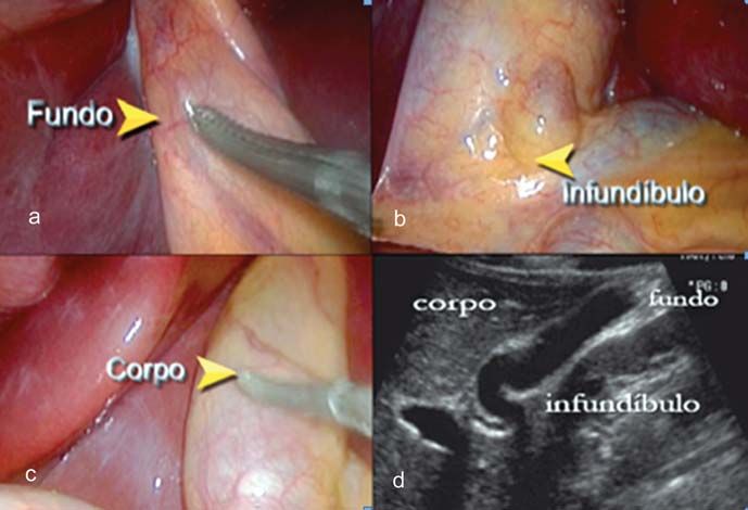
gallbladder is divided into the infundibu-
lum, body and fundus (Figure 1), and its
walls comprise four layers: a mucosa formed
by a simple columnar epithelium and by a
basal lamina; a second layer comprising
irregular muscular tissue; a third layer con-
stituted by loose connective tissue; and a
last layer formed by the serosa(8–10). Its
function is to store the bile, and presents a
volume of 30 to 50 ml(6).
Gallbladder US is routinely performed
with a convex transducer. In order to ac-
quire appropriate images, a systematic
scanning should be carried out with longi-
tudinal and cross sectional views of the
organ, evaluating its shape, dimensions,
wall thickness, regularity and texture pat- Figure 1. Laparoscopic anatomy (a,b,c) compared with sonographic anatomy (d) demonstrating gall-
tern of its walls and contents, besides bladder infundibulum, body and fundus.
locoregional and Doppler velocimetric al-
terations(8). In order to assist the sono- ening” is related to erroneous insonation by frequently greater than 10 mm(7). The pres-
graphic evaluation, the apparatuses are the transducer. In this case, the performance ence of some associated signs allows the
equipped with resources that enhance the of maneuvers changing the decubitus is observer to direct the diagnosis towards a
methods accuracy, such as the harmonic useful in the correct definition of the gall- more specific etiology(9–11). Among such
imaging, which allows increased lateral bladder wall thickness. An important dif- signs, the following can be mentioned: bil-
resolution, signal-noise and contrast-noise ferential diagnosis in these cases is the iary tract dilation, presence of a static gall-
ratios(9). functional change of the gallbladder (10,11) stone, perivesicular fluid, hilar lymph node
Sonographic images provide a faithful (Figure 2). enlargement, perivesicular fat heterogene-
representation of the gallbladder which can Gallbladder wall thickening is classi- ity and increased gallbladder transverse
be correlated with its anatomical structure. fied as mild (between 4 and 7 mm), marked diameter. The disorders that cause gallblad-
By means of US it is possible to identify (> 7 mm), and in focal or diffuse. As a rule, der wall thickening may be classified as in-
three layers: the innermost, corresponding systemic diseases such as heart, renal or flammatory, neoplastic and systemic, and
to the mucosa, that is linear, echogenic and hepatic failure cause diffuse and less marked their differentiation may be obtained by
presents a regular surface; the second one, thickening, contrary to tumor lesions that means of a combined evaluation of clini-
corresponding to the muscular layer, is thin cause focal and more exuberant thickening, cal and imaging findings.
and slightly hypoechoic; and outermost
layer corresponding to the organ’s serosa,
that is linear, echogenic and regular(1,9).
According to several authors(1,2), the up-
per limit for normality of the gallbladder
wall thickness is 3 mm. However, in pa-
tients under inappropriate fasting, the pa-
rietal thickness may exceed such a limit
because of the organ’s smooth muscle con-
traction(8). So, 8-hour fasting before the ex-
amination is recommended, particularly in
cases where the gallbladder is the focus of
the study. The main differential diagnosis
of parietal thickening is that of functional
changes of the organ, in which one ob- a b
serves a persistently withered gallbladder,
Figure 2. Patient with dyspepsia. Observe the withered gallbladder with thickened wall (arrow) at the first
even at a re-evaluation after extended fast- evaluation (a), appearance which is maintained after 12-hour fasting (arrowheads on b). The sonographic
ing(9). Another cause of “pseudothick- appearance suggests gallbladder dysmotility.
382 Radiol Bras. 2011 Nov/Dez;44(6):381–387
Barbosa ABR et al. Gallbladder wall thickening

INFLAMMATORY CAUSES                              inflammatory signs and gallbladder wall               ence of a pancreatic head tumor or primary
                                                 thickening, similar to acute cholecystitis.           sclerosing cholangitis(11) (Figure 5).
Acute calculous cholecystitis                    Magnetic resonance imaging (MRI) and
    It is the most common inflammatory           MRI cholangiography are very useful in                Chronic calculous cholecystitis
complication that affects the gallbladder,       such cases, particularly to rule out the pres-            It consists of an inflammatory process
and is related to choledocholithiasis in 90–                                                           of the gallbladder, originated from a tran-
95% of the cases. It is the fourth most com-                                                           sitory gallbladder obstruction, causing in-
mon cause of acute abdomen requiring hos-                                                              flammation and fibrosis(11,12). Porcelain
pitalization(3). In 95% of cases it is caused                                                          gallbladder is a rare presentation of chronic


by persistent obstruction by stones in the                                                             cholecystitis, where the gallbladder walls
infundibulum or in the cystic duct. In spite                                                           are partially or completely calcified. In
of not being pathognomonic, acute calcu-                                                               spite of the lack of consensus, many au-
lous cholecystitis is the main cause of gall-                                                          thors consider that the inflammatory pro-
bladder wall thickening at US. In general,                                                             cess represents a risk factor for gallbladder
the gallbladder wall is less than 7 mm thick,                                                          carcinoma, and, even being it an acciden-
presenting regular contour and trilaminar                                                              tal finding in asymptomatic patients sub-
appearance(3,9,11). Such echotextural ap-                                                              mitted to routine US examinations, many
                                                 Figure 3. Emphysematous cholecystitis. Observe
pearance of the gallbladder walls may            echogenic parietal images of the gallbladder, with    advocate the prophylactic cholecystec-
change, for example in cases of emphyse-         reverberation compatible with gas (arrow).            tomy(1,3,13) (Figure 4).
matous cholecystitis, where echogenic pa-
rietal images with acoustic reverberation
compatible with gas are observed(12) (Fig-
ure 3).
    Other sonographic findings are impor-
tant, as they increase the method specific-
ity, such as: impacted calculus in the com-
mon bile duct with upstream dilatation, in-
fundibular calculi, tense gallbladder with
a transverse diameter > 4 cm (hydrops of
the gallbladder), positive painful decom-
pression at the cystic point (sonographic
Murphy’s sign), presence of perivesicular
fluid and hyperflow from its walls at Dop-
pler(5) (Figure 4).
    The US sensitivity ranges between 80%         a                                                           b
and 100%, and specificity ranges between
                                                 Figure 4. Female, 45-year-old patient with severe abdominal pain in the right hypochondrium, irradiat-
60% and 100%. The positive predictive            ing to the scapular region. Positive sonographic Murphy’s sign. Figures (a,b) demonstrate tense gallblad-
value in the identification of calculi is 88%,   der with thickened walls and presence of gallstones.
increasing to 92% as associated with
sonographic Murphy’s sign. Gallbladder
                                                   a                                                               b
wall thickening associated with the
Murphy’s sign has a predictive value of up
to 94%(11,12).
    Another rare condition that determines
gallbladder wall thickening associated with
inflammatory process is the Mirizzi syn-
drome. In such a situation, an impacted
gallstone located in the gallbladder neck or
in the cystic duct causes dilatation of the
biliary tract, causing compression of the
common hepatic duct or secondary inflam-
mation, producing edema or fibrosis on the
duct wall. At US, besides the impacted
                                                 Figure 5. Mirizzi syndrome. a: Oblique coronal T2-weighted sequence. b: MRI cholangiography with vol-
calculus, one observes a distal common           ume rendering. In this case, the presence of impacted gallstone in the cystic duct, causing dilatation of
bile duct with normal caliper, peribiliary       the biliary tract and compression of the common hepatic duct.

Radiol Bras. 2011 Nov/Dez;44(6):381–387                                                                                                             383
Barbosa ABR et al. Gallbladder wall thickening
Acalculous cholecystitis
Acalculous cholecystitis is an uncom-
mon and severe entity, affecting patients
with diabetes and in poor general condi-
tions. It is more common in hospitalized
patients (undergoing mechanical ventila-
tion and hyperalimentation therapy) and
trauma victims, or in extensive burn pa-
tients, with a high mortality rate. Such a
condition was described in 1970, in seri-
ously wounded soldiers during the Vietnam a b
war(14).
Figure 7. Diffuse and unilateral pyelonephritis. Upper abdominal US (a) did not identify gallstones or
During the interpretation of the sono-
increase in gallbladder’s diameter, however the walls were thickened. At contrast-enhanced abdominal
graphic findings, i.e., gallbladder wall thick- CT (b), heterogeneous nephrogram and increased volume of the right kidney are observed, leading to
ening, tense and distended gallbladder, and gallbladder wall thickening (arrow).
presence of perivesicular fluid, the corre-
lation with the clinical context is of utmost with adenocarcinoma(17,18). At macroscopic Cholesterol polyp
importance for a correct diagnosis(3,14). The examination, one observes a nodular thick- Focal and nodular gallbladder wall
absence of sonographic Murphy’s sign does ening of the walls in association with the thickening represent approximately 50% of
not rule out the diagnosis(14,15) (Figure 6). presence of calculi and possible loco- all polypoid lesions, and most of the times
regional infiltration. Lymph nodes enlarge- do not present a malignant potential(1). The
ment and coexistence with gallbladder can- patients do not present any symptoms and
cer may also be found. at US, a static echogenic well defined
Its main sonographic sign is diffuse nodular image is identified. The main dif-
gallbladder wall thickening, besides hypo- ferential diagnoses include adenoma and
echoic nodules, which may be found in up adenocarcinoma(1,19).
to 35% of the patients(3,17,18). Such cases Two-dimensional US is not capable of
may be undistinguishable from the infiltra- differentiating small neoplastic polypoid
tive presentation of gallbladder carci- lesions from non-neoplastic ones, but some
noma(18). Clinically, it manifests with a studies have demonstrated the usefulness
clinical picture of cholecystitis in women of three-dimensional US in the differential
aged from 60 to 70 years. diagnosis between polyps (Figure 8). In
Figure 6. Acalculous cholecystitis. Observe diffuse
gallbladder wall thickening, with flow at color Dop- Adenomyomatosis of the gallbladder such cases, MRI may be very useful in that
pler and a minor, adjacent fluid collection. All these differentiation(20).
findings are frequently observed in cholecystitis, and
It is characterized by excessive prolif-
in the present case is not associated with the pres- eration of the surface epithelium towards Porcelain gallbladder
ence of gallstones. the Rokitansky-Aschoff sinuses, determin- It is an uncommon chronic cholecysti-
ing wall thickening that may be focal, seg- tis variation characterized by extensive cal-
Acalculous cholecystitis is frequently mental or diffuse(5). The main sonographic
misdiagnosed, as some sonographers presentation corresponds to segmental pa-
equivocally attribute to chronic acalculous rietal thickening with multiple echogenic
cholecystitis, the secondary thickening de- intramural foci, which cause posterior re-
termined by systemic pathologies such as verberation artifacts, known as comet-tail
pyelonephritis(16), for example. Another artifacts. Other signs such as gallbladder
common erroneous interpretation occurs in distension, presence of perivesicular fluid
cases of acute cholecystitis caused by non- or positive sonographic Murphy’s sign are
visualized small obstructive calculi in the not observed(3,18).
common bile duct(3) (Figure 7). It is a benign non-inflammatory condi-
tion of the gallbladder, found in 8.7% of the
Xanthogranulomatous cholecystitis cholecystectomies(5). It manifests with per-
It is an uncommon pathology, described sistent pain in the right hypochondrium,
in the early 1980’s as a pseudotumor pre- and is most commonly found in women, in Figure 8. Gallbladder cholesterolosis with polypoid
sentation of chronic calculous cholecysti- association with gallstones in 90% of the image in the fundus. Echogenic points with rever-
beration on the gallbladder walls, corresponding to
tis, secondary to bile extravasation through cases. Upon symptoms persistence, chole- cholesterolosis (arrowheads) associated with poly-
the gallbladder walls, frequently associated cystectomy is indicated(5,15). poid lesion in the gallbladder fundus (arrow).
384 Radiol Bras. 2011 Nov/Dez;44(6):381–387Barbosa ABR et al. Gallbladder wall thickening
cification of the gallbladder walls, which times achieved at advanced stages of the parietal lesion projecting towards its lu-
can be partially or completely affected. The disease, because of the scarcity of symp- men. Its most frequent presentation is a
term “porcelain” is utilized because of its toms. When present, the initial symptoms large solid lesion in the gallbladder fossa
consistency and appearance (Figure 9). Its are nonspecific and include weight loss, in association with calculi and extending to
prevalence in cholecystectomies is 0.06% abdominal pain, fever and jaundice(1,2,7,16) the liver and adjacent organs (Figure 10).
to 0.8%. In 95% of cases, association with and are frequently associated with the pres- When focal or asymmetric wall thick-
cholelithiasis is found. It is five times more ence of calculi (73% to 98%)(6). Only 1% ening > 10 mm is found, the possibility of
common in women than in men, and is most of the cholecystectomies performed for neoplasia is high. In such cases the charac-
frequent in the fifth and sixth decades of cholelithiasis result in incidental finding of terization of other factors such as loco-
life(7,12). Early in the 20th century, one be- gallbladder carcinoma(2). The most signifi- regional lymph nodes enlargement en-
lieved that there was an association with cant risk factor is the presence of chronic hances the diagnostic hypothesis. Com-
neoplasia, but most recently, studies have inflammatory process usually triggered by puted tomography (CT) presents a charac-
not confirmed the initial findings, thus dem- the calculi. The main differential diagnoses teristic enhancement pattern that is typical
onstrating a low incidence of coexistence include complicated acute cholecystitis, of lesions suspicious for malignancy, with
of neoplasia with porcelain gallbladder(20,21). hepatocellular carcinoma and metastasis to iodinated contrast uptake in the arterial
the gallbladder fossa. phase, becoming isodense in the equilib-
NEOPLASTIC CAUSES Adenocarcinoma is the malignant his- rium phase(22,23). Magnetic resonance im-
tological type of tumor that most frequently aging demonstrates hyperintense and het-
Gallbladder carcinoma affects the gallbladder, occurring in 90% of erogeneous images on T2-weighted se-
It is the most common neoplasia of the cases. Such tumor generally presents three quences and hypointense on T1-weighted
biliary system, with 2.5 new cases per image patterns: a) mass occupying and sequences, with post-contrast enhance-
100,000 inhabitants per year. It has a high obscuring the gallbladder bed; b) focal or ment. In the cases of diffuse thickening
mortality rate as its diagnosis is most of diffuse parietal thickening; c) polypoid with uniform infiltration, its imaging ap-
pearance is similar to that of chronic chole-
cystitis(21).
The accurate differentiation between
malignant and benign polypoid lesions can-
not be made by means of US alone. Gen-
erally, malignant polyps are > 1 cm and
seldom present calcification and necrosis.
They present early and prolonged contrast
uptake after administration of gadolinium,
contrary to benign lesions, which present
early contrast uptake with subsequent
washout(21–23).
a b Metastasis to the gallbladder
Figure 9. Abdominal US and radiograph of a 50-year-old patient presenting with abdominal discomfort. Some tumors, such as carcinoid tumor,
Observe porcelain gallbladder (arrows), with thin calcifications on its wall. lymphoma, breast carcinoma and sarcomas
a b c
Figure 10. Gallbladder carcinoma associated with lithiasis. Observe dilatation of intrahepatic biliary ducts (a – arrows), lymph node enlargement in the hepatic
hilum (b – arrow) and ill-defined lesion associated with gallstones (c).
Radiol Bras. 2011 Nov/Dez;44(6):381–387 385Barbosa ABR et al. Gallbladder wall thickening
metastasize to the gallbladder, thus being mine gallbladder wall thickening(3,4,9,20,25) in ascites(7). Hypoalbuminemia is the ab-
a possible cause of gallbladder wall thick- (Figure 8). normality most frequently associated with
ening. Among them, the most common one In cases of viral hepatitis, regular and dif- ascites in children(1). Ascites may be a con-
is the melanoma, representing approxi- fuse gallbladder wall thickening is observed. sequence of benign and malignant disor-
mately 50% of cases(24). Such lesions are The gallbladder is withered, in association ders. Some reports suggest that the gall-
undistinguishable from primary neoplasia, with the presence of ganglia adjacent to the bladder wall thickening is more frequently
but are much less frequently found and not hilum and hepatomegaly, besides malaise, found in benign diseases, while malignant
associated with gallstones. fatigue, arthralgia and jaundice(2,17). conditions do not cause gallbladder wall
Cases of transinfection by hepatitis in- thickening (Figure 12).
clude diseases such as acquired immune Cardiac liver is a clinical condition
SYSTEMIC CAUSES
deficiency syndrome (AIDS), dengue and found in individuals presenting with right
Inflammatory processes located in the malaria. In patients with AIDS, such find- heart failure. The physiopathological
right hypochondrium that do not originate ing may be secondary to the utilization of mechanism of the gallbladder wall thick-
from the gallbladder and systemic disorders antiretroviral drugs, worsened nutritional ening is related to increased intrahepatic
may mimic acute cholecystitis, determining status and opportunistic infections of the pressure, determining edema in the second
symmetrical and diffuse wall thickening. biliary tract(17) (Figure 11). layer of the gallbladder wall associated
Such changes are caused by the extension Hepatic dysfunctions such as cirrhosis, with preservation of the hyperechogenic
of the inflammation or by the increase in malnutrition and ascites, cause parietal appearance of the mucosa. Other findings,
the portal venous pressure associated with thickening secondary to hypoalbuminemia such as ectasia of hepatic veins and lower
decrease in the intravascular osmotic pres-
sure, causing parietal edema. The assess-
ment of a patient presenting with one of
such disorders should be undertaken with
caution in order to avoid erroneous inter-
pretation of a picture of acalculous chole-
cystitis. Clinical and laboratory tests results,
signs of pneumoperitoneum, inflamed di-
verticula in the right/transverse colon, in-
flammation of the appendix with upper lo-
cation, pyelonephritis signs allow the defi-
nition of the anatomical diagnosis. Even so,
in several situations, the previous epidemio-
logical history plays an extremely relevant
Figure 11. Female patient, sugar cane field worker, presenting with fever, malaise, chills and jaundice.
role for the diagnostic orientation, more-
Ultrasonography (a,b) demonstrated reactive gallbladder wall thickening due to systemic causes. Echogenic
over in cases of infectious diseases(4,6,23). inner layer (mucosa) is preserved. Presence of Plasmodium sp was identified in the peripheral blood.
In the evaluation of such disorders, the
key sonographic finding is the preservation
of mucosal regularity and echogenicity, i.e.,
of the first layer. The wall thickening oc-
curs at the expense of the hypoechoic layer
corresponding to edema of the muscular
Figure 12. Examples of systemic
layer and of the conjunctive tissue(1,4,5). causes of gallbladder wall thick-
Acute pancreatitis is a common disease ening. Patient with long-term al-
in our environment and its main causes are coholism presenting with in-
creased abdominal volume and
those of biliary and alcoholic origin. Ap- hematemesis. Gallbladder wall
proximately 64% of the patients presenting thickening secondary to ascites is
with pancreatitis evolve with gallbladder observed (a), associated with
chronic hepatopathy. The three-
wall thickening(3,23) secondary to extension dimensional appearance of the
of the inflammatory process towards gallbladder (b) confirms the find-
locoregional structures(3–5). ings at the conventional mode.
AIDS patient undergoing anti-
Based on this physiopathological prin- retroviral therapy presenting with
ciple, any inflammatory process located in abdominal pain (c). Ultrasonog-
the right hypochondrium, such as perfo- raphy demonstrated gallbladder
wall thickening with reactive as-
rated duodenal ulcer, acute diverticulitis, pect. The echogenic inner layer is
appendicitis and pyelonephritis, can deter- clearly preserved (arrow).
386 Radiol Bras. 2011 Nov/Dez;44(6):381–387Barbosa ABR et al. Gallbladder wall thickening
vena cava, Doppler velocimetric alterations 5. Rosenthal SJ, Cox GG, Wetzel LH, et al. Pitfalls sonography in intensive care unit patients. AJR
and hepatomegaly are also frequently and differential diagnosis in biliary sonography. Am J Roentgenol. 2000;174:973–7.
Radiographics. 1990;10:285–311. 16. Campos FA, Rosas GQ, Goldenberg D, et al. Fre-
found in this clinical condition(6).
6. Watanabe Y, Nagayama M, Okumura A, et al. MR qüência dos sinais de pielonefrite aguda em pa-
imaging of acute biliary disorders. Radiographics. cientes submetidos a tomografia computadori-
2007;27:477–95. zada. Radiol Bras. 2007;40:309–14.
CONCLUSION 7. van Breda Vriesman AC, Engelbrecht MR, Smith- 17. Parra JA, Acinas O, Bueno J, et al. Xanthogranulo-
uis RH, et al. Diffuse gallbladder wall thickening: matous cholecystitis: clinical, sonographic, and
Ultrasonography is the method of differential diagnosis. AJR Am J Roentgenol. CT findings in 26 patients. AJR Am J Roentgenol.
choice for the study of the gallbladder, with 2007;188:495–501. 2000;174:979–83.
a high sensitivity in the detection of gall- 8. AIUM practice guideline for the performance of 18. Ros PR, Goodman ZD. Xanthogranulomatous
bladder wall thickening. Such a finding is an ultrasound examination of the abdomen and/ cholecystitis versus gallbladder carcinoma. Radi-
or retroperitoneum. J. Ultrasound Med. 2008;27: ology. 1997;203:10–2.
not a synonymous for acute cholecystitis. 319–26. 19. Boscak AR, Al-Hawary M, Ramsburgh SR. Best
The correlation with other sonographic, 9. Hong HS, Han JK, Kim TK, et al. Ultrasono- cases of the AFIP: Adenomyomatosis of gallblad-
clinical, laboratory and epidemiological graphic evaluation of the gallbladder: comparison der. Radiographics. 2006;26:941–6.
of fundamental, tissue harmonic, and pulse inver- 20. Xu HX, Yin XY, Lu MD, et al. Comparison of
findings is of utmost importance in order
sion harmonic imaging. J Ultrasound Med. 2001; three- and two-dimensional sonography in diag-
to avoid unnecessary cholecystectomies. 20:35–41. nosis of gallbladder diseases: preliminary expe-
10. Feldman MK, Katyal S, Blackwood MS. US ar- rience. J Ultrasound Med. 2003;22:181–91.
REFERENCES
tifacts. Radiographics. 2009;29:1179–89. 21. Catalano OA, Sahani DV, Kalva SP, et al. MR
1. Levy AD, Murakata LA, Abbott RM, et al. From 11. Smith EA, Dillman JR, Elsayes KM, et al. Cross- Imaging of the gallbladder: a pictorial essay.
the archives of the AFIP: Benign tumors and tu-
sectional imaging of acute and chronic gallblad- Radiographics. 2008;28:135–55.
morlike lesions of the gallbladder and extrahe-
der inflammatory disease. AJR Am J Roentgenol. 22. Zissin R, Osadchy A, Shapiro-Feinberg M, et al.
patic bile ducts: radiologic-pathologic correlation.
2009;192:188–96. CT of a thickened-wall gall bladder. Br J Radiol.
Radiographics. 2002;22:387–413.
12. Handler SJ. Ultrasound of gallbladder wall thick- 2003;76:137–43.
2. Wibbenmeyer LA, Sharafuddin MJ, Wolverson
ening and its relation to cholecystitis. AJR Am J 23. Yun EJ, Cho SG, Park S, et al. Gallbladder carci-
MK, et al. Sonographic diagnosis of unsuspected
Roentgenol. 1979;132:581–5. noma and chronic cholecystitis: differentiation
gallbladder cancer: imaging findings in compari-
son with benign gallbladder conditions. AJR Am 13. Riñón C, de Mingo L, Cortés MJ, et al. Preopera- with two-phase spiral CT. Abdom Imaging.
J Roentgenol. 1995;165:1169–74. tory sonography efficiency in paediatric patients 2004;29:102–8.
3. Spence SC, Teichgraeber D, Chandrasekhar C. with cholelithiasis undergoing laparoscopic 24. Martel JP, McLean CA, Rankin RN. Melanoma
Emergent right upper quadrant sonography. J cholecystectomy. Cir Pediatr. 2009;22:34–8. of the gallbladder. Radiographics. 2009;29:291–
Ultrasound Med. 2009;28:479–96. 14. Lindberg EF, Grinnan GL, Smith L. Acalculous 6.
4. Patriquin HB, DiPietro M, Barber FE, et al. cholecystitis in Viet Nam casualties. Ann Surg. 25. Hanbidge AE, Buckler PM, O’Malley ME, et al.
Sonography of thickened gallbladder wall: causes 1970;171:152–7. From the RSNA refresher courses: imaging evalu-
in children. AJR Am J Roentgenol. 1983;141:57– 15. Boland GW, Slater G, Lu DS, et al. Prevalence and ation for acute pain in the right upper quadrant.
60. significance of gallbladder abnormalities seen on Radiographics. 2004;24:1117–35.
Radiol Bras. 2011 Nov/Dez;44(6):381–387 387You can also read