Genetic landscape of 6089 inherited retinal dystrophies affected cases in Spain and their therapeutic and extended epidemiological implications
←
→
Page content transcription
If your browser does not render page correctly, please read the page content below
www.nature.com/scientificreports
OPEN Genetic landscape of 6089
inherited retinal dystrophies
affected cases in Spain and their
therapeutic and extended
epidemiological implications
Irene Perea‑Romero1,2,59, Gema Gordo1,59, Ionut F. Iancu1,2,59, Marta Del Pozo‑Valero1,2,
Berta Almoguera1,2, Fiona Blanco‑Kelly1,2, Ester Carreño3, Belen Jimenez‑Rolando3,
Rosario Lopez‑Rodriguez1, Isabel Lorda‑Sanchez1,2, Inmaculada Martin‑Merida1,2,
Lucia Pérez de Ayala1, Rosa Riveiro‑Alvarez1,2, Elvira Rodriguez‑Pinilla1,
Saoud Tahsin‑Swafiri1,2, Maria J. Trujillo‑Tiebas1,2, The ESRETNET Study Group*, The
ERDC Study Group*, The Associated Clinical Study Group*, Blanca Garcia‑Sandoval2,3,
Pablo Minguez1,2, Almudena Avila‑Fernandez1,2, Marta Corton1,2,59* & Carmen Ayuso1,2,59*
Inherited retinal diseases (IRDs), defined by dysfunction or progressive loss of photoreceptors,
are disorders characterized by elevated heterogeneity, both at the clinical and genetic levels. Our
main goal was to address the genetic landscape of IRD in the largest cohort of Spanish patients
reported to date. A retrospective hospital-based cross-sectional study was carried out on 6089 IRD
affected individuals (from 4403 unrelated families), referred for genetic testing from all the Spanish
autonomous communities. Clinical, demographic and familiar data were collected from each patient,
including family pedigree, age of appearance of visual symptoms, presence of any systemic findings
and geographical origin. Genetic studies were performed to the 3951 families with available DNA
using different molecular techniques. Overall, 53.2% (2100/3951) of the studied families were
genetically characterized, and 1549 different likely causative variants in 142 genes were identified. The
most common phenotype encountered is retinitis pigmentosa (RP) (55.6% of families, 2447/4403).
The most recurrently mutated genes were PRPH2, ABCA4 and RS1 in autosomal dominant (AD),
autosomal recessive (AR) and X-linked (XL) NON-RP cases, respectively; RHO, USH2A and RPGR in
AD, AR and XL for non-syndromic RP; and USH2A and MYO7A in syndromic IRD. Pathogenic variants
c.3386G > T (p.Arg1129Leu) in ABCA4 and c.2276G > T (p.Cys759Phe) in USH2A were the most frequent
variants identified. Our study provides the general landscape for IRD in Spain, reporting the largest
cohort ever presented. Our results have important implications for genetic diagnosis, counselling and
new therapeutic strategies to both the Spanish population and other related populations.
Inherited retinal diseases (IRDs) are one of the most heterogeneous clinical and genetical disorders known among
all human medical conditions, characterized by the progressive loss of photoreceptor cells, resulting in severe
visual impairment1. IRDs are classified as rare diseases, and their estimated prevalence is about 1 in 1 0002–40001.
IRDs can be classified according to different clinical or genetic criteria, based upon the primary retinal cell
1
Department of Genetics, Health Research Institute-Fundación Jiménez Díaz University Hospital, Universidad
Autónoma de Madrid (IIS-FJD, UAM), Madrid, Spain. 2Center for Biomedical Network Research on Rare Diseases
(CIBERER), Instituto de Salud Carlos III, Madrid, Spain. 3Department of Ophthalmology, Health Research
Institute-Fundación Jiménez Díaz University Hospital, Universidad Autónoma de Madrid (IIS-FJD, UAM), Madrid,
Spain. 59These authors contributed equally: Irene Perea-Romero, Gema Gordo, Ionut F. Iancu, Marta Corton
and Carmen Ayuso. *A list of authors and their affiliations appears at the end of the paper. *email: mcorton@
quironsalud.es; cayuso@fjd.es
Scientific Reports | (2021) 11:1526 | https://doi.org/10.1038/s41598-021-81093-y 1
Vol.:(0123456789)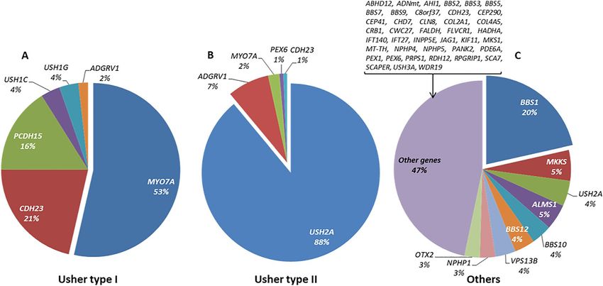
www.nature.com/scientificreports/
affected (rods, cones, retinal pigment epithelium (RPE), bipolar cells or ganglion cells), the ophthalmological
findings and/or the affected gene found after the genetic testing.
All modes of inheritance can be observed (autosomal dominant (AD), autosomal recessive (AR), X-linked
(XL), including rare non-Mendelian forms such as mitochondrial or digenic inheritance patterns). Age of onset of
first symptoms (from early childhood to adulthood), rate of progression, association with extra-ocular symptoms
(non-syndromic versus syndromic forms) or causative gene can also help to subclassify the different phenotypes3.
The most prevailing form of IRDs is Retinitis Pigmentosa (RP [MIM: 268000]), which is estimated to affect
approximately 1.5 million people worldwide4. RP begins with the degeneration of rod photoreceptors, resulting
in night blindness and characteristic pigmentary changes in the peripheral retina. This is also considered a rod-
cone dystrophy due to the subsequent cone photoreceptor death in later stages. Other forms of IRD, including
cone-dominated diseases, are characterized by photophobia, reduced visual acuity and impaired colour vision (i.e.
cone dystrophy (CD), achromatopsia, blue cone monochromatism), as well as generalized retinal degeneration
involving simultaneously both cones and rods such as in rod-cone or cone-rod dystrophies (CRD). The most
severe form of non-syndromic IRDs is Leber congenital amaurosis (LCA) characterized by congenital or early
childhood blindness. Other IRD forms are characterized by central vision loss affecting primarily the macula and
are therefore acknowledged as Macular Dystrophies (MD), such as Stargardt disease (STGD1 [MIM: 248200])
and Best Vitelliform Macular Dystrophy (VMD2 [MIM: 153700])1.
Conversely, syndromic IRDs are subclassified according to the type of syndrome. The most prevalent is Usher
syndrome (sensorineural hearing loss and RP) which can be further subclassified to Usher type I (USH1 [MIM:
276900]), type II (USH2 [MIM: 276901]) and type III (USH3 [MIM: 276902]), and other syndromes such as
Bardet-Biedl (BBS [MIM: 209900]) or Alström (ALMS [MIM: 203800])3.
Since the identification of rhodopsin (RHO [MIM: 180380]) in 1990 as the first gene involved in the develop-
ment of AD-RP5, 270 genes have been additionally described as causative of IRDs (RetNet, Retinal Information
Network; https://sph.uth.edu/retnet/; accessed on March 2020). Some of these genes have been reported in only
few families worldwide; therefore, their individual contribution to IRD prevalence is relatively small. The most
prevalent IRD-causing genes across all populations are ABCA4 (MIM: 601691), RHO, USH2A (MIM: 608400)
and RPGR (MIM: 312610), which account for high percentages of some of the IRD subtypes, i.e. 70–71% in
STGD1/AR-MD/AR-CRD6, 19–25% of AD-RP, 10% of AR-RP, and 70% of XL-RP, r espectively1. In addition,
although the majority of the causative variants are private, some are more frequent, especially in Spanish families,
such as USH2A (GenBank: NM_206933.3) c.2299delG (p.Glu767SerfsTer21) and c.2276G > T (p.Cys759Phe)7 or
ABCA4 (GenBank: NM_000350.3) c.3386G > T (p.Arg1129Leu) and c.5882G > A (p.Gly1961Glu)6.
The aim of this study is to present a comprehensive overview of the largest cohort of IRD patients ever
reported worldwide and related to IRD in the Spanish population. The presented data includes the presumed
inheritance pattern for the different phenotypic subtypes, mutational spectrum, prevalence of genes carrying
likely pathogenic variants and the recurrence of disease related variants.
Results
Prevalence of IRD in Spain. The number of cases diagnosed as having IRD in our hospital (until August
2019) was 6089, and the last Spanish population registry accounted for 46,722,980 habitants giving us a minimal
prevalence of 1:7673 (confidence interval (CI):1:7485–1:7871). Regional distribution of cases and prevalence can
be seen in Fig. 1A,B. Considering a worldwide IRD prevalence of 1:10002–40001, our cohort would represent
20–53% of the total patients with IRD in Spain as shown in Supplementary Table S1.
For non-syndromic IRD, our cohort was grouped in 2 categories: 1703 affected individuals (1335 families), in
cone-dominated phenotypes—hereafter “NON-RP”—and 3561 affected cases from 2447 unrelated families within
primarily rod affection—hereafter “RP”. This resulted in a minimal prevalence of 1:27,436 (CI:1:26,192–1:28,804)
and 1:13,121 (CI:12,704–13,566) for NON-RP and RP, respectively. Additionally, syndromic IRD forms repre-
sented the smallest fraction accounting for 13.6% of the patients with 825 affected individuals (621 families),
resulting in a minimal prevalence of 1:56,634 (CI:1:53,016–1:60,781).
In our cohort, we have a higher proportion of cases from Madrid area (26.7%; 1625/6089).
Initial classification of IRD families by clinical type and suspected mode of inheritance prior
to genetic testing. Non-syndromic NON-RP and RP cases were categorized by the mode of inheritance
(Fig. 2A-I and A-II). Syndromic IRD were categorized by the specific type of suspected syndrome (Fig. 2A-III),
instead of inheritance type, given that most of them were sporadic (53.6%) or had recessive inheritance (40.2%).
The remaining 6.2% corresponded to dominant (0.5%), X-linked (0.7%), mitochondrial inherited disease (0.2%)
or non-classificable cases (4.8%).
According to this “a priori” diagnosis based on the clinical and familial history of the patients, the main
inheritance pattern in non-syndromic NON-RP and RP was recessive or sporadic, representing the 68% and
75% of cases, respectively. Autosomal dominant and X-linked forms accounted for 21% and 8% for NON-RP
and 15% and 8% for RP, respectively (Fig. 2A-I). Families with no familiar data were annotated as unclassified.
Non-syndromic RP represents the most common phenotype, representing 55.6% of families in our cohort.
In the present cohort, 47% of the syndromic IRD index cases (270/577) suffered from USH2, followed by 17%
USH1 (98/577), as well as other very rare syndromes like some atypical forms of Usher syndrome (3%; 16/577)
and ciliopathies such as BBS or ALMS (16%; 90/577). A miscellanea of non-ciliopathic syndromes or unclassified
symptoms were presented in 103 index cases.
Molecular studies. Diagnostic yield. Genetic testing was performed in a total of 3951 index cases with
available DNA6–11 (89.7% of the total cohort), including 1291 NON-RP, 2083 RP, and 577 syndromic IRD pa-
Scientific Reports | (2021) 11:1526 | https://doi.org/10.1038/s41598-021-81093-y 2
Vol:.(1234567890)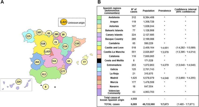
www.nature.com/scientificreports/
Figure 1. IRD affected cases distribution and estimated prevalence in Spain. (A) Distribution of RP/NON-RP
affected cases across Spain. Total cases: 6089 (known Spanish origin: 4668; unknown origin: 1421). Spanish map
modified using image editor from https://www.veomapas.com/mapa-mudo-de-las-comunidades-autonomas-
de-espana-m103.html. (B) IRD estimated prevalence in Spanish regions. Data was obtained from the number of
cases we had in our Hospital Service and the recorded population in the different regions. *Inconclusive data.
tients as shown in Supplementary Fig. S1. The genetic analysis procedures evolved over time since novel ge-
netic approaches were implemented in our laboratory. A definite genetic diagnosis was established for 53.2%
(2100/3951) of cases. For NON-RP families, 754 out of 1291 (58.4%) obtained a genetic diagnosis; in RP the
molecular cause was identified in 1038 out of 2083 (49.8%). Similarly, 53.4% (308/577) of syndromic IRD fami-
lies were genetically solved (Fig. 2B).
Final inheritance pattern and reclassification of characterized families. The identification of the
causative gene allowed us to reclassify the inheritance mode in 8.2% (146/1792) of the NON-RP and RP fami-
lies, thus establishing the final inheritance type (Supplementary Table S2). A comparison between the “a priori”
suspected inheritance based on the pedigree and the final inheritance suggested by the molecular diagnosis was
performed in characterized NON-RP and RP families (Fig. 2C). As expected, most sporadic NON-RP (n = 378)
and RP cases (n = 379) were confirmed as having AR inheritance after the genetic testing. The rest of the S cases
were reclassified to AD (n = 43) and XL (n = 36) (Supplementary Table S2). Twenty-four cases (11 NON-RP and
13 RP) with an initial unknown mode of inheritance were classified as: AD (n = 5), AR (n = 16), and XL (n = 3)
after the molecular testing.
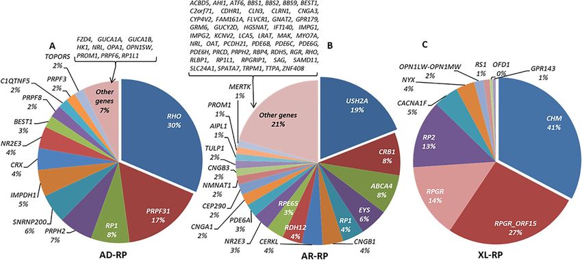
Gene landscape. In total, 1549 different pathogenic and likely pathogenic variants were identified in 142 differ-
ent genes. These included SNVs (Single Nucleotide Variants) and CNVs (Copy Number Variants). As showed in
Figs. 3, 4, 5, there was a wide spectrum of genes implicated in IRD, 121 of them represented in 1% or less of the
cohort. The 5 most frequent mutated genes were ABCA4, USH2A, RS1 (MIM: 300839), CRB1 (MIM: 604210)
and RHO.
In NON-RP families, PRPH2 (MIM: 179605), ABCA4 and RS1 were the most commonly mutated genes,
explaining 42.2%, 82.7% and 93.3% of the AD, AR and XL forms, respectively as shown in Supplementary
Table S3.
For non-syndromic RP, 207 AD-RP families were genetically characterized with heterozygous variants identi-
fied in one of 23 genes found. The most frequent mutated genes were RHO (65/207; 31.4%) and PRPF31 (MIM:
606419) (34/207; 16.4%). In AR-RP families, the most common gene mutated was USH2A (127/666; 19.1%).
However, the number of other disease genes detected was very high (N = 70) in the 666 families characterized.
For XL-RP families, both the number of cases and variety of genes were low (N = 9) as shown in Supplementary
Table S4. For these patients, 41.2% (68/165) of the index cases (all males) carried a hemizygous pathogenic variant
in RPGR (44 RPGR_ORF15 and 24 in the rest of RPGR regions), and 32.7% (54/165) in CHM (MIM: 300390).
A total of 53 cases out of the total 2100 (2.5%) were clinically reconsidered and reclassified after genetic test-
ing: 12 NON-RP were reclassified as RP (9/754; 1.2%; characterized with RHO, FSCN2, PRPF8, AHI1, CNGB1,
TRPM1 and CHM) or as syndromic IRD (3/754; 0.4%; COL11A1, CDH3 and MYO7A) and 37 RP as NON-RP
Scientific Reports | (2021) 11:1526 | https://doi.org/10.1038/s41598-021-81093-y 3
Vol.:(0123456789)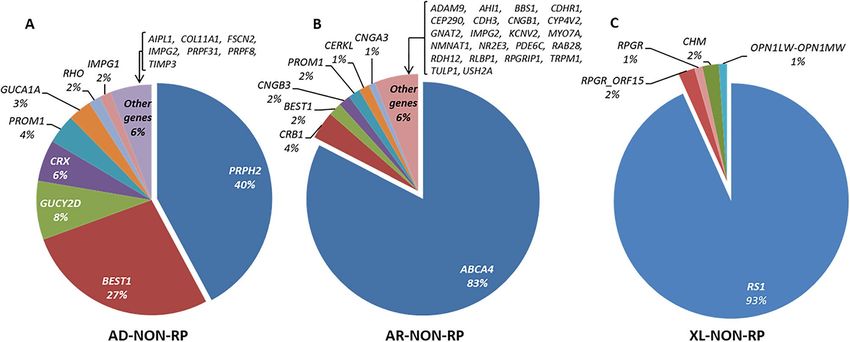
www.nature.com/scientificreports/

                                            Figure 2.  “A priori” and final classification of IRD affected cases. (A) “A priori” classification of IRD families
                                            with data obtained from the clinical and family history of the patients, before performing molecular tests:
                                            NON-RP (I), RP (II), and syndromic IRD (III), and subclassification according to the inheritance type in case of
                                            RP and NON-RP, and type of syndrome in case of syndromic IRD. Data obtained with the clinical and familiar
                                            history of the patients, before performing molecular tests. AD Autosomal Dominant, AR autosomal recessive,
                                            S sporadic, XL X-linked. (B) Proportion of genetically solved NON-RP, RP, syndromic IRD and total IRD.
                                            The diagnostic ratio in the different group of NON-RP and RP by the type of “a priori” inheritance and in the
                                            different group of syndromic IRD by type of syndrome is indicated. (C) Genetically solved families. Comparison
                                            of inheritance classification before (light gray) and after (dark gray) the molecular study was performed.

                                            Figure 3.  Classification of mutated genes in the genetically solved NON-RP affected cases. Below each gene is
                                            given the percentage that each gene was mutated in the cohort. Total characterized families: AD-NON-RP: 121;
                                            AR-NON-RP: 544; XL-NON-RP: 89.

          Scientific Reports |   (2021) 11:1526 |                https://doi.org/10.1038/s41598-021-81093-y                                                  4

Vol:.(1234567890)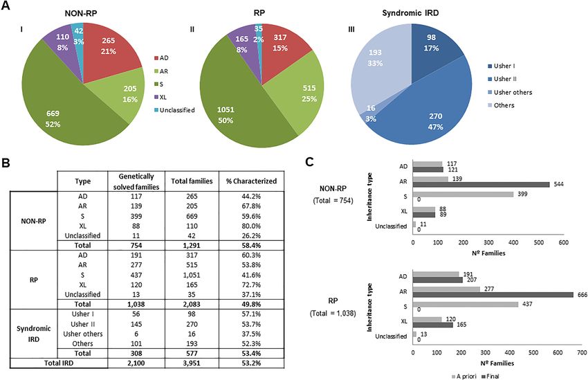
www.nature.com/scientificreports/

                                  Figure 4.  Classification of mutated genes in the genetically solved RP affected cases. Below each gene is given
                                  the percentage that each gene was mutated in the cohort. Total characterized families: AD-RP: 207; AR-RP: 666;
                                  XL-RP: 165.

                                  Figure 5.  Classification of mutated genes in the genetically solved syndromic IRD affected cases. Below each
                                  gene is given the percentage that each gene was mutated in the cohort. Total characterized families: Usher I: 56;
                                  Usher II: 145; Others (including atypical Usher): 107.

                                  (32/1038; 3.1%; C1QTNF5, GUCA1A, CNGB3, CNGA3, ACBD5, GNAT2, PDE6C, ATF6, PDE6H, RS1 and
                                  OPN1LW-OPN1MW), syndromic IRD (1/1038; 0.1%; MYO7A) and other visual diseases, such as exudative
                                  familiar vitreoretinopathy, optic atrophy, albinism and gyrate atrophy (4/1038; 0.4%; FZD4, OPA1, GPR143 and
                                  OAT). Moreover, 4 syndromic cases were reclassified to RP group (4/308; 1.3%; RDH12, PDE6A and RPGRIP1).
                                      Among syndromic IRD families, the causative gene was identified in 56 families with a diagnosis of USH1,
                                  145 of USH2, 6 of atypical Usher, and 101 of other syndromes including Bardet-Biedl or Alström syndrome,
                                  respectively. In USH1, biallelic variants in MYO7A (MIM: 276903) were identified in 30 out of 56 patients
                                  (53.5%). USH2A defects were the main cause of USH2 in 90% (129/145) of the patients. The group “others” was
                                  a clinically heterogeneous group of non-Usher cases with a total of 48 involved genes, with BBS1 (MIM: 209901)
                                  being the most frequent one (N = 23), as shown in Supplementary Table S5.
                                      In addition to clinical and genetic heterogeneity, the group of “others” included in syndromic IRD also
                                  presented unusual modes of inheritance, such as triallelism. In our cohort, 4 possible triallelic cases have been

Scientific Reports |   (2021) 11:1526 |                https://doi.org/10.1038/s41598-021-81093-y                                                     5

                                                                                                                                              Vol.:(0123456789)
www.nature.com/scientificreports/
Nº of total alleles of this
Spanish region Gene Nucleotide change Amino acid change Nº of alleles gene found in the region Frequency in the region
ABCA4 c.3386G > T p.Arg1129Leu 18 19.40%
Basque Country 93
ABCA4 c.5882G > A p.Gly1961Glu 5 5.40%
Castile-La Mancha ABCA4 c.3386G > T p.Arg1129Leu 8 60 13.30%
NON-RP
Madrid ABCA4 c.3386G > T p.Arg1129Leu 47 373 12.60%
ABCA4 c.3386G > T p.Arg1129Leu 8 11.30%
Murcia 71
ABCA4 c.5882G > A p.Gly1961Glu 7 9.90%
USH2A c.2276G > T p.Cys759Phe 11 13.30%
Murcia 83
PRCD c.64C > T p.Arg22Ter 6 7.20%
Non-syndromic RP and Canary Islands NR2E3 c.932G > A p.Arg311Gln 8 83 9.60%
syndromic IRD Castile-La Mancha USH2A c.2276G > T p.Cys759Phe 18 202 8.90%
Madrid USH2A c.2276G > T p.Cys759Phe 50 714 7.00%
Extremadura USH2A c.2276G > T p.Cys759Phe 5 87 5.70%
Table 1. Variants detected in > 5% of NON-RP, RP and syndromic IRD by Spanish regions. Regions with
variants detected below this percentage are not represented in the table, and neither are regions with less than
50 mutated alleles in total or variants that were presented less than 5 times. In bold the variant which not
appeared as one of the most frequent variants in Supplementary Table S6.
identified, all of them diagnosed with BBS, 3 patients carrying biallelic BBS1 variants together with one allele in
MKKS (MIM: 604896)9,12 and one additional case carrying biallelic MKKS variants and one allele in BBS5 (MIM:
603650). The phenotypic modifier effect of the triallelism could only be stablished in two of the 4 families. One
has two affected siblings with different phenotypic severity that correlates with the presence of the third allele;
and in the other both affected showed triallelism and have the same clinical manifestation. Although, it could
not be stablished in the rest of the families, as they were sporadic cases.
Most frequent variants. Our findings reflect the high allelic heterogeneity in IRD. We identified 458 different
disease-causing variants in 45 genes in cases “a priori” classified as NON-RP, as well as 836 in 94 genes in the
“a priori” RP cases and 295 in 55 genes in the “a priori” syndromic IRD. The most common pathogenic variant
detected in our NON-RP cohort was the previously known missense change ABCA4 c.3386G > T (p.Arg1129Leu)
(180 mutated alleles of 3,618; 5% of the total pathogenic alleles), presented in 21.5% (162/754) of the character-
ized families, in homozygous or compound heterozygous state in 18 and 144 families, respectively (Supplemen-
tary Table S6). Among the RP families, the most prevalent pathogenic variant was the missense change USH2A
c.2276G > T (p.Cys759Phe), identified in 106 alleles in 8.4% of the solved families (87/1038); in 19 cases in
homozygous and in 68 cases in compound heterozygous state. In addition, there were 15 other variants present
in more than 10 genes (Supplementary Table S6). Some of the mutated genes overlap in NON-RP (Fig. 3), RP
(Fig. 4) and syndromic IRD (Fig. 5).
Disease‑causing variant distribution in Spain. Analysis of causing variants by the different Spanish regions
resulted in a wide variety of disease-causing variants. Table 1 shows variants detected in more than 5% of char-
acterized families, by Spanish regions. All these variants are depicted in the Supplementary Table S6 of NON-RP
and RP most frequent causing variants, except for the nonsense variant PRCD (OMIM: 610598) (GenBank:
NM_001077620.3) c.64C > T (p.Arg22Ter), that was found in homozygosity in 3 families, representing 7.2%
(6/83) of the identified alleles from Murcia.
Discussion
This is the first and largest comprehensive study addressing the prevalence and epidemiology of IRD in the Span-
ish population. The cohort here described, comprising 6089 cases from 4403 unrelated families, is not based on
a national registry of IRD patients, but it is the outcome of a very wide recruiting effort of a single center over
the last 28 years. An increasing number of centers are currently performing clinical and/or genetic diagnosis of
IRD in Spain, therefore our cohort did not reflect all of IRD patients in our country. Hence, to date, no accurate
data about the IRD prevalence in the Spanish population is available.
In terms of representation of patients from the different Spanish regions, our cohort reflects a biased recruit-
ment, being enriched with patients from Madrid and the surrounding regions (i.e. Castile and Leon, Castile-La
Mancha, and Extremadura) probably due to the fact that our hospital has been their referral center during
most of the time of the study. Other areas like Andalusia, Catalonia, Navarre or the Valencian Community had
different referral centers and genetic testing is performed locally. In spite of these limitations, the large sample
size of our cohort and the exhaustive molecular analysis performed over the years, together with an overall low
genetic heterogeneity in the Spanish population13, have allowed a straightforward extrapolation of prevalent
genes and/or variants in IRD. Considering a worldwide prevalence of 1:10002–40001 and an estimated Spanish
population of 46.7 million, our cohort would represent 20–53% of the total patients with IRD in Spain. Despite
numerous studies about the characteristics of the different IRDs in Spain, such as NON-RP and RP have been
Scientific Reports | (2021) 11:1526 | https://doi.org/10.1038/s41598-021-81093-y 6
Vol:.(1234567890)www.nature.com/scientificreports/
partially published, still no global overview of NON-RP and RP diseases using a representative cohort has been
addressed yet before in our country.
Several studies on IRD have been performed globally (Supplementary Table S7) and, in the two last years,
some including big cohorts14–16 or meta-analysis17 have been published, reporting more than 125 genes explaining
55–62% of the families using several molecular techniques to achieve t hat14–16 as it could be also seen in this study.
Other studies focused on stablishing the prevalence of IRD in certain regions has been performed in Western
countries and in cohorts of non-syndromic RP, including Western Australia (1:6000)18 or Maine (1:4756)19, as
well as in cohorts with general IRD and an estimated prevalence of 1:3454 in D enmark20 or 1:3856 in N orway21.
However, this prevalence has been reported in areas and populations with low rates of consanguinity and could
be higher when consanguinity rate i ncreases2, which is not the common scenario in Spain nowadays.
Our study identified AR inheritance as the most common mode of inheritance for non-syndromic IRD,
explaining up to 70–75% NON-RP and RP subcohorts (Fig. 2A-I and A-II). By contrast, only 7% of our NON-
RP and RP families are explained by X-linked genes. These results were consistent with previous studies
published18–22. Besides, some cases could be explained by different molecular mechanisms as the pseudodomi-
nance, incomplete penetrance or the presence of two variants in an AR gene in AD a priori families8, so extended
segregation analysis within these families are needed. Additional non-Mendelian transmission patterns were
only found in exceptionally rare cases with syndromic IRD, including 3 families carrying variants affecting the
mitochondrial DNA and 4 cases with apparent triallelism in BBS-associated genes. Within syndromic IRD group,
most of the cases were explained by AR biallelic monogenic inheritance. Similar to previous published studies
from other countries3,23, Usher syndrome was the most prevalent form of syndromic IRD in our cohort, and
more specifically, USH2, representing almost half of the total syndromic IRD families.
The overall diagnostic rate of 53.2% obtained here is similar to other studies previously reported (50–70%)24–26.
Molecular studies allowed the identification of the genes responsible for the disease and the reclassification of
the inheritance type. In our work, 8.2% of the patients were reclassified after the detection of the disease-causing
variants in genes with a specific inheritance pattern. All of those were or have been previously validated. Moreo-
ver, in all the characterized sporadic cases a more accurate genetic classification and counselling could be d one8.
Additionally, a 2.5% were clinically reclassified after the genetic testing, due to a poor clinical data acquisition
at the origin center. So, identification of the genetic cause of the disease represents a hallmark for the patients,
firstly, regarding genetic counselling and the risk of affectation for other relatives; and secondly, given the pos-
sibility of future recruitments for clinical trials targeting specific genes and variants.
A total of 142 different genes were identified as the cause of IRD in our study, but it is important to notice
that each subgroup of the cohort (AD, AR and XL NON-RP and RP) has an enrichment of characterized cases
in specific genes.
For instance, PRPH2 was mutated in more than a third of AD-NON-RP families, followed by BEST1 (MIM:
607854). As expected, ABCA4 was the most prevalent gene in AR-NON-RP families. Recent studies in N orway21
and Korea22 also identified this gene as one of the most prevalent mutated genes. A study published by Birtel
et al.27 in patients with MD and cone/cone-rod dystrophy showed a similar distribution of mutated genes, with
ABCA4, PRPH2 and BEST1 responsible for 74% of their solved cases. For the XL-NON-RP subcohort, RS1 was
the most frequently mutated gene.
Non-syndromic RP presented a wider spectrum of causative genes, with RHO, USH2A and RPGR (RPGR_
ORF15 and the rest of RPGR regions) being the most prevalent ones in AD-RP, AR-RP and XL-RP subcohorts,
respectively. Our findings are in line with those published in other s tudies8,28. For instance, Hartong et al.3,
showed as well MYO7A, USH2A and BBS1 to be the most frequently mutated genes in USH1, USH2 and BBS,
respectively. Other studies in different populations highlighted different genes as the most representative in their
IRD cohorts. For example, Eisenberger et al.24 described RP1 (MIM: 603937) (11.3%) and EYS (MIM: 612424)
(9.4%) as the most frequent genes in German patients with AR-RP, and Kim et al.22 detected that EYS (22%)
and PDE6B (MIM: 180072) (17%) are most frequently involved in AR-RP in Korean patients. EYS was also the
most prevalent causative gene in the Japanese population studied by Maeda et al.29, implicated in 21 out of 33
AR-RP patients (63.6%), whereas in our population EYS was mutated in 5.5% of the families with “a priori”
AR-RP diagnosis, being the fourth most frequent gene, after USH2A, CRB1 and ABCA4. However, the order
of the causative genes in AR-RP changes after reviewing the clinical data of ABCA4 related IRD patients, since
they were mostly reclassified as NON-RP, downgrading EYS as the third most common gene in AR-RP in our
population. This result supports an eastward gradient in the frequency of EYS variants in patients throughout
the world and within Europe, being more frequent in Germany than in Spain.
The most frequent causing variants detected in our study appeared, as expected, in ABCA4 and USH2A, the
most prevalent mutated genes in the Spanish population6–8,30. ABCA4 c.3386G > T (p.Arg1129Leu) is a variant
almost exclusively found in Spanish NON-RP patients6,30, being probably a Spanish founder mutation31,32. How-
ever, USH2A c.2276G > T (p.Cys759Phe) is not exclusive from the Spanish population and has been reported
in other p opulations33.
According to the geographical distribution of the variants within the country, no differences between regions
were observed. In NON-RP, the two most common ABCA4 variants were also the most represented in regions
with variant frequencies above 5%. Meanwhile, in RP we found a higher representation of the most common
USH2A variant, which appeared above 5% of the total alleles in four regions. Finally, two variants appeared
to be more frequent in some regions, i.e. PRCD c.64C > T (p.Arg22Ter) in Murcia and NR2E3 (MIM: 604485)
(GenBank: NM_014249.4) c.932G > A (p.Arg311Gln) in the Canary Islands, where a founder effect could be
happening.
Our results delineate the genetic background of the Spanish IRD patients, indicating a wide range of causa-
tive genes involved in the disease. Some of the causing variants identified are also frequent in Europe. Some
examples include the ABCA4 c.5882G > A (p.Gly1961Glu), reported with high prevalence in the Italian, German
Scientific Reports | (2021) 11:1526 | https://doi.org/10.1038/s41598-021-81093-y 7
Vol.:(0123456789)www.nature.com/scientificreports/
and Spanish p opulations30,34,35; the c.2276G > T (p.Cys759Phe) as one of the most frequent variants in USH2A,
especially in European c ountries36, USH2A c.2299delG (p.Glu767SerfsTer21), which is possibly an ancestral Euro-
pean pathogenic variant37; BBS1 (GenBank: NM_024649.5) c.1169T > G (p.Met390Arg), identified previously
by Mykytyn et al.38 in 22 North American BBS probands with North European ancestry; and CRB1 (GenBank:
NM_201253.3) c.2843G > A (p.Cys948Tyr), identified repeatedly in different European c ountries39,40. Finally,
RHO (GenBank: NM_000539.3) c.1040C > T (p.Pro347Leu), described in the Italian and French p opulations41,42
5,43,44
and also in non-European cohorts .
Variants found in individuals from East Mediterranean and Middle-Eastern countries also appeared in
our Spanish cohort: PRCD c.64C > T (p.Arg22Ter) found by Sharon et al.14 in homozygosis in 15 Israeli Mus-
lim Arab families, and by Beheshtian et al.45 in a Persian family; and FAM161A (MIM: 613596) (GenBank:
NM_001201543.2) c.1355_1356delCA (p.Thr452SerfsTer3), which was identified in Jewish families mainly
originating from North African c ountries46. As mentioned above, the variant in PRCD was found with a higher
frequency in the region of Murcia, and this could be due to the settlement of Muslim populations during several
centuries during the Middle A ges13. FAM161A does not have a significant specific geographical distribution in
Spain.
Remarkably, we identified three pathogenic variants with high frequency in Spain: ABCA4 c.3386G > T
(p.Arg1129Leu), previously mentioned; CERKL (MIM: 608381) (GenBank: NM_201548.5) c.847C > T
(p.Leu283Phe), first described by Tuson et al.47, and RP1 c.1625C > G (p.Ser542Ter) previously described origi-
nally as a Spanish founder pathogenic variant. These three variants had been scarcely reported outside the Span-
ish population. In the case of RP1 c.1625C > G (p.Ser542Ter) v ariant48, because of its presence in 11 out of 244
unrelated families, we can extrapolate that it may very well account for approximately 4.5% of all AR-RP cases
in the Spanish population. Other groups also identified this variant in Swiss p atients26.
In conclusion, this study shows the general landscape of the genetic underpinnings of IRD in Spain and will
help design clinical and preventive healthcare approaches to this disorder in our country.
Materials and methods
Cohort description. A retrospective analysis was performed including all IRD patients from our Spanish
registry at the Fundación Jiménez Díaz University Hospital (FJD, Madrid, Spain) from 1991 until August 2019.
This patient registry includes: all patients referred to the Genetic Service at the FJD for genetic diagnostic testing
and/or counselling due to a previous clinical suspicion of IRD, and patients without genetic analysis in our unit
but identified in the shared electronic clinical history of our same-company hospitals using ICD (International
Classification of Diseases) terms. The complete cohort contains 6089 IRD affected cases (including index cases
and affected relatives) belonging to 4403 unrelated families as shown in Supplementary Fig. S1.
This study was approved by the Ethics Committee of the FJD under approval number 134/2016_FJD and
fulfilled all the tenets of the Declaration of Helsinki and its further reviews. A written informed consent form
was obtained from all the patients or their legal guardians.
IRD classification and diagnostic criteria. During this study, different clinical, demographic and famil-
iar data were collected, including (i) family pedigree; (ii) age at onset of visual acuity loss, extent of visual field
loss, night blindness and/or other early symptoms of retinal dystrophy; (iii) presence of any systemic findings
suggestive of syndromic forms of IRD; (iv) geographical origin of the patients.
Clinical diagnosis was based on ophthalmic examination, including measurement of best-corrected visual
acuity, visual field testing, fundus examination and, if possible, full-field electroretinography, fundus autofluores-
cence and spectral domain optical coherence tomography scan. NON-RP and RP include non-syndromic IRD,
and their clinical classification was done according to previously described c riteria6,8. NON-RP group include
most patients with CD, CRD and achromatopsia, although some of them were included in the RP group due to
incomplete phenotyping at the moment of the diagnosis. Non-syndromic LCA cases were also included in RP.
For NON-RP and RP families, an “a priori” inheritance pattern (AD/AR/XL/sporadic (S)) was established
according to previously described c riteria1. The subgroup of XL-RP also included choroideremia cases.
For cases not extensively described in the first referral, a generic classification was made as NON-RP or RP.
Criteria for syndromic IRD diagnosis were previously d escribed7,9.
Information about the geographic origin of all the IRD cases from Spain was available in 4668. They are
distributed throughout the 17 different Spanish communities (Fig. 1A).
Inheritance reclassification of IRD cases. After molecular diagnosis, inherited patterns were reviewed
and compared with “a priori” data of each family. Statistical analysis between these data sets to assess the global
association in the NON-RP group was made using the Fisher’s exact test with a p equal to 0.497. Whereas for the
RP group, Chi-square test was used with a p below 0.001. Comparisons for each type of inheritance have also
been made with the Fisher’s exact test in the NON-RP Unclassified subgroup and with the Chi-square test in
the rest. Fisher’s exact test was used in those cases in which more than 20% of expected values were below than
5, or at least one of the expected frequencies was below 1. Regarding the significance levels chosen, we have a
global comparison for which the significance level is the usual threshold of 0.05 and p-value is not corrected, and
several post-hoc comparisons for which Bonferroni’s multiple comparisons adjustment is applied, multiplying
the p-values by the number of comparisons.
Prevalence of IRD. In this study, we performed a retrospective analysis of the largest cohort of patients with
IRD from Spain, whom were recruited during a period of 28 years by a single center, the FJD. The FJD is a center
of reference for molecular diagnosis of IRD from all over the country, especially in some specific autonomous
Scientific Reports | (2021) 11:1526 | https://doi.org/10.1038/s41598-021-81093-y 8
Vol:.(1234567890)www.nature.com/scientificreports/
communities, like Castile and Leon, Castile-La Mancha, Extremadura or Madrid. On the other hand, as we
take into account other Spanish regions, there are other centers of reference and the prevalence data obtained is
inconclusive.
Prevalence was calculated for each clinical type of the disease by dividing the total number of diagnosed
IRD cases by the total population in Spain, published by the Spanish Statistical Office (INE; http://www.ine.es)
in January 2019.
Genomic screening. Genomic DNA samples were obtained from the FJD Biobank from a total of 3951
families (89.7%), including 1291 NON-RP, 2083 non-syndromic RP and 577 syndromic IRD families. Molecular
studies were performed using different molecular techniques as shown in Supplementary Table S8. According
to the technology available and the knowledge on the genetic determinants of IRD at the time of the diagnosis,
a maximum of 291 different genes involved in IRD were processed for the molecular characterization (Supple-
mentary S1 Appendix). In these studies, index cases were initially screened, analysed following the American
College of Medical Genetics and Genomics (ACMG; https://www.acmg.net/docs/standards_guidelines_for_
the_interpretation_of_sequence_variants.pdf) variants classification guidelines. If potentially disease-causing
variants were found, segregation analysis was performed when DNA samples from relatives were available.
In the general description of mutated genes and frequent pathogenic variants, only fully molecularly char-
acterized index cases were considered. Patients with a heterozygote allele in a recessive gene were counted as
uncharacterized.
The frequency of recurrent IRD causing variants was established considering not only the total Spanish
population, but also the different geographical regions of Spain (Fig. 1A), in order to assess the possibility of
identifying any endemic or founder effects. Pathogenic variants with a prevalence above 5% in a particular region
were recorded, and only those with a higher prevalence were considered for further analysis.
Data availability
Part of the NGS data are available in public, open access repositories such as the European Genome-Phenome
Archive (EGA; https://www.ebi.ac.uk/ega/home; EGAD00001005746 and EGAD00001005498), RD-Connect
(https://rd-connect.eu/) and the Collaborative Spanish Variant Server (CSVS; http://csvs.babelomics.org/) as
aggregated data. The rest of the data are available upon reasonable request.
Received: 28 October 2020; Accepted: 31 December 2020
References
1. Ayuso, C. & Millan, J. M. Retinitis pigmentosa and allied conditions today: A paradigm of translational research. Genome Med. 2,
34. https://doi.org/10.1186/gm155 (2010).
2. Hanany, M., Rivolta, C. & Sharon, D. Worldwide carrier frequency and genetic prevalence of autosomal recessive inherited retinal
diseases. Proc. Natl. Acad. Sci. U. S. A. 117, 2710–2716. https://doi.org/10.1073/pnas.1913179117 (2020).
3. Hartong, D. T., Berson, E. L. & Dryja, T. P. Retinitis pigmentosa. Lancet 368, 1795–1809. https://doi.org/10.1016/S0140-6736(06)
69740-7 (2006).
4. Vaidya, P. & Vaidya, A. Retinitis pigmentosa: Disease encumbrance in the eurozone. Int. J. Ophthalmol. Clin. Res. 2, 030. https://
doi.org/10.23937/2378-346x/1410030 (2015).
5. Dryja, T. P. et al. Mutations within the rhodopsin gene in patients with autosomal dominant retinitis pigmentosa. N. Engl. J. Med.
323, 1302–1307. https://doi.org/10.1056/NEJM199011083231903 (1990).
6. Riveiro-Alvarez, R. et al. Outcome of ABCA4 disease-associated alleles in autosomal recessive retinal dystrophies: Retrospective
analysis in 420 Spanish families. Ophthalmology 120, 2332–2337. https://doi.org/10.1016/j.ophtha.2013.04.002 (2013).
7. Blanco-Kelly, F. et al. Clinical aspects of Usher syndrome and the USH2A gene in a cohort of 433 patients. JAMA Ophthalmol. 133,
157–164. https://doi.org/10.1001/jamaophthalmol.2014.4498 (2015).
8. Martin-Merida, I. et al. Genomic landscape of sporadic retinitis pigmentosa: Findings from 877 Spanish Cases. Ophthalmology
126, 1181–1188. https://doi.org/10.1016/j.ophtha.2019.03.018 (2019).
9. Sanchez-Navarro, I. et al. Combining targeted panel-based resequencing and copy-number variation analysis for the diagnosis
of inherited syndromic retinopathies and associated ciliopathies. Sci. Rep. 8, 5285. https://doi.org/10.1038/s41598-018-23520-1
(2018).
10. Martin-Merida, I. et al. Toward the mutational landscape of autosomal dominant retinitis pigmentosa: A comprehensive analysis
of 258 Spanish families. Investig. Ophthalmol. Vis. Sci. 59, 2345–2354. https://doi.org/10.1167/iovs.18-23854 (2018).
11. Avila-Fernandez, A. et al. Mutation analysis of 272 Spanish families affected by autosomal recessive retinitis pigmentosa using a
genotyping microarray. Mol. Vis. 16, 2550–2558 (2010).
12. Castro-Sanchez, S. et al. Exploring genotype-phenotype relationships in Bardet–Biedl syndrome families. J. Med. Genet. 52,
503–513. https://doi.org/10.1136/jmedgenet-2015-103099 (2015).
13. Gayan, J. et al. Genetic structure of the Spanish population. BMC Genom. 11, 326. https://doi.org/10.1186/1471-2164-11-326
(2010).
14. Sharon, D. et al. A nationwide genetic analysis of inherited retinal diseases in Israel as assessed by the Israeli inherited retinal
disease consortium (IIRDC). Hum. Mutat. https://doi.org/10.1002/humu.23903 (2019).
15. Pontikos, N. et al. Genetic basis of inherited retinal disease in a molecularly characterized cohort of more than 3000 families from
the United Kingdom. Ophthalmology 127, 1384–1394. https://doi.org/10.1016/j.ophtha.2020.04.008 (2020).
16. Goetz, K. E. et al. Genetic testing for inherited eye conditions in over 6000 individuals through the eyeGENE network. Am. J. Med.
Genet. C Semin. Med. Genet. 184, 828–837. https://doi.org/10.1002/ajmg.c.31843 (2020).
17. Jaffal, L. et al. The genetics of rod-cone dystrophy in Arab countries: A systematic review. Eur. J. Hum. Genet. https://doi.org/10.
1038/s41431-020-00754-0 (2020).
18. Chelva, E. et al. A retinitis pigmentosa register for western Australia. Aust. N. Z. J. Ophthalmol. 20, 311–317 (1992).
19. Bunker, C. H., Berson, E. L., Bromley, W. C., Hayes, R. P. & Roderick, T. H. Prevalence of retinitis pigmentosa in Maine. Am. J.
Ophthalmol. 97, 357–365. https://doi.org/10.1016/0002-9394(84)90636-6 (1984).
20. Bertelsen, M., Jensen, H., Bregnhoj, J. F. & Rosenberg, T. Prevalence of generalized retinal dystrophy in Denmark. Ophthalmic
Epidemiol. 21, 217–223. https://doi.org/10.3109/09286586.2014.929710 (2014).
Scientific Reports | (2021) 11:1526 | https://doi.org/10.1038/s41598-021-81093-y 9
Vol.:(0123456789)www.nature.com/scientificreports/
21. Holtan, J. P., Selmer, K. K., Heimdal, K. R. & Bragadottir, R. Inherited retinal disease in Norway—A characterization of current
clinical and genetic knowledge. Acta Ophthalmol. https://doi.org/10.1111/aos.14218 (2019).
22. Kim, M. S. et al. Genetic mutation profiles in Korean patients with inherited retinal diseases. J. Korean Med. Sci. 34, e161. https://
doi.org/10.3346/jkms.2019.34.e161 (2019).
23. Motta, F. L., Martin, R. P., Filippelli-Silva, R., Salles, M. V. & Sallum, J. M. F. Relative frequency of inherited retinal dystrophies in
Brazil. Sci. Rep. 8, 15939. https://doi.org/10.1038/s41598-018-34380-0 (2018).
24. Eisenberger, T. et al. Increasing the yield in targeted next-generation sequencing by implicating CNV analysis, non-coding exons
and the overall variant load: The example of retinal dystrophies. PLoS ONE 8, e78496. https://doi.org/10.1371/journal.pone.00784
96 (2013).
25. Huang, X. F. et al. Genotype–phenotype correlation and mutation spectrum in a large cohort of patients with inherited retinal
dystrophy revealed by next-generation sequencing. Genet. Med. 17, 271–278. https://doi.org/10.1038/gim.2014.138 (2015).
26. Tiwari, A. et al. Next generation sequencing based identification of disease-associated mutations in Swiss patients with retinal
dystrophies. Sci. Rep. 6, 28755. https://doi.org/10.1038/srep28755 (2016).
27. Birtel, J. et al. Clinical and genetic characteristics of 251 consecutive patients with macular and cone/cone-rod dystrophy. Sci. Rep.
8, 4824. https://doi.org/10.1038/s41598-018-22096-0 (2018).
28. Bravo-Gil, N. et al. Unravelling the genetic basis of simplex Retinitis Pigmentosa cases. Sci. Rep. 7, 41937. https://doi.org/10.1038/
srep41937 (2017).
29. Maeda, A. et al. Development of a molecular diagnostic test for Retinitis Pigmentosa in the Japanese population. Jpn. J. Ophthalmol.
62, 451–457. https://doi.org/10.1007/s10384-018-0601-x (2018).
30. Riveiro-Alvarez, R. et al. Frequency of ABCA4 mutations in 278 Spanish controls: An insight into the prevalence of autosomal
recessive Stargardt disease. Br. J. Ophthalmol. 93, 1359–1364. https://doi.org/10.1136/bjo.2008.148155 (2009).
31. Del Pozo-Valero, M. et al. Genotype–phenotype correlations in a Spanish cohort of 506 families with biallelic ABCA4 pathogenic
variants. Am. J. Ophthalmol. 219, 195–204. https://doi.org/10.1016/j.ajo.2020.06.027 (2020).
32. Cremers, F. P. M., Lee, W., Collin, R. W. J. & Allikmets, R. Clinical spectrum, genetic complexity and therapeutic approaches for
retinal disease caused by ABCA4 mutations. Prog. Retin. Eye Res. 79, 100861. https://doi.org/10.1016/j.preteyeres.2020.100861
(2020).
33. Seyedahmadi, B. J., Rivolta, C., Keene, J. A., Berson, E. L. & Dryja, T. P. Comprehensive screening of the USH2A gene in Usher
syndrome type II and non-syndromic recessive retinitis pigmentosa. Exp. Eye Res. 79, 167–173. https://doi.org/10.1016/j.exer.
2004.03.005 (2004).
34. Rivera, A. et al. A comprehensive survey of sequence variation in the ABCA4 (ABCR) gene in Stargardt disease and age-related
macular degeneration. Am. J. Hum. Genet. 67, 800–813. https://doi.org/10.1086/303090 (2000).
35. Passerini, I. et al. Novel mutations in of the ABCR gene in Italian patients with Stargardt disease. Eye (Lond) 24, 158–164. https://
doi.org/10.1038/eye.2009.35 (2010).
36. Dreyer, B. et al. Identification of novel USH2A mutations: Implications for the structure of USH2A protein. Eur. J. Hum. Genet0
8, 500–506. https://doi.org/10.1038/sj.ejhg.5200491 (2000).
37. Aller, E. et al. The USH2A c.2299delG mutation: dating its common origin in a Southern European population. Eur. J. Hum. Genet.
18, 788–793. https://doi.org/10.1038/ejhg.2010.14 (2010).
38. Mykytyn, K. et al. Identification of the gene (BBS1) most commonly involved in Bardet–Biedl syndrome, a complex human obesity
syndrome. Nat. Genet. 31, 435–438. https://doi.org/10.1038/ng935 (2002).
39. Kousal, B. et al. Phenotypic features of CRB1-associated early-onset severe retinal dystrophy and the different molecular approaches
to identifying the disease-causing variants. Graefes Arch. Clin. Exp. Ophthalmol. 254, 1833–1839. https://doi.org/10.1007/s00417-
016-3358-2 (2016).
40. Bujakowska, K. et al. CRB1 mutations in inherited retinal dystrophies. Hum. Mutat. 33, 306–315. https://doi.org/10.1002/humu.
21653 (2012).
41. Ziviello, C. et al. Molecular genetics of autosomal dominant retinitis pigmentosa (ADRP): A comprehensive study of 43 Italian
families. J. Med. Genet. 42, e47. https://doi.org/10.1136/jmg.2005.031682 (2005).
42. Audo, I. et al. Spectrum of rhodopsin mutations in French autosomal dominant rod-cone dystrophy patients. Investig. Ophthalmol.
Vis. Sci. 51, 3687–3700. https://doi.org/10.1167/iovs.09-4766 (2010).
43. Roshandel, D. et al. Rhodopsin gene mutation analysis in Iranian patients with autosomal dominant retinitis pigmentosa. Int.
Ophthalmol. https://doi.org/10.1007/s10792-019-01099-4 (2019).
44. Yang, G. et al. Spectrum of rhodopsin gene mutations in Chinese patients with retinitis pigmentosa. Mol. Vis. 20, 1132–1136 (2014).
45. Beheshtian, M. et al. Impact of whole exome sequencing among Iranian patients with autosomal recessive retinitis pigmentosa.
Arch. Iran. Med. 18, 776–785 (2015).
46. Bandah-Rozenfeld, D. et al. Homozygosity mapping reveals null mutations in FAM161A as a cause of autosomal-recessive retinitis
pigmentosa. Am. J. Hum. Genet. 87, 382–391. https://doi.org/10.1016/j.ajhg.2010.07.022 (2010).
47. Tuson, M., Marfany, G. & Gonzalez-Duarte, R. Mutation of CERKL, a novel human ceramide kinase gene, causes autosomal
recessive retinitis pigmentosa (RP26). Am. J. Hum. Genet. 74, 128–138. https://doi.org/10.1086/381055 (2004).
48. Avila-Fernandez, A. et al. Identification of an RP1 prevalent founder mutation and related phenotype in Spanish patients with
early-onset autosomal recessive retinitis. Ophthalmology 119, 2616–2621. https://doi.org/10.1016/j.ophtha.2012.06.033 (2012).
Acknowledgements
The authors would like to thank Ignacio Mahillo for the statistical support, and all patients who participated in
the study.
Author contributions
Conceptualization, I.P.R., G.G., I.F.I., M.P.V., B.A., F.B.K., E.C., B.J.R., I.M.M., R.R.A., B.G.S., P.M., A.A.F., M.C.
and C.A.; Data curation, I.P.R., G.G., I.F.I., M.P.V., L.O., L.P.A., C.L. and C.A.; Formal analysis, I.P.R., G.G., I.F.I.,
M.P.V., B.A., F.B.K., A.B.A., R.C.M., E.C., R.F.S., J.G.M., I.G.V., A.G.P., L.H.B., F.I.B., B.J.R., E.L., M.A.L.M., R.L.R.,
I.L.S., I.M.M., A.M.R., L.O., L.P.A., R.R.A., M.R.A., E.R.P., C.S.J., S.T.S., M.J.T.T., C.V.M., C.V., O.Z., D.A.G., A.A.,
R.P.C., I.S.N., R.A., S.B., F.P.M.C., R.W.J.C., E.D.B., H.H., S.K., C.R., D.S., G.A., M.B., M.C., J.M.M., D.V., B.G.S.,
P.M., A.A.F., M.C. and C.A.; Funding acquisition, C.A.; Investigation, I.P.R., G.G., I.F.I., M.P.V., B.A., F.B.K.,
A.B.A., R.C.M., E.C., R.F.S., J.G.M., I.G.V., A.G.P., L.H.B., F.I.B., B.J.R., E.L., M.A.L.M., R.L.R., I.L.S., I.M.M.,
A.M.R., L.O., L.P.A., R.R.A., M.R.A., E.R.P., C.S.J., S.T.S., M.J.T.T., C.V.M., C.V., O.Z., D.A.G., J.A.L., A.A., D.C.,
P.F.S.J., L.G.M., M.G.H., C.L., M.I.L.M., R.P.C., L.R.J.D.S., C.R., R.S.A., I.S.N., S.D.T., E.V., R.A., S.B., F.P.M.C.,
R.W.J.C., E.D.B., H.H., S.K., C.R., D.S., G.A., M.B., M.C., J.M.M., D.V., B.G.S., P.M., A.A.F., M.C. and C.A.;
Methodology, I.P.R., G.G., I.F.I., M.P.V., D.A.G., J.A.L., A.A., D.C., P.F.S.J., L.G.M., M.G.H., C.L., M.I.L.M., R.P.C.,
L.R.J.D.S., C.R., R.S.A., I.S.N., S.D.T., E.V., E.A., M.J.G., G.G.G., I.H., T.J., R.A., S.B., F.P.M.C., R.W.J.C., E.D.B.,
Scientific Reports | (2021) 11:1526 | https://doi.org/10.1038/s41598-021-81093-y 10
Vol:.(1234567890)www.nature.com/scientificreports/
H.H., S.K., C.R., D.S., G.A., M.B., M.C., J.M.M., D.V., B.G.S., P.M., A.A.F., M.C. and C.A.; Project administration,
C.A.; Resources, F.B.K., E.C., B.J.R., I.L.S., E.R.P., S.T.S., E.A., M.C.A.C., M.J.B.M., S.B., C.B.L., S.B., J.C.M., C.C.,
C.C.P, M.F.B., A.F.R., E.G.G., M.J.G., M.G.B., L.M.G.C., G.G.G., B.G., B.G.F., N.G., E.G.N., I.H., I.H.A., C.I., S.I.A.,
T.J., I.L.R., M.A.L.A., V.L.G., F.L.G., L.M., P.M.P., M.M.I., R.O.I., F.P.M., G.P.N., F.J.R.F., R.R.L., M.R.P., L.R.P.,
B.R.S., J.R., N.R., R.S.V., A.S., I.V.P., E.V.D., R.A., S.B., F.P.M.C., R.W.J.C., E.D.B., H.H., S.K., C.R., D.S., G.A.,
M.B., M.C., J.M.M., D.V., B.G.S., P.M. and C.A.; Software, I.F.I., B.A., R.A., S.A., F.P.M.C., R.W.J.C., E.D.B., H.H.,
S.K., C.R., D.S. and P.M.; Supervision, M.C. and C.A.; Validation, I.P.R., G.G., I.F.I., M.P.V., B.A., F.B.K., A.B.A.,
R.C.M., E.C., R.F.S., J.G.M., I.G.V., A.G.P., L.H.B., F.I.B., B.J.R., E.L., M.A.L.M., R.L.R., I.L.S., I.M.M., A.M.R.,
L.O., L.P.A., R.R.A., M.R.A., E.R.P., C.S.J., S.T.S., M.J.T.T., C.V.M., C.V., O.Z., D.A.G., J.A.L., A.A., D.C., P.F.S.J.,
L.G.M., M.G.H., C.L., M.I.L.M., R.P.C., L.R.J.D.S., C.R., R.S.A., I.S.N., S.D.T., E.V., E.A., M.C.A.C., M.J.B.M.,
S.B., C.B.L., S.B., J.C.M., C.C., C.C.P., M.F.B., A.F.R., E.G.G., M.J.G., M.G.B., L.M.G.C., G.G.G., B.G., B.G.F., N.G.,
E.G.N., I.H., I.H.A., C.I., S.I.A., T.J., I.L.R., M.A.L.A., V.L.G., F.L.G., L.M., P.M.P., M.M.I., R.O.I., F.P.M., G.P.N.,
F.J.R.F., R.R.L., M.R.P., L.R.P., B.R.S., J.R., N.R., R.S.V., A.S., I.V.P., E.V.D., G.A., M.B., M.C., J.M.M., D.V., B.G.S.,
P.M., A.A.F., M.C. and C.A.; Visualization, I.P.R., G.G., I.F.I., M.C. and C.A.; Writing—original draft, I.P.R., G.G.,
I.F.I., M.P.V., M.G.H., M.C. and C.A.; Writing—review & editing, all authors.
Funding
This work was supported by the Instituto de Salud Carlos III (ISCIII) of the Spanish Ministry of Health (FIS;
PI16/00425 and PI19/00321), Centro de Investigación Biomédica en Red Enfermedades Raras (CIBERER,
06/07/0036), IIS-FJD BioBank (PT13/0010/0012), Comunidad de Madrid (CAM, RAREGenomics Project,
B2017/BMD-3721), European Regional Development Fund (FEDER), the Organización Nacional de Ciegos
Españoles (ONCE), Fundación Ramón Areces, Fundación Conchita Rábago and the University Chair UAM-IIS-
FJD of Genomic Medicine. Irene Perea-Romero is supported by a PhD fellowship from the predoctoral Program
from ISCIII (FI17/00192). Ionut F. Iancu is supported by a grant from the Comunidad de Madrid (CAM, PEJ-
2017-AI/BMD7256). Marta del Pozo-Valero is supported by a PhD grant from the Fundación Conchita Rábago.
Berta Almoguera is supported by a Juan Rodes program from ISCIII (JR17/00020). Pablo Minguez is supported
by a Miguel Servet program from ISCIII (CP16/00116). Marta Corton is supported by a Miguel Servet program
from ISCIII (CPII17/00006). The funders played no role in study design, data collection, data analysis, manuscript
preparation and/or publication decisions.
Competing interests
The authors declare no competing interests.
Additional information
Supplementary Information The online version contains supplementary material available at https://doi.org/
10.1038/s41598-021-81093-y.
Correspondence and requests for materials should be addressed to M.C. or C.A.
Reprints and permissions information is available at www.nature.com/reprints.
Publisher’s note Springer Nature remains neutral with regard to jurisdictional claims in published maps and
institutional affiliations.
Open Access This article is licensed under a Creative Commons Attribution 4.0 International
License, which permits use, sharing, adaptation, distribution and reproduction in any medium or
format, as long as you give appropriate credit to the original author(s) and the source, provide a link to the
Creative Commons licence, and indicate if changes were made. The images or other third party material in this
article are included in the article’s Creative Commons licence, unless indicated otherwise in a credit line to the
material. If material is not included in the article’s Creative Commons licence and your intended use is not
permitted by statutory regulation or exceeds the permitted use, you will need to obtain permission directly from
the copyright holder. To view a copy of this licence, visit http://creativecommons.org/licenses/by/4.0/.
© The Author(s) 2021
The ESRETNET Study Group
Ana Bustamante‑Aragones1,2, Rocio Cardero‑Merlo1,2, Ruth Fernandez‑Sanchez1, Jesus
Gallego‑Merlo1,2, Ines Garcia‑Vara1, Ascension Gimenez‑Pardo1,2, Laura Horcajada‑Burgos1,
Fernando Infantes‑Barbero1,2, Esther Lantero1, Miguel A. Lopez‑Martinez1, Andrea
Martinez‑Ramas1, Lorena Ondo1, Marta Rodriguez de Alba1,2, Carolina Sanchez‑Jimeno1,2,
Camilo Velez‑Monsalve1, Cristina Villaverde1,2, Olga Zurita1,2, Domingo Aguilera‑Garcia1,
Jana Aguirre‑Lamban1, Ana Arteche1, Diego Cantalapiedra1, Patricia Fernandez‑San Jose1,
Liliana Galbis‑Martinez1, Maria Garcia‑Hoyos1, Carlos Lombardia1, Maria I. Lopez‑Molina1,
Raquel Perez‑Carro1, Luciana R. J. Da Silva1, Carmen Ramos1, Rocio Sanchez‑Alcudia1,
Iker Sanchez‑Navarro1, Sorina D. Tatu1, Elena Vallespin1, Elena Aller2,4, Sara Bernal2,9,
Scientific Reports | (2021) 11:1526 | https://doi.org/10.1038/s41598-021-81093-y 11
Vol.:(0123456789)You can also read