MUTATIONAL LANDSCAPE AND IN SILICO STRUCTURE MODELS OF SARS-COV-2 SPIKE RECEPTOR BINDING DOMAIN REVEAL KEY MOLECULAR DETERMINANTS FOR VIRUS-HOST ...
←
→
Page content transcription
If your browser does not render page correctly, please read the page content below

Nelson-Sathi et al. BMC Molecular and Cell Biology                 (2022) 23:2
https://doi.org/10.1186/s12860-021-00403-4
                                                                                                                                         BMC Molecular and
                                                                                                                                         Cell Biology

 RESEARCH                                                                                                                                              Open Access

Mutational landscape and in silico structure
models of SARS-CoV-2 spike receptor
binding domain reveal key molecular
determinants for virus-host interaction
Shijulal Nelson-Sathi1*, P. K. Umasankar1*, E. Sreekumar1, R. Radhakrishnan Nair1, Iype Joseph1,
Sai Ravi Chandra Nori1, Jamiema Sara Philip1, Roshny Prasad1, K. V. Navyasree1, Shikha Ramesh1, Heera Pillai1,
Sanu Ghosh1, T. R. Santosh Kumar1 and M. Radhakrishna Pillai2

  Abstract
  Background: SARS-CoV-2, the causative agent of COVID-19 pandemic is a RNA virus prone to mutations. Formation
  of a stable binding interface between the Receptor Binding Domain (RBD) of SARS-CoV-2 Spike (S) protein and
  Angiotensin-Converting Enzyme 2 (ACE2) of host is pivotal for viral entry. RBD has been shown to mutate
  frequently during pandemic. Although, a few mutations in RBD exhibit enhanced transmission rates leading to rise
  of new variants of concern, most RBD mutations show sustained ACE2 binding and virus infectivity. Yet, how all
  these mutations make the binding interface constantly favourable for virus remain enigmatic. This study aims to
  delineate molecular rearrangements in the binding interface of SARS-CoV-2 RBD mutants.
  Results: Here, we have generated a mutational and structural landscape of SARS-CoV-2 RBD in first six months of
  the pandemic. We analyzed 31,403 SARS-CoV-2 genomes randomly across the globe, and identified 444 non-
  synonymous mutations in RBD that cause 49 distinct amino acid substitutions in contact and non-contact amino
  acid residues. Molecular phylogenetic analysis suggested independent emergence of RBD mutants. Structural
  mapping of these mutations on the SARS-CoV-2 Wuhan reference strain RBD and structural comparison with RBDs
  from bat-CoV, SARS-CoV, and pangolin-CoV, all bound to human or mouse ACE2, revealed several changes in the
  interfacial interactions in all three binding clusters. Interestingly, interactions mediated via N487 residue in cluster-I
  and Y449, G496, T500, G502 residues in cluster-III remained largely unchanged in all RBD mutants. Further analysis
  showed that these interactions are evolutionarily conserved in sarbecoviruses which use ACE2 for entry.
  Importantly, despite extensive changes in the interface, RBD-ACE2 stability and binding affinities were maintained in
  all the analyzed mutants. Taken together, these findings reveal how SARS-CoV-2 uses its RBD residues to constantly
  remodel the binding interface.

* Correspondence: shijulalns@rgcb.res.in; umasankarpk@rgcb.res.in
1
 Corona Research & Intervention Group, Rajiv Gandhi Centre for
Biotechnology, Thiruvananthapuram 695014, India
Full list of author information is available at the end of the article

                                           © The Author(s). 2022 Open Access This article is licensed under a Creative Commons Attribution 4.0 International License,
                                           which permits use, sharing, adaptation, distribution and reproduction in any medium or format, as long as you give
                                           appropriate credit to the original author(s) and the source, provide a link to the Creative Commons licence, and indicate if
                                           changes were made. The images or other third party material in this article are included in the article's Creative Commons
                                           licence, unless indicated otherwise in a credit line to the material. If material is not included in the article's Creative Commons
                                           licence and your intended use is not permitted by statutory regulation or exceeds the permitted use, you will need to obtain
                                           permission directly from the copyright holder. To view a copy of this licence, visit http://creativecommons.org/licenses/by/4.0/.
                                           The Creative Commons Public Domain Dedication waiver (http://creativecommons.org/publicdomain/zero/1.0/) applies to the
                                           data made available in this article, unless otherwise stated in a credit line to the data.
Nelson-Sathi et al. BMC Molecular and Cell Biology   (2022) 23:2                                                    Page 2 of 12

 Conclusion: Our study broadly signifies understanding virus-host binding interfaces and their alterations during
 pandemic. Our findings propose a possible interface remodelling mechanism used by SARS-CoV-2 to escape
 deleterious mutations. Future investigations will focus on functional validation of in-silico findings and on
 investigating interface remodelling mechanisms across sarbecoviruses. Thus, in long run, this study may provide
 novel clues to therapeutically target RBD-ACE2 interface for pan-sarbecovirus infections.
 Keywords: SARS-CoV-2, Spike protein, Receptor binding domain, Mutations, ACE2, Binding interface

Background                                                         These structures provided valuable clues regarding mo-
The Severe Acute Respiratory Syndrome Coronavirus-2                lecular architecture of the binding interface. A total of 21
(SARS-CoV-2) has brought in a new normal to the                    contact residues were identified on RBD which interacts
world, by causing the COVID-19 disease [1]. It has                 with 20 residues of the ACE2 peptidase domain. Most of
already curbed many lives to date and the emerging new             these ACE2 engaging residues were found to be confined
variants have also become a matter of great concern [2].           to a variable loop region within RBD called Receptor bind-
COVID-19 is a kind of pneumonia that affects the re-               ing motif (RBM) [9, 13, 15].
spiratory system, in severe cases cause hypoxemia and                SARS-CoV-2 genome has been predicted to mutate
respiratory failure [3]. It has been reported that the dis-        with ~ 1.12 × 10⁻³nucleotide substitutions per site per
ease spreads through the aerosols released from an in-             year [22, 23]. Mutational landscape of SARS-CoV-2 after
fected individual while coughing, sneezing, and talking,           an year of pandemic revealed considerable changes in
etc. The spread of the disease occurs when these in-               the original Wuhan strain with 27 proteins mutating at
fected droplets are inhaled by a healthy individual.               different rates [24]. Among these, S-protein has been
Moreover, the disease is also shown to spread through              identified to be one of the highly mutated proteins with
the fomites of the patient [4].                                    4% mutations observed in the first quarter of the pan-
   The SARS-CoV-2 belongs to the Sarbecovirus sub-                 demic as reported in Koyama et al., 2020 [25] and in our
genus of Coronaviridaefamily. The other members of the             own study. Significantly, some of these S-protein muta-
family include the SARS-CoV, MERS-CoV, bat-CoV,                    tions dominated and contributed to the emergence of
pangolin-CoV and other endemic human coronaviruses                 new variants of concern globally. For instance, the muta-
[5]. The SARS-CoV-2, in specific, has four structural              tions ΔH69–V70, ΔY144, N501Y, A570D, D614G,
and 16 non-structural proteins that are important for              P681H, T716I, S982A, and D1118H were associated with
viral replication and propagation. The structural proteins         the alpha variant B.1.1.7 in United Kingdom [26]; S13I,
include the Spike protein (S-protein), Membrane protein            W152C, L452R, and D614G with the epsilon variant
(M-protein), Envelope protein (E-protein), and Nucleo-             B.1.429 in United States [27]; ΔL242–L244, L18F, D80A,
capsid protein (N-protein), and the non-structural pro-            D215G, R246I, K417N, E484K, N501Y, D614G, and
teins include Nsp 1–16 [6].                                        A701V with the beta variant B.1.351 in South Africa
   The S-protein is responsible for viral entry; thus, has         [28]; L18F, T20N, P26S, D138Y, R190S, K417T, E484K,
been the main target of diagnostics and therapeutics for           N501Y, D614G, H655Y, T1027I and V1176F with the
COVID-19 [7, 8]. This homo-trimeric transmembrane                  gamma variant P.1 in Brazil [29], and T19R, V70F, T95I,
protein is bipartite consisting of S1 and S2 subunits [9].         G142D, E156G, F157G, R158G, A222V, W258L, K417N,
The virus uses S1 and S2 subunits to bind to host and to           L452R, T478K, D614G, P681R, D950N, E484Q and
fuse to the host cell membrane [10]. The fusion occurs             L452R with the delta variant B.1.617 in India [30, 31].
after cleavage via one of the host proteases- TMPRSS2                A few S-protein mutations have also been experimen-
at cell surface, cathepsin-L in endolysosomes or furin             tally demonstrated to significantly improve virus transmis-
like enzymes during trafficking in the producer cell [11].         sibility and immune evasion [2]. Experiments with the
These sequential steps ultimately facilitate SARS-CoV-2            lung cell line Calu-3 showed that the D614G, a mutation
entry into the respiratory system [12].                            outside the RBD region can induce conformational
   To initiate viral entry, a region in S1, spanning from          changes in S-protein which render enhanced stability for
Arg319 to Phe541 called Receptor Binding Domain                    ACE2 binding leading to increased viral fitness and infect-
(RBD) must interact with the N-terminal peptidase do-              ivity [32–36]. Similarly, mutations within the RBD-
main of Angiotensin Converting Enzyme-2 (hACE-2).                  N439K, Y453F, S477N, E484K and N501Y, have also been
   3-D structures of S-protein trimer or RBD of the SARS-          shown to increase ACE2 binding affinity and improved
CoV-2 Wuhan reference strain bound to human ACE2                   viral transmissibility in humans and mink [37–39]. In
has been extensively elucidated via X-ray crystallography          addition, SARS-CoV-2 bearing N501Y, L452R and E484K
[13, 14] cryo-EM [9, 15–18] and MD simulations [19–21].            mutations which overlap with major epitope regions on

Nelson-Sathi et al. BMC Molecular and Cell Biology         (2022) 23:2                                                                      Page 3 of 12

RBD have been shown to escape from highly neutralizing                        from SARS-CoV-2 genomes. Using unbiased and stringent
COVID-19 convalescent plasma [2, 40, 41].                                     filtering criteria, we analyzed 31,403 genomes deposited in
  Several independent studies reporting the mutational                        GISAID till 29th June 2020. Altogether, 444 non-
landscape of SARS-CoV-2 S- protein suggested that ma-                         synonymous mutations in RBD were identified that belong
jority of mutations accumulated on RBD could be neutral                       to viral genomes from 30 countries. Overall, RBD muta-
in nature [42–47] likely favouring sustained viral spread                     tions accounted for ~ 9% of the total non-synonymous mu-
during the pandemic [48, 49]. But, the structural basis for                   tations in S-protein. These mutations were found to
this neutral effect is currently unclear. We ask the follow-                  substitute 49 amino acid residues in which 23 residues lie
ing questions. What type of mutations accumulate on                           within RBM (Fig. 1). These include contact residues that
SARS-CoV-2 RBD. What are the molecular changes in-                            directly engage ACE2 (G446, L455, A475, G476, E484,
duced by these mutations on the binding interface. Can                        F490 and Q493) and non-contact residues that are present
we gain valuable insights into the structural mechanism of                    in the near binding vicinity. Hot spot mutations were also
RBD-ACE2 interface formation in SARS-CoV-2 and in                             identified that caused recurrent substitutions of amino acid
other sarbecoviruses. Hence, in this study, we set out to                     residues in the same position (N354, P384, Q414, I468,
investigate the possible structural mechanism behind RBD                      S477, V483, F490, A520, P521 and A522). Each RBD muta-
mutation effect. We have used high-fidelity bioinformatics                    tion was found to be unique to the genome; a combination
pipeline, in silico- structure modelling/mutagenesis and                      of mutations was never observed in our analysis.
molecular dynamic (MD) simulations, to analyze RBD
mutations and corresponding structural rearrangements
in the binding interface in the first six months (January–                    Evolutionary pattern of RBD variants
June 2020) of the pandemic.                                                   To see the evolutionary trend in RBD mutations, we
                                                                              compared RBDs from SARS-CoV-2, the related SARS-
Results and discussion                                                        CoV and the bat coronavirus RaTG13, a sister lineage of
Non-synonymous RBD mutational profile                                         SARS-CoV-2. SARS-CoV-2 RBD is 73.4% identical to
To capture mutations that affect binding interface, we                        SARS-CoV and 90.1% identical to RaTG13 [50, 51]
searched for non-synonymous mutations in RBD sequences                        (Fig. 2A).

 Fig. 1 Matrix representing amino acid substitutions present in RBD domain of SARS-CoV-2 S protein of 31,403 genomes. Name of countries and
 the number of mutants vs. genomes sampled are given on the Y-axis and the relevant amino acid residues (single letter code) in the reference strain
 are given on the X-axis. Mutated amino acid residues and their frequency of occurrence are provided in matrix cells. Light green colored matrix cells
 represent non- interface mutations and dark green color matrix cells represent interface-mutations in the RBD domain of spike protein. Mutations,
 which are present, at least in two independent genomes at the same position are represented in the matrix along with their positions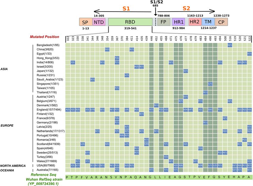
Nelson-Sathi et al. BMC Molecular and Cell Biology   (2022) 23:2                                                    Page 4 of 12

  We identified several RBD mutations on residues that             shown to weaken RBD-ACE2 binding [53]. Similarly, an
are unique to SARS-CoV-2 (N439K, V483A/F/I, E484D,                 adaptive mutation in the F486 interacting residue on
F490S/L, Q493L and S494P) or are conserved in all three            ACE2 (L79T) abolished stable interface formation in
viruses. In addition, we observed mutations in SARS-               mouse [52] (Fig. 3B, C). In addition, a natural F > L substi-
CoV-2 that interchange residues to that in SARS-CoV or             tution in SARS-CoV prevents formation of a complete
RaTG13 (R346K, N354D, N439K, L452R, E471V and                      hydrophobic binding pocket in the interface leading to re-
S477G). Interestingly, most of these reversion mutations           duced ACE2 binding affinity and infectivity of the virus
were located in the RBM region and thus may have im-               [54]. Thus, it appears that the disrupted cluster-I interac-
plications in viral tropism [30, 37]. We performed phylo-          tions in mutants may be critical for virus transmissibility
genomic analysis to understand the evolutionary pattern            [43, 44]. Intriguingly, a new hydrogen bond between RBD
of RBD variants during pandemic. In the phylogenetic               Y489-ACE2 Y83 was observed in cluster-I mutants. Also,
tree, we observed an unbiased distribution of RBD vari-            RBD N487-ACE2 Q24, Y83 and RBD F486-ACE2 M82 in-
ants among distinct SARS-CoV-2 clades (19A, 19B, 20A,              teractions remained largely unaffected in all the mutants
20B, 20C). This likely indicates independent emergence             (Fig. 3B, C and Additional File 1: Table S1). Together, it
of these mutants during pandemic (Fig. 2B).                        suggests the possibility of compensatory mechanisms to
  Interestingly, L452R, R346K and F490S mutations ob-              maintain hydrophobicity in RBD cluster-I.
served in our study sustained in delta, mu and lambda var-            Cluster-II is stabilized by polar /charged residue inter-
iants respectively which evolved later during the                  actions via hydrogen bonds, van der Waals forces and
pandemic. However, E484D mutation which likely did not             salt bridges. The major bonds form between RBD:K417,
affect viral infectivity in the beginning of pandemic,             L455- ACE2:D30; RBD:E484-ACE2:K31; RBD:Y453-
evolved with an acidic to basic residue change (E484K) in          ACE2:H34 and RBD:Q493- ACE2:E35 [14]. Mutational
beta, gamma and mu variants and then with a neutral resi-          studies have identified K417, L455 and E484 as en-
due (E484Q) in the delta variants. Likewise, T478I muta-           hancers of ACE2 binding [53, 55]. The presence of
tion evolved to T478K in the delta variants of concern.            unique K417-D30 salt bridge in SARS-CoV-2 has been
Overall, these findings suggest the role of RBD residues in        shown to significantly enhance receptor binding and in-
shaping a unique pattern for SARS-CoV-2 evolution.                 fectivity [56]. Further, K417 salt bridge and E484 van der
                                                                   Waal’s interactions were abolished in mouse due to
Structural implications of RBD mutations                           adaptive mutations on ACE2 (D30N, K31N) [52] (Fig.
Structurally, RBM scaffold resembles a concave arch that           3B, C). Surprisingly, these key interactions were missing
makes three contact points with ACE2 α- helix; Cluster-            in majority of mutants in our analysis (Fig. 3B, C and
I, II and III. Cluster-I and Cluster-III are on two ends           Additional File 1: Table S1). Nevertheless, interactions
and Cluster-II is towards the middle of the interface              with ACE2 K31, a hotspot of binding for SARS-CoV-2
(Fig. 3A and B). It has been shown that certain adaptive           and SARS-CoV [10, 57, 58] were maintained in all the
mutations in the RBD binding residues of mouse ACE2                mutants. Here, the van der Waal’s forces in the reference
destabilize the interface rendering the organism resistant         Wuhan strain were found to be replaced with a new set
to infection from SARS-like coronaviruses [52]. Hence,             of hydrogen bonds formed via Q493/L490 residues in
to gain insights into relevant interactions that can create        these mutants likely strengthening the hotspot interac-
a stable interface, we included RBD-mouse ACE2 com-                tions (Fig. 3B, C and Additional File 1: Table S1).
plex in our analysis. In this structural background, we               Cluster-III interactions involving RBD:Y449, Q498-
analyzed the impact of RBM mutations on each of the                ACE2:D38; RBD:T500, N501-ACE2: Y41; RBD:Q498,
binding clusters in SARS-CoV-2 by using two in silico              G446- ACE2:Q42; RBD:T500- ACE2:L45, N330 and
approaches: structural modelling and mutagenesis (Fig.             RBD:G502, G496- ACE2:K353 are known to model the
3B, C and Additional File 1: Table S1).                            binding interface in SARS-CoV-2. Several studies re-
   Each mutation was modelled based on the reported                ported Q498 and N501 RBM residues as high affinity
crystal structures of SARS-CoV-2 RBD-ACE2 bound                    binders [14]. The absence of their hydrogen bond inter-
complex [14]. The key interactions that stabilize cluster-I        actions in mouse interface further confirms the import-
in SARS-CoV-2 are formed between RBD:A475, G476-                   ance of these residues [52] (Fig. 3B, C). Moreover, single
ACE2:S19; RBD:N487-ACE2:Q24, Y83 and RBD:F486-                     N501Y mutation showed 10-fold increase in ACE2 bind-
ACE2:L79, M82 [14]. We observed that RBD:A475, G476-               ing in the SARS-CoV-2 variants of concern [39]. Surpris-
ACE2:S19 and RBD:F486-ACE2:L79 interactions com-                   ingly, the cluster of interactions mediated by Q498 and
pletely disappeared in several mutants (Fig. 3B, C and             N501 were absent in some mutants (Fig. 3B, C and Add-
Additional File 1: Table S1). A475, G476 and F486 are              itional File 1: Table S1). A few mutants also exhibited
considered critical hotspot residues for ACE2 binding              partial disruption of T500 interactions with ACE2. How-
[42]. Further mutations in A475 and G476 have been                 ever, interactions involving the ACE2 critical residues
Nelson-Sathi et al. BMC Molecular and Cell Biology   (2022) 23:2   Page 5 of 12

 Fig. 2 (See legend on next page.)
Nelson-Sathi et al. BMC Molecular and Cell Biology      (2022) 23:2                                                                   Page 6 of 12

 (See figure on previous page.)
 Fig. 2 (A) Conservation of Receptor Binding Domain (RBD) of SARS-CoV-2 with its close relatives, SARS-CoV and Bat RaTG13. The blue colored
 region shows RBD and the yellow highlighted region within RBD is the Receptor Binding Motif (RBM). The mutated residues are highlighted in
 light blue and substitutions are marked below. Non-conserved residues are highlighted in grey color. Interacting residues are marked with black
 asterisk and the mutated interactions are in red asterisk symbol. (B) The carton model representation of SARS-CoV-2 RBM highlighting mutated
 interacting residues and most frequent mutations (red color) in RBM. (C) Maximum Likelihood Phylogenetic tree of 494 SARS-CoV-2 isolates
 containing RBD mutations. The outer circle represents the RBD mutations

D38, Y41, N330 and the hotspot residue K353 were all                       ACE2 independent sarbecoviruses showed 20-30fold
retained in all the mutants indicating significance of                     lower Δlog10 (Kd) values validating our analysis. The
these amino acids in shaping the interface (Fig. 3B, C                     binding affinity differences (Δlog10 (KD) obtained from
and Additional File 1: Table S1). Thus our findings sug-                   our analyses were comparable up to 60% with the ex-
gest that the binding interface of SARS-CoV-2 remodels                     perimental values reported in other studies [43, 44]. The
constantly regardless of the position and number of                        40% mismatch may attribute to differences in affinity,
RBD mutations.                                                             avidity and conformation of trimeric spike (used for ex-
                                                                           periments) versus monomeric RBD (used for in-silico
Stability of RBD-ACE2 complexes                                            analysis). We did not observe a significant correlation
MD simulations of wild type and mutated RBD- ACE2                          between binding energies and mutations in contact ver-
complexes were carried out for 50 ns to analyse the stabil-                sus non-contact residues. This suggests that mutations
ity. The root mean square deviation (RMSD) for each com-                   on any RBM residues could impact spatial arrangements
plex was calculated. RMSD was found to be below 3 Å in                     of backbone leading to altered binding affinities. Overall,
wild type and mutants suggesting good stability of the com-                our observation were consistent with recent studies
plexes during simulations (Fig. 4A). The fluctuations in                   showing that the whole RBM, and not the ACE2 binding
each residue of RBD over time were also analysed by plot-                  residues alone, was necessary to complement viral entry
ting the Root Mean Square Fluctuation (RMSF) graph (Fig.                   of ACE2 independent sarbecoviruses [59, 60].
4B). The β-sheets were less fluctuating throughout the
simulation. The higher peaks were mainly observed in the                   Possible structural mechanism of RBD-ACE2 interface
loop regions in both wild type and mutant RBDs, indicating                 formation in sarbecoviruses
more fluctuations in loop regions than structured regions.                 Based on our structural analysis of RBD mutants, we sur-
However, the overall values were below 3 Å further suggest-                mised that the RBD contact residues which remained un-
ing that the RBD-ACE2 complexes remained stable and                        affected in all the mutants- N487, F486 and Y489 residues
bound together throughout the simulation.                                  in cluster-I, E484, F490 and Q493 and Y449, G496, T500,
                                                                           G502 in cluster-III could possibly play a crucial role in the
Binding affinity of RBD mutants                                            formation of a stable binding interface. Given the spatial
Binding affinities were derived from both modelled and                     arrangement, these residues appear critical in directly an-
mutated structures. For detailed comparison, we also cal-                  choring the RBM loop to ACE2 from both ends. This may
culated binding affinities from modelled RBDs of ACE2                      help initiate interface formation that structurally favours
dependent (SARS-CoV, pangolin-CoV) and ACE2 inde-                          viral entry [61]. The significant changes in cluster-II inter-
pendent (BM48–31, Rf1, Rp3, HKU3–1) sarbecoviruses                         actions indicate they are dispensable for anchoring but
[59]. The differences in binding affinities with respect to                might be important for remodelling the interface. Intri-
wild type SARS-CoV-2 Δlog10 (Kd) were calculated by the                    guingly, the corresponding residues on ACE2 (Q24, M82,
following equation and plotted as shown in Fig. 5:                         Y83, D38, Y41, N330, K353) that interact with these RBD
                                                                           residues have been shown to be crucial for interspecies
    Δ log10 ðKDÞ ¼ log10 ðKDÞ wild - log10 ðKDÞ mutants                    transmission of sarbecoviruses [61–63]. To understand
                                                                   ð1Þ     structural evolution of RBD-ACE2 interface, we looked at
                                                                           the conservation of these residues across ACE2-
                                                                           dependent and ACE2-independent sarbecoviruses (Fig. 6).
  SARS-CoV and pangolin CoV were on the two ends of                        N487 was highly conserved in all sarbecoviruses whereas
the spectrum showing ~ 7 fold decrease and increase in                     Y449, and T500 were present only in ACE2-dependent
the binding affinity respectively compared to wild type                    sarbecoviruses. Interestingly, Y489, G496, G502 and F486
SARS-CoV-2. Δlog10 (Kd) values of all observed RBD                         which changed to L in SARS-CoV were found to be con-
fall within this range with the lowest affinity mutant                     served in BM48–31. BM48–31 is a sarbecovirus distinct
close to that of SARS-CoV and the highest affinity mu-                     from other ACE2-independent viruses and likely shows
tant close to pangolin CoV (Fig. 5). As expected, all the                  evolution toward ACE2 dependency [64, 65]. A large
Nelson-Sathi et al. BMC Molecular and Cell Biology (2022) 23:2 Page 7 of 12 Fig. 3 Molecular rearrangements in RBD-ACE2 interface. (A) The cartoon model representation of SARS-CoV-2 RBM highlighting mutated interacting residues and most frequent mutations (red color) in RBM. (B) List of cluster specific molecular interactions of hACE2, mACE2, and mutated RBD-ACE2 complexes. Hydrogen bonds are marked in red, van der Waal’s interactions in blue and salt bridges in green. (C) Structural visualization of key interactions listed in (B). RBD is represented in green and ACE2 in gold. The hydrogen bond interactions between ACE2 and RBD are shown as dotted lines deletion near RBM cluster-I responsible for disruption of observed in mutants are both conserved in BM48–31 (Fig. ACE2 interaction is not present in BM48–31 [64]. In 6). Together, it supports possible evolution of a favourable addition, E484 residue which is important for stabilizing ACE2 binding interface in this virus [65]. Many residues cluster-II in SARS-CoV-2 and its replacement L492 important in interface formation and remodelling are also
Nelson-Sathi et al. BMC Molecular and Cell Biology    (2022) 23:2                                                                Page 8 of 12

 Fig. 4 Structural stability of SARS CoV-2 wild type and mutant RBD –ACE2 complexes. (A) Root mean square deviation (RMSD) of wild type and
 mutant RBD with ACE2 complexes. (B) Root mean square fluctuation (RMSF) of RBD wild type and mutant structures. Each mutant and wild type
 are separately colour coded

part of distinct epitopes present on RBD. Significantly,                landscape and the corresponding structural landscape is
mutations in A475, F486, E484 etc., have been shown to                  a novel approach to completely understand the virus
be immune evasive [41, 66]. Hence, interface remodelling                and its interaction with the host. The predicted mechan-
mediated by these residues may help virus to simultan-                  ism of interface remodelling in our study may be useful
eously sustain ACE2 binding and escape neutralizing anti-               to design novel strategies to combat coronavirus infec-
bodies [55].                                                            tions in general. Overall, our study proposes the signifi-
                                                                        cance of understanding structural evolution of protein
Conclusions                                                             interfaces during pandemics.
Currently, all SARS-CoV-2 immunogens and testing re-
agents are based on the Wuhan reference sequence.                       Materials and methods
Thus, growing number of mutations in the reference                      Mutational analysis
strain is wreaking a global havoc regarding efficacy of                 A total of 55,485 spike proteins of SARS-CoV-2 were
vaccines and therapeutics. Our elucidation of mutational                directly downloaded on 29th June 2020 from the GISA
Nelson-Sathi et al. BMC Molecular and Cell Biology (2022) 23:2 Page 9 of 12
Fig. 5 Bar graph representing variations in binding affinity differences among all RBD mutants and other coronaviruses. Orange, green and blue
bars indicate Δlog10 (Kd) values obtained from modelling, mutagenesis and functional studies reported
ID database. We removed the partial sequences, se- affinity towards the human ACE2 (hACE2) receptor. The
quences greater than 1% unidentified ‘X’ amino acids crystal structure of the SARS-CoV-2 RBD-hACE2 recep-
and sequences from low quality genomes. Further, tor complex was downloaded from Protein Data Bank
31,403 spike protein sequences along with Wuhan refer- (PDB ID: 6LZG) and the mutagenesis analysis was per-
ence spike protein (YP_009724390.1) were aligned using formed using Pymol [71]. As an alternative approach, we
Mafft (maxiterate 1000 and global pair-ginsi) [67]. The also modelled the mutants of SARS-CoV-2 RBD-ACE
alignments were visualized in Jalview [68] and the amino complex and other coronaviruses spike RBD bound with
acid substitutions in each position were extracted using hACE2 receptors using Swiss model [72]. In addition,
custom python script. We ignored the substitutions that homology modelling of Mouse ACE2 (mACE2) structure
were present in only one genome and unidentified was performed in Swiss-Model using SARS-CoV-2 RBD-
amino acid X. The mutations that are present in at least hACE2 as template. The YASARA server [73] was used
two independent genomes in a particular position were for the energy minimization of analysed structures. The
further considered. These two criteria were used to Z-dock webserver [74] was used for docking the mACE-2
avoid mutations due to sequencing errors. The mutated and the spike protein RBD of SARS-CoV-2. The binding
amino acids were further tabulated and plotted as a affinity of the wild, mutated and docked structures was
matrix using R script. calculated using PRODIGY web server [75]. The hydrogen
bond and salt bridge interactions were calculated using
Phylogeny reconstruction Protein Interaction Calculator [76] and the van der Waals
For the Maximum-likelihood phylogeny reconstruction, interactions were calculated using Ligplot [77]. All the vi-
we have used the SARS-CoV-2 genomes containing RBD sualizations were done using Pymol [71].
mutations, and 10 genomes were sampled as representa-
tives for each known subtype with Wuhan RefSeq strain MD simulations
as root. Sequences were aligned using Mafft (maxiterate The stability of the wild type and mutated structures
1000 and global pair-ginsi), and phylogeny was recon- were analysed by Molecular Dynamic (MD) Simulations
structed using IQ-Tree [69]. The best evolutionary using Desmond (Desmond, Schrödinger, LLC, NY, USA)
model (GTR + F + I + G4) was picked using the Model [78]. The wild type and mutated SARS CoV-2 RBD –
Finder program [70]. ACE2 complex were prepared by Schrodinger Maestro
Protein Preparation wizard. The water molecules were
Structural analysis removed and optimized the structures by adding Hydro-
The structural analysis of the mutated spike glycoprotein gen atoms. The system was solvated using TIP3P water
of SARS-CoV-2 RBD domain was done to assess the im- model and neutralized by adding Na/Cl ions and mini-
pact of interface amino acid residue mutations on binding mized using OPLS3e force field. The Nose-Hoover chainNelson-Sathi et al. BMC Molecular and Cell Biology (2022) 23:2 Page 10 of 12
Fig. 6 Multiple sequence alignment of RBD across sarbecoviruses. The blue highlighted box denotes RBM. Black asterisks indicate RBD residues
that directly bind to ACE2. Red asterisks denote mutations on the binding residues analyzed in this study. The mutated residues are highlighted
in light blue and substitutions are marked below. Binding residues in cluster-I, II and III are marked in red, green and blue bars on the top. 1A,B
2A,B and 3A,B indicates the RBD epitopes present in SARS-CoV-2
thermostat method and Martyna-Toubias-Klein barostat Authors’ contributions
method were used to maintain the temperature and Conceptualization, S.N.S., U.P.K., I.J. and M.R.P.; methodology, S.N.S., U.P.K., E.S.,
J.S.P., R.P., N.K.V., S.R. and M.R.P.; software, S.N.S., U.P.K., S.R.C.N., J.S.P., R.P. and
pressure of the system respectively. A 50-ns simulation M.R.P.; validation, S.N.S., U.P.K., S.R.C.N., J.S.P., R.P. and M.R.P.; formal analysis,
for each mutant and wild type RBD-ACE2 complex were S.NS., U.P.K., S.R.C.N., J.S.P., R.P., N.K.V., S.R. and S.K.T.R.; investigation, S.N.S,
done in an NPT Ensemble of 300 K at 1.01325 bar. U.P.K., S.R.C.N., J.S.P., S.K.T.R. and M.R.P.; resources, S.N.S., U.P.K., R.N., I.J.,
S.R.C.N., J.S.P., R.P., H.P., S.G., S.K.T.R. and M.R.P.; data curation, S.N.S., U.P.K.,
S.R.C.N., J.S.P., R.P. and N.K.V.; writing—original draft preparation, S.N.S., U.P.K.,
Abbreviations S.R.C.N., J.S.P., R.P., N.K.V. and S.R.; writing—review and editing, S.N.S., U.P.K.,
S: Spike; RBD: Receptor Binding Domain; ACE2: Angiotensin-Converting E.S., S.K.T.R. and M.R.P,; visualization, S.N.S, U.P.K., S.R.C.N., J.S.P., R.P. and N.K.V..;
Enzyme 2; RBM: Receptor binding motif; hACE2: human ACE2; supervision, S.N.S., U.P.K., E.S., S.K.T.R and M.R.P.; project administration, S.N.S.,
mACE2: Mouse ACE2 U.P.K., E.S., R.N., I.J., H.P., S.G., S.K.T.R. and M.R.P.; funding acquisition, S.N.S.,
U.P.K. and M.R.P. All authors read and approved the final manuscript.
Supplementary Information
The online version contains supplementary material available at https://doi. Funding
org/10.1186/s12860-021-00403-4. This research was funded by the INSPIRE Faculty Fellowship [DST/INSPIRE/04/
2015/002935]; and Biotechnology Industry Research Assistance Council [BT/
Additional file 1: Table S1. List of clusterwise interfacial interactions PR40330/COT/142/16/2020]; and Ramalingaswami Fellowship by the
between wild type or mutant SARS CoV-2 RBD protein and human or Department of Biotechnology [BT/RLF/Re-entry/18/2014].
mouse ACE2.
Availability of data and materials
Acknowledgments The SARS-CoV-2 datasets analysed during the current study are available in
The authors wish to acknowledge John B Johnson, Mahendran KR and Sara the GISAID-EpiCoV databases.
Jones for critical comments. (https://www.gisaid.org/).Nelson-Sathi et al. BMC Molecular and Cell Biology (2022) 23:2 Page 11 of 12
Declarations three hinges. Science. 2020;370(6513):203–8. https://doi.org/10.1126/
science.abd5223.
Ethics approval and consent to participate 19. Wang Y, Liu M, Gao J. Enhanced receptor binding of SARS-CoV-2 through
Not applicable. networks of hydrogen-bonding and hydrophobic interactions. Proc Natl
Acad Sci. 2020;117(25):13967–74. https://doi.org/10.1073/pnas.2008209117.
Consent for publication 20. Ali A, Vijayan R. Dynamics of the ACE2–SARS-CoV-2/SARS-CoV spike protein
Not applicable. interface reveal unique mechanisms. Sci Rep. 2020;10:14214.
21. Chan KK, Dorosky D, Sharma P, Abbasi SA, Dye JM, Kranz DM, et al.
Competing interests Engineering human ACE2 to optimize binding to the spike protein of SARS
The authors declare no conflict of interest. coronavirus 2. Science. 2020;369(6508):1261–5. https://doi.org/10.1126/
science.abc0870.
Author details 22. Ray D, Le L, Andricioaei I. Distant residues modulate conformational
1
Corona Research & Intervention Group, Rajiv Gandhi Centre for opening in SARS-CoV-2 spike protein. BioRxiv. 2021;2020(2012):2007.415596.
Biotechnology, Thiruvananthapuram 695014, India. 2SAGENOME Private 23. Simmonds P. Rampant C→ U hypermutation in the genomes of SARS-CoV-
Limited, BioNest, Kochi 683503, India. 2 and other coronaviruses: causes and consequences for their short-and
long-term evolutionary trajectories. Msphere. 2020;5(3):e00408–20. https://
Received: 3 March 2021 Accepted: 23 December 2021 doi.org/10.1128/mSphere.00408-20.
24. Vilar S, Isom DG. One year of SARS-CoV-2: how much has the virus
changed? Biology. 2021;10(2):91. https://doi.org/10.3390/biology10020091.
25. Koyama T, Platt D, Parida L. Variant analysis of SARS-CoV-2 genomes. Bull
References
World Health Organ. 2020;98(7):495–504. https://doi.org/10.2471/BLT.20.253591.
1. WHO Coronavirus (COVID-19) Dashboard with vaccination data. WHO; 2021.
26. Shen X, Tang H, McDanal C, Wagh K, Fischer W, Theiler J, et al. SARS-CoV-2
https://covid19.who.int.
variant B. 1.1. 7 is susceptible to neutralizing antibodies elicited by ancestral
2. Harvey WT, Carabelli AM, Jackson B, Gupta RK, Thomson EC, Harrison EM,
spike vaccines. Cell Host Microbe. 2021;29(4):529–39 e523.
et al. SARS-CoV-2 variants, spike mutations and immune escape. Nat Rev
Microbiol. 2021;19(7):409–24. https://doi.org/10.1038/s41579-021-00573-0. 27. Zhang W, Davis BD, Chen SS, Martinez JMS, Plummer JT, Vail E. Emergence
3. Budinger GS, Misharin AV, Ridge KM, Singer BD, Wunderink RG. Distinctive of a novel SARS-CoV-2 variant in Southern California. Jama. 2021;325(13):
features of severe SARS-CoV-2 pneumonia. J Clin Investig. 2021;131(14): 1324–6. https://doi.org/10.1001/jama.2021.1612.
e149412. https://doi.org/10.1172/JCI149412. 28. Tegally H, Wilkinson E, Giovanetti M, Iranzadeh A, Fonseca V, Giandhari J,
4. Port JR, Yinda CK, Owusu IO, Holbrook M, Fischer R, Bushmaker T, et al. et al. Detection of a SARS-CoV-2 variant of concern in South Africa. Nature.
SARS-CoV-2 disease severity and transmission efficiency is increased for 2021;592(7854):438–43. https://doi.org/10.1038/s41586-021-03402-9.
airborne compared to fomite exposure in Syrian hamsters. Nat Commun. 29. Dejnirattisai W, Zhou D, Supasa P, Liu C, Mentzer AJ, Ginn HM, et al.
2021;12(1):1–15. https://doi.org/10.1038/s41467-021-25156-8. Antibody evasion by the P. 1 strain of SARS-CoV-2. Cell. 2021;184(11):2939–
5. Wu F, Zhao S, Yu B, Chen Y-M, Wang W, Song Z-G, et al. A new coronavirus 54 e2939.
associated with human respiratory disease in China. Nature. 2020;579(7798): 30. Cherian S, Potdar V, Jadhav S, Yadav P, Gupta N, Das M, et al. SARS-CoV-2
265–9. https://doi.org/10.1038/s41586-020-2008-3. spike mutations, L452R, T478K, E484Q and P681R, in the second wave of
6. Gordon DE, Jang GM, Bouhaddou M, Xu J, Obernier K, White KM, et al. A COVID-19 in Maharashtra, India. Microorganisms. 2021;9(7):1542. https://doi.
SARS-CoV-2 protein interaction map reveals targets for drug repurposing. org/10.3390/microorganisms9071542.
Nature. 2020;583(7816):459–68. https://doi.org/10.1038/s41586-020-2286-9. 31. Dhar MS, Marwal R, Radhakrishnan V, Ponnusamy K, Jolly B, Bhoyar RC, et al.
7. Dai L, Gao GF. Viral targets for vaccines against COVID-19. Nat Rev Immunol. Genomic characterization and Epidemiology of an emerging SARS-CoV-2
2021;21(2):73–82. https://doi.org/10.1038/s41577-020-00480-0. variant in Delhi, India. medRxiv. 2021.
8. Raghuvamsi PV, Tulsian NK, Samsudin F, Qian X, Purushotorman K, Yue G, 32. Korber B, Fischer WM, Gnanakaran S, Yoon H, Theiler J, Abfalterer W, et al.
et al. SARS-CoV-2 S protein: ACE2 interaction reveals novel allosteric targets. Tracking changes in SARS-CoV-2 spike: evidence that D614G increases
Elife. 2021;10:e63646. https://doi.org/10.7554/eLife.63646. infectivity of the COVID-19 virus. Cell. 2020;182(4):812–27 e819.
9. Walls AC, Park Y-J, Tortorici MA, Wall A, McGuire AT, Veesler D. Structure, 33. Plante JA, Liu Y, Liu J, Xia H, Johnson BA, Lokugamage KG, et al. Spike
function, and antigenicity of the SARS-CoV-2 spike glycoprotein. Cell. 2020; mutation D614G alters SARS-CoV-2 fitness. Nature. 2021;592(7852):116–21.
181(2):281–92 e286. https://doi.org/10.1038/s41586-020-2895-3.
10. Shang J, Wan Y, Luo C, Ye G, Geng Q, Auerbach A, et al. Cell entry 34. Volz E, Hill V, McCrone JT, Price A, Jorgensen D, O’Toole Á, et al. Evaluating
mechanisms of SARS-CoV-2. Proc Natl Acad Sci. 2020;117(21):11727–34. the effects of SARS-CoV-2 spike mutation D614G on transmissibility and
https://doi.org/10.1073/pnas.2003138117. pathogenicity. Cell. 2021;184(1):64–75 e11.
11. Hoffmann M, Pöhlmann S. How SARS-CoV-2 makes the cut. Nat Microbiol. 35. Zhang L, Jackson CB, Mou H, Ojha A, Peng H, Quinlan BD, Rangarajan ES,
2021;6(7):828–9. https://doi.org/10.1038/s41564-021-00931-x. Pan A, Vanderheiden A, Suthar MS: SARS-CoV-2 spike-protein D614G
12. Jackson CB, Farzan M, Chen B, Choe H. Mechanisms of SARS-CoV-2 entry mutation increases virion spike density and infectivity. Nat Commun 2020,
into cells. Nat Rev Mol Cell Biol. 2021;23(1):1–18. https://doi.org/10.1038/s41 11(1):1–9, 6013, https://doi.org/10.1038/s41467-020-19808-4.
580-021-00418-x. 36. Zhang J, Cai Y, Xiao T, Lu J, Peng H, Sterling SM, et al. Structural impact on
13. Lan J, Ge J, Yu J, Shan S, Zhou H, Fan S, et al. Structure of the SARS-CoV-2 SARS-CoV-2 spike protein by D614G substitution. Science. 2021;372(6541):
spike receptor-binding domain bound to the ACE2 receptor. Nature. 2020; 525–30. https://doi.org/10.1126/science.abf2303.
581(7807):215–20. https://doi.org/10.1038/s41586-020-2180-5. 37. Thomson EC, Rosen LE, Shepherd JG, Spreafico R, da Silva FA,
14. Wang Q, Zhang Y, Wu L, Niu S, Song C, Zhang Z, et al. Structural and Wojcechowskyj JA, et al. Circulating SARS-CoV-2 spike N439K variants
functional basis of SARS-CoV-2 entry by using human ACE2. Cell. 2020; maintain fitness while evading antibody-mediated immunity. Cell. 2021;
181(4):894–904 e899. 184(5):1171–87 e1120.
15. Wrapp D, Wang N, Corbett KS, Goldsmith JA, Hsieh C-L, Abiona O, et al. 38. Tian F, Tong B, Sun L, Shi S, Zheng B, Wang Z, et al. N501Y mutation of
Cryo-EM structure of the 2019-nCoV spike in the prefusion conformation. spike protein in SARS-CoV-2 strengthens its binding to receptor ACE2. Elife.
Science. 2020;367(6483):1260–3. https://doi.org/10.1126/science.abb2507. 2021;10:e69091. https://doi.org/10.7554/eLife.69091.
16. Ke Z, Oton J, Qu K, Cortese M, Zila V, McKeane L, et al. Structures and 39. Barton MI, MacGowan SA, Kutuzov MA, Dushek O, Barton GJ, van der
distributions of SARS-CoV-2 spike proteins on intact virions. Nature. 2020; Merwe PA. Effects of common mutations in the SARS-CoV-2 spike RBD and
588(7838):498–502. https://doi.org/10.1038/s41586-020-2665-2. its ligand the human ACE2 receptor on binding affinity and kinetics. Elife.
17. Fan X, Cao D, Kong L, Zhang X: Cryo-EM analysis of the post-fusion 2021;10:e70658. https://doi.org/10.7554/eLife.70658.
structure of the SARS-CoV spike glycoprotein. Nat Commun 2020, 11(1):1– 40. Andreano E, Piccini G, Licastro D, Casalino L, Johnson NV, Paciello I, et al.
10, 3618, https://doi.org/10.1038/s41467-020-17371-6. SARS-CoV-2 escape from a highly neutralizing COVID-19 convalescent
18. Turoňová B, Sikora M, Schürmann C, Hagen WJ, Welsch S, Blanc FE, et al. In plasma. Proc Natl Acad Sci. 2021;118(36). https://doi.org/10.1073/pnas.21031
situ structural analysis of SARS-CoV-2 spike reveals flexibility mediated by 54118.Nelson-Sathi et al. BMC Molecular and Cell Biology (2022) 23:2 Page 12 of 12
41. Greaney AJ, Starr TN, Gilchuk P, Zost SJ, Binshtein E, Loes AN, et al. analysis of ACE2 in vertebrates. Proc Natl Acad Sci. 2020;117(36):22311–22.
Complete mapping of mutations to the SARS-CoV-2 spike receptor-binding https://doi.org/10.1073/pnas.2010146117.
domain that escape antibody recognition. Cell host & microbe. 2021;29(1): 62. Liu Y, Hu G, Wang Y, Ren W, Zhao X, Ji F, et al. Functional and genetic
44–57 e49. analysis of viral receptor ACE2 orthologs reveals a broad potential host
42. Ghorbani M, Brooks BR, Klauda JB. Critical sequence hotspots for binding of range of SARS-CoV-2. Proc Natl Acad Sci. 2021;118(12). https://doi.org/10.1
novel coronavirus to angiotensin converter enzyme as evaluated by 073/pnas.2025373118.
molecular simulations. J Phys Chem B. 2020;124(45):10034–47. https://doi. 63. Zhang H-L, Li Y-M, Sun J, Zhang Y-Y, Wang T-Y, Sun M-X, et al. Evaluating
org/10.1021/acs.jpcb.0c05994. angiotensin-converting enzyme 2-mediated SARS-CoV-2 entry across
43. Li Q, Wu J, Nie J, Zhang L, Hao H, Liu S, et al. The impact of mutations in species. J Biol Chem. 2021;296:100435. https://doi.org/10.1016/j.jbc.2021.1
SARS-CoV-2 spike on viral infectivity and antigenicity. Cell. 2020;182(5):1284– 00435.
94 e1289. 64. Wells HL, Letko M, Lasso G, Ssebide B, Nziza J, Byarugaba DK, et al. The
44. Starr TN, Greaney AJ, Hilton SK, Ellis D, Crawford KH, Dingens AS, et al. Deep evolutionary history of ACE2 usage within the coronavirus subgenus
mutational scanning of SARS-CoV-2 receptor binding domain reveals Sarbecovirus. Virus Evol. 2021;7(1):veab007.
constraints on folding and ACE2 binding. Cell. 2020;182(5):1295–310 e1220. 65. Li F. Structure, function, and evolution of coronavirus spike proteins. Ann
45. Ashoor D, Khalaf NB, Marzouq M, Jarjanazi H, Chelif S, Fathallah MD. A Rev Virol. 2016;3(1):237–61. https://doi.org/10.1146/annurev-virology-110615-
computational approach to evaluate the combined effect of SARS-CoV-2 042301.
RBD mutations and ACE2 receptor genetic variants on infectivity: The 66. Yi C, Sun X, Ye J, Ding L, Liu M, Yang Z, et al. Key residues of the receptor
COVID-19 host-pathogen nexus. bioRxiv. 2021;2020:2010–23 352344. binding motif in the spike protein of SARS-CoV-2 that interact with ACE2
46. Gobeil S, Janowska K, McDowell S, Mansouri K, Parks R, Stalls V, et al. Effect and neutralizing antibodies. Cell Mol Immunol. 2020;17(6):621–30. https://
of natural mutations of SARS-CoV-2 on spike structure, conformation and doi.org/10.1038/s41423-020-0458-z.
antigenicity. bioRxiv. 2021;373(6555):eabi6226. 67. Katoh K, Misawa K, Ki K, Miyata T. MAFFT: a novel method for rapid multiple
47. Schrörs B, Riesgo-Ferreiro P, Sorn P, Gudimella R, Bukur T, Rösler T, et al. sequence alignment based on fast Fourier transform. Nucleic Acids Res.
Large-scale analysis of SARS-CoV-2 spike-glycoprotein mutants 2002;30(14):3059–66. https://doi.org/10.1093/nar/gkf436.
demonstrates the need for continuous screening of virus isolates. PLoS 68. Waterhouse AM, Procter JB, Martin DM, Clamp M, Barton GJ. Jalview version
ONE. 2021;16(9):e0249254. https://doi.org/10.1371/journal.pone.0249254. 2—a multiple sequence alignment editor and analysis workbench.
48. López-Cortés GI, Palacios-Pérez M, Zamudio GS, Veledíaz HF, Ortega E, José Bioinformatics. 2009;25(9):1189–91. https://doi.org/10.1093/bioinformatics/
MV. Neutral evolution test of the spike protein of SARS-CoV-2 and its btp033.
implications in the binding to ACE2. Sci Rep. 2021;11(1):18847. 69. Nguyen L-T, Schmidt HA, Von Haeseler A, Minh BQ. IQ-TREE: a fast and
49. Marquioni VM, de Aguiar MA. Modeling neutral viral mutations in the effective stochastic algorithm for estimating maximum-likelihood
spread of SARS-CoV-2 epidemics. PLoS ONE. 2021;16(7):e0255438. https:// phylogenies. Mol Biol Evol. 2015;32(1):268–74. https://doi.org/10.1093/
doi.org/10.1371/journal.pone.0255438. molbev/msu300.
50. Liu Z, Xiao X, Wei X, Li J, Yang J, Tan H, et al. Composition and divergence 70. Kalyaanamoorthy S, Minh BQ, Wong TK, Von Haeseler A, Jermiin LS.
of coronavirus spike proteins and host ACE2 receptors predict potential ModelFinder: fast model selection for accurate phylogenetic estimates. Nat
intermediate hosts of SARS-CoV-2. J Med Virol. 2020;92(6):595–601. https:// Methods. 2017;14(6):587–9. https://doi.org/10.1038/nmeth.4285.
doi.org/10.1002/jmv.25726. 71. DeLano WL. Pymol: An open-source molecular graphics tool. CCP4
51. Jaimes JA, André NM, Chappie JS, Millet JK, Whittaker GR. Phylogenetic Newsletter on protein crystallography. 2002;40(1):82–92.
analysis and structural modeling of SARS-CoV-2 spike protein reveals an 72. Schwede T, Kopp J, Guex N, Peitsch MC. SWISS-MODEL: an automated
evolutionary distinct and proteolytically sensitive activation loop. J Mol Biol. protein homology-modeling server. Nucleic Acids Res. 2003;31(13):3381–5.
2020;432(10):3309–25. https://doi.org/10.1016/j.jmb.2020.04.009. https://doi.org/10.1093/nar/gkg520.
52. Zhao X, Chen D, Szabla R, Zheng M, Li G, Du P, et al. Broad and differential 73. Krieger E, Koraimann G, Vriend G. Increasing the precision of comparative
animal angiotensin-converting enzyme 2 receptor usage by SARS-CoV-2. J models with YASARA NOVA—a self-parameterizing force field. Proteins Struct
Virol. 2020;94(18):e00940–20. https://doi.org/10.1128/JVI.00940-20. Funct Bioinforma. 2002;47(3):393–402. https://doi.org/10.1002/prot.10104.
53. Yang Y, Zhang Y, Qu Y, Zhang C, Liu X-W, Zhao M, et al. Key residues of the 74. Pierce BG, Wiehe K, Hwang H, Kim B-H, Vreven T, Weng Z. ZDOCK server:
receptor binding domain in the spike protein of SARS-CoV-2 mediating the interactive docking prediction of protein–protein complexes and symmetric
interactions with ACE2: a molecular dynamics study. Nanoscale. 2021;13(20): multimers. Bioinformatics. 2014;30(12):1771–3. https://doi.org/10.1093/
9364–70. https://doi.org/10.1039/D1NR01672E. bioinformatics/btu097.
75. Xue LC, Rodrigues JP, Kastritis PL, Bonvin AM, Vangone A. PRODIGY: a web
54. Wan Y, Graham R, Baric R, Li F. An analysis based on decade-long structural
server for predicting the binding affinity of protein–protein complexes.
studies of SARS 3, JVI accepted manuscript posted online 29 January 2020. J
Bioinformatics. 2016;32(23):3676–8. https://doi.org/10.1093/bioinformatics/
Virol. 2020;94(7):e00127–0. https://doi.org/10.1128/JVI.00127-20.
btw514.
55. Starr TN, Greaney AJ, Addetia A, Hannon WW, Choudhary MC, Dingens AS,
76. Tina K, Bhadra R, Srinivasan N. PIC: protein interactions calculator. Nucleic
et al. Prospective mapping of viral mutations that escape antibodies used
Acids Res. 2007;35:W473–6.
to treat COVID-19. Science. 2021;371(6531):850–4. https://doi.org/10.1126/
77. Wallace AC, Laskowski RA, Thornton JM. LIGPLOT: a program to generate
science.abf9302.
schematic diagrams of protein-ligand interactions. Protein Eng Des Sel.
56. Laffeber C, de Koning K, Kanaar R, Lebbink JH. Experimental evidence for
1995;8(2):127–34. https://doi.org/10.1093/protein/8.2.127.
enhanced receptor binding by rapidly spreading SARS-CoV-2 variants. J Mol
78. Bowers KJ, Chow DE, Xu H, Dror RO, Eastwood MP, Gregersen BA, et al.
Biol. 2021;433(15):167058. https://doi.org/10.1016/j.jmb.2021.167058.
ACM/IEEE conference on supercomputing. IEEE. 2006:43–3.
57. Li F. Structural analysis of major species barriers between humans and palm
civets for severe acute respiratory syndrome coronavirus infections. J Virol.
2008;82(14):6984–91. https://doi.org/10.1128/JVI.00442-08. Publisher’s Note
58. Wu K, Peng G, Wilken M, Geraghty RJ, Li F. Mechanisms of host receptor Springer Nature remains neutral with regard to jurisdictional claims in
adaptation by severe acute respiratory syndrome coronavirus. J Biol Chem. published maps and institutional affiliations.
2012;287(12):8904–11. https://doi.org/10.1074/jbc.M111.325803.
59. Boni MF, Lemey P, Jiang X, Lam TT-Y, Perry BW, Castoe TA, et al.
Evolutionary origins of the SARS-CoV-2 sarbecovirus lineage responsible for
the COVID-19 pandemic. Nat Microbiol. 2020;5(11):1408–17. https://doi.org/1
0.1038/s41564-020-0771-4.
60. Letko M, Marzi A, Munster V. Functional assessment of cell entry and
receptor usage for SARS-CoV-2 and other lineage B betacoronaviruses. Nat
Microbiol. 2020;5(4):562–9. https://doi.org/10.1038/s41564-020-0688-y.
61. Damas J, Hughes GM, Keough KC, Painter CA, Persky NS, Corbo M, et al.
Broad host range of SARS-CoV-2 predicted by comparative and structuralYou can also read