IRF-8 regulates expansion of myeloid-derived suppressor cells and Foxp3+regulatory T cells and modulates Th2 immune responses to gastrointestinal ...
←
→
Page content transcription
If your browser does not render page correctly, please read the page content below
IRF-8 regulates expansion of myeloid-derived suppressor cells and
Foxp3+regulatory T cells and modulates Th2 immune responses to
gastrointestinal nematode infection
Valanparambil, R. M., Tam, M., Gros, P. P., Auger, J. P., Segura, M., Gros, P., Jardim, A., Geary, T. G., Ozato,
K., & Stevenson,
       +         M. M. (2017). IRF-8 regulates expansion of myeloid-derived suppressor cells and
Foxp3 regulatory T cells and modulates Th2 immune responses to gastrointestinal nematode infection. PLoS
Pathogens, 13(10), [e1006647]. https://doi.org/10.1371/journal.ppat.1006647
Published in:
PLoS Pathogens

Document Version:
Publisher's PDF, also known as Version of record

Queen's University Belfast - Research Portal:
Link to publication record in Queen's University Belfast Research Portal

Publisher rights
Copyright 2017 the authors.
This is an open access article published under a Creative Commons Attribution License (https://creativecommons.org/licenses/by/4.0/),
which permits unrestricted use, distribution and reproduction in any medium, provided the author and source are cited.

General rights
Copyright for the publications made accessible via the Queen's University Belfast Research Portal is retained by the author(s) and / or other
copyright owners and it is a condition of accessing these publications that users recognise and abide by the legal requirements associated
with these rights.

Take down policy
The Research Portal is Queen's institutional repository that provides access to Queen's research output. Every effort has been made to
ensure that content in the Research Portal does not infringe any person's rights, or applicable UK laws. If you discover content in the
Research Portal that you believe breaches copyright or violates any law, please contact openaccess@qub.ac.uk.

Download date:29. Jan. 2022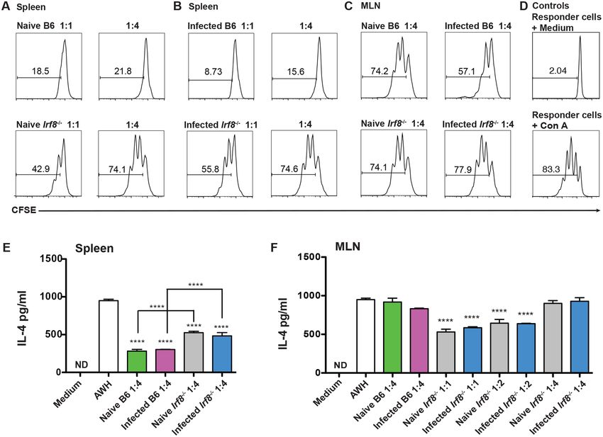
RESEARCH ARTICLE

                                                         IRF-8 regulates expansion of myeloid-derived
                                                         suppressor cells and Foxp3+ regulatory T cells
                                                         and modulates Th2 immune responses to
                                                         gastrointestinal nematode infection
                                                         Rajesh M. Valanparambil1,2,3, Mifong Tam2, Pierre-Paul Gros2, Jean-Philippe Auger4,
                                                         Mariela Segura3,4, Philippe Gros5, Armando Jardim3, Timothy G. Geary3, Keiko Ozato6,
                                                         Mary M. Stevenson1,2,3,7*
a1111111111
                                                         1 Division of Experimental Medicine, Department of Medicine, McGill University, Montreal, Quebec, Canada,
a1111111111
                                                         2 The Research Institute of the McGill University Health Centre, Montreal, Quebec, Canada, 3 Centre for
a1111111111                                              Host-Parasite Interactions, Institute of Parasitology, McGill University, Ste-Anne de Bellevue, Quebec,
a1111111111                                              Canada, 4 Department of Pathology and Microbiology, Faculty of Veterinary Medicine, University of Montreal,
a1111111111                                              St. Hyacinthe, Quebec, Canada, 5 Department of Biochemistry, McGill University, Montreal, Quebec,
                                                         Canada, 6 Division of Developmental Biology, National Institute of Child Health and Human Development,
                                                         NIH, Bethesda MD, United States of America, 7 Department of Microbiology and Immunology, McGill
                                                         University, Montreal, Quebec, Canada

                                                         * mary.stevenson@mcgill.ca
    OPEN ACCESS

Citation: Valanparambil RM, Tam M, Gros P-P,
Auger J-P, Segura M, Gros P, et al. (2017) IRF-8
regulates expansion of myeloid-derived suppressor
                                                         Abstract
cells and Foxp3+ regulatory T cells and modulates
                                                         Interferon regulatory factor-8 (IRF-8) is critical for Th1 cell differentiation and negatively reg-
Th2 immune responses to gastrointestinal
nematode infection. PLoS Pathog 13(10):                  ulates myeloid cell development including myeloid-derived suppressor cells (MDSC).
e1006647. https://doi.org/10.1371/journal.               MDSC expand during infection with various pathogens including the gastrointestinal (GI)
ppat.1006647                                             nematode Heligmosomoides polygyrus bakeri (Hpb). We investigated if IRF-8 contributes to
Editor: P’ng Loke, New York University, UNITED           Th2 immunity to Hpb infection. Irf8 expression was down-regulated in MDSC from Hpb-
STATES                                                   infected C57BL/6 (B6) mice. IRF-8 deficient Irf8-/- and BXH-2 mice had significantly higher
Received: November 7, 2016                               adult worm burdens than B6 mice after primary or challenge Hpb infection. During primary
Accepted: September 12, 2017                             infection, MDSC expanded to a significantly greater extent in mesenteric lymph nodes
                                                         (MLN) and spleens of Irf8-/- and BXH-2 than B6 mice. CD4+GATA3+ T cells numbers were
Published: October 2, 2017
                                                         comparable in MLN of infected B6 and IRF-8 deficient mice, but MLN cells from infected
Copyright: This is an open access article, free of all
                                                         IRF-8 deficient mice secreted significantly less parasite-specific IL-4 ex vivo. The numbers
copyright, and may be freely reproduced,
distributed, transmitted, modified, built upon, or       of alternatively activated macrophages in MLN and serum levels of Hpb-specific IgG1 and
otherwise used by anyone for any lawful purpose.         IgE were also significantly less in infected Irf8-/- than B6 mice. The frequencies of antigen-
The work is made available under the Creative            experienced CD4+CD11ahiCD49dhi cells that were CD44hiCD62L- were similar in MLN of
Commons CC0 public domain dedication.
                                                         infected Irf8-/- and B6 mice, but the proportions of CD4+GATA3+ and CD4+IL-4+ T cells were
Data Availability Statement: All relevant data are       lower in infected Irf8-/- mice. CD11b+Gr1+ cells from naïve or infected Irf8-/- mice suppressed
within the paper and its Supporting Information
files.
                                                         CD4+ T cell proliferation and parasite-specific IL-4 secretion in vitro albeit less efficiently
                                                         than B6 mice. Surprisingly, there were significantly more CD4+ T cells in infected Irf8-/- mice,
Funding: This work was supported by grants from
the Natural Sciences and Engineering Research
                                                         with a higher frequency of CD4+CD25+Foxp3+ T (Tregs) cells and significantly higher num-
Council of Canada (http://www.nserc-crsng.gc.ca/         bers of Tregs than B6 mice. In vivo depletion of MDSC and/or Tregs in Irf8-/- mice did not
index_eng) (#342150 to MS, #326946 to TGG, and           affect adult worm burdens, but Treg depletion resulted in higher egg production and
#238294 to AJ), National Institute of Health
(https://www.niaid.nih.gov) RO1-AI035327-19 to

PLOS Pathogens | https://doi.org/10.1371/journal.ppat.1006647 October 2, 2017                                                                                 1 / 22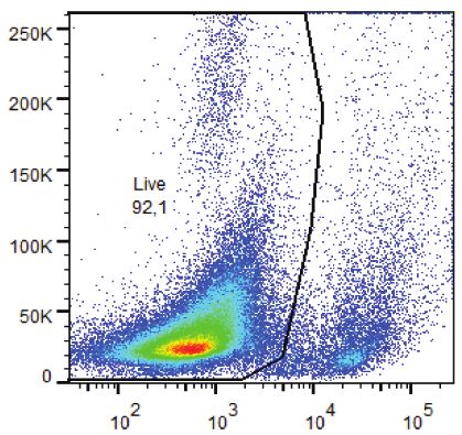
IRF-8 modulates Th2 immunity to a GI nematode

PG, Intramural Program of the National Institute of    enhanced parasite-specific IL-5, IL-13, and IL-6 secretion ex vivo. Our data thus provide a
Child Health and Development, National Institutes      previously unrecognized role for IRF-8 in Th2 immunity to a GI nematode.
of Health (https://www.nichd.nih.gov) to KO, and
the Canadian Institutes of Health Research (http://
www.cihr-irsc.gc.ca/e) MOP-81169 and MOP-
130369 to MMS. RMV was supported by a
Studentship from the Research Institute of the
McGill University Health Centre (http://www.
                                                         Author summary
rimuhc.ca/). The Centre for Host-Parasite                We investigated if IRF-8, which is critical for Th1 immunity and negatively regulates mye-
Interactions is supported by Fonds de Québec de         loid cell development including MDSC, contributes to Th2 immunity to the gastrointesti-
recherche sur la nature et les technologies (http://
                                                         nal nematode Heligmosomoides polygyrus bakeri (Hpb). Irf8 expression was down-
www.frqnt.gouv.qc.ca/en). The funders had no role
in the study design, data collection and analysis,       regulated in MDSC from infected C57BL/6 (B6) mice. Hpb-infected IRF-8 deficient mice
decision to publish, or preparation of the               had significantly higher adult worm burdens than B6 mice. There were significantly more
manuscript.                                              MDSC, fewer alternatively activated macrophages, lower serum levels of Hpb-specific
Competing interests: The authors have declared           antibodies in infected IRF-8 deficient than B6 mice, and MLN cells from infected IRF-8
that no competing interests exist.                       deficient mice secreted less parasite-specific IL-4 ex vivo. There were similar frequencies
                                                         of antigen-experienced CD4+CD11ahiCD49dhi T cells in MLN that were CD44hiCD62L-
                                                         in infected Irf8-/- and B6 mice, but lower proportions of CD4+GATA3+ and CD4+IL-4+ T
                                                         cells in Irf8-/- mice. Infected Irf8-/- mice had a higher frequency of CD4+Foxp3+ T (Tregs)
                                                         cells and significantly higher numbers of Tregs compared to infected B6 mice. MDSC
                                                         from infected Irf8-/- mice suppressed CD4+ T cell effector functions in vitro albeit less effi-
                                                         ciently than B6 mice. Treg and/or MDSC depletion did not affect adult worm burdens in
                                                         infected Irf8-/- mice, but Treg depletion partially restored Th2 cytokine responses. These
                                                         data highlight the importance of IRF-8 in Th2 immunity to Hpb infection.

                                                       Introduction
                                                       Interferon regulatory factor (IRF)-8 is a member of the IRF family of transcription factors and
                                                       plays an important role in regulating proinflammatory cytokines especially IL-12p40, which is
                                                       critical for Th1 cell differentiation [1]. IRF-8 is essential for the development of various mye-
                                                       loid-derived cells including macrophages, dendritic cells (DC), eosinophils, and basophils, but
                                                       negatively regulates neutrophil differentiation [2, 3]. Through its IRF-8 association domain
                                                       (IAD), IRF-8 interacts with other transcription factors, such as PU-1, IRF-1, IRF-4, and IRF-2,
                                                       and plays an important role in immunity against tumors and infections with intracellular path-
                                                       ogens, including bacteria, viruses, and protozoan parasites [4–6]. Irf8-/- mice develop a disease
                                                       similar to chronic myeloid leukemia characterized by expansion of immature Gr1+ granulo-
                                                       cytes [7]. Partial or total loss-of-function of IRF-8 results in decreased resistance to infections
                                                       with intracellular pathogens such as Toxoplasma gondii in mice and Mycobacterium tuberculo-
                                                       sis in humans [8, 9].
                                                           BXH-2 mice, a recombinant inbred strain generated by a cross between C57BL/6 (B6) and
                                                       C3H/HeJ mice, carry an arginine-to-cysteine substitution at position 294 in the IAD of the Irf8
                                                       gene [10, 11]. In the presence of this mutation, IRF-8 is unable to bind to its partner transcrip-
                                                       tion factors resulting in a phenotype similar to Irf8-/- mice. BXH-2 mice display increased mye-
                                                       loproliferation of CD11b+Gr1+ cells with splenomegaly and lymph node enlargement [10, 12].
                                                       Like Irf8-/- mice, BXH-2 mice are highly susceptible to infections with intracellular pathogens
                                                       including M. bovis (BCG), Salmonella enterica serovar Typhimurium, and Plasmodium cha-
                                                       baudi AS as well as M. tuberculosis [13, 14].
                                                           During infection, IRF-8 is induced in antigen presenting cells (APC) by IFN-γ and TLR
                                                       ligands, binds to IRF-4, and stimulates IL-12 as well as IL-18 secretion, resulting in CD4+ Th1

PLOS Pathogens | https://doi.org/10.1371/journal.ppat.1006647 October 2, 2017                                                                        2 / 22
IRF-8 modulates Th2 immunity to a GI nematode

                                            cell differentiation and elimination of intracellular pathogens [15, 16]. The severe immunode-
                                            ficiency and failure to produce IFN-γ and Th1-associated cytokines evident in Irf8-/- and BXH-
                                            2 mice is considered to be due to a deficiency in CD11c+CD8α+ DC responsible for producing
                                            high levels of IL-12 as well as type 1 interferon [12, 17]. On one hand, Irf8-/- mice are resistant
                                            to experimental autoimmune encephalomyelitis (EAE) due to a deficiency in Th17 cell expan-
                                            sion [18]. On the other hand, Ouyang et al observed that IRF-8 deficient mice have enhanced
                                            Th17 cell differentiation in vitro and adoptive transfer of naïve CD4+ T cells from Irf8-/- mice
                                            results in enhanced Th17 responses and more severe intestinal inflammation in a Rag-/- mouse
                                            model of colitis than cells from control mice [19]. These data highlight the importance of IRF-
                                            8 in Th1- and Th17-mediated immune responses, but the effect of IRF-8 deficiency on Th17
                                            cell differentiation appears to be variable depending on the experimental model used.
                                                Earlier, Giese et al observed that Irf8-/- mice are susceptible to Leishmania major infection
                                            compared to wild-type (WT) B6 mice, which are resistant [15]. Draining lymph node and
                                            spleen cells from infected Irf8-/- mice produce high levels of antigen-specific IL-4, the signature
                                            Th2 cytokine, and little or no IFN-γ, compared to cells from infected B6 mice. Based on these
                                            findings, it was concluded that a deficiency in IRF-8 leads to Th2 cell differentiation as a
                                            default pathway due to deficient IL-12 secretion by APC [15]. These data indicate that IRF-8 is
                                            critical for Th1- and Th17-mediated immune responses, but its role in Th2 responses has not
                                            been investigated.
                                                Heligmosomoides polygyrus bakeri (Hpb) is a natural pathogen of mice that serves as a labo-
                                            ratory model of gastrointestinal (GI) nematode infection [20]. Hpb infection is associated with
                                            strong immunosuppressive effects and induces regulatory T cells (Tregs), tolerogenic DC, reg-
                                            ulatory B cells, and high levels of the immunoregulatory cytokines IL-10 and TGFβ [20–23].
                                            Primary Hpb infection is chronic and can last up to several months in some inbred mouse
                                            strains, including B6 mice [21]. Adult worms are rapidly cleared after challenge infection due
                                            to a highly polarized Th2 immune response critically dependent on IL-4-producing
                                            CD4+GATA3+ T cells, alternatively activated macrophages (AAMØ), B cells, and parasite-spe-
                                            cific IgG1 [22]. Together, these cells and their effector molecules play important roles in elimi-
                                            nating the infection.
                                                Recently, we showed that F4/80-CD11b+Gr1hi myeloid-derived suppressor cells (MDSC)
                                            increase dramatically during primary Hpb infection in B6 mice [24]. We also demonstrated
                                            that MDSC contribute to chronic infection characteristic of these hosts by suppressing para-
                                            site-specific Th2 responses. MDSC are a heterogeneous population of immature myeloid cells
                                            that suppress effector CD4+ and CD8+ T cell responses, including proliferation and cytokine
                                            production [25]. Interestingly, MDSC are present in high numbers in naïve Irf8-/- mice [26].
                                            MDSC expansion is associated with faster tumor growth in Irf8-/- mice than in WT B6 mice,
                                            while expansion of MDSC is diminished during tumor growth in transgenic mice that over-
                                            express Irf8. The expression of Irf8 is down-regulated in breast cancer patients, and the level
                                            correlates inversely with the frequency of peripheral MDSC. Together, these data indicate IRF-
                                            8 negatively regulate MDSC.
                                                Here, we investigated the contribution of IRF-8 to the development of Th2 immunity to
                                            Hpb infection. Irf8 expression was down-regulated in B6 MDSC after primary Hpb infection,
                                            suggesting a role for this transcription factor in regulating Th2 immune responses. IRF-8 defi-
                                            cient Irf8-/- and BXH-2 mice harboured significantly higher adult worm burdens than B6 mice
                                            after primary or challenge Hpb infection. During primary infection, high worm burdens in
                                            IRF-8-deficient mice were associated with marked expansion of MDSC in mesenteric lymph
                                            node (MLN) and spleen. MLN and spleen cells from infected Irf8-/- mice secreted significantly
                                            lower levels of parasite-specific IL-4 ex vivo than infected B6 mice. Further analysis revealed
                                            that deficient IL-4 secretion by cells from infected Irf8-/- mice was not due to differentiation of

PLOS Pathogens | https://doi.org/10.1371/journal.ppat.1006647 October 2, 2017                                                             3 / 22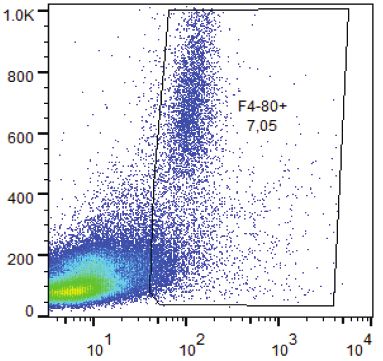
IRF-8 modulates Th2 immunity to a GI nematode

                                            naïve CD4+ T cells to Th1/Th17 cells. Importantly, lower proportions of CD4+ T cells
                                            expressed GATA3 and IL-4, but a higher proportion of CD4+ T were Foxp3+ in Irf8-/- com-
                                            pared to B6 mice during Hpb infection. Indeed, MDSC from naïve and infected Irf8-/- mice
                                            suppressed antigen-specific IL-4 secretion in vitro. Administration of 5-FU to infected Irf8-/-
                                            mice significantly decreased MDSC in MLN, but there were no effects on the adult worm bur-
                                            den or egg production. We also observed significantly higher numbers of total CD4+ T cells
                                            and CD4+CD25+Foxp3+ cells in Irf8-/- mice compared to B6 mice during Hpb infection.
                                            Depletion of Tregs in infected Irf8-/- mice enhanced Th2 cytokine responses without affecting
                                            the adult worm burden but egg production was significantly increased.

                                            Results
                                            Irf8 expression is down-regulated in MDSC during Hpb infection and
                                            contributes to higher adult worm burdens
                                            We recently showed that MDSC increase significantly in local and systemic lymphoid tissues
                                            in B6 mice during primary Hpb infection and contribute to chronic infection by suppressing
                                            Th2 responses [24]. Irf8 levels are decreased in MDSC recovered from tumor-bearing mice
                                            and cancer patients [26]. To determine if Irf8 expression is modulated during Hpb infection,
                                            we analyzed the levels in tissues as well as purified CD11b+Gr1+ cells from naïve and infected
                                            B6 mice by qRT-PCR. There were no differences in Irf8 expression in MLN or spleen from
                                            naïve versus Hpb-infected mice (S1 Fig). However, Irf8 expression was significantly down-reg-
                                            ulated in purified CD11b+Gr1+ cells from infected compared to naïve B6 mice (Fig 1A).
                                                To investigate if IRF-8 plays a role in Th2 immunity, WT B6 and Irf8-/- mice were infected
                                            with Hpb and the adult worm burden was determined on day 14 after primary infection. A sig-
                                            nificantly higher number of adult worms was recovered from Irf8-/- compared to B6 mice (Fig
                                            1B). We confirmed the importance of IRF-8 deficiency in controlling primary Hpb infection
                                            by examining the adult worm burden in BXH-2 mice that bear a mutation in the IAD region
                                            of the Irf8 gene [11]. Consistent with our findings in Irf8-/- mice, BXH-2 mice harboured sig-
                                            nificantly more adult worms than progenitor B6 or C3H/HeJ mice (Fig 1B). We also deter-
                                            mined if a deficiency in IRF-8 perturbed protective Th2 immunity required for eliminating
                                            adult worms after challenge Hpb infection. Two weeks after re-infection of anthelmintic-
                                            treated mice, Irf8-/- as well as BXH-2 (Fig 1C) mice had significantly more adult worms in the
                                            small intestine than B6 mice. These data indicate that Irf8 expression is down-regulated during
                                            primary Hpb infection and a partial or total loss-of-function in IRF-8 affects immune control
                                            of the adult worm burden after both primary and challenge Hpb infections.

                                            IRF-8 deficiency is associated with expansion of MDSC and decreased
                                            CD4+ Th2 cell effector function after Hpb infection
                                            To determine if differences in immune cell populations in lymphoid tissues, especially in
                                            MLN, contributed to higher worm burdens in IRF-8 deficient mice, we examined the numbers
                                            and cellular composition of MLN and spleen from naïve and Hpb-infected B6 versus similar
                                            groups of Irf8-/- and BXH-2 mice. There were significantly higher numbers of total cells in
                                            MLN and spleen of naïve Irf8-/-, but not in BXH-2 mice, than B6 mice (S2 Fig). On day 14 after
                                            primary or challenge infection, there were marked increases in the total cellularity of lymphoid
                                            tissues from B6 as well as IRF-8 deficient mice with significantly higher cell numbers in the
                                            MLN and spleen of Irf8-/- compared to B6 mice.
                                                As previously reported, the number of F4/80-CD11bhiGr1hi cells was significantly higher in
                                            the spleen of naïve Irf8-/- compared to B6 mice (S3A Fig) [26]. In addition, we observed a

PLOS Pathogens | https://doi.org/10.1371/journal.ppat.1006647 October 2, 2017                                                           4 / 22
IRF-8 modulates Th2 immunity to a GI nematode
Fig 1. Irf8 expression is down-regulated in MDSC and contributes to higher worm burdens after Hpb infection. (A) Irf8 expression in
CD11b+Gr1+ cells purified from spleens of naïve and infected C57BL/6 (B6) mice on day 7 p.i. Each point represents cells pooled from 2 naïve mice
or one infected B6 mouse. Representative results of one of two replicate experiments are shown. Data are presented as relative quantity of Irf8
normalized against the endogenous control Actb. (B) Adult worm burden after primary Hpb infection in WT B6 and progenitor C3H/HeJ mice
compared to Irf8-/- and IRF-8 deficient BXH-2 mice. The worm burdens of individual mice pooled from independent experiments are shown. (C) Adult
worm burden after challenge Hpb infection in WT B6, Irf8-/- and BXH-2 mice. The worm burdens of individual mice pooled from independent
experiments are shown. Data are presented as mean ± SEM. ns, not significant; **, p 0.01; ***, p 0.001; ****, p 0.0001.
https://doi.org/10.1371/journal.ppat.1006647.g001
significantly higher frequency and number of these cells in MLN of naïve Irf8-/- than B6 mice
(S3A & S4 Figs). There was also a significantly higher frequency of MDSC in MLN of naïve
BXH-2 vs. B6 mice although the number of MDSC was similar (S3B & S4 Figs). Similar to
naïve Irf8-/- mice, there was a significantly higher number of MDSC in the spleen of naïve
BXH-2 than B6 mice.
Consistent with our previous observation, the frequency of F4/80-CD11bhiGr1hi cells
increased significantly in MLN of infected compared to naïve B6 mice on day 14 post infection
(p.i.) (S4 Fig) [24]. In contrast, the frequency of MDSC decreased during Hpb infection in
IRF-8 deficient mice compared to naïve mice (S4 Fig). Importantly, there were significantly
higher numbers of F4/80-CD11bhiGr1hi cells in the MLN of infected Irf8-/- and BXH-2 mice
compared to B6 mice (Fig 2A–2C). Interestingly, the numbers of CD4+GATA3+ T cells in
MLN were similar in infected Irf8-/- and B6 mice (Fig 2D), but the number of F4/
80+CD11b+CD206+ AAMØ was significantly lower in infected Irf8-/- mice (Fig 2E). Similar
findings were observed for CD4+GATA3+ cells and AAMØ in MLN of BXH-2 compared to
B6 mice after primary Hpb infection (S3C and S3D Fig).
Despite similar numbers of CD4+GATA3+ T cells in the MLN of Hpb-infected B6 and IRF-
8-deficient mice, IL-4 secretion ex vivo was significantly lower in response to AWH by MLN
as well as spleen cells from infected Irf8-/- compared to B6 mice (Fig 3A and 3B). Likewise,
MLN cells from infected BXH-2 mice produced significantly lower levels of AWH-specific IL-
4 ex vivo than infected B6 mice (S3E Fig). In addition, serum levels of HES-specific IgG1 and
IgE were significantly lower in Hpb-infected Irf8-/- compared to B6 mice (Fig 3C).
To further characterize the CD4+ T cell response in IRF-8 deficient mice after Hpb infec-
tion, we used the surrogate marker approach described by McDermott at al to identify anti-
gen-experienced CD4+ T cells [27]. Previous studies in mice infected with a variety of
pathogens have validated this approach, which is based on increased co-expression of the
PLOS Pathogens | https://doi.org/10.1371/journal.ppat.1006647 October 2, 2017 5 / 22IRF-8 modulates Th2 immunity to a GI nematode
Fig 2. Immunophenotype of cells in MLN of C57BL/6 and IRF-8 deficient mice after primary Hpb infection. (A)
Gating strategy used to analyze F4/80-CD11bhiGr1hi (MDSC) and F4/80+CD11b+CD206+ (AAMØ) cells by flow
cytometry. Total numbers of F4/80-CD11bhiGr1hi cells in MLN of (B) C57BL/6 (B6) and Irf8-/- mice and (C) B6 and
BXH-2 mice on day 14 p.i. Total numbers of (D) CD4+GATA3+ T cells and (E) F4/80+CD11b+CD206+ cells in MLN of
PLOS Pathogens | https://doi.org/10.1371/journal.ppat.1006647 October 2, 2017 6 / 22IRF-8 modulates Th2 immunity to a GI nematode
B6 and Irf8-/- mice on day 14 p.i. n = 5 mice/group. Data are representative of three replicate experiments in B6 and
Irf8-/- mice and two replicate experiments in B6 and BXH-2 mice. Data are presented as mean ± SEM. ns, not
significant; *, p 0.05; **, p 0.01.
https://doi.org/10.1371/journal.ppat.1006647.g002
integrins CD11a and CD49d, to track CD4+ T cell responses even without knowledge of patho-
gen-specific epitopes [28–30]. As expected, there were low frequencies of CD4+CD11ahiCD49-
dhi cells in MLN of naïve B6 and Irf8-/- mice (Fig 3D upper panels). After Hpb infection, the
frequency of this population increased to similar levels in MLN of B6 and Irf8-/- mice (Fig 3D
lower panels). Between 50–60% of CD4+CD11ahiCD49dhi cells in naïve B6 and Irf8-/- mice
were CD44hiCD62L- and the frequency of this population increased in B6 as well as Irf8-/- mice
to 70–80% during Hpb infection.
To address the possibility that CD4+ T cells differentiated to Th cell subsets other than Th2
cells in Irf8-/- mice during Hpb infection, we analyzed the expression of transcription factors
and cytokines that distinguish various Th cell subsets, including Tregs, in MLN CD4+ T cells.
Immunophenotyping of gated CD4+ T cells showed that 73% of total CD4+ T cells in MLN of
B6 mice and 76% in Irf8-/- mice were undifferentiated on day 14 p.i. (Fig 3E). During Hpb
infection, 11% of the total CD4+ T cells were GATA3+ and 9% were Foxp3+ in B6 mice, while
7% expressed GATA3 and 12% expressed Foxp3 in Irf8-/- mice. There were no significant dif-
ferences between the proportions of CD4+GATA3+ and CD4+Foxp3+ T cells between infected
B6 and Irf8-/- mice, but the ratio of the proportions of Tregs to Th2 cells was significantly
higher in infected Irf8-/- compared to B6 mice (Fig 3F). Consistent with the findings described
above that MLN cells from infected Irf8-/- mice secreted significantly lower levels of parasite-
specific IL-4 ex vivo than cells from infected B6 mice, only 1% of CD4+ T cells were IL-4+ in
Irf8-/- compared to 2% in B6 mice during Hpb infection. The proportions of CD4+ T cells that
expressed ROR-γt, IL-17 or IFN-γ were low in MLN of both genotypes after infection. These
data indicate CD4+ T cells in infected Irf8-/- mice did not differentiate to the Th1 or Th17 path-
way as a default, but that Tregs may be the dominate CD4+ T cell population in these mice dur-
ing Hpb infection.
Suppression of CD4+ T cell effector function by MDSC from Irf8-/- mice
Previously, we showed that purified CD11b+Gr1+ cells from Hpb-infected B6 mice potently
suppress antigen-specific CD4+ T cell proliferation and IL-4 secretion in vitro [24]. To investi-
gate if MDSC from Irf8-/- mice suppress CD4+ T cell proliferation, purified CD11b+Gr1+ cells
from naïve and infected B6 or Irf8-/- mice were co-cultured with CFSE-labeled spleen cells
from naïve B6 mice. Following stimulation with Con A, proliferation was examined in gated
CD4+ T cells by CFSE staining. On a cell-per-cell basis, CD11b+Gr1+ cells recovered from the
spleens of naïve and infected Irf8-/- mice were less suppressive than CD11b+Gr1+ cells from B6
mice at ratios of MDSC to responder cells of 1:1 and 1:4 (Fig 4A and 4B) as well as 1:8 (S5 Fig).
Nevertheless, MDSC from either genotype suppressed CD4+ T cell proliferation to Con A
in a dose-dependent manner compared to the control (Fig 4D). The low numbers of
CD11b+Gr1+ cells obtained from MLN permitted us to compare the suppressive activity of
MDSC from B6 vs. Irf8-/- mice at only a 1:4 ratio. Although CD11b+Gr1+ cells purified from
MLN of naïve B6 or Irf8-/- mice suppressed Con A-induced proliferation to a similar albeit
negligible extent compared to the control culture, CD11b+Gr1+ cells from infected B6 mice
were more suppressive than cells from infected Irf8-/- mice (Fig 4C).
We also compared the ability of MDSC from B6 and Irf8-/- mice to suppress Hpb-specific
IL-4 secretion. Purified CD11b+Gr1+ cells were co-cultured with spleen cells from infected B6
PLOS Pathogens | https://doi.org/10.1371/journal.ppat.1006647 October 2, 2017 7 / 22IRF-8 modulates Th2 immunity to a GI nematode
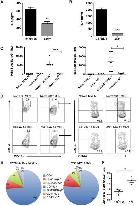
Fig 3. Analysis of CD4+ Th2 cell effector function in Hpb-infected C57BL/6 and Irf8-/- mice. (A) MLN and (B)
spleen cells obtained from C57BL/6 (B6) and Irf8-/- mice (n = 5 mice per group) on day 14 p.i. were stimulated ex
vivo with 50 μg AWH. Supernatants were collected 48 h later and IL-4 levels determined by ELISA. Data
representative of two to three replicate experiments are presented as mean ± SEM. **; p 0.01; ***, p0.001.
(C) HES-specific IgG1 and IgE titers in the sera of naïve and infected B6 and Irf8-/- mice on day 14 p.i. Each point
PLOS Pathogens | https://doi.org/10.1371/journal.ppat.1006647 October 2, 2017 8 / 22IRF-8 modulates Th2 immunity to a GI nematode
represents one mouse. Mean ± SEM for each group is shown. *, p 0.05; ***, p0.001. (D) MLN cells were
obtained from naïve and infected B6 or Irf8-/- mice on day 14 p.i. (n = 3 mice per group). CD11a and CD49d
expression were analyzed on gated CD4+ T cells by flow cytometry. CD11ahiCD49dhi cells were gated and the
expression of CD44 and CD62L was analyzed. Representative contour plots of one mouse from each group are
shown for co-expression of CD11a and CD49d and CD44 and CD62L. Data presented for B6 mice are from one
of 2 replicate experiments performed. (E) Gated CD4+ T cells obtained from naïve and infected B6 or Irf8-/- mice
on day 14 p.i. (n = 3 mice/group) were analyzed for intracellular expression of GATA3, Foxp3, IL-4, ROR-γt, IFN-
γ, and IL-17 by flow cytometry. The proportions of each cell type are shown as a percent of total CD4+ T cells. (F)
The ratios of the proportions of CD4+Foxp3+ to CD4+GATA3+ T cells were determined for C57BL/6 and Irf8-/-
mice based on the data shown in panel E. Each point represents one mouse. Mean ± SEM for each group is
shown. *, p 0.05.
https://doi.org/10.1371/journal.ppat.1006647.g003
Fig 4. CD4+ T cells are suppressed by MDSC from naïve or Hpb-infected C57BL/6 and Irf8-/- mice. Purified CD11b+Gr1+ cells from spleen of naïve
(A) and infected (B) C57BL/6 (B6) or Irf8-/- mice were co-cultured with CFSE-labeled spleen cells from naïve B6 mice and stimulated with 2 μg/ml Con A.
(C) Purified CD11b+Gr1+ cells from MLN of naïve or infected C57BL/6 (B6) and Irf8-/- mice were co-cultured with CFSE-labeled spleen cells from naïve B6
mice and stimulated with 2 μg/ml Con A. (D) Control cultures containing responder cells alone (top) or responder cells stimulated with 2 μg/ml Con A
(bottom). CFSE dilution in panels A-D was analyzed in gated CD4+ T cells by flow cytometry. The data shown are representative of two replicate
experiments. Purified CD11b+Gr1+ cells from spleens (E) or MLN (F) of naïve or infected B6 and Irf8-/- mice were co-cultured with spleen cells from
infected B6 mice and stimulated with medium or 50 μg AWH. IL-4 levels were determined in 48 h supernatants by ELISA. The ratio of MDSC:responder
cells is indicated for each panel. The data shown are representative of two to three replicate experiments and are presented as mean ± SEM. ND, not
detectable; ****, p 0.0001.
https://doi.org/10.1371/journal.ppat.1006647.g004
PLOS Pathogens | https://doi.org/10.1371/journal.ppat.1006647 October 2, 2017 9 / 22IRF-8 modulates Th2 immunity to a GI nematode
Fig 5. Effect of MDSC depletion on worm burdens in Hpb-infected C57BL/6 and Irf8-/- mice. Numbers of F4/80-CD11bhiGr1hi cells in MLN of infected
C57BL/6 (B6) mice treated with PBS or 5-FU on (A) day 7 or (B) day 14 p.i. (C) Adult worm burdens on day 14 p.i. of individual B6 mice treated with PBS or
5-FU. Numbers of (D) F4/80-CD11bhiGr1hi and (E) CD4+GATA3+ T cells in MLN of infected Irf8-/- mice treated with PBS or 5-FU on day 14 p.i. (F) Adult
worm burdens on day 14 p.i. and (G) fecal egg counts on day 13 p.i. of individual infected Irf8-/- mice treated with PBS or 5-FU. Data are representative of
two replicate experiments each with n = 5 mice per group. Data are presented as mean ± SEM. ns, not significant; *, p 0.05; **, p 0.01.
https://doi.org/10.1371/journal.ppat.1006647.g005
mice and stimulated with AWH. Spleen CD11b+Gr1+ cells from B6 as well as Irf8-/- mice,
either naïve or infected, significantly suppressed IL-4 secretion compared to spleen cells stimu-
lated with AWH alone (Fig 4E). However, IL-4 secretion was significantly lower in co-cultures
with CD11b+Gr1+ cells from naïve or infected B6 compared to similar groups of Irf8-/- mice.
Importantly, CD11b+Gr1+ cells purified from the MLN of naïve or infected Irf8-/- mice signifi-
cantly suppressed AWH-induced IL-4 secretion at ratios of 1:1 and 1:2 MDSC to responder
cells compared to responder cells stimulated with AWH in the absence of MDSC (Fig 4F).
MDSC depletion does not alter adult worm burdens in infected Irf8-/-
mice
Administration of the chemotherapeutic agent 5-fluorouracil (5-FU) to tumor-bearing mice
significantly decreases MDSC in tissue and peripheral blood and restores anti-cancer immune
responses [31, 32]. To determine if 5-FU treatment depletes MDSC and enhances Th2 immu-
nity in Hpb-infected mice, B6 mice were treated i.p. with 5-FU or PBS as a control on day -2
prior to infection and on days 5 and 12 p.i. MDSC were significantly decreased in MLN of
5-FU-treated compared to PBS-treated B6 mice on day 7 p.i. (Fig 5A). Consistent with previ-
ous findings in tumor-bearing mice treated with anti-Gr-1 mAb to deplete MDSC, MDSC
depletion was transient in 5-FU-treated mice and the numbers of MDSC in MLN rebounded
and were significantly higher compared to PBS-treated B6 mice on day 14 p.i. (Fig 5B) [33].
PLOS Pathogens | https://doi.org/10.1371/journal.ppat.1006647 October 2, 2017 10 / 22IRF-8 modulates Th2 immunity to a GI nematode
Despite this, the adult worm burden was significantly but modestly decreased after 5-FU treat-
ment compared to PBS control (Fig 5C).
To investigate the effects of MDSC depletion in Hpb-infected Irf8-/- mice, we used a similar
protocol to administer 5-FU to these mice. MDSC were significantly reduced in 5-FU treated
Irf8-/- mice compared to PBS control mice on day 14 p.i. (Fig 5D). MDSC depletion did not
alter the numbers of CD4+GATA3+ T cells (Fig 5E), adult worm burden (Fig 5F) or egg pro-
duction (Fig 5G) in infected Irf8-/- mice.
Higher numbers of CD4+Foxp3+ Tregs are associated with decreased
Th2 responses in Hpb-infected Irf8-/- mice
After Hpb infection, Foxp3+ Tregs expand in local and systemic lymphoid tissues but the role
of these cells remains ambiguous [34, 35]. Surprisingly, there were significantly higher num-
bers of total CD4+ T cells in MLN of Irf8-/- than B6 mice on day 14 p.i. (Fig 6A) despite similar
numbers of CD4+GATA3+ T cells in infected mice of both genotypes (Fig 2D). At this time,
the number of CD4+CD25+Foxp3+ T cells was significantly higher in infected Irf8-/- compared
to B6 mice (Fig 6B). We performed a kinetic analysis of Th2 versus Tregs in B6 compared to
Irf8-/- mice during infection. The numbers of CD4+CD25+Foxp3+ T cells were similar in naïve
B6 and Irf8-/- mice, but this population expanded early during infection in both B6 and Irf8-/-
mice (Fig 6C). On day 7 p.i., there was approximately a 2-fold increase in CD4+CD25+Foxp3+
T cells in B6 mice and while there was over a 5-fold increase in Irf8-/- mice. By day 14 p.i.,
Irf8-/- mice had approximately twice as many CD4+CD25+Foxp3+ T cells in the MLN as B6
mice. The marked expansion of Tregs in infected Irf8-/- mice led to significantly higher num-
bers of CD4+CD25+Foxp3+ T cells in Irf8-/- than B6 mice, resulting in higher ratios of Foxp3+
Tregs to Th2 cells in Hpb-infected Irf8-/- compared to B6 mice on day 7 as well as day 14 p.i.,
consistent with the data shown in Fig 3F.
To investigate if Foxp3+ Tregs contributed to high adult worm burdens in Irf8-/- mice, we
treated Hpb-infected Irf8-/- mice with anti-CD25 mAb or isotype control rat IgG and deter-
mined the effects on Th2 responses as well as on the adult worm burden and egg production.
Administration of anti-CD25 mAb significantly reduced CD4+CD25+Foxp3+ cells in Hpb-
infected Irf8-/- mice (Fig 6D), while the numbers of CD4+GATA3+ T cells (Fig 6E), AAMØ
(Fig 6F), and MDSC (Fig 6G) were similar to isotype control mice. Depletion of Foxp3+ Tregs
did not affect the adult worm burden, but egg production was significantly higher in anti-
CD25 mAb treated Irf8-/- mice compared to isotype controls indicating increased worm fecun-
dity (Fig 6H and 6I). We also determined the profile and levels of cytokines secreted ex vivo by
MLN cells from anti-CD25 mAb treated and isotype control Irf8-/- mice in response to AWH.
There were significant increases in the Th2 cytokines IL-5 and IL-13 and the pro-inflamma-
tory cytokine IL-6 in the supernatants of infected Irf8-/- mice treated with anti-CD25 mAb
compared to isotype control mice while there were no differences in IL-4, IL-10, or TGFβ (Fig
7 & S6 Fig).
To investigate the effects of depletion of both MDSC and Tregs in IRF-8 deficient mice dur-
ing Hpb infection, Irf8-/- mice were treated in tandem with both 5-FU and anti-CD25 mAb as
described for single depletions. Control mice were treated with PBS and isotype control anti-
body. F4/80-CD11bhiGr1hi cells as well as CD4+CD25+Foxp3+ cells were significantly reduced
in 5-FU and anti-CD25 mAb treated mice (S7A and S7B Fig). Depletion of both MDSC and
Tregs in infected Irf8-/- mice did not affect either the adult worm burden, but egg production
was significantly increased similar to mice treated with anti-CD25 mAb alone (S7C and S7D
Fig). In contrast to significantly increased levels of IL-5 and IL-13 secreted ex vivo in response
to AWH by MLN cells from Treg-depleted compared to control mice shown in Fig 7, serum
PLOS Pathogens | https://doi.org/10.1371/journal.ppat.1006647 October 2, 2017 11 / 22IRF-8 modulates Th2 immunity to a GI nematode
Fig 6. CD4+CD25+Foxp3+ T cell expansion is significantly greater in Irf8-/- than C57BL/6 mice during Hpb infection. (A)
Numbers of total CD4+ T cells in MLN of naïve and infected C57BL/6 (B6) or Irf8-/- mice on days 7 and 14 p.i. (B) Numbers of
CD4+CD25+Foxp3+ cells in MLN of infected B6 and Irf8-/-.mice on day 14 p.i. (C) Numbers of CD4+GATA3+ and CD4+CD25+Foxp3+ T
PLOS Pathogens | https://doi.org/10.1371/journal.ppat.1006647 October 2, 2017 12 / 22IRF-8 modulates Th2 immunity to a GI nematode
cells in MLN of naïve B6 and Irf8-/-.mice and on days 7 and 14 p.i. (D) Numbers of CD4+CD25+Foxp3+ T cells in MLN of Irf8-/-.mice
treated with isotype control antibody or anti-CD25 mAb on day 14 p.i. Numbers of (E) CD4+GATA3+ T cells, (F) F4/80+CD11c+CD206+
cells, and (G) F4/80-CD11bhiGr1hi cells in MLN of Irf8-/-.mice treated with isotype control antibody or anti-CD25 mAb on day 14 p.i. (H)
Adult worm burdens on day 14 p.i. and (I) fecal egg counts on day 13 p.i. of Irf8-/- mice treated with isotype control antibody or anti-CD25
mAb. Data from individual mice (n = 5 mice per group) are presented in panels A and D-I. Data are presented as mean ± SEM.
**, p 0.01; ***, p 0.001; ****, p 0.0001.
https://doi.org/10.1371/journal.ppat.1006647.g006
levels of Th2 cytokines were similar in Irf8-/- mice treated with anti-CD25 mAb alone or both
anti-CD25 mAb and 5-FU compared to control Irf8-/- mice (S7E–S7G Fig). These date suggest
that changes in cytokine levels may vary in supernatants vs. sera of Hpb-infected mice due to
differences in kinetics of Th2 cytokine production. Altogether, our data indicate that IRF-8
regulates the expansion of MDSC and Foxp3+ Tregs during Hpb infection and modulates Th2
immunity essential for expulsion of adult worms. Depletion of either MDSC or Tregs or both,
however, does not completely eliminate the high adult worm burdens in Irf8-/- mice suggesting
that either the depletions were incomplete or that another cell type may be involved.
Discussion
A partial or complete deficiency in IRF-8 in mice results in increased susceptibility to intracel-
lular pathogens including T. gondii, M. tuberculosis, Salmonella, and P. chabaudi AS [36]. Con-
trol of these pathogens is dependent on Th1-mediated immune responses, which are deficient
in Irf8-/- and BXH-2 mice. Similar effects have also been reported in a pediatric patient homo-
zygous for a loss-of-function mutation in the IRF8 gene. A deficiency in DC subsets expressing
CD8α especially CD11c+B220-CD8α+ DC, the major APC producing IL-12p40 in response to
microbial products, is considered to be the underlying defect responsible for the lack of Th1
responses to intracellular pathogens in IRF-8 deficient hosts [12, 17, 37]. Th17 responses are
also aberrant in Irf8-/- mice but the mechanism(s) is only partially understood [18, 19].
In addition to other abnormalities in myeloid-derived cell development, naïve Irf8-/- mice
have a dramatically higher number of MDSC than WT B6 mice [26]. Importantly, significantly
fewer MDSC are apparent in tissues and tumors of transgenic mice that over-express Irf8 com-
pared to WT B6 mice. In addition, Irf8 expression is down-regulated in MDSC during tumor
growth and the frequency of MDSC in patients with breast cancer correlates inversely with
IRF-8 levels in these cells [26]. The importance of IRF-8 in regulating MDSC expansion during
tumorigenesis is supported by the finding that tumor growth occurs more rapidly in Irf8-/-
than B6 mice.
Based on the findings described above and our previous observation that MDSC increase in
B6 mice during Hpb infection, we investigated if IRF-8 contributes to Th2 immunity to infec-
tion with a GI nematode [24, 26]. First, we examined Irf8 expression in MDSC during Hpb
infection. Irf8 expression was significantly down-regulated in MDSC from Hpb-infected com-
pared to naïve B6 mice during primary infection. We also observed that adult worm burdens
were significantly higher in mice with a total (Irf8-/- mice) or partial (BXH-2 mice) loss of func-
tional IRF-8 after both primary and challenge Hpb infections. These data provide compelling
evidence that IRF-8 plays an important role in Th2 immunity to Hpb infection.
Analysis of immune cell populations in local and systemic lymphoid tissues from IRF-
8-deficient hosts showed significantly higher numbers of total cells and MDSC in Irf8-/- and
BXH-2 compared to B6 mice especially in infected Irf8-/- mice. Consistent with previous obser-
vations, we found significantly higher numbers of MDSC in the MLN as well as the spleen of
naïve Irf8-/- mice [26]. We also observed that naïve BXH-2 have significantly higher numbers
of MDSC in the spleen compared to naïve B6 mice. After Hpb infection, MDSC increased in
the MLN of Irf8-/- and BXH-2 mice as we previously observed in B6 mice [24]. There were,
PLOS Pathogens | https://doi.org/10.1371/journal.ppat.1006647 October 2, 2017 13 / 22IRF-8 modulates Th2 immunity to a GI nematode
Fig 7. Antigen-specific cytokine secretion by MLN cells from infected Irf8-/- mice treated with anti-CD25 mAb. Single cell suspensions of
MLN were prepared from infected Irf8-/- mice treated with isotype control antibody or anti-CD25 mAb on day 14 p.i. The cells were stimulated in vitro
with 50 μg AWH, supernatants were harvested 48 h later, and cytokine levels were determined using a Bio-Plex assay. (A) IL-4, (B) IL-5, (C) IL-13,
(D) IL-6, and (E) IL-10 levels. Data from individual mice are presented as pg/ml and were calculated based on internal standards for each cytokine
with the mean ± SEM for each group shown. n = 5 mice per group. *, p 0.05; **, p 0.01.
https://doi.org/10.1371/journal.ppat.1006647.g007
however, significantly higher numbers of MDSC in infected Irf8-/- and BXH-2 mice compared
to B6 mice, with Irf8-/- mice having the largest increase in MDSC among the strains examined.
Our findings thus confirm and extend the observations of Waight et al in tumor-bearing mice
to IRF-8 deficient mice infected with a GI nematode [26].
Despite marked expansion of MDSC in infected IRF-8 deficient mice, CD4+GATA3+ T cell
numbers were similar in infected Irf8-/-, BXH-2 and B6 mice. Nevertheless, the numbers of
AAMØ in MLN and serum levels of HES-specific IgG1 and IgE were significantly lower in
association with significantly lower parasite-specific IL-4 secretion ex vivo by MLN cells from
infected Irf8-/- and BXH-2 mice compared to B6 mice. Analysis of co-expression of CD11a and
CD49d, considered to be surrogate markers of antigen-experienced CD4+ T cells, showed that
the frequencies of CD4+CD11ahiCD49dhi cells increased to similar levels in B6 and Irf8-/- mice
during Hpb infection. The majority of these cells were CD44hiCD62L- in infected mice of both
genotypes. During infection, 11% of total CD4+ T cells in MLN of B6 mice were GATA3+
while 12% of total CD4+ T cells expressed Foxp3 in infected Irf8-/- mice. A lower proportion of
CD4+ T cells from Irf8-/- mice expressed intracellular IL-4 compared to B6 mice consistent
with significantly lower IL-4 secretion ex vivo in response to AWH.
Previous studies by Waight et al demonstrated that purified CD11b+Gr1+ cells from Irf8-/-
mice strongly inhibit polyclonal as well as allogeneic CD4+ T cell proliferation in vitro com-
pared to WT B6 mice [26]. We observed that CD11b+Gr1+ cells purified from MLN and
spleens of naïve and infected Irf8-/- mice were less potent on a cell-per-cell basis than cells
from similar groups of B6 mice in suppressing Con A-induced CD4+ T cell proliferation.
PLOS Pathogens | https://doi.org/10.1371/journal.ppat.1006647 October 2, 2017 14 / 22IRF-8 modulates Th2 immunity to a GI nematode
Previously, we showed that CD11b+Gr1+ cells from mucosal and systemic tissues of infected
B6 mice significantly suppress IL-4 secretion in response to AWH in co-cultures even at a
ratio of 1 MDSC:16 responder cells [24]. Similar to differences in the ability to suppress poly-
clonal CD4+ T cell proliferation, CD11b+Gr1+ cells from infected B6 mice suppressed antigen-
specific IL-4 secretion to significantly lower levels than cells from Irf8-/- mice. Together, these
findings suggest the suppressive ability of MDSC from Irf8-/- mice relative to MDSC from WT
B6 mice may vary depending on the experimental model used.
Administration of 5-FU to tumor-bearing mice results in significant decreases in MDSC
and enhanced CD8+ T cell effector responses [31]. We observed that MDSC were significantly
reduced in MLN of 5-FU treated B6 mice on day 7 p.i. In our hands, depletion was transient
and the numbers of MDSC rebounded and were significantly increased compared to PBS con-
trols on day 14 p.i. Even so, the adult worm burden was significantly lower in 5-FU treated
compared to control B6 mice. This observation is consistent with our previous finding that
adoptive transfer of CD11b+Gr1+ cells from Hpb-infected B6 mice results in a significantly
higher worm burden and increased egg production in recipient mice [24]. These data indicate
there is an inverse correlation between the number of MDSC and the adult worm burden in
line with our findings in the present study in IRF-8 deficient mice. Altogether, these data sup-
port the contention that MDSC suppress Th2 immunity to Hpb infection resulting in higher
adult worm burdens.
Although 5-FU administration to Hpb-infected Irf8-/- mice significantly decreased MDSC
in the MLN through day 14 p.i., there were no effects on CD4+GATA3+ T cells, adult worm
burden, or egg production. This finding suggested to us that MDSC alone may not be respon-
sible for the increased susceptibility of IRF-8-deficient mice to Hpb infection. Importantly,
there were significantly more total CD4+ T cells despite similar numbers of CD4+GATA3+ T
cells in MLN of IRF-8-deficient mice compared to B6 mice after Hpb infection. This difference
was due to a significantly higher number of CD4+CD25+Foxp3+ cells in infected Irf8-/- than B6
mice. Indeed, CD4+CD25+Foxp3+ Tregs constituted a larger proportion of the total CD4+ T
cells in MLN of infected Irf8-/- than B6 mice, resulting in a higher ratio of this T cell population
to CD4+GATA3+ Th2 cells in Irf8-/- mice.
Previous studies indicate that Foxp3+ Tregs expand in local lymphoid tissues, including the
lamina propria, Peyer’s patches, and MLN, as well as systemically in the spleen of Hpb-infected
B6 and BALB/c mice [34, 35, 38]. Although Foxp3+ Tregs contribute to suppressed Th2 cyto-
kine responses during Hpb infection, their effect on adult worm expulsion is variable [35, 39].
Several strategies have been used to increase Foxp3+ Tregs or to partially or completely deplete
these cells in infected BALB/c and B6 mice [35, 39]. Together, the results of these studies pro-
vide evidence of a complex role for Foxp3+ Tregs in immunity to Hpb infection. The overall
effect of increasing Foxp3+ Tregs or depleting these cells differs not only on the genetic back-
ground of the host but also on the depletion strategy used. Recently, it was proposed that a low
number of Foxp3+ Tregs may be beneficial in providing a balance between a protective Th2
response and harmful pro-inflammatory response [35].
In the present study, we depleted Foxp3+ Tregs in infected Irf8-/- mice using repeated treat-
ments with an anti-CD25 mAb prior to infection, on the day of infection and day 7 p.i. Consis-
tent with previous studies, there were significant increases in Th2 cytokines as well as IL-6
compared to isotype control, infected Irf8-/- mice, but there were no effects on the adult worm
burden or the numbers of CD4+GATA3+ T cells or AAMØ [35, 38, 39]. Interestingly, egg pro-
duction was significantly increased in Treg-depleted compared to isotype control Irf8-/- mice
indicating increased worm fecundity in the depleted mice, but the basis of the increase is not
clear. Depletion of both MDSC and Foxp3+ Tregs using 5-FU and anti-CD25 mAb did not
PLOS Pathogens | https://doi.org/10.1371/journal.ppat.1006647 October 2, 2017 15 / 22IRF-8 modulates Th2 immunity to a GI nematode
affect serum levels of Th2 cytokines or the adult worm burden in Hpb-infected Irf8-/- mice, but
the number of eggs/g feces was significantly increased.
In conclusion, our data demonstrate that a high adult worm burden in Hpb-infected IRF-8
deficient mice is associated with decreased AWH-specific IL-4 secretion by MLN cells ex vivo,
fewer AAMØ, and marked expansion of MDSC in MLN. Compared to infected B6 mice, Irf8-/-
mice had significantly lower serum levels of HES-specific IgG1 and IgE and significantly
higher numbers of CD4+ T cells and Foxp3+ Tregs in MLN during Hpb infection. Although
MDSC as well as Foxp3+ Tregs suppress critical Th2 responses, depleting these cell populations
individually or in tandem did not promote adult worm expulsion in infected Irf8-/- mice. This
may be due to the limitations of using 5-FU and anti-CD25 mAb to deplete MDSC and Foxp3
Tregs, respectively [31, 40]. Nevertheless, our data provide novel information supporting a
role for IRF-8 in the development of Th2 immunity to a GI nematode infection.
Materials and methods
Ethics statement
The animal studies reported in this paper were approved and conducted in accordance with
the guidelines and recommendations of The Animal Care and Use Committee of McGill Uni-
versity (#2015–7592). Animals were sacrificed by CO2 overdose under isoflurane anesthesia.
Mice, parasite and infection
Male and female B6 mice, 8–10 weeks old, were purchased from Charles River Laboratories
(St. Constant, QC). Irf8-/- mice, generated as previously described, were bred and maintained
at the Research Institute of the McGill University Health Centre from breeding stock gener-
ously provided by Dr. Keiko Ozato (NICHD, NIH) [7]. BXH-2 mice were obtained from the
colony maintained by Dr. P. Gros at McGill University [13]. Hpb was maintained and propa-
gated as described previously [41]. Mice were age- and sex-matched for all experiments. For
primary infection, mice were infected per os (p.o.) with 200 infective third-stage Hpb larvae
(L3). To determine egg production, fecal pellets were collected on day 13 p.i. and eggs were
enumerated by the McMaster method [41]. To assess parasite burden, mice were sacrificed on
day 14 p.i., the intestines harvested, and the number of adult worms in each mouse was
counted by dissection microscopy. For challenge infection, mice were infected as described
above and treated p.o. with a single dose of pyrantel pamoate (Combantrin; Pfizer Canada,
Montreal, QC) at 100 mg/kg body weight on day 14 p.i. to eliminate adult worms. Drug-cured
mice were allowed to rest for 5 weeks and then re-infected p.o. with 200 L3. Adult worm
homogenate (AWH) was prepared as described previously [41], the protein concentration
determined using a Bradford protein assay kit (Bio-Rad, Hercules, CA), and stored at -20˚C
until use.
Cell preparation
MLN and spleens were harvested from naïve and Hpb-infected mice on the indicated days p.i.
Briefly, single cell suspensions were prepared using a sterile wire mesh. The cells were centri-
fuged, re-suspended in 0.175 M NH4Cl to lyse red blood cells, washed, and re-suspended in
RPMI 1640 medium (Life Technologies, Burlington, ON, Canada) supplemented with 5%
heat-inactivated fetal calf serum (HyClone Laboratories, Logan, UT), 25 mM HEPES (Life
Technologies), 0.12% gentamicin (Life Technologies), and 2 mM glutamine (Life Technolo-
gies) (complete medium). Dead cells were removed using a dead cell removal kit from Miltenyi
Biotec (Auburn, CA) according to the manufacturer’s instructions. Immune cell populations
PLOS Pathogens | https://doi.org/10.1371/journal.ppat.1006647 October 2, 2017 16 / 22IRF-8 modulates Th2 immunity to a GI nematode
were purified using antibody-conjugated magnetic beads according to the manufacturer’s
instructions (Miltenyi Biotec). To obtain CD11b+Gr1+ cells, MLN and spleen cells were
enriched by negative selection of CD11c+ cells using anti-CD11c-conjugated beads followed
by positive selection of CD11b+ cells using anti-CD11b-conjugated beads. The resultant cell
population had a purity of >96% for CD11c-CD11b+Gr1+ cells as determined by flow
cytometry.
To determine cytokine secretion in response to AWH, single cell suspensions of MLN cells
were adjusted to 5 x 10⁶ cells/ml in complete medium. Aliquots of 5 x 10⁶ cells were added to
48-well tissue culture plates and stimulated in triplicates with medium or 50 μg/ml AWH. The
cultures were incubated at 37˚C in 5% CO2 for 48 h, and supernatants were collected for cyto-
kine determination by ELISA or multiplex assay.
In vitro suppression assays
To assess suppression of Con A-induced proliferation, MLN and spleens were harvested from
naïve and infected B6 or Irf8-/- mice on day 7 p.i. CD11b+Gr1+ cells were purified as described
above and co-cultured with spleen cells from naïve B6 mice labeled with CFSE (Life Technolo-
gies) as previously described [41]. Aliquots of 1 x 10⁶ CFSE-labeled spleen cells in complete
RPMI 1640 medium were added to 96-well plates and CD11b+Gr1+ cells were added at the
indicated ratios. The co-cultures were stimulated with 2 μg/ml Con A (Sigma, St. Louis, MO)
for 72 h at 37˚C in 5% CO2, and the cells were harvested and stained with APC-conjugated
anti-CD4 mAb (clone GK1.5; eBioscience, San Diego, CA). CD4+ T cells were gated, the CFSE
dilution analyzed by flow cytometry, and cell cycle progression was determined using FlowJo
software (FlowJo LLC, Ashland, OR). Data are presented as the percentage of total CD4+ T
cells that proliferated. For suppression of IL-4 secretion in response to AWH, CD11b+Gr1+
cells purified from MLN or spleen of naive and infected B6 or Irf8-/- mice on day 7 p.i. were
co-cultured in complete medium at the indicated ratios with 1 x 10⁶ spleen cells obtained from
Hpb-infected B6 mice on day 14 p.i. The cultures were stimulated with 50 μg/ml AWH for 48
h, the supernatants were harvested, and IL-4 levels were determined by ELISA.
Immunophenotyping
Single cell suspensions of MLN and spleen were prepared as described above. Aliquots of 1–2
x 106 cells were stained with viability dye (eFluor 780 or eFluor 506; eBioscience, San Diego,
CA), and Fc receptors blocked with anti-mouse CD16/CD32 (clone 2.4G2; BD Biosciences,
San Jose, CA). To identify MDSC and AAMØ, cells were stained with PE-conjugated anti-F4/
80 (clone BMB; eBioscience), APC-conjugated anti-CD11b (clone M1/70; eBioscience), and
PerCP-Cy5.5-conjugated anti-Gr1 (clone RB6-8C5; eBioscience) mAbs. Surfaced stained cells
were fixed using a fixation and permeabilization kit (eBioscience) prior to intracellular staining
with FITC-conjugated anti-CD206 mAb (clone C068C2; BioLegend). To identify Th cell sub-
sets, cells were surface-stained with FITC-conjugated anti-CD4 mAb (clone GK1.5;
eBioscience), fixed and permeabilized, and stained intracellularly with PE-conjugated anti-
GATA3 (clone TWAJ; eBioscience), PE-conjugated anti-Foxp3 (Clone FJK-16a; eBioscience),
or PE-conjugated anti-RORγt (clone B2D; eBioscience) mAb. In some experiments, cells were
surface stained with the following antibodies from BioLegend: FITC-conjugated anti-CD4
(Clone GK1.5), PerCP/Cy5.5-conjugated anti-CD11a (Clone M17/4), PE-conjugated anti-
CD49d (Clone 9C10), APC-conjugated anti-CD44 (Clone 1M7), and BV421-conjugated anti-
CD62L (Clone MEL-14). For intracellular cytokine staining (ICS), cells were stimulated in
vitro with PMA (50 ηg/ml) and ionomycin (1 μg/ml) in the presence of brefeldin A (5 μg/ml)
and incubated at 37˚C in 5% CO2 for 5 h. Stimulated cells were stained with viability dye
PLOS Pathogens | https://doi.org/10.1371/journal.ppat.1006647 October 2, 2017 17 / 22IRF-8 modulates Th2 immunity to a GI nematode
followed by anti-mouse CD16/CD32 to block Fc receptors and surface stained with FITC-con-
jugated anti-CD4 (Clone GK1.5, eBioscience) mAb. The cells were fixed, permeabilized, and
stained intracellularly with APC-conjugated anti-IL-4 (Clone 11B11; eBioscience), PE-conju-
gated anti-IFN-γ (Clone XMG1.2; eBioscience), or APC-conjugated anti-IL-17 (Clone
TC11.18H10.1; eBioscience) mAb. UltraComp eBeads (eBioscience) were stained with each
fluorochrome in the panel of markers as compensation controls. Flow cytometry was per-
formed using a LSR Fortessa (BD Biosciences) and the data were analyzed using FlowJo soft-
ware (FlowJo, LLC).
qRT-PCR
To analyze Irf8 expression in tissue, MLN and spleen were harvested from naïve and infected
B6 mice on day 7 p.i and immediately snap-frozen in liquid nitrogen. Frozen tissues were
stored at -80˚C until processing. To analyze Irf8 expression in MDSC, MLN and spleen were
harvested from separate groups of naïve and infected B6 mice on day 7 p.i. Single cell suspen-
sions were prepared from tissues of individual mice and CD11c-CD11b+Gr1+ cells were puri-
fied as described above. Even with pooling, insufficient CD11b+Gr1+ cells were obtained from
MLN of naïve or infected mice for analysis of Irf8 expression by qRT-PCR. To obtain 2 x 10⁶
MDSC for analysis, CD11b+Gr1+ cells purified from the spleens of two naïve B6 mice were
pooled while 2 x 10⁶ CD11b+Gr1+ cells from the spleen of individual infected mice were ana-
lyzed separately.
Total RNA was extracted from frozen tissues and purified CD11b+Gr1+ cells using TRIzol
reagent (Life Technologies) according to the manufacturer’s instructions. RNA integrity was
confirmed by denaturing agarose gel electrophoresis, followed by staining with ethidium bro-
mide, and visualized on a UV trans-illuminator. Purity and quantification of RNA were deter-
mined by UV absorbance ratio of A260/A280. First strand cDNA synthesis was performed using
1 μg purified RNA in 20 μl reaction using iScript reverse transcriptase kit (Bio-Rad, Hercules,
CA) according to the manufacturer’s instructions. A no-enzyme RT control was also prepared
for each sample. First strand synthesis products of all samples including the no-RT controls were
analyzed immediately by qRT-PCR using TaqMan probe-based gene expression assays (Applied
Biosystems, Waltham, MA) for Irf8 (assay ID Mn00492567_m1) and Actb (beta-actin, assay ID
Mn00607939_s1) as an endogenous control. qRT-PCR reactions in 20 μl in triplicates were pre-
pared in a MicroAmp optical 96-well plate (Applied Biosystems) using 1 ηg cDNA and Taqman
Universal PCR Master Mix (Applied Biosystems). No template controls were also included for
each gene assayed on the plate. Reactions were performed in a StepOne real-time PCR instru-
ment (Applied Biosystems) using a pre-set, standard run thermal cycling condition of 40 cycles.
Data were analyzed using the StepOne software (Applied Biosystems). Relative quantification of
gene expression was determined using the comparative Ct (ΔΔCt) method.
In vivo depletion of MDSC
For in vivo depletion of MDSC, B6 and Irf8-/- mice were treated i.p. with 200 μl 5-FU (Sigma,
St. Louis, MO) at a dose of 50 mg/kg on day -2 before infection and days 5 and 12 p.i. [31].
Control mice were injected i.p. with 200 μl PBS on the days corresponding to 5-FU treatment.
The extent of MDSC depletion was verified in MLN cells obtained from 5-FU treated and PBS
control mice on day 14 p.i. by flow cytometry as described for immunophenotyping.
In vivo depletion of CD25+ cells
To deplete Tregs, Irf8-/- mice were treated i.p. with 1 mg of neutralizing anti-CD25 mAb
(clone PC61, rat IgG1) or control rat IgG (isotype control) (Bio X Cell; West Lebanon, NH) on
PLOS Pathogens | https://doi.org/10.1371/journal.ppat.1006647 October 2, 2017 18 / 22You can also read