Functional constipation induces bladder overactivity associated with upregulations of Htr2 and Trpv2 pathways - Nature
←
→
Page content transcription
If your browser does not render page correctly, please read the page content below

www.nature.com/scientificreports
OPEN Functional constipation induces
bladder overactivity associated
with upregulations of Htr2
and Trpv2 pathways
Nao Iguchi1, Alonso Carrasco Jr.2,3, Alison X. Xie1, Ricardo H. Pineda1, Anna P. Malykhina1 &
Duncan T. Wilcox1,2*
Bladder and bowel dysfunction (BBD) is a common yet underdiagnosed paediatric entity that
describes lower urinary tract symptoms (LUTS) accompanied by abnormal bowel patterns
manifested as constipation and/or encopresis. LUTS usually manifest as urgency, urinary frequency,
incontinence, and urinary tract infections (UTI). Despite increasing recognition of BBD as a risk
factor for long-term urinary tract problems including recurrent UTI, vesicoureteral reflux, and renal
scarring, the mechanisms underlying BBD have been unclear, and treatment remains empirical. We
investigated how constipation affects the lower urinary tract function using a juvenile murine model
of functional constipation. Following four days of functional constipation, animals developed LUTS
including urinary frequency and detrusor overactivity evaluated by awake cystometry. Physiological
examination of detrusor function in vitro using isolated bladder strips, demonstrated a significant
increase in spontaneous contractions without affecting contractile force in response to electrical field
stimulation, carbachol, and KCl. A significant upregulation of serotonin receptors, Htr2a and Htr2c,
was observed in the bladders from mice with constipation, paralleled with augmented spontaneous
contractions after pre-incubation of the bladder strips with 0.5 µM of serotonin. These results suggest
that constipation induced detrusor overactivity and increased excitatory serotonin receptor activation
in the urinary bladder, which contributes to the development of BBD.
Functional urologic and bowel problems are common in childhood, account for up to 40% of consultations in
paediatric urology, and can cause both physical and psychosocial distress for children and families1. The clinical
spectrum of paediatric lower urinary tract dysfunction (LUTD) is wide, including over- or under-active bladder,
frequent- or infrequent voiding, voiding postponement, urgency, enuresis and nocturia. LUTD may be associated
with other conditions including neuropsychiatric problems and gastrointestinal dysfunctions, constipation being
the commonest comorbidity of LUTD. Constipation is typically defined as infrequent bowel evacuations (less
than three bowel movements per week), abnormally large stools, and difficult or painful defecation. Functional
constipation is a common problem in childhood with the prevalence of up to one-third of c hildren2. Constipated
children were reported to be 6.8 times more likely to have LUTD compared to those with normal bowel func-
tion, and up to 84% of functionally constipated children suffer from encopresis which is also often associated
with LUTD3–5. With increasing recognition of the prevailing comorbidity of LUTD and bowel disturbances in
children, the condition has been labelled with a standardized term, bladder and bowel dysfunction (BBD) by the
International Children’s Continence Society in 2 0146. Due to a diverse spectrum, BBD is often not recognized by
child, family or the referring professional, instead, the secondary symptoms of urinary and/or faecal incontinence
or urinary tract infection (UTI), prompt the child to be evaluated by s pecialists7–9. Hence the diagnosis of BBD
is often delayed, and the treatment mainly being symptomatic management, which increase a risk of prolonged
and more serious conditions such as recurrent UTI, faecal impaction, rectal prolapse, vesicoureteral reflux, renal
failure, and irritable bowel syndrome8,10–13. In addition, clinical studies found that adults with LUTD and/or
1
Division of Urology, Department of Surgery, University of Colorado Denver School of Medicine, 12700 E 19th
Avenue, Aurora, CO 80045, USA. 2Children’s Hospital Colorado, 13123 E 16th Avenue, B463, Aurora, CO 80045,
USA. 3Children’s Mercy Kansas City, 2401 Gillham Rd, Kansas City, MO 64108, USA. *email: Duncan.Wilcox@
childrenscolorado.org
Scientific Reports | (2021) 11:1149 | https://doi.org/10.1038/s41598-020-80794-0 1
Vol.:(0123456789)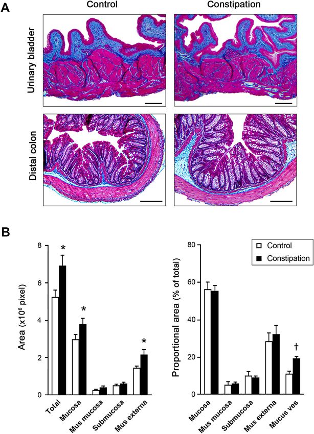
www.nature.com/scientificreports/
bowel symptoms often experienced BBD symptoms in childhood, underscoring the importance of recognizing
BBD early on and to address both, LUT and gastrointestinal dysfunctions, s imultaneously11,14–16.
The aetiology of BBD seems to be multifactorial, and the mechanical compression of large stool in the colon
or rectum during constipation is considered to be a principal cause for LUTD development17. Physical pressure
onto the LUT decreases the functional capacity of the urinary bladder, constricts the urethra, thereby resulting
in urgency, dysfunctional voiding, urinary retention and urinary incontinence18. This explains the improvement
of lower urinary tract symptoms (LUTS) following the medical relief of constipation in the majority of cases with
BBD symptoms8,19. However, other studies found no improvement or persistent LUTS with release of intra-rectal
pressure in children with B BD20–22.
Normal function of the urinary bladder and gastrointestinal tract depends on their components including
the nerves, muscle, connective tissues and mucosa, and the coordinated interactions among them. Prolonged
BBD causes muscle instability in both bladder and colon, suggesting that BBD affects the muscles and nerves
that control normal bowel and bladder function. One proposed mechanism is the development of bladder-bowel
cross-organ sensitization via overlapping neural pathways at both peripheral and central l evel23,24. Other possibil-
ity includes histological and/or molecular changes in the tissues including inflammation, fibrosis, alterations in
intercellular connections, signalling molecules, receptors and ion channels15,25–28. As such, the pathophysiological
mechanisms underlying BBD are complex, yet not understood very well. In the current study, we examined the
effects of functional constipation on the LUT early in life using a murine model of constipation at young age.
Materials and methods
Animals. Male C57BL/6J mice (4-week-old) were divided into two groups: constipation model, and sham
group. Constipation was induced as previously described by Heredia et al.29. Briefly, a lubricated 2.0 mm diam-
eter polyethylene tubing was inserted into the anus of mice (1 cm) under anaesthesia. After injection of 2%
lidocaine into the skin next to anal opening, a purse-string suture (size 7–0, Prolene, Ethicon, Somerville, NJ,
USA) was snugly placed in the external sphincter region around the tubing, and then the tubing was removed.
To create functional constipation model (N = 42), the suture remained in place for four days. Sham operated ani-
mals underwent the same procedure except for the immediate removal of the suture after its placement (control
group, N = 36). Mice were maintained in the animal facility (14-h light: 10-h dark cycle) with access to water and
chow ad libitum. Rectal sutures and perianal area were checked to ensure the integrity of the model. The colo-
rectum and urinary bladders were harvested for examination at four days after the surgery. Urine samples were
collected from the bladders (N = 12 per group) and tested with urinalysis strips (U031-131, ACON laboratories,
San Diego, CA, USA). All animal procedures were reviewed and approved by the Institutional Animal Care and
Use Committee (IACUC) of the University of Colorado Denver. All experiments were performed in accordance
with relevant guidelines and regulations, as well in compliance with the ARRIVE guidelines 2.0 (https://arriv
eguidelines.org/arrive-guidelines).
Micturition pattern evaluation. In order to evaluate voiding patterns, void spot assays (VSA) were con-
ducted at four days after the surgery, as previously described (N = 22 per group)30. Each mouse had free access to
water but no food during the 3 h test period. All VSA experiments were started at 10 am. The urine spots on the
filter paper were imaged using ultraviolet light on a transilluminator, and the number and area of the spots were
analysed using Adobe Photoshop CS6 (Adobe Systems, San Jose, CA, USA). Faecal pellets were collected and
air dried at least 16 h following VSA (N = 8 per group), and the number and the weight of pellets were recorded.
Urodynamic evaluation of bladder function. For urodynamic evaluation, mice underwent surgical
catheter implantation in the bladder as previously described15 immediately before undergoing the constipa-
tion or sham surgery. Cystometry was performed in unanesthetized unrestrained mice 4 days after the surgery
reviously15. The tip of the exteriorized bladder catheter located at the base of the
(N = 4 per group) as described p
mouse neck was connected to a pressure transducer and an infusion pump of the cystometry station (Catamount
Research and Development, St. Albans, VT, USA). Room temperature saline was infused into the bladder at
the rate of 10 μl/min. Each animal was observed for minimum four voiding cycles of reproducible micturition
patterns. Urodynamic values recorded continuously during testing, and four parameters; maximum intravesical
pressure at micturition, functional bladder capacity, voided volume, and the number of non-void contractions
(NVC) per voiding cycle were analysed using Cystometry Analysis Software (SOF-552, Catamount Research and
Development). The NVC were defined as intravesical pressure rises greater than one-third of average maximal
voiding pressure in each animal without triggering micturition.
Physiological evaluation of detrusor function in vitro. In vitro physiological evaluation of detrusor
contractility and the baseline spontaneous activity was conducted using freshly isolated bladder strips from mice
in each group as described p reviously30. Spontaneous contractions in each bladder strip were collected for 2 min
after initial equilibration for 30 min in Tyrode’s solution. Detrusor contractility was examined in the responses
to a series of electrical field stimulation (EFS, 70 V, 2–32 Hz), the acetylcholine receptor agonist carbachol (CCh,
1–100 µM), high KCl (125 mM replaced NaCl in Tyrode’s solution), a mixture of purinoceptor agonists (ADP/
ATP; ADP-γ-s, ATP-γ-s and αβ-methylene ATP,10 µM each) (n = 14–15 per group). Contractile responses to
EFS were also recorded after 20 min of incubation with the following substances: (1) ADP/ATP to desensitize
purinoceptors, and (2) atropine (muscarinic receptor antagonist, 1 µM) (n = 10–12 per group). Peak force of
the contractile response was calculated in grams of tension per weight of individual bladder strip. Spontaneous
contractions in the bladder strips were collected for 2 min after 30 min of (1) initial equilibration in Tyrode’s
solution, (2) incubation with 5-hydroxytryptamine (5-HT) at 0.5 µM, and (3) incubation with a mixture of 5-HT
Scientific Reports | (2021) 11:1149 | https://doi.org/10.1038/s41598-020-80794-0 2
Vol:.(1234567890)www.nature.com/scientificreports/
Primary Vendor and Catalog No Application and dilution Validation
26
Desmin Novus Biologicals, NB120-15200 1:200 (IF)
Santa Cruz Biotechnology, sc-32233 1:500 (WB)
Gapdh
Proteintech, HRP-60004 1:5,000 (WB)
48
Htr2a Santa Cruz Biotechnology, sc-166775 1:100 (IF)
49
Santa Cruz Biotechnology, sc-17797 1:100 (IF)
Htr2c
LSBio, LS-C386171 1:500 (WB) Supplement 1
50
Uchl1 Santa Cruz Biotechnology, sc-23852 1:50 (IF)
Secondary Conjugate Vendor and Catalog No Application and dilution
Goat IgG DyLight488 Rockland Immunochemicals, 605-741-125 1:5,000 (IF)
HRP Jackson ImmunoResearch, 715-035-150 1:10,000 (WB)
Mouse IgG DyLight549 Rockland Immunochemicals, 610-142-002 1:5,000 (IF)
DyLight549 Rockland Immunochemicals, 610-742-124 1:5,000 (IF)
HRP Novus Biologicals, NB7160 1:10,000 (WB)
Rabbit IgG
DyLight488 Rockland Immunochemicals, 611-141-122 1:5,000 (IF)
Table 1. Antibodies.
at 0.5 µM and a selective Htr2 receptor antagonist, ketanserin (+)-tartrate31 (Sigma-Aldrich, St. Louis, MO,
USA) at 200 nM (n = 16 per group). All data analyses were performed using PowerLab Lab-Chart version 8.1.9
(ADInstruments, Colorado Springs, CO, USA). Calcium imaging of denuded detrusor sections from each group
of mice (~ 6 mm × 10 mm, n = 3) was performed with 10 µM Cal-520 AM (abcam, Cambridge, MA, USA) in
Tyrode’s buffer according to the manufacturer’s protocol. The relative amplitude of Ca2+ transients was expressed
as F/F0, where F represents the total fluorescence in an event of fluorescent increase, and F0 represent the basal
fluorescence. Images were taken every 0.3 s for 3 min.
Histological analysis. Paraformaldehyde-fixed paraffin sections (5 µm thickness) of the urinary bladders
and the distal colon from each group were stained with Masson’s trichrome staining32. Area measurement of
the distal colon sections was performed for the 4 tissue layers, mucosa, muscularis mucosa, submucosa, and
muscularis externa, as well as the oval shaped non-staining area in the mucosa which was considered as mucus
vesicles. All measurements were conducted in a blind fashion to avoid biased interpretation. For immunostain-
ing, sections were subjected to heat-induced antigen retrieval (10 mM Tris, 1 mM EDTA, and 0.05% Tween 20,
pH 9.0). All antibodies used in this study are listed in Table 1. Control experiments performed without primary
antibodies showed neither nonspecific labelling nor cross-reactivity between secondary antibodies. Sections
from at least three animals in each group were analysed for reproducibility.
Gene expression analysis. Total RNA isolated from the urinary bladders (N = 5 per group) was tran-
scribed into cDNA and used in real-time quantitative PCR (qRT-PCR) as previously d escribed15. Expression
levels of each gene were calculated as fold changes based on ∆∆Ct values. Data were normalized to the mean of
three housekeeping genes: β-actin (Actb), Glyceraldehyde 3-phosphate dehydrogenase (Gapdh) and TATA box-
binding protein (Tbp). Western blot analysis was performed to assess the protein expression level as previously
described (N = 4 per group)15. The antibodies used in this study are listed in Table 1. The signals specific for each
antibody were quantified using Fiji ImageJ software (Version 1.53c, National Institutes of Health, Bethesda, MD,
USA) and normalized with Gapdh.
Measurement of serum serotonin levels. Blood serum samples were collected from each group of mice
(N = 15 per group) with BD Microtainer serum separator tubes (Becton, Dickinson and Company, Franklin
Lakes, NJ, USA) and flash frozen. Serotonin ELISA Kit (Aviva Systems Biology Corporation, San Diego, CA,
USA) was used to quantify serotonin concentration in mice serum samples. All ELISA measurements were made
in duplicate.
Statistical analysis. All data were analysed using two-tailed unpaired t-test using GraphPad Prism 8.4.3
(GraphPad Software, La Jolla, CA, USA) between two groups. GraphPad outlier calculator (GraphPad Software,
https://www.graphpad.com/quickcalcs/Grubbs1.cfm) was used to detect outliers, which were excluded from the
analysis in detrusor physiology test in vitro. A probability value of p < 0.05 was regarded as significant. Results
are expressed as means ± standard error of the mean (SE).
Results
Functional constipation increases urinary frequency and bladder instability in mice. Partial
obstruction of the external anal sphincter caused a growth retardation, and a profound enlargement of the colo-
rectum with faecal impaction in juvenile mice (Table 2). The bladder-body weight ratio and morphology of the
bladder were comparable between the groups. No signs of fibrosis, inflammatory cell infiltration or disturbance
in the organ walls was detected in both the bladder and the distal colon from both groups of mice (Fig. 1A).
Scientific Reports | (2021) 11:1149 | https://doi.org/10.1038/s41598-020-80794-0 3
Vol.:(0123456789)www.nature.com/scientificreports/
Colon
Body (g) Bladder (% of body) Weight (mg) Length (mm) Width (mm) Stool (mg)
Control 16.6 ± 0.9 0.09 ± 0.00 167 ± 7 6.5 ± 0.2 1.4 ± 0.1 101 ± 12
Constipation 13.4 ± 0.6** 0.10 ± 0.00 273 ± 22** 6.6 ± 0.2 3.1 ± 0.2** 349 ± 40**
Table 2. Morphological parameters of the urinary bladder and colon in the control and the constipation
groups. Mean ± SE, **p < 0.005 vs. the control group.
However, the total area of the distal colon sections was significantly larger in the constipation group compared
to the control. The area analysis of each layers in the colon showed that all 4 layers, mucosa, muscularis mucosa,
submucosa, and muscularis externa, were increased in a similar level. The proportion of each layer in the cor-
responding section was similar between the two groups. We also observed a marked increase in the oval shaped
structure in the mucosa which is likely mucus vesicle in the goblet cells in the colon sections from the con-
stipation group when compared to the control group (Fig. 1B). In addition to the faecal impaction, the faecal
output during VSA experiments was decreased to less than a half in the experimental group compared to the
sham operated control group (4.0 ± 1.0 mg vs. 8.6 ± 0.5 mg, p = 0.0025). Furthermore, the sign of faecal inconti-
nence (loose stool spots) was observed on filter papers in 10 mice from the constipation group, but none from
the control group. These results verified that our murine model replicated clinical characteristics of functional
constipation in children. Urinalysis showed normal values for all parameters (ascorbic acid, glucose, bilirubin,
ketones, specific gravity, blood, pH, protein, urobilinogen, nitrites, leukocyte, albumin, and creatinine), in all
urine samples tested (N = 12 per group), indicating that no UTI occurred during the study period. The total
number of urine spots in the constipation group was 2.7-fold higher than in the control group (p = 0.0016). Mice
with constipation voided more frequently in small volume (< 50 µl per void, 11.2 ± 2.4 vs. 2.1 ± 0.6, p = 0.0005),
while less large voids (≥ 50 µl per void, 1.7 ± 0.4 vs. 2.8 ± 0.3, p = 0.024) compared to the control group (Fig. 2).
The volumes of total void and the mean of the large urine spots were decreased in the constipation group com-
pared with those in the control animals (259 ± 36 µl vs. 347 ± 33 µl, p > 0.05 and 84 ± 6 µl vs. 126 ± 13 µl, p = 0.012,
respectively). To further evaluate how constipation impacts LUT function, urodynamic studies were conducted
in each group of animals. Urodynamic parameters were clearly distinguished between the two groups (Fig. 3
and Table 3). Functional bladder capacity was significantly decreased by approximately 30% in the constipation
group in comparison to the control (p = 0.041), consistent with the data from VSA experiments. The voiding
efficiency and the maximal intravesical pressure at micturition (Pves max) were comparable between the two
groups. Intravesical pressure traces in the constipation group revealed bladder instability with frequent NVCs
along with continuous small spikes of pressure which were minimal in the control group (p = 0.006).
Functional constipation induced detrusor overactivity. Bladder strips showed comparable con-
tractile responses to EFS, CCh, ADP/ATP and KCl between the two groups. Inhibition of purinergic or mus-
carinic receptors suppressed EFS-evoked contractile response at similar level in bladder strips from both groups
(Fig. 4A,B). However, the analyses of basal activity of the bladder strips revealed a profound increase in spontane-
ous contractions in the constipation group. Both frequency and amplitude of the spontaneous contractions were
elevated compared to the control group (9.2 ± 0.5 vs. 6.7 ± 0.4 min−1, p = 0.0015, and 35.6 ± 5.8 vs.14.2 ± 1.0 mg,
p = 0.0013, respectively) (Fig. 4C). Likewise, a significant elevation of unprovoked spontaneous intracellular C a2+
transients was observed in detrusor myocytes from the constipation group compared to those from the control
animals (incidence, 6.7 ± 1.7 min−1 vs. 3.4 ± 1.2 min−1, p = 0.037, and F/F0, 7.9 ± 1.6 vs. 3.8 ± 0.8, p = 0.044).
Functional constipation induced an upregulation of serotonin receptors in the bladder. In
order to explore the potential molecular candidates contributing to constipation-induced detrusor overactivity,
qRT-PCR analyses were performed for the genes involved in neuromuscular function in the bladder, as well as
the genes associated with bladder o veractivity26–28,33. Three genes, 5-HT receptor subtype 2a and 2c (Htr2a and
Htr2c) and Transient receptor potential vanilloid 2 (Trpv2) were significantly upregulated in the constipation
group in comparison to the control group (Fig. 5A). No significant changes in expression level were observed in
other tested genes including muscle contractile factors (Desmin, myosin light chain kinase, Mylk, and myosin
heavy chain, Mhc), ion channels (voltage-gated calcium channels, Cav1.2, Cav 3.1 and Cav 3.2, and potassium
channels, Bkα, Bkβ1 and Bkβ4), neurotransmitter receptors (muscarinic receptors, Chrm2 and Chrm3, adr-
energic receptors, Adrβ2 and Adrβ3, and purinoceptors, P2x1-3,7), and a cholinergic/motor neuron marker
(Chat). No changes in the expression level in other classes of 5-HT receptors, Htr1, Htr3, Htr4 and Htr7, and
Trp channels, Trpv1, Trpv4,Trpa1 and Trpm8, which are also reported to be expressed in the human and murine
bladders, were noted28,33,34. Western blotting revealed that Htr2a and Htr2c proteins were elevated by 1.5- and
1.4-fold (p = 0.032 and p = 0.042) at the protein level in the bladder after four days of functional constipation
(Fig. 5B,C). We could not determine the level of Trpv2 protein in the bladder as three tested commercially avail-
able antibodies did not detect any specific signal with the molecular weight close to the predicted (86 kDa) even
for the protein samples from mouse spleen used as a positive control. Immunofluorescence labelling detected
Htr2a immunoreactivity (IR) in the urothelial cell cytoplasm and in the detrusor layer in the bladders from the
control group (Fig. 6a–d). By contrast, the Htr2a IR was more robust in the constipation group than that in the
control group (Fig. 6e–h). The Htr2a IR in the detrusor layer was in a small punctate pattern, appeared to be
on the edge of detrusor cells labelled with the antibody against Desmin. The Desmin IR was observed in the
Scientific Reports | (2021) 11:1149 | https://doi.org/10.1038/s41598-020-80794-0 4
Vol:.(1234567890)www.nature.com/scientificreports/
Figure 1. Bladder and colon histology. (A) Representative images of cross sections of the bladder (upper
panels) and colon (lower panels) from mice in the control (left) and the constipation (right) groups. Masson’s
trichrome staining: Collagen is represented by the blue staining. Bars, 200 µm. (B) The area measurement of
total colon tissue section and the four layers (mucosa, muscularis mucosa, submucosa and muscularis externa)
of the colonic wall from each group of mice (left). The proportional area of each layer relative to the entire tissue
section and the mucus vesicles relative to the mucosa (right). Mean ± SE. *p < 0.05, †p < 0.0005 vs. the control
group. Figures were prepared using Adobe Photoshop CS6 and GraphPad Prism 8.4.3 (https://www.graphpad.
com/scientific-software/prism/).
Scientific Reports | (2021) 11:1149 | https://doi.org/10.1038/s41598-020-80794-0 5
Vol.:(0123456789)www.nature.com/scientificreports/
Figure 2. Micturition patterns at 4 days after the surgery. (A) Representative void spot assay images from
each group. (B) Number of the urine spots (left), of large (≥ 50 μl) and small voids (< 50 μl). The voided
volume (right) of total and per void in the large void spots. White and black bars represent the control and the
constipation mice, respectively. Mean ± SE. *p < 0.05, **p < 0.005, †p < 0.0005 vs. the control mice. Figures were
prepared using Adobe Photoshop CS6 and GraphPad Prism 8.4.3 (https://www.graphpad.com/scientific-softw
are/prism/).
detrusor myocytes at the similar level in both groups. The Htr2c IR was distributed throughout the cytoplasm
of detrusor myocytes, while no signal in the urothelial and lamina propria layers (Fig. 6i–p). The Htr2c IR did
not show overlap with the IR with the antibody against Uchl1 (also known as Pgp9.5), a pan-neuronal marker.
The Uchl1 IR was observed mainly in the lamina propria layer and between the muscle bundles in the detrusor
layer in similar manner in both groups of mice. The Htr2c IR signal was more intense in the bladders from the
constipation group compared with the control group. These results demonstrate that functional constipation
induced an upregulation of 5-HT receptors, Htr2a and Htr2c in the urinary bladder.
Scientific Reports | (2021) 11:1149 | https://doi.org/10.1038/s41598-020-80794-0 6
Vol:.(1234567890)www.nature.com/scientificreports/
Figure 3. Functional bladder analyses in cystometry. Representative cystometrogram trace from
unanesthetized, unrestrained mice in the control (left) and the constipation group (right) during a continuous
intravesical infusion (10 μl/min) of room temperature saline. Volume infused (top), intravesical pressure
(Pves, middle) and voided volume (bottom) are shown. Arrowheads indicate examples of non-voiding bladder
contractions. Figures were prepared using Cystometry Analysis Software (SOF-552, https://www.med-associates
.com/product/cystometry-analysis-data-analysis-software/) and Adobe Photoshop CS6.
Infused vol. (µl) Void vol. (µl) Pves max (mmHg) Voiding efficiency (%) Non-void contractions
Control 106 ± 11 109 ± 6 17.0 ± 0.5 105 ± 8 0.1 ± 0.1
Constipation 79 ± 5* 72 ± 7† 15.6 ± 2.0 99 ± 7 0.8 ± 0.2*
Table 3. Comparison of urodynamic parameters between the groups. The constipation group showed a
significant decrease in bladder capacity, voided volume, and a significant increase in the number of non-void
contractions compared to the control group (N = 4 per each group). Mean ± SE, *p < 0.05 and †p < 0.005 vs. the
control group, Pves max, maximum intravesical pressure at micturition.
5‑HT exacerbated constipation‑induced detrusor overactivity. To investigate how the Htr2 upreg-
ulation in the bladder induced by functional constipation impacts detrusor function, the responses of the blad-
der strips to 5-HT were evaluated. The 5-HT caused a small contractile response that was less than half of the
response to EFS at 2 Hz, with a trend of higher force produced in the constipation group especially at lower
concentrations (0.1 and 0.5 µM) compared to the control group (Fig. 7A). On the contrary, the baseline sponta-
neous activity of the bladder strips was profoundly affected by pre-incubation with 5-HT (Fig. 7B). Serum 5-HT
concentration showed no difference between control and the constipation group (104 ± 5 vs. 96 ± 4 ng/ml, equal
to 0.59 ± 0.03 vs. 0.57 ± 0.02 µM, p = 0.502) measured by ELISA (Fig. 7C), which correspond to the normal serum
5-HT level (101–283 ng/ml equals to 0.57–1.61 µM)35. Accordingly, we selected the concentration of 0.5 µM to
further examine the effect of 5-HT on the bladder strips. Pre-incubation of the bladder strips with 0.5 µM 5-HT
showed the similar pattern as that without 5-HT, more frequent and larger spontaneous contractions in the
constipation group compared to those in control group (4.7 ± 0.5 vs. 7.5 ± 0.3 min−1, p < 0.0001, and 22.6 ± 3.0
vs. 55.1 ± 4.6 mg, p < 0.0001, respectively) (Fig. 7D). In comparison to that in absence of 5-HT, the change in
Scientific Reports | (2021) 11:1149 | https://doi.org/10.1038/s41598-020-80794-0 7
Vol.:(0123456789)www.nature.com/scientificreports/
Figure 4. Detrusor contractility evaluation. (A) Peak contractile force in response to electric field stimulation
(EFS, left), carbachol (CCh, centre), and KCl and ADP/ATP (right). (B) Relative contribution of purinergic
and muscarinic pathways to EFS-evoked contractility. The force was normalized with tissue weight. (C) The
frequency (left) and amplitude (right) of spontaneous contractions. White and grey circles represent the control
and the constipation groups, respectively. Mean ± SE. **p < 0.005, vs. the control mice. Figures were prepared
using Adobe Photoshop CS6 and GraphPad Prism 8.4.3 (https://www.graphpad.com/scientific-software/prism/).
the amplitude was statistically significant in both the control (1.3-fold, p = 0.043) and the constipation groups
(2.1-fold, p = 0.019). The change in the frequency of spontaneous contractions was statistically significant in the
constipation group (1.3-fold, p < 0.0001) but not in the control group (1.2-fold, p = 0.220). The selective Htr2
receptor antagonist, ketanserin (+)-tartrate31 treatment in presence of 5-HT reversed the spontaneous contrac-
tion responses similar to those in absence of 5-HT in both control and the constipation groups (4.0 ± 0.4 vs.
5.8 ± 0.3 min−1, p = 0.003, and 23.1 ± 3.0 vs. 42.9 ± 4.6 mg, p = 0.020). The changes of spontaneous contractions
between with and without ketanserin were statistically significant in both the frequency (0.8-fold, p = 0.001) and
the amplitude (0.8-fold, p = 0.008) in the constipation groups, but not in the control group (0.9- and 1.1-fold,
p > 0.05).
Discussion
In the present study, we investigated the impact of functional constipation onto the urinary bladder at histologi-
cal, physiological, and molecular levels. The murine model employed in this study recapitulated clinical and
pathological features of functional constipation in children including faecal impaction, faecal incontinence, colo-
rectal distension, and growth retardation5,36. Spontaneous voiding behaviour tests demonstrated that constipation
led to urinary frequency with decreased volumes per-void in mice. It is possible that large stool in the colorectum
takes up abdominal space and prevents the bladder to expand fully, therefore contributing to the decreased func-
tional bladder capacity. Cystometry, an independent in vivo bladder function test, confirmed urinary frequency
accompanied with a decreased functional bladder capacity in the constipation group. Additionally, increased
NVC suggested bladder overactivity. These results suggested that constipation at young age can induce LUTS
and contribute to the development of BBD. During urodynamic tests in awake mice, the control animals usually
Scientific Reports | (2021) 11:1149 | https://doi.org/10.1038/s41598-020-80794-0 8
Vol:.(1234567890)www.nature.com/scientificreports/
Figure 5. Gene expression analysis in the bladders. (A) The level of mRNA expression of each gene is presented
as the fold difference to that in the control group (N = 5 per group). (B) The protein expression comparison.
Representative Western blotting results for Htr2a (top left), Htr2c (top right) and Gapdh (bottom). (C)
Summary of relative protein expression levels of Htr2a and Htr2c normalized with Gapdh (right). Mean ± SE,
*p < 0.05 vs. the control group. Figures were prepared using Adobe Photoshop CS6 and GraphPad Prism 8.4.3
(https://www.graphpad.com/scientific-software/prism/).
moved to a corner of the cage immediately before they started voiding as a part of voiding behaviour in mice as
described previously37. However, the same behaviour was observed only in about half of the micturition cycles
recorded in each mouse with constipation. We consider that this behavioural phenotype in mice with constipation
was a manifestation of urinary urgency and/or incontinence, another LUTS common in BBD cases.
Constipation is also associated with increased occurrence of UTI in children, and UTI can cause symptoms
similar to those of overactive b ladder38. The urinalysis results showed normal parameters for both groups,
indicating that UTI, and bladder lesions were not responsible for LUTS observed in the constipation group.
Cystometry showed that the voided volume was approximately equal to the volume infused in each micturition
cycle, and no intermittent void was detected in both groups of mice. Altogether, these results suggest that neither
bladder outlet obstruction nor detrusor-sphincter dyssynergia developed in the constipated animals tested in
urodynamic study. Histological examination also confirmed the absence of detrusor hypertrophy or fibrosis in
the bladder, suggesting no bladder outlet obstruction developed along constipation in this study32. Based on the
current results, we propose three potential explanations for constipation-induced bladder overactivity observed
during awake cystometry. First, the bladder experienced an additional external pressure from the overdistended
colorectum, which contributed to the rises in intravesical pressure without causing micturition (NVCs). Second,
the convergent neurons that receive afferent inputs from both the colon and the bladder were likely continuously
stimulated during constipation and sent aberrant signals to the bladder causing an increase in sensation and
overactive symptoms. Studies has been shown that colorectal distension or inflammation alters bladder sensa-
tions and detrusor activities, and vice versa in both humans and experimental animals39, suggesting the existence
of "cross-organ sensitization" between the colon and bladder. This might be one of the reasons why successful
treatment of constipation leads to an improvement of LUTS in many patients as previously reported8,19. However,
Scientific Reports | (2021) 11:1149 | https://doi.org/10.1038/s41598-020-80794-0 9
Vol.:(0123456789)www.nature.com/scientificreports/
Figure 6. Distribution of Htr2a and Htr2c in the bladders. Representative immunohistochemical images with
antibodies against Htr2a (red) and Desmin (green) (a–h), or Htr2c (red) and Uchl1 (green) (i–p) along nuclei
staining using DAPI (blue). U, urothelia, L, lamina propria, and D, detrusor muscle layers. Bars, 100 µm (a, e, i
and m) and 25 µm (d, h, l and p). Figures were prepared using Adobe Photoshop CS6.
not all children with BBD improve LUTS after successful resolution of constipation21,22. This prompted us to
hypothesize the third possibility that constipation, especially in chronic condition, may trigger alterations in
the bladder physiology itself (e.g., release of certain neurotransmitters, receptors, ion channels, and other mol-
ecules which regulate detrusor function). Our physiological studies in vitro revealed a prominent augmentation
of spontaneous contractions of the bladder strips alongside enhanced spontaneous excitation of the detrusor
muscle cells in mice with constipation. These results provided evidence that constipation negatively impacted
the detrusor itself, and likely altered its physiology independent from the neural inputs. On the other hand, the
detrusor contractile responses evoked by EFS, carbachol and KCl as well as the proportion of purinergic and
muscarinic contribution to EFS-triggered contractility did not altered following constipation. This result sug-
gested that constipation had no significant impact on the contractile apparatus, the balance between excitatory
and inhibitory neurotransmitter release as a whole, or their receptors in the bladder.
Scientific Reports | (2021) 11:1149 | https://doi.org/10.1038/s41598-020-80794-0 10
Vol:.(1234567890)www.nature.com/scientificreports/
Figure 7. The effect of 5-HT on the bladder strips. (A) Peak contractile force in response to 5-HT. (B)
Representative traces of basal activity of the bladder strips from each group before and after incubation with
0.5 µM 5-HT (upper and lower panels, respectively). (C) Serum 5-HT level from each group. the amplitude, and
the frequency of spontaneous contractions of the bladder strips. D, the frequency (left) and amplitude (right) of
spontaneous contractions of the bladder strips in presence or absence of 5-HT and ketanserin (Htr2 antagonist).
Open and grey circles represent the control and the constipation groups (n = 16 per group), respectively.
Mean ± SE, *p < 0.05, **p < 0.005 between the groups. Figures were prepared using Adobe Photoshop CS6 and
GraphPad Prism 8.4.3 (https://www.graphpad.com/scientific-software/prism/).
Molecular studies revealed comparable expression levels of many genes involved in regulation of detrusor
contractile function including muscle contractile apparatus, ion channels, neurotransmitter receptors as well
as a cholinergic neuron marker in the bladder, corresponding to the data from physiological evaluation of the
bladder. Yet, two 5-HT receptors, Htr2a and Htr2c, were significantly upregulated in the bladder following four
days of constipation. Accumulating evidence suggests that 5-HT contributes to LUT function by modulating
the activity of the detrusor, external sphincter, urothelium, and neural pathways at both central and peripheral
levels33,34,40. The effect of selective serotonin uptake inhibitors (SSRIs) as well as agents acts on 5-HT receptors
on LUT function has been extensively studied in both clinical and experimental settings, however, demonstrated
contradictory results41–43. The discrepancies among studies may be due to differences in the pharmacological
properties, affinities to different receptors of each agent, availability of receptors, and age and gender of study
cohorts42–44. Htr2 receptors couple to G q/11 protein which activates inositol 1,4,5-triphosphate pathway leading
to intracellular Ca2+ increase33. Our in vitro experiments demonstrated 5-HT at 0.5 µM further augmented
Scientific Reports | (2021) 11:1149 | https://doi.org/10.1038/s41598-020-80794-0 11
Vol.:(0123456789)www.nature.com/scientificreports/
overactivity in the bladder strips from the constipation group, which was reversed by the Htr2 antagonist. These
results suggest that the increase in the functional Htr2 receptors expressed in the bladder contribute to the
constipation-induced detrusor overactivity and related LUTS at peripheral level (in situ). This notion agrees
with the association between bladder hyperactivity and Htr2 upregulation in the bladder described previously44.
Both groups of animals had a normal level of serum 5-HT and the comparable expression of 5-HT transporter,
Sert, in the bladder, suggesting that 5-HT bioavailability was unaffected by constipation. Therefore, the detru-
sor cells could be activated and contract spontaneously at the normal level of 5-HT through Htr2 following a
period of constipation. Other 5-HT receptor subtypes (Htr1, Htr3, Htr4 and Htr7) did not show difference in the
expression level in the bladder following constipation, suggesting their contribution to 5-HT induced detrusor
hyperactivity may be m inor28,33,34.
The comparable intensity and distribution of Uchl1 IR in the bladders between the two groups indicate
that constipation did not alter bladder innervation at appreciable level. The bladder strips from both groups
generated equal contractile responses to EFS alongside the equal level of Chat gene expression, suggesting that
the proportion of motor and sensory neurons in the bladder was not affected following constipation. Lines of
evidence demonstrated that Htr3 plays a predominant role in bladder afferent firing and micturition reflexes at
spinal and spinobulbospinal levels40,45,46, consequently, an elevated activation of Htr3 pathway facilitates blad-
der overactivity. Our data showed an equal level of Htr3a receptor expression in the bladder from both groups,
suggesting the involvement of Htr3 pathway in bladder overactivity observed in the constipation group to be
minor. However, it is possible that constipation induced changes in afferent activity at the level of dorsal root
ganglia or spinal cord, and therefore contributes to bladder overactivity observed in the constipation group. We
did not address this hypothesis in this study, and it needs to be evaluated in future studies.
An ion channel, Trpv2 also showed a significant elevation in the bladder following four days of constipation.
Tprv2 is a member of transient receptor potential (TRP) cation channel family with high Ca2+ permeability, and
is expressed in the bladder along with several other TRP channels including Trpv1, Trpv4, Trpa1, and T rpm828.
TRP channels are expressed in nerve fibres and urothelium in the bladder wall, and suggested to act as sensors of
stretch and/or chemical irritation28. Yet, the functional significance of Trpv2 in bladder sensation is considered
to be minor as the receptor was shown to be not essential for heat or mechanical nociception or hypersensitivity
in the adult mouse. On the other hand, several lines of evidence indicate that Trpv2 plays a role in Ca2+ influx in
different types of myocytes, and there is an association between an upregulation of Trpv2 and abnormal or leaky
Ca2+ influx in pathologic skeletal m uscle47. Since Trpv2 is expressed in detrusor cells in addition to nerves and
28
urothelium , we speculated that the upregulation of this receptor may contribute to the elevated spontaneous
Ca2+ transients in detrusor cells and associated unstable baseline detrusor excitability detected in the constipa-
tion group. This conjecture requires to be tested in future studies.
The present study provides initial evidence of association between functional constipation and LUTD, includ-
ing the changes in detrusor responses to different stimuli and symptoms of bladder overactivity. The elevated
expression of Htr2 and Trpv2 in the urinary bladder might account for impaired intracellular Ca2+ regulation in
the detrusor myocytes, and be associate with the persistence of LUTS even after successful treatments of con-
BD20–22. The data obtained in this study suggest that local administration of selective antagonists
stipation in B
of Htr2 and Trpv2 may offer therapeutic options for BBD patients. Future studies focused on long-term effects
of functional constipation on LUT function, as well as a possible prevention and reversibility/progression, will
allow for deeper understanding of the mechanisms underlying BBD.
Received: 8 June 2020; Accepted: 23 December 2020
References
1. Wolfe-Christensen, C. et al. Bladder and bowel dysfunction: evidence for multidisciplinary care. J. Urol. 190, 1864–1868. https://
doi.org/10.1016/j.juro.2013.05.012 (2013).
2. Biggs, W. S. & Dery, W. H. Evaluation and treatment of constipation in infants and children. Am. Fam. Physician 73, 469–477
(2006).
3. Sampaio, C. et al. Constipation and lower urinary tract dysfunction in children and adolescents: a population-based study. Front.
Pediatr. 4, 101. https://doi.org/10.3389/fped.2016.00101 (2016).
4. Combs, A. J., Van Batavia, J. P., Chan, J. & Glassberg, K. I. Dysfunctional elimination syndromes—how closely linked are constipa-
tion and encopresis with specific lower urinary tract conditions?. J. Urol. 190, 1015–1020. https: //doi.org/10.1016/j.juro.2013.03.111
(2013).
5. Xinias, I. & Mavroudi, A. Constipation in childhood. An update on evaluation and management. Hippokratia 19, 11–19 (2015).
6. Austin, P. F. et al. The standardization of terminology of lower urinary tract function in children and adolescents: update report
from the Standardization Committee of the International Children’s Continence Society. J. Urol. 191, 1863–1865. https://doi.
org/10.1016/j.juro.2014.01.110 (2014).
7. Lindgren, H., Nejstgaard, M. C., Salo, M. & Stenstrom, P. Evaluation of bowel function in healthy children: untreated constipation
is common. Acta Paediatr. 107, 875–885. https://doi.org/10.1111/apa.14225 (2018).
8. Santos, J. D., Lopes, R. I. & Koyle, M. A. Bladder and bowel dysfunction in children: an update on the diagnosis and treatment of
a common, but underdiagnosed pediatric problem. Can. Urol. Assoc. J. 11, S64–S72. https://doi.org/10.5489/cuaj.4411 (2017).
9. Loening-Baucke, V. Prevalence rates for constipation and faecal and urinary incontinence. Arch. Dis. Child 92, 486–489. https://
doi.org/10.1136/adc.2006.098335 (2007).
10. Podzemny, V., Pescatori, L. C. & Pescatori, M. Management of obstructed defecation. World J. Gastroenterol. 21, 1053–1060. https
://doi.org/10.3748/wjg.v21.i4.1053 (2015).
11. Fitzgerald, M. P. et al. Childhood urinary symptoms predict adult overactive bladder symptoms. J. Urol. 175, 989–993. https://doi.
org/10.1016/S0022-5347(05)00416-7 (2006).
Scientific Reports | (2021) 11:1149 | https://doi.org/10.1038/s41598-020-80794-0 12
Vol:.(1234567890)www.nature.com/scientificreports/
12. Doiron, R. C., Kogan, B. A., Tolls, V., Irvine-Bird, K. & Nickel, J. C. Childhood bladder and bowel dysfunction predicts irritable
bowel syndrome phenotype in adult interstitial cystitis/bladder pain syndrome patients. Can. Urol. Assoc. J. 11, 255–259. https://
doi.org/10.5489/cuaj.4251 (2017).
13. Bongers, M. E., van den Berg, M. M., Reitsma, J. B., Voskuijl, W. P. & Benninga, M. A. A randomized controlled trial of enemas in
combination with oral laxative therapy for children with chronic constipation. Clin. Gastroenterol. Hepatol. 7, 1069–1074. https://
doi.org/10.1016/j.cgh.2009.06.018 (2009).
14. van Ginkel, R. et al. Childhood constipation: longitudinal follow-up beyond puberty. Gastroenterology 125, 357–363. https://doi.
org/10.1016/s0016-5085(03)00888-6 (2003).
15. Iguchi, N., Malykhina, A. P. & Wilcox, D. T. Early life voiding dysfunction leads to lower urinary tract dysfunction through altera-
tion of muscarinic and purinergic signaling in the bladder. Am. J. Physiol. Renal Physiol. 315, F1320–F1328. https: //doi.org/10.1152/
ajprenal.00154.2018 (2018).
16. Costantini, E. et al. Urological dysfunction in young women: an inheritance of childhood?. BJU Int. https://doi.org/10.1111/
bju.14081(2017).
17. Averbeck, M. A. & Madersbacher, H. Constipation and LUTS - how do they affect each other?. Int. Braz. J. Urol. 37, 16–28 (2011).
18. Halachmi, S. & Farhat, W. A. Interactions of constipation, dysfunctional elimination syndrome, and vesicoureteral reflux. Adv.
Urol. https://doi.org/10.1155/2008/828275 (2008).
19. Charach, G., Greenstein, A., Rabinovich, P., Groskopf, I. & Weintraub, M. Alleviating constipation in the elderly improves lower
urinary tract symptoms. Gerontology 47, 72–76. https://doi.org/10.1159/000052776 (2001).
20. Burgers, R. et al. Effect of rectal distention on lower urinary tract function in children. J. Urol. 184, 1680–1685. https://doi.
org/10.1016/j.juro.2010.03.120 (2010).
21. Lucanto, C., Bauer, S. B., Hyman, P. E., Flores, A. F. & Di Lorenzo, C. Function of hollow viscera in children with constipation and
voiding difficulties. Dig. Dis. Sci. 45, 1274–1280 (2000).
22. Veiga, M. L. et al. Parasacral transcutaneous electrical nerve stimulation for overactive bladder in constipated children: the role
of constipation. J. Pediatr. Urol. 12(396), e391-396. https://doi.org/10.1016/j.jpurol.2016.04.047 (2016).
23. Christianson, J. A. et al. Convergence of bladder and colon sensory innervation occurs at the primary afferent level. Pain 128,
235–243. https://doi.org/10.1016/j.pain.2006.09.023 (2007).
24. Valentino, R. J., Wood, S. K., Wein, A. J. & Zderic, S. A. The bladder-brain connection: putative role of corticotropin-releasing
factor. Nat. Rev. Urol. 8, 19–28. https://doi.org/10.1038/nrurol.2010.203 (2011).
25. Feichter, S., Meier-Ruge, W. A. & Bruder, E. The histopathology of gastrointestinal motility disorders in children. Semin. Pediatr.
Surg. 18, 206–211. https://doi.org/10.1053/j.sempedsurg.2009.07.002 (2009).
26. Iguchi, N. et al. Doxorubicin induces detrusor smooth muscle impairments through myosin dysregulation, leading to a risk of
lower urinary tract dysfunction. Am. J.. Physiol. Renal Physiol. 317, F197–F206. https: //doi.org/10.1152/ajpren
al.00090. 2019 (2019).
27. Neuhaus, J., Pfeiffer, F., Wolburg, H., Horn, L. C. & Dorschner, W. Alterations in connexin expression in the bladder of patients
with urge symptoms. BJU Int. 96, 670–676. https://doi.org/10.1111/j.1464-410X.2005.05703.x (2005).
28. Merrill, L., Gonzalez, E. J., Girard, B. M. & Vizzard, M. A. Receptors, channels, and signalling in the urothelial sensory system in
the bladder. Nat. Rev. Urol. 13, 193–204. https://doi.org/10.1038/nrurol.2016.13 (2016).
29. Heredia, D. J., Grainger, N., McCann, C. J. & Smith, T. K. Insights from a novel model of slow-transit constipation generated by
partial outlet obstruction in the murine large intestine. Am. J. Physiol. Gastrointest. Liver Physiol. 303, G1004-1016. https://doi.
org/10.1152/ajpgi.00238.2012 (2012).
30. Iguchi, N., Donmez, M. I., Malykhina, A. P., Carrasco, A. Jr. & Wilcox, D. T. Preventative effects of a HIF inhibitor, 17-DMAG,
on partial bladder outlet obstruction-induced bladder dysfunction. Am. J. Physiol. Renal Physiol. 313, F1149–F1160. https://doi.
org/10.1152/ajprenal.00240.2017 (2017).
31. Levin, E. D. et al. Ketanserin, a 5-HT2 receptor antagonist, decreases nicotine self-administration in rats. Eur. J. Pharmacol.. 600,
93–97. https://doi.org/10.1016/j.ejphar.2008.10.016 (2008).
32. Iguchi, N., Malykhina, A. P. & Wilcox, D. T. Inhibition of HIF reduces bladder hypertrophy and improves bladder function in
murine model of partial bladder outlet obstruction. J. Urol. 195, 1250–1256. https://doi.org/10.1016/j.juro.2015.08.001 (2016).
33. Matsumoto-Miyai, K., Yoshizumi, M. & Kawatani, M. Regulatory effects of 5-hydroxytryptamine receptors on voiding function.
Adv. Ther. 32(Suppl 1), 3–15. https://doi.org/10.1007/s12325-015-0240-2 (2015).
34. D’Agostino, G., Condino, A. M., Gallinari, P., Franceschetti, G. P. & Tonini, M. Characterization of prejunctional serotonin recep-
tors modulating [3H]acetylcholine release in the human detrusor. J. Pharmacol. Exp. Ther. 316, 129–135. https://doi.org/10.1124/
jpet.105.092551 (2006).
35. https://www.ucsfbenioff childrens.org/tests/003562.html.
36. Chao, H. C. et al. The impact of constipation on growth in children. Pediatr. Res. 64, 308–311. https://doi.org/10.1203/PDR.0b013
e31817995aa (2008).
37. Hill, W. G., Zeidel, M. L., Bjorling, D. E. & Vezina, C. M. Void spot assay: recommendations on the use of a simple micturition
assay for mice. Am. J. Physiol. Renal Physiol. 315, F1422–F1429. https://doi.org/10.1152/ajprenal.00350.2018 (2018).
38. Nik-Ahd, F., Lenore Ackerman, A. & Anger, J. Recurrent urinary tract infections in females and the overlap with overactive blad-
der. Curr. Urol. Rep. 19, 94. https://doi.org/10.1007/s11934-018-0839-3 (2018).
39. Panicker, J. N. et al. Bladder-bowel interactions: Do we understand pelvic organ cross-sensitization? International Consultation
on Incontinence Research Society (ICI-RS) 2018. Neurourol. Urodyn. 38(Suppl 5), S25–S34. https://doi.org/10.1002/nau.24111
(2019).
40. Konthapakdee, N. et al. Serotonin exerts a direct modulatory role on bladder afferent firing in mice. J. Physiol. 597, 5247–5264.
https://doi.org/10.1113/JP278751 (2019).
41. Movig, K. L., Leufkens, H. G., Belitser, S. V., Lenderink, A. W. & Egberts, A. C. Selective serotonin reuptake inhibitor-induced
urinary incontinence. Pharmacoepidemiol. Drug Saf. 11, 271–279. https://doi.org/10.1002/pds.705 (2002).
42. Solmaz, V. et al. Evaluation of overactive bladder in male antidepressant users: a prospective study. Int.. Neurourol. J. 21, 62–67.
https://doi.org/10.5213/inj.1732652.326 (2017).
43. Sacco, E. & Bientinesi, R. Innovative pharmacotherapies for women with overactive bladder: where are we now and what is in the
pipeline?. Int. Urogynecol. J. 26, 629–640. https://doi.org/10.1007/s00192-014-2557-9 (2015).
44. Sakai, T., Kasahara, K., Tomita, K., Ikegaki, I. & Kuriyama, H. 5-Hydroxytryptamine-induced bladder hyperactivity via the 5-HT2A
receptor in partial bladder outlet obstruction in rats. Am. J. Physiol. Renal Physiol. 304, F1020-1027. https://doi.org/10.1152/ajpre
nal.00365.2012 (2013).
45. Hall, J. D., DeWitte, C., Ness, T. J. & Robbins, M. T. Serotonin enhances urinary bladder nociceptive processing via a 5-HT3 recep-
tor mechanism. Neurosci. Lett. 604, 97–102. https://doi.org/10.1016/j.neulet.2015.07.048 (2015).
46. Schwen, Z. et al. Involvement of 5-HT3 receptors in pudendal inhibition of bladder overactivity in cats. Am. J. Physiol. Renal
Physiol. 305, F663-671. https://doi.org/10.1152/ajprenal.00105.2013 (2013).
47. Peralvarez-Marin, A., Donate-Macian, P. & Gaudet, R. What do we know about the transient receptor potential vanilloid 2 (TRPV2)
ion channel?. FEBS J. 280, 5471–5487. https://doi.org/10.1111/febs.12302 (2013).
48. Aira, Z. et al. Time-dependent cross talk between spinal serotonin 5-HT2A receptor and mGluR1 subserves spinal hyperexcitability
and neuropathic pain after nerve injury. J. Neurosci. 32, 13568–13581. https://doi.org/10.1523/JNEUROSCI.1364-12.2012 (2012).
Scientific Reports | (2021) 11:1149 | https://doi.org/10.1038/s41598-020-80794-0 13
Vol.:(0123456789)www.nature.com/scientificreports/
49. Price, A. E., Sholler, D. J., Stutz, S. J., Anastasio, N. C. & Cunningham, K. A. Endogenous serotonin 5-HT2A and 5-HT2C recep-
tors associate in the medial prefrontal cortex. ACS Chem. Neurosci. 10, 3241–3248. https://doi.org/10.1021/acschemneuro.8b006
69 (2019).
50. Yamauchi, T., Sakurai, M., Abe, K., Matsumiya, G. & Sawa, Y. Ubiquitin-mediated stress response in the spinal cord after transient
ischemia. Stroke 39, 1883–1889. https://doi.org/10.1161/STROKEAHA.106.455832 (2008).
Acknowledgements
The authors thank Dr. M. Írfan Dönmez from University of Colorado Denver, USA for providing technical sup-
port, and Akane Ito from Nara Women’s University, Nara, Japan, for assistance with data analyses.
Author contributions
N.I., A.P.M. and D.T.W. conceived/designed the experiments; N.I. and A.C. generated the murine model of func-
tional constipation and performed void spot assays; N.I. performed urodynamic evaluation of bladder function,
tissue harvest, histology, in vitro physiological evaluation of detrusor muscle, gene expression studies, prepared
figures, and wrote the manuscript; R.H.P. performed calcium imaging of the detrusor muscles; A.X.X., A.P.M.
and D.T.W. edited the manuscript. All authors reviewed the manuscript.
Funding
This study was supported by University of Colorado, School of Medicine Academic Enrichment Seed Funds
(D.T.W.) and Ponzio Family Endowment Fund (D.T.W.).
Competing interests
The authors declare no competing interests.
Additional information
Supplementary Information The online version contains supplementary material available at https://doi.
org/10.1038/s41598-020-80794-0.
Correspondence and requests for materials should be addressed to D.T.W.
Reprints and permissions information is available at www.nature.com/reprints.
Publisher’s note Springer Nature remains neutral with regard to jurisdictional claims in published maps and
institutional affiliations.
Open Access This article is licensed under a Creative Commons Attribution 4.0 International
License, which permits use, sharing, adaptation, distribution and reproduction in any medium or
format, as long as you give appropriate credit to the original author(s) and the source, provide a link to the
Creative Commons licence, and indicate if changes were made. The images or other third party material in this
article are included in the article’s Creative Commons licence, unless indicated otherwise in a credit line to the
material. If material is not included in the article’s Creative Commons licence and your intended use is not
permitted by statutory regulation or exceeds the permitted use, you will need to obtain permission directly from
the copyright holder. To view a copy of this licence, visit http://creativecommons.org/licenses/by/4.0/.
© The Author(s) 2021
Scientific Reports | (2021) 11:1149 | https://doi.org/10.1038/s41598-020-80794-0 14
Vol:.(1234567890)You can also read