Geroprotectors and Skeletal Health: Beyond the Headlines
←
→
Page content transcription
If your browser does not render page correctly, please read the page content below
REVIEW
                                                                                                                                         published: 09 February 2022
                                                                                                                                       doi: 10.3389/fcell.2022.682045

                                           Geroprotectors and Skeletal Health:
                                           Beyond the Headlines
                                           Alexandra Rayson 1, Maya Boudiffa 1, Maneeha Naveed 1, Jon Griffin 2, Enrico Dall’Ara 1,3 and
                                           Ilaria Bellantuono 1*
                                           1
                                             Healthy Lifespan Institute, Department of Oncology and Metabolism, The Medical School, Sheffield, United Kingdom, 2Healthy
                                           Lifespan Institute, Department of Molecular Biology and Biotechnology, The University of Sheffield, Sheffield, United Kingdom,
                                           3
                                             Insigneo Institute for in silico Medicine, Sheffield, United Kingdom

                                           Osteoporosis and osteoarthritis are the most common age-related diseases of the
                                           musculoskeletal system. They are responsible for high level of healthcare use and are
                                           often associated with comorbidities. Mechanisms of ageing such as senescence,
                                           inflammation and autophagy are common drivers for both diseases and molecules
                                           targeting those mechanisms (geroprotectors) have potential to prevent both diseases
                                           and their co-morbidities. However, studies to test the efficacy of geroprotectors on bone
                                           and joints are scant. The limited studies available show promising results to prevent and
                                           reverse Osteoporosis-like disease. In contrast, the effects on the development of
                                           Osteoarthritis-like disease in ageing mice has been disappointing thus far. Here we
                                           review the literature and report novel data on the effect of geroprotectors for
                           Edited by:      Osteoporosis and Osteoarthritis, we challenge the notion that extension of lifespan
                 Elizabeth Lara Ostler,    correlates with extension of healthspan in all tissues and we highlight the need for
                University of Brighton,
                      United Kingdom       more thorough studies to test the effects of geroprotectors on skeletal health in ageing
                         Reviewed by:      organisms.
                       Ioannis Kanakis,
University of Chester, United Kingdom      Keywords: aging, senescence, osteoarthritis, osteoporosis, geroprotectors, mouse models
                     Alexey Moskalev,
 Komi Scientific Center (RAS), Russia
                   *Correspondence:
                                           INTRODUCTION
                     Ilaria Bellantuono
         I.bellantuono@sheffield.ac.uk      In the United Kingdom, musculoskeletal disorders are responsible for approximately one third of
                                           General Practitioner consultations and for a NHS budget of nearly £5 billions/annum (Executive,
                    Specialty section:     2015). In addition, an estimated 8.9 million working days were lost in 2020 for musculoskeletal
         This article was submitted to     disorders, accounting for 34% of all working days lost due to ill-health in the United Kingdom alone
             Cell Growth and Division,     (Health and Safety Executive, 2020). As a whole, musculoskeletal disorders cause more functional
               a section of the journal    limitations in the adult population in the western world than any other group of disorders (Woolf
  Frontiers in Cell and Developmental      and Pfleger, 2003).
                               Biology
                                              The two most frequent musculoskeletal diseases are osteoarthritis (OA) and osteoporosis (OP).
         Received: 17 March 2021           OA, the most common age-related joint pathology, is characterized by cartilage degradation and
        Accepted: 10 January 2022
                                           inflammation in the joint, thickening of the bone plate, changes in subchondral bone and formation
       Published: 09 February 2022
                                           of osteophytes (Lotz and Caramés, 2011). Symptomatic knee OA occurs in 10% of men and 13% of
                              Citation:    women aged 60 years or older (Zhang and Jordan, 2010). The number of people affected with
   Rayson A, Boudiffa M, Naveed M,
                                           symptomatic OA is likely to increase due to the aging of the population and the obesity epidemic, an
Griffin J, Dall’Ara E and Bellantuono I
 (2022) Geroprotectors and Skeletal
                                           important risk factor driving OA (Raud et al., 2020). Patients with OA have higher levels of
       Health: Beyond the Headlines.       comorbidity compared to those of similar age without OA (Kadam et al., 2004). OA is significantly
     Front. Cell Dev. Biol. 10:682045.     associated with other musculoskeletal diseases such as other arthropathies, synovial and tendon
      doi: 10.3389/fcell.2022.682045       disorders and non-musculoskeletal comorbidities such as gastritis, intestinal diverticula and

Frontiers in Cell and Developmental Biology | www.frontiersin.org                 1                                        February 2022 | Volume 10 | Article 682045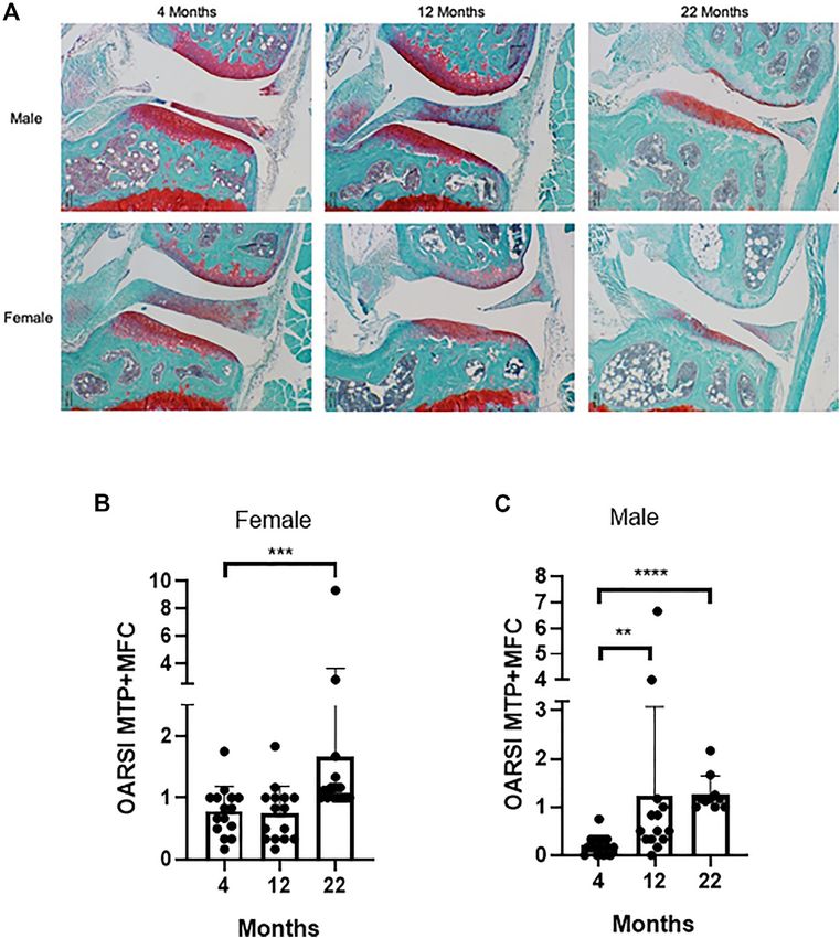
Rayson et al.                                                                                          Geroprotectors in Osteoarthritis and Osteoporosis

ischemic heart disease (Kadam et al., 2004). There is no effective           Therefore, targeting mechanisms of ageing may offer new
cure for OA. Current management of OA is limited to symptoms’                opportunities for treatment for OA.
alleviation (Bijlsma et al., 2011) followed by joint replacement
when pharmacological management of the pain is no longer                     Osteoporosis and Ageing
effective.                                                                   The adult skeleton is continuously remodelled by osteoclasts,
    OP is characterised by significant bone loss and increased risk           which resorb bone, osteoblasts, which form new bone and
of fractures. Osteoporosis is defined by the World Health                     osteocytes. Osteocytes derives from osteoblasts and are
Organization when the mineral bone density of a person is 2.5                contained in the bone matrix. Through secreted factors they
standard deviations below the young normal mean. It is most                  coordinate the activity of osteoclasts and osteoblasts in response
frequent in post-menopausal women and it affects around one in               to physical and hormonal stimuli. Osteocytes, Osteoblasts and
five men and one in two women over the age of 50 (Poole and                   their precursors secrete RANK-L which binds to RANK (receptor
Compston, 2006). Although present drugs reduce the risk of                   activator of nuclear factor κ-B) receptor on osteoclasts precursors,
fractures, the number needed to treat (i.e., the number of patients          initiating their proliferation and differentiation to mature
that need to be treated for one to benefit compared with a control            osteoclasts able to resorb bone. Osteoclast activation is
in a clinical trial) to prevent a fracture is >50 over 1–3 year period       inhibited by another protein known as osteoprotegerin (OPG)
(Crandall et al., 2014), suggesting the need to find new more                 produced by osteocytes and osteoblasts, which acts as a decoy
effective interventions for OP. Furthermore, 92% of patients                 RANK-L and therefore competes with RANK-L for receptors.
affected with osteoporosis present other age-related                         Although RANK-L and macrophage colony stimulating factor
comorbidities that can include cardiovascular, neurological and              (M-CSF) are essential for osteoclastogenesis, additional cytokines
gastric conditions (Salive, 2013).                                           such as TNF-alpha and IL-1 are likely to contribute to the
    The frequent association of both OP and OA with co-                      regulation of osteoclast formation both in physiological and
morbidities often results in problems of polypharmacy,                       pathological condition such as oestrogen deficiency in
including increased adverse events and reduced efficacy of                    postmenopausal women. Features of bone ageing include
their treatments due to drug-drug interaction or disease-drug                reduction in bone mass and bone mineral content, changes in
interactions (Van Der Heide et al., 2018). Up until now, research            bone shape and structure with loss of trabecular bone, thinning of
has focused predominantly on the identification of drugs for the              cortical bone and increased porosity, enlargement of the
maintenance of function of single tissues (i.e., bone or cartilage)          medullary cavity, higher levels of bone marrow fat, and
or the identification of treatments for individual musculoskeletal            increase in bone turnover (Pignolo et al., 2019). At the cellular
diseases. However, as both conditions are associated with high               level, there is an increase in osteoclast resorption and a decrease in
level of comorbidities and polypharmacy, this approach is                    osteoblast bone formation, leading to a reduction in bone density
ineffective (Tinetti et al., 2012) and approaches which target               and increased risk of fracture. Increased age has long been
clusters of diseases would be an advantage.                                  associated with reduced bone mass, which is largely thought to
                                                                             be due to hormonal deficiency, mainly oestrogen due to
Osteoarthritis and Ageing                                                    menopause. However, age-associated bone loss occurs even in
The onset of OA is characterised by alteration in the extracellular          individuals with normal levels of sex steroids (Riggs et al., 2008)
matrix (ECM) produced by chondrocytes, which stimulates their                and there is a close association between the effects of loss of
increased proliferative response, in an attempt to restore articular         oestrogen and dysregulation of mechanisms driving ageing.
cartilage (Goldring, 2000). This leads to formation of                       Similarly, to OA dysregulation of mechanisms of ageing such
chondrocyte clusters and increased synthesis of irregular                    as inflammation, autophagy, increased oxidative stress and
matrix components such as proteoglycans and collagen                         senescence have also been associated with OP (Farr and
(Rothwell and Bentley, 1973). With advancing age and OA                      Khosla, 2019; Yin et al., 2019). Some of these mechanisms
progression chondrocytes show hallmarks of ageing, such as                   have been shown to be deficient in presence of decreased
mitochondrial dysfunction and increased oxidative stress,                    oestrogen. In an ovariectomised rat model, a significant
senescence and inflammation (Loeser et al., 2002; Grishko                     reduction in levels of autophagy in osteocytes correlated with
et al., 2009; Goldring and Otero, 2011; Loeser et al., 2016) as              an increase in oxidative stress and bone loss (Yang et al., 2014). In
well as aging associated changes in autophagy (Caramés et al.,               addition, ovariectomy (OVX) resulted in significant acceleration
2010). This results in a reduced ability to produce ECM and an               of the epigenetic clock, the DNA methylation changes occurring
increase in catabolic processes largely mediated by                          with age (Stubbs et al., 2017) suggesting a close link between
proinflammatory cytokines and mediators such as                               oestrogen deficiency and ageing, the two main drivers of OP.
metalloproteinases (Burrage et al., 2006). In turn, the ECM                  Therefore, ways to target mechanisms of ageing may offer new
becomes more vulnerable to damage, leading to the onset of                   opportunities for the development of improved treatment in OP.
OA, increased cartilage degradation and disease progression. The
importance of ageing in driving the disease is highlighted by the
fact that aged mice show signs of cartilage degradation and                  GEROPROTECTORS
develop the full osteoarthritis phenotype faster and more
aggressively than young mice after destabilization of the                    Recent work has shown that it is possible to prevent or even
medial meniscus or following injury (Huang et al., 2017).                    reverse the dysregulation of oxidative stress, autophagy and the

Frontiers in Cell and Developmental Biology | www.frontiersin.org        2                                   February 2022 | Volume 10 | Article 682045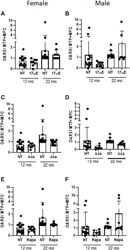
Rayson et al.                                                                                           Geroprotectors in Osteoarthritis and Osteoporosis

occurrence of senescence using a new class of drugs called                     Ashabi et al., 2015). There are multiple evidence in animal models
geroprotectors. Geroprotectors are drugs that delay or reverse                 and retrospective human studies that metformin has positive
ageing processes and in doing so target the major risk factors for             effects on multiple age-related diseases [reviewed in (Morsli and
age-related diseases. They promise to promote health span of                   Bellantuono, 2021)]. Indeed a clinical trial, using Metformin to
more than one organ system at the same time in animal models                   extend survival and reduce the incidence of multiple diseases (the
(Figueira et al., 2016; Bellantuono, 2018). Studies in model                   TAME study) has obtained FDA approval (Barzilai et al., 2016).
organisms or retrospective studies in patients show that they                      Less studied but interesting in the context of OA is Acarbose
can ameliorate tissue dysfunction and reduce the onset and                     (ACA), an intestinal α-glucosidase inhibitor, FDA-approved to
severity of many diseases [reviewed in (Morsli and                             treat diabetes and acts by inhibiting digestion of complex
Bellantuono, 2021)]. Over 200 compounds have been classified                    carbohydrates and reducing postprandial hyperglycaemia
as geroprotectors, each reported to slow ageing and/or extend                  (Dinicolantonio et al., 2015). The mechanisms by which ACA
lifespan in a variety of organisms (geroprotectors.org).                       leads to lifespan extension are not well understood. It is
    Such drugs could have distinct advantages over present                     considered a calorie restriction mimetic and it has been
treatments in OP and offer new opportunities for OA due to                     reported to improve parameters of health, including reduced
the fact that they may be able to prevent both OP and OA and                   incidence of lung tumours in males mice, reduced liver
their co-morbidities. However, the effects of geroprotectors on                degeneration in both sexes and glomerulosclerosis in female
skeletal health have received little attention compared to other               mice (Harrison et al., 2019), improved neuromuscular function
organ systems with the assumption that these drugs will work                   in females, balance/coordination and grip strength in both sexes
equally well for all tissues. Here we review the evidence available            (Herrera et al., 2020). Age-related cardiac hypertrophy was seen
to address whether geroprotectors have potential for the care of               only in male mice, and this male-specific ageing effect was
skeletal age-related diseases and their co-morbidities. We focus               attenuated by ACA (Herrera et al., 2020).
on drugs with a good safety profile, which have been shown to                       Similarly, less known but tested in the context of OA is 17α-
target ageing pathways, extend the lifespan and healthspan in                  estradiol (17α-E2), a naturally occurring enantiomer of 17β-
animal models and have some evidence of improving health in                    estradiol (17β-E2), yet appears to be non-feminizing due to
humans by demonstrating protection from multiple-age-related                   minimal activation of classical oestrogen receptors, ERα and
diseases (Partridge et al., 2020) and for which there are well                 ERβ (Stout et al., 2016). It has been shown to extend lifespan
designed studies in animal models of OP and OA or clinical data                in male mice (Harrison et al., 2014), ameliorate age-associated
available.                                                                     metabolic and inflammatory dysfunction (Stout et al., 2017) and
    Among the most studied geroprotector is Rapamycin. It is an                improve male glucose tolerance across much of adult life (Garratt
inhibitor of the mTOR signalling pathway, a nutrient sensing                   et al., 2017). When administered in later life it maintains body
pathway closely associated with ageing and longevity (Johnson                  weight, with larger muscle mass and fibres, increased grip
et al., 2013; Saxton and Sabatini, 2017). mTOR is a highly                     strength and coordination (Garratt et al., 2019). The metabolic
conserved biological pathway, encompassing two distinct                        improvements are sex-specific and influenced by gonadal
complexes mTORC1 and mTORC2. The two complexes differ                          hormones (Garratt et al., 2017). Little is known on the
in composition and function, with mTORC1 having Raptor and                     molecular basis of 17α-E2 on lifespan and healthspan. The
mTORC2 containing Rictor (Saxton and Sabatini, 2017).                          metabolic improvements appear to be associated with
mTORC1 is acutely inhibited by Rapamycin, whereas                              enhanced hepatic mTORC2 signalling, increased AKT activity
mTORC2 requires chronic exposure to be affected (Li et al.,                    and phosphorylation of FOXO1 (Garratt et al., 2017), increased
2014). It targets multiple mechanisms of ageing including                      AMPKα and reduced mTOR complex 1 activity in visceral
autophagy, oxidative stress, DNA repair (Li et al., 2014;                      adipose tissue (Stout et al., 2017). These latter changes were
Figueira et al., 2016). More recently Rapamycin has also been                  not found in liver or quadriceps muscle (Stout et al., 2017).
shown to inhibit the Senescence Associated Secretory Phenotype                     Of interest, particularly in the context of OA, is Glucosamine
(SASP) (Wang et al., 2017), composed of pro-inflammatory and                    (GluN), an amino-monosaccharide derived principally from
tissue remodelling factors and secreted by senescent cells. Already            chitin, a compound found in the exoskeleton of marine
in clinical use as an immunosuppressor, Rapamycin has been                     invertebrate. It is a component of glycoproteins, proteoglycans
tested extensively in animal models of ageing and age-related                  and glycosaminoglycans. The main compounds containing GluN
diseases [reviewed in (Morsli and Bellantuono, 2021)]. An                      are glucosamine hydrochloride, glucosamine sulphate,
analogue of Rapamycin, RAD001, has been tested in clinical                     N-Acetylglucosamine (GlcNAc). Those compounds have
trials at a substantially lower dose to elicit geroprotective effects to       different phamarcokinetic and pharmacodynamics and seem to
delay immunosenescence with excellent tolerability (Mannick                    act through different mechanisms, which may account in part for
et al., 2014; Mannick et al., 2018).                                           the heterogeneity of response observed in the different studies.
    Metformin, used in the treatment of type 2 diabetes (T2D)                  For example glucosamine sulphate requires a stabiliser in the
(Bosi, 2009; Aroda et al., 2017), has also been extensively                    form of salt and is therefore less pure than glucosamine
investigated for its additional mechanisms of action related to                hydrochloride, necessitating higher dosage (Owens et al.,
ageing (Valencia et al., 2017; Kulkarni et al., 2020). This includes           2004). Glucosamine (sulphate/hydrochloride) have been shown
inhibition of inflammation, reduction in DNA damage and                         to extend lifespan in C. Elegans and in ageing mice by mimicking
inhibition of SASP (Algire et al., 2012; Moiseeva et al., 2013;                similar effects to a low carbohydrate diet. Indeed, it has been

Frontiers in Cell and Developmental Biology | www.frontiersin.org          3                                 February 2022 | Volume 10 | Article 682045
Rayson et al. Geroprotectors in Osteoarthritis and Osteoporosis
shown to activate AMPK, which in turn promotes mitochondrial be causal to multiple age-associated tissue dysfunction or age-
biogenesis, increase aminoacid transport and inhibits glycolysis related diseases in animal models (Jeyapalan et al., 2007; Wang
(Weimer et al., 2014). GlcNAc has also been seen to promote et al., 2009). Senolytics work by targeting specific survival
lifespan extension in C. Elegans but by promoting proteasome pathway and selectively inducing senescent cell death
activity and autophagy (Denzel et al., 2014). Treatment with (Kirkland et al., 2017). The senolytics Dasatinib, a tyrosine
GlcNAc showed an increase in mobility function in models of kinase inhibitor, and Navitoclax, a BCL2 family inhibitor, are
Parkinson’s and Alzhemer’s disease, suggesting effects on among those most studied. They both have anti-neoplastic
healthspan as well (Denzel et al., 2014). In addition, activity and side effects include increased risk of bleeding,
examination of abitual use of glucosamine supplements in a immunosuppression and increased risk of infections (Wilson
retrospective study showed a lower incidence of coronary et al., 2010; Futosi et al., 2012; Rodriguez et al., 2012).
heart disease and stroke (Ma et al., 2019), and a reduction in Dasatinib has little or no senolytic activity on its own but has
mortality due to cancer, respiratory, digestive and cardiovascular senolytic activity only when given in combination with Quercetin
diseases (Li Z.-H. et al., 2020). However, glucosamine users were (Zhu et al., 2015). Quercetin and Fisetin (another senolytic) are
also more active and often took additional supplements flavonoids found in plants and fruits and sold as food
compared to the control group, making it difficult to assess supplements for their antioxidant activities with a good safety
whether these effects were due to GluN uptake per se. GluN profile. The dose required to elicit senolytic activity is higher than
has been shown to have a good safety profile and may have that which is recommended as a food supplement and
promise as geroprotector (Zeng et al., 2015) but well controlled appropriate long-term safety studies are required. Several
prospective preclinical and clinical studies with long duration are clinical trials with these drugs are ongoing.
required to assess whether any of the GluN compounds improves
health span safely. Geroprotectors to Target OA
Spermidine is also emerging for its geroprotective properties. In vitro studies on the effects of geroprotectors are limited.
It is a naturally occurring polyamine, and its concentration has Rapamycin has been found to modulate chondrocytes’
been shown to decline with age in both males and females rat survival, cell death and senescence [reviewed in (Pal et al.,
tissues and in erythrocytes (Jänne et al., 1964). Administration of 2015)]. In addition, it has been shown to increase expression
spermidine extends lifespan in aged cells, C. Elegans, Drosophila of autophagy genes in human and mouse chondrocytes (Zhang
(Eisenberg et al., 2009) and mice (Eisenberg et al., 2016) and it has et al., 2015) and in bovine cartilage explants (Caramés et al.,
been shown to improve some parameters of health. It increase 2012b). This increase was associated with an increased expression
B cell function in aged mice and humans (Zhang et al., 2019), of two major proteins of the cartilage matrix, aggrecan and type II
cardiac function and arterial stiffness in aged mice (Eisenberg collagene, and a decreased expression of metalloproteinases and
et al., 2016) and synapse ageing in drosophila (Maglione et al., chemokines (Zhang et al., 2015). Similarly, metformin has been
2019). Dhal salt sensitive rats fed high salt diet receiving shown to increase chondrocyte survival and delay senescence by
spermidine showed reduced high blood pressure, delayed increasing AMPK and reducing TORC1 signalling (Feng et al.,
progression of heart failure and renal abnormalities seen in 2020) resulting in attenuated aggrecanase activity and
presence of hypertension (Eisenberg et al., 2016). In addition, proteoglycan breakdown in chondrocytes grown in presence of
administration of spermidine reduced the severity of liver lesions inflammatory cytokines (Li H. et al., 2020). Senolytics Navitoclax,
in a mouse model of liver cirrhosis (Yue et al., 2017) and of retinal Fisetin and UBX0101 showed a reduction in markers of
ganglion cell death in a mouse model of optic nerve injury (Noro inflammation, an increase in markers of matrix deposition
et al., 2015). In a community-based cohort study participants (e.g., glycosaminoglycans) (Jeon et al., 2017; Zheng et al.,
taking spermidine showed significant lower all-cause mortality 2017; Yang et al., 2020) and an improvement in chondrocytes
(Kiechl et al., 2018). These effects seem to be the result of proliferation (Jeon et al., 2017) in human OA chondrocytes.
increased autophagy, mitophagy and mitochondrial biogenesis Glucosamine has been shown to have anabolic effects by
(Eisenberg et al., 2016). However, changes in other mechanisms inducing the production of hyaluronic acid in human
associated with ageing have also been seen in presence of chondrocytes and synovial cells and regulate expression of
spermidine supplementation such as decreased histon H3 inflammatory cytokines. A summary of the key in vitro studies
acetylation with possible consequences for gene expression is here (Henrotin et al., 2012). Spermidine has only recently been
(Eisenberg et al., 2009). Safety profile will require appropriate studied in the context of OA. In vitro it shows chondro-protective
assessment in prospective studies as the results in animal models effects from oxidative damage and cell death, and anti-
have been obtained using a wide range of doses, some of which inflammatory properties (Silvestri et al., 2018; D’adamo et al.,
may show toxicity when translating into humans. 2020).
Senolytics [reviewed in (Kirkland and Tchkonia, 2020)] are In vivo most studies testing geroprotectors to delay or reverse
among the most promising geroprotectors due to their the onset of OA are mainly performed in rodent models at young
intermittent mode of administration to periodically eliminate age following induction of OA by destabilization of the meniscus
newly formed senescent cells. This has the potential to reduce side (Table 1). Administration of Rapamycin for 10 weeks at 1 mg/kg/
effects and be more cost-effective. Senescent cells are day after transection of the medial meniscal tibial ligament and
characterised by irreversible cell cycle arrest and the presence the medial collateral ligament in 8 weeks old mice decreased
of SASP. They accumulate with age and they have been shown to cartilage degradation (Caramés et al., 2012a). Similarly, intra-
Frontiers in Cell and Developmental Biology | www.frontiersin.org 4 February 2022 | Volume 10 | Article 682045Rayson et al. Geroprotectors in Osteoarthritis and Osteoporosis
TABLE 1 | Summary of studies testing geroprotectors to attenuate OA in experimental models and patients.
Geroprotector Model Age at the start Key findings (compared Reference
of the experiment to controls)
Rapamycin 1 mg/kg/day i.p. Starting at the Mice C57Bl/6—Transection 8 weeks ↓Cartilage loss Caramés et al. (2012a)
time of MMTL + MCL of MMTL and MCL
Rapamycin (10 µl of 10 µM solution) 2×/ Mice C57Bl/6—DMM 10 weeks ↓Cartilage loss Takayama et al. (2014)
week intra-articular injection starting at the
time of DMM
Rapamycin (100 ng−1 µg) intra-articular Mice C57Bl/6—DMM 8 weeks ↓Cartilage loss Matsuzaki et al. (2014)
gelatin hydrogel starting at the time of DMM
Rapamycin14 mg/kg/day Mice UM-HET Assessed at natural No difference in OA score Ewart et al. (2020)
death
Acarbose 1,000 mg/kg/day Mice UM-HET Assessed at natural No difference in OA score Ewart et al. (2020)
death
17-α-estradiol 14.4 mg/kg/day Mice UM-HET Assessed at natural No difference in OA score Ewart et al. (2020)
death
Metformin Mice C57Bl/6—DMM 8–10 weeks ↓Cartilage loss. ↑paw Li et al. (2020a)
Intra-gastric 200 mg/kg/day starting withdrawal threshold ↓weight-
3 days post-DMM bearing asymmetry
Intra-articular 0.1 mmol/kg twice/week
post-DMM
Metformin 205 mg/kg/day in drinking water Mice C57Bl/6—DMM 10 weeks ↓Cartilage loss Li et al. (2020b)
2 weeks prior to DMM ↓Synovitis
2 weeks post-DMM ↓Osteophytes
No effect on subchondral bone
mass
↑paw withdrawal threshold
↑spontanous activity
Metformin 51.7 mg/kg/day in drinking Rhesus macaques—PMM 8.5–11.5 years ↓Cartilage loss Li et al. (2020b)
water 1 month post-PMM ↓subchondral bone mass
↑standing and walking time
Fisetin 20 mg/kg/day gavage immediately Mice C57Bl/6—DMM 10 weeks ↓Cartilage loss Zheng et al. (2017)
after DMM ↓subchondral bone mass
↓Synovitis
Navitoclax 0.25, 1, 5 µM intra-articular SD Rat—DMM 4–6 weeks ↓Cartilage loss Yang et al. (2020)
injection at the time of DMM twice/week for ↓subchondral bone mass
2 weeks ↓osteophytes
UBX0101 intra-articular (10 µl of Mice C57Bl/6—ACLT 10 weeks ↓Cartilage loss Jeon et al. (2017)
0.2–5 mM) ↓subchondral bone mass
14 days post-ACLT ↓Pain
42 days post-ACLT ↓osteophytes (only with
d14 post-ACLT treatment)
UBX0101 intra-articular (10 µl of 1 mM) Mice C57Bl/6—ACLT 19 months ↓Pain Jeon et al. (2017)
2 weeks post-ACLT No change in cartilage loss,
subchondral bone mass
UBX0101 intra-articular Phase II clinical trial—OA N/A No difference in pain Inc, (2020)
patients
Glucosamine Sulphate oral 250 mg/kg/day Wistar Rats ACLT N/A ↓Pain Wen et al. (2010)
starting at 5 weeks post ACLT for 10 days ↓Cartilage loss
Glucosamine Hydrochloride oral (approx. Wistar Rats ACLT 10 weeks ↓Cartilage loss Naito et al. (2010)
1,000 mg/kg/day) for 8 weeks ↓Bone erosion
Glucosamine Systematic review and Approx. 40–70 years No difference observed on (Liu et al., 2018) (Runhaar et al.,
meta-analysis of clinical old (when available) pain scores 2017) (Zhu et al., 2018) (Wandel
trials et al., 2010)
(Continued on following page)
Frontiers in Cell and Developmental Biology | www.frontiersin.org 5 February 2022 | Volume 10 | Article 682045Rayson et al. Geroprotectors in Osteoarthritis and Osteoporosis
TABLE 1 | (Continued) Summary of studies testing geroprotectors to attenuate OA in experimental models and patients.
Geroprotector Model Age at the start Key findings (compared Reference
of the experiment to controls)
Glucosamine Systematic review and N/A ↓VAS pain (Ogata et al., 2018)
meta-analysis of clinical No effect on WOMAC (Simental-Mendía et al., 2018)
trials
Spermidine 0.3–3–6 mM/day for C57BL6 + ACLT 12 weeks ↓Cartilage loss Chen et al. (2020)
4–8 weeks ↓Osteophytes (@3–6 mM)
↓inflammation
↓MMP13
↑Aggecan/collagenII
MMTL, medial meniscotibial ligament; MCL, medial collateral ligament; DMM, destabilization of the medial meniscus, PMM, partial medial meniscectomy; ACLT, anterior cruciate ligament
transection, i.p, Intra-peritoneal; SD, Sprague-Dawley; VAS, Visual Analogue Scale; WOMAC, Western Ontario and McMaster Universities Osteoarthritis Index.
articular administration of rapamycin twice a week for 8 weeks in chondrocytes and ability to produce matrix with age, impacting
10 weeks old mice following destabilization of the medial further on responses. Similarly a phase I clinical trial with
meniscus (DMM) or injection of hydrogel containing UBX0101 in patients with moderate to severe OA did not
rapamycin in the joint in 8 weeks old DMM mice showed a show any improvement in pain score used as primary
reduction of OARSI score (Matsuzaki et al., 2014; Takayama outcome (Inc, 2020).
et al., 2014). In all these studies rapamycin was given immediately Similarly little or no response was observed by our group and
after the DMM and prior to the establishment of damage in others when the effect of Acarbose, 17-α-estradiol and
young animals. A similar experimental design has been used to Rapamycin were assessed in aged UM-HET mice. UM-HET3
test other geroprotectors such as Fisetin, Metformin, Spermidine are produced by a cross between (BALB/cByJ × C57BL/6J) F1
or Navitoclax with similar results (Zheng et al., 2017; Li J. et al., mothers and (C3H/HeJ × DBA/2J) F1 fathers and have been used
2020; Chen et al., 2020; Yang et al., 2020). However, the at the National Institute on Ageing Intervention Testing
administration of Metformin (4 mg/day in drinking water, Programme (NIA-ITP) (Nadon et al., 2017).
until the animals were sacrificed), was also administered All three compounds have been shown to extend lifespan in
2 weeks after DMM surgery and caused partial but significant these mice, although with some sex differences (Harrison et al.,
reduction in cartilage degradation, suggesting that Metformin 2009; Miller et al., 2011; Harrison et al., 2014; Miller et al., 2014;
may be beneficial even when given at very early stages of damage Strong et al., 2016; Harrison et al., 2019). Similarly to what
(Li J. et al., 2020). This has been confirmed in adult Rhesus reported in the study by (Ewart et al., 2020) our unpublished
Macaques where Metformin was administered 1 month after data showed a significant increase in cartilage degradation with
surgery and significantly alleviated cartilage degradation and age in these mice in both males and females (Figures 1, 2 and
subchondral bone thickening with a reduction in pain-related Supplementary Material). However, none of the treatments
behaviour and improvement in the duration of standing and showed any significant improvement in cartilage degradation
walking (Li J. et al., 2020). Similarly, testing of glucosamine in measured by OARSI scores in both males and females (Figures
young rats showed attenuation of cartilage degradation following 3A–C and Supplementary Material) assessed at 12 and
transection of the anterior cruciate ligament using either GluN 22 months of age. Treatment started at 4 months of age with
sulphate (Wen et al., 2010) or hydrochloride (Naito et al., 2010) the exception of 17-α-E2, which started at 10 months of age when
even when given 5 weeks post-induction of OA at high dose the oestrus cycle starts reducing to avoid a potential interference
(Naito et al., 2010). with sexual development (Nelson et al., 1982). Drugs were used at
The effects of geroprotectors become less effective when the same concentrations shown to elicit lifespan extension (1,000,
administered in older mice or in young mice when the disease 14, and 14.4 mg per kg of diet for acarbose, rapamycin and 17-α-
is already well established. Intra-articular treatment with a new E2, respectively). These data are in line with what reported by
senolytic UBX0101 prevented OA disease in young mice. Ewart et al. (2020) when testing the effect of acarbose and 17-α-
However, administered when OA was established only led to E2estradiol in the same mice using a different experimental
improvement in some aspects of the disease such as cartilage design. In the latter, mice were collected at the time of
structure and pain, but no rescue of subchondral bone death, whereas in our experiments the mice were culled at
remodelling and osteophyte formation (Jeon et al., 2017). fixed time points, reducing some of the variability that may
When the disease was triggered in 19-month-old mice have arisen with the previous experimental design. It is still
administration of the same molecule in advance stages of the possible that our study was underpowered. However, even if
disease showed no improvement in cartilage structure despite this was the case, it indicates that any effect is very small. It is
evidence of senescent cell clearance from the articular cartilage possible that these drugs may work in aged mice only
(Jeon et al., 2017). This was associated with a lack of increased in situation of challenge (e.g., following DMM surgery) and
expression of prochondrogenic genes, suggesting that there may therefore they should be tested in mice following induction of
be an age-related decline in the proliferative capacities of articular OA disease in older age.
Frontiers in Cell and Developmental Biology | www.frontiersin.org 6 February 2022 | Volume 10 | Article 682045Rayson et al. Geroprotectors in Osteoarthritis and Osteoporosis
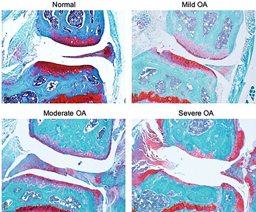
FIGURE 1 | Representative images of joint pathology in UM-HET3 mice. Normal includes joints with an OARSI score of 0–.5, mild 1–2, moderate 3–4 and
severe 5–6.
No studies are available to test the effects of Glucosamine in assessment of the effects of each drugs in all tissues including
aged animals. However, there is an abundance of studies in the skeletal tissues with natural ageing and in models of
patients affected by OA with very contrasting results as disease.
highlighted by meta-analysis and systematic reviews (Wandel
et al., 2010; Runhaar et al., 2017; Liu et al., 2018; Ogata et al., 2018; Geroprotectors to Target OP
Zhu et al., 2018). The discrepancy seems to lie in whether the In vitro studies are primarily focused on the effects of rapamycin
trials contained biases (e.g., trials led by industry were more likely on osteoaclasts and osteoblasts. Based on studies using mouse,
to show positive results) (Vlad et al., 2007) or the sample size rabbit and human cells Rapamycin has been shown to reduce
(trials with over 100 patients seem to show no effect of osteoclasts’ formation, survival and activity (Glantschnig et al.,
glucosamine on pain) (Wandel et al., 2010; Runhaar et al., 2003; Kneissel et al., 2004; Browne et al., 2017). Effects on
2017). In addition, the formulation of glucosamine produced osteoblasts’ proliferation, survival and differentiation are
by Rottapharm seemed to be the most effective (Towheed et al., inconclusive with differences in reports depending on the dose
2005; Vlad et al., 2007; Runhaar et al., 2017). However, it is used and the species from which the cells were derived and
unclear whether this is due to the formulation or to the fact that whether they were primary or cell lines (Kneissel et al., 2004;
trials with the Rottapharm formulation targeted patients at early Singha et al., 2008; Xian et al., 2012; Huang et al., 2015; Browne
stages of disease. Whilst recent meta-analysis concluded that et al., 2017; Wu et al., 2019). This is particularly true for its effects
studies have shown modest or no efficacy of glucosamine on on differentiation. For example the analogue of rapamycin
pain or other parameters of OA questions remain whether the Everolimus showed no effect on the osteoblast marker
treatment should be tested for longer, with a higher dose (Mccarty Alkaline phosphatase (ALP) when hMSC were induced to
et al., 2019) and whether patients should be stratified based on differentiate to the osteoblastic lineage for 7 days at 1 nM but
severity of the disease and/or age, a factor that is never considered showed a reduction in ALP expression at higher concentration of
in the analysis. 10 and 100 nM (Browne et al., 2017). In contrast Runx2 and
Overall, these studies highlight that reducing the severity of Osteocalcin, two other markers of osteoblasts differentiation were
OA in older organisms may be challenging and not sufficient increased at 1 and 10 nM in the same human osteoblasts cultures
on its own. These studies challenge the notion that extension of but were decreased in murine cultures (Browne et al., 2017).
lifespan can be considered an indirect measure of health span There are many reasons for these discrepancies such as
for all tissues. They highlight the need for a comprehensive osteoblasts differentiation may proceed at different rates in
Frontiers in Cell and Developmental Biology | www.frontiersin.org 7 February 2022 | Volume 10 | Article 682045Rayson et al. Geroprotectors in Osteoarthritis and Osteoporosis
FIGURE 2 | UM-HET3 mice develop joint pathology with age (A) Representative examples of joint pathology in male and female UM-HET3 mice at different ages;
(B) Cartilage changes in female mice at 4 months (n = 16), 12 months (n = 15) and 22 months (n = 18); (C) Cartilage changes in male mice at 4 (n = 17), 12 (n = 14) and
(n = 10) 22 months. Values are the mean ± SD of OARSI score for the medial tibia plateau (MTP) plus the medial femoral condyle (MFC). Data were analysed by Kruskal-
Wallis test and Dunn’s multiple comparisons test, **p < .01, ***p < .001, ****p < .0001.
human, mouse cultures and cell lines and markers of osteoblasts effects of hydrogen peroxide on survival and proliferation of
differentiation are dynamic, i.e., they can be upregulated and osteoblast (Jiang et al., 2018).
downregulated at different rates over the period of observation. Most in vivo studies (summarised in Table 2) have used
Assessments of markers over multiple time points may be Rapamycin or one of its derivatives, Everolimus to test their
required to shed some light. effects on bone loss. Rapamycin and Everolimus have been shown
In vitro studies with other geroprotectors are very scant. to delay bone loss in mice in situations of challenge, i.e., in models
Fisetin has been shown to inhibit osteoclasts’ formation and of ovariectomy, iron load, cancer bone disease or ageing (Kneissel
differentiation but effects on osteoblasts were not reported et al., 2004; Luo et al., 2016; Browne et al., 2017; Wu et al., 2019).
(Léotoing et al., 2013). Navitoclax reduced senescent cell These effects are primarily the result of inhibition of osteoclasts
burden but it also negatively impacted on the number of bone formation and activity with the exception of the study utilising
progenitors and osteoblasts in culture inducing apoptosis the iron load model where no difference has been observed in the
(Sharma et al., 2020). Spermidine reduced osteoclasts number of osteoclasts but an increase in ALP+ osteoblasts has
differentiation but did not affect their survival and had no been reported. A study in 24 months old rats receiving
effect on survival and differentiation of osteoblasts (Yamamoto Rapamycin at 1 mg/kg/day for 12 weeks showed positive
et al., 2012). N-Acetyl glucosamine increased osteoblasts effects on osteoblasts activity with an increase in serum
differentiation and mineralization and attenuated the negative osteocalcin and mineral apposition rates (Luo et al., 2016).
Frontiers in Cell and Developmental Biology | www.frontiersin.org 8 February 2022 | Volume 10 | Article 682045Rayson et al. Geroprotectors in Osteoarthritis and Osteoporosis FIGURE 3 | No effect on joint pathology in UMHET3 mice following treatment with 17-α-Estradiol, Acarbose and Rapamycin (A) Cartilage changes in female mice at 12 months (NT n = 15; 17 αE n = 8) and 22 months (NT n = 18; 17 αE n = 9) following treatment with 17-α-Estradiol (17 αE); (B) Cartilage changes in male mice at 12 months (NT n = 14; 17 αE n = 9) and 22 months (NT n = 10, 17 αE n = 8) following treatment with 17 αE; (C) Cartilage changes in female mice at 12 months (NT n = 15; ACA n = 8) and 22 months (NT n = 18; ACA n = 12) following treatment with Acarbose (ACA); (D) Cartilage changes in male mice at 12 months NT n = 14; ACA n = 7) and 22 months (NT n = 14; ACA n = 8) following treatment with ACA; (E) Cartilage changes in female mice at 12 months (NT n = 15; Rapa n = 8) and 22 months (NT n = 18; Rapa n = 12) following treatment with Rapamycin (Rapa); (F) Cartilage changes in male mice at 12 months (NT n = 14; Rapa n = 6) and 22 months (NT n = 10; Rapa n = 9) following treatment with Rapamycin (Rapa). Values are the mean ± SD of OARSI score for the medial tibia plateau (MTP) plus the medial femoral condyle (MFC). Data were analysed by Kruskal-Wallis test and Dunn’s multiple comparisons test, *p < 0.05, **p < 0.01, ***p < 0.001, ****p < 0.0001. NT, not treated. The discrepancy in recording positive effects on osteoblasts’ Of interest is the fact that most studies report outcomes only in activity may be due to the short length of administration of trabecular bone and do not assess cortical bone, despite both Rapamycin in some of the studies (4–8 weeks). Whilst being important to confer bone strength. Kneissel et al. (2004) osteoclasts have shorter lifespan in vivo (2 weeks) osteoblasts (Kneissel et al., 2004) reported a partial protection in trabecular turnover takes approximately 3 months (Manolagas, 2000) and bone but not in cortical bone following treatment of 9 months old therefore it is possible that only those studies assessing rats with Everolimus for 4–8 weeks at the dose of 3 mg/kg/day. osteoblastogenesis for longer periods of time were able to These data suggest that the effects may be limited to the detect an effect. trabecular bone, the more metabolic active part of the bone. Frontiers in Cell and Developmental Biology | www.frontiersin.org 9 February 2022 | Volume 10 | Article 682045
Rayson et al. Geroprotectors in Osteoarthritis and Osteoporosis
TABLE 2 | Summary of in vivo testing of geroprotectors to attenuate bone loss in experimental models.
Geroprotector Model Age at the start Key findings (compared Reference
of the to controls)
experiment
Rapamycin SD rats 24 months ↑BMD Luo et al. (2016)
1 mg/kg weight/day, i.p. 12 weeks ↑Trabecular BV/TV and number,
thickness
↑MAR
↓N Oc and serum Tracp 5b
↑serum OCN
Everolimus Wistar Rats—OVX 9 months Attenuated cancellous bone loss Kneissel et al.
0.5 mg/kg/day ↓trabecular number (2004)
1.5 mg/kg/day No effect on cortical bone
3.0 mg/kg/day ↓N Oc
Gavage No difference in cancellous bone
4–8 weeks treatment formation rates
Everolimus i.p Mice NMRI nude + MDA- 6 weeks OVX model Browne et al.
2 days post tumor injection or OVX 1 mg/kg/day for 4 weeks MB-231 9 weeks ↑BMD (2017)
Mice C57BL/6 + OVX ↑Trabecular BV/TV
↓N Oc
Nude tumour model
↓N tumor lesions
↑BMD
↑Trabecular BV/TV
↓N Oc
Rapamycin Mice Hepcidin knockout 8 weeks ↑BMD Wu et al. (2019)
3 mg/kg/day, i.p. for 2 months C57Bl/6 + OVX ↑Trabecular BV/TV
No difference in cortical bone
↑ N ALP + Ob
No diff in N Oc
Dasatinib (5 mg/kg)and Quercetin (50 mg/kg)monthly for Mice C57Bl/6 20 months Vertebrae Farr et al. (2017)
4 months by gavage ↑Trabecular BV/TV, number and
thickness
↓N Oc
No difference in Ob numbers, BFR,
MAR
Femur
↑cortical thickness
↑strength
↓endocortical N Oc
↑endocortical N Ob
Fisetin Mice C57Bl/6 + OVX 8 weeks ↑BMD Léotoing et al.
5 mg/kg/day or 50 mg/kg/day for 1 week by gavage prior to C57Bl/6 + LPS ↑serum OCN (2013)
OVX followed by 5 mg/kg/day ↑BMD
25 mg/kg/day for 4 weeks by gavage
5 mg/kg/day
25 mg/kg/day 50 mg/kg/day for 3 weeks by gavage
Navitoclax Mice C57Bl/6 24 months ↑Trabecular BV/TV Sharma et al.
50 mg/kg/day for 2 weeks by gavage (2020)
N-Acetyl Glucosamine Sprague-Dawley Rats 12 weeks ↑BV/TV Jiang et al. (2018)
100 mg/kg/day ↑Trabecular bone area
250 mg/kg/day for 12 weeks
Spermidine 0.3–3 mM/day drinking water C57BL6 mice + OVX 8 weeks ↑BV/TV Yamamoto et al.
↓N Oc (2012)
SD, Sprague-Dawley; i.p, intra-peritoneal; BMD, Bone mineral density; MAR, Mineral apposition rates; BFR, Bone Formation Rates; Oc, Osteoclasts; OCN, osteocalcin; Ob, Osteoblasts;
ALP, Alkaline phosphatase; LPS, lypopolysaccharide.
Frontiers in Cell and Developmental Biology | www.frontiersin.org 10 February 2022 | Volume 10 | Article 682045Rayson et al. Geroprotectors in Osteoarthritis and Osteoporosis Long-term studies with Rapamycin and its derivatives are Studies on spermidine and Glucosamine are still in their infancy required to assess its effect on osteoblastogenesis and whether and limited to young mice. Spermidine was administered at both cortical and trabecular bone benefit from the intervention 0.3–3 mM/day orally to 8 weeks old ovariectomised C57BL6 when exposed for prolonged periods. Careful consideration needs mice and analysed 28 days after OVX. Analysis of vertebral to be given to the dose and time of administration and the type of bone showed an increase in BV/TV associated with a decreased mTOR inhibitor as prolonged administration of Rapamycin may in the number of osteoclasts and no effects on osteoblasts have side effects. Intermittent dosing has been proposed to avoid (Yamamoto et al., 2012). N-Acethyl Glucosamine was adverse events (Arriola Apelo et al., 2016). However, regimen of administered at 250 mg/kg and 100 mg/kg/day to 12 weeks old Rapamycin 2 mg/kg once every 5 days has been shown to inhibit ovarectomised Sprague-Dawley rats for 12 weeks. An increase in mTORC1 complex but loss of glucose tolerance persisted in the bone mineral density and trabecular bone area was observed. This same way than what was observed when given daily (Houde et al., was associated with signs of increased osteoblasts differentiation 2010). In humans no major side effects have been seen with and mineralizations (Yamamoto et al., 2012). Effects on osteoclasts weekly dosing of Everolimus and this was enough to improve were no reported. Although these studies are promising, more in immune responses (Mannick et al., 2014). However, Everolimus depth studies in aged mice are required to assess whether these administered at a weekly dose did not produce any difference on agents hold promise for attenuating bone loss with age. bone parameters (Kneissel et al., 2004), suggesting that daily dose may be required to detect effects. However, Everolimus may still be preferable to Rapamycin. Indeed when given daily it had CONCLUSION reduced impact on glucose tolerance compared to daily Rapamycin despite being equally efficacious in inhibiting Geroprotectors potentially have additional benefits to treat OA and proteins of the TORC1 complex (Arriola Apelo et al., 2016). OP and their co-morbidities. However, few studies focus on The effect with senolytics has shown mixed results. skeletal health despite their burden of disease. Only one study Pharmacologic clearance of senescence cells in aged mice with the combination of senolytics DQ shows signs of (20 months) treated with Dasatinib and Quercetin (DQ) for improvement in a model of bone loss and no improvement has 4 months by single monthly administration showed been demonstrated so far in aged models of OA. These studies improvement of both the trabecular and cortical bone in highlight that extension of lifespan cannot be considered a femur and vertebrae (Farr et al., 2017). DQ suppressed surrogate marker for extension of health span in all tissues and resorption by reducing osteoclast numbers and improved thorough studies in aged models of OP and OA are required to osteoblast numbers on the cortical bone surface but not on the assess the real benefit of geroprotectors to improve skeletal health. trabecular bone surface (Farr et al., 2017). When Fisetin was given to 8 weeks old mice, 1 week before OVX, an increase in trabecular bone volume fraction, thickness and AUTHOR CONTRIBUTIONS number were observed 4 weeks after OVX (Léotoing et al., 2013). A similar effect was also reported when using a model of AR, MB, and JG performed the experiments, analysed the data, inflammation-induced bone loss by Lypopolysaccharide injection reviewed and approved the manuscript. ED designed the study, (Léotoing et al., 2013). However, it is unlikely that these effects are reviewed and approved the manuscript. MN wrote and approved due to Fisetin’s senolytic activity. Very low levels of senescent cells the manuscript. IB designed the experiments, analysed the data, have been reported in mice before 8 months of age (Farr et al., 2017). wrote and approved the manuscript. Studies in aged mice are required to determine whether Fisetin has senolytic effects and prevent bone loss observed with age. Detrimental effects to trabecular bone were reported in aged FUNDING male and female C57BL/6 mice (24 months old), when they were treated with the senolytic drug Navitoclax once daily for 2 weeks This work was supported by the Biotechnology and Biological with signs of apoptosis on bone cells (Sharma et al., 2020). The Science Research Council Grant Ref N BB/R001510/1. same dose was used in the study by Chang et al. (Chang et al., 2016) to eliminate senescent cells. Indeed it showed improved proliferation and regeneration ability of hematopoietic stem cells ACKNOWLEDGMENTS (HSC), compatible with a reversal of HSC to a more youthful phenotype (Chang et al., 2016). However, Navitoclax was We are grateful to Richard Miller at the University of Michigan administered only for 7 days in the study by Chang et al. for donating mouse tissues from the UM-HET3 mice. (Chang et al., 2016) as opposed to 14 days in the study by (Sharma et al., 2020). This may account for the toxicity observed. The toxicity of Navitoclax is well known and SUPPLEMENTARY MATERIAL therefore improved regimen should be tested, particularly with the new galacto-conjugated Navitoclax, where the drug can be The Supplementary Material for this article can be found online at: preferentially activated by SA-β-gal activity primarily in https://www.frontiersin.org/articles/10.3389/fcell.2022.682045/ senescent cells (González-Gualda et al., 2020). full#supplementary-material Frontiers in Cell and Developmental Biology | www.frontiersin.org 11 February 2022 | Volume 10 | Article 682045
Rayson et al.                                                                                                                        Geroprotectors in Osteoarthritis and Osteoporosis

REFERENCES                                                                                            Protein Quality Control and Prolong Life. Cell 156, 1167–1178. doi:10.
                                                                                                      1016/j.cell.2014.01.061
                                                                                                   Dinicolantonio, J. J., Bhutani, J., and O’keefe, J. H. (2015). Acarbose: Safe and
Algire, C., Moiseeva, O., Deschênes-Simard, X., Amrein, L., Petruccelli, L., Birman,                  Effective for Lowering Postprandial Hyperglycaemia and Improving
    E., et al. (2012). Metformin Reduces Endogenous Reactive Oxygen Species and                       Cardiovascular Outcomes. Open Heart 2, e000327. doi:10.1136/openhrt-
    Associated DNA Damage. Cancer Prev. Res. 5, 536–543. doi:10.1158/1940-                            2015-000327
    6207.capr-11-0536                                                                              Eisenberg, T., Abdellatif, M., Schroeder, S., Primessnig, U., Stekovic, S., Pendl, T.,
Aroda, V. R., Knowler, W. C., Knowler, W. C., Crandall, J. P., Perreault, L.,                         et al. (2016). Cardioprotection and Lifespan Extension by the Natural
    Edelstein, S. L., et al. (2017). For the Diabetes Prevention Program                              Polyamine Spermidine. Nat. Med. 22, 1428–1438. doi:10.1038/nm.4222
    ResearchMetformin for Diabetes Prevention: Insights Gained from the                            Eisenberg, T., Knauer, H., Schauer, A., Büttner, S., Ruckenstuhl, C., Carmona-
    Diabetes Prevention Program/Diabetes Prevention Program Outcomes                                  Gutierrez, D., et al. (2009). Induction of Autophagy by Spermidine Promotes
    Study. Diabetologia 60, 1601–1611. doi:10.1007/s00125-017-4361-9                                  Longevity. Nat. Cel Biol. 11, 1305–1314. doi:10.1038/ncb1975
Arriola Apelo, S. I., Neuman, J. C., Baar, E. L., Syed, F. A., Cummings, N. E., Brar,              Ewart, D., Harper, L., Gravely, A., Miller, R. A., Carlson, C. S., and Loeser, R. F.
    H. K., et al. (2016). Alternative Rapamycin Treatment Regimens Mitigate the                       (2020). Naturally Occurring Osteoarthritis in Male Mice with an Extended
    Impact of Rapamycin on Glucose Homeostasis and the Immune System. Aging                           Lifespan. Connect. Tissue Res. 61, 95–103. doi:10.1080/03008207.2019.
    Cell 15, 28–38. doi:10.1111/acel.12405                                                            1635590
Ashabi, G., Khalaj, L., Khodagholi, F., Goudarzvand, M., and Sarkaki, A. (2015).                   Executive, L. H. A. S. (2015). Work Related Musculoskeletal Disorder Statistics (WRMSDs)
    Pre-treatment with Metformin Activates Nrf2 Antioxidant Pathways and                              in Great Britain 2014/15. [Online]. Available: https://www.england.nhs.uk/ourwork/
    Inhibits Inflammatory Responses through Induction of AMPK after                                    clinical-policy/ltc/our-work-on-long-term-conditions/musculoskeletal/.[Accessed].
    Transient Global Cerebral Ischemia. Metab. Brain Dis. 30, 747–754. doi:10.                     Farr, J. N., and Khosla, S. (2019). Cellular Senescence in Bone. Bone 121, 121–133.
    1007/s11011-014-9632-2                                                                            doi:10.1016/j.bone.2019.01.015
Barzilai, N., Crandall, J. P., Kritchevsky, S. B., and Espeland, M. A. (2016).                     Farr, J. N., Xu, M., Weivoda, M. M., Monroe, D. G., Fraser, D. G., Onken, J. L., et al.
    Metformin as a Tool to Target Aging. Cel Metab. 23, 1060–1065. doi:10.                            (2017). Targeting Cellular Senescence Prevents Age-Related Bone Loss in Mice.
    1016/j.cmet.2016.05.011                                                                           Nat. Med. 23, 1072–1079. doi:10.1038/nm.4385
Bellantuono, I. (2018). Find Drugs that Delay many Diseases of Old Age. Nature                     Feng, X., Pan, J., Li, J., Zeng, C., Qi, W., Shao, Y., et al. (2020). Metformin
    554, 293–295. doi:10.1038/d41586-018-01668-0                                                      Attenuates Cartilage Degeneration in an Experimental Osteoarthritis Model by
Bijlsma, J. W., Berenbaum, F., and Lafeber, F. P. (2011). Osteoarthritis: an Update                   Regulating AMPK/mTOR. Aging 12, 1087–1103. doi:10.18632/aging.102635
    with Relevance for Clinical Practice. Lancet 377, 2115–2126. doi:10.1016/s0140-                Figueira, I., Fernandes, A., Mladenovic Djordjevic, A., Lopez-Contreras, A.,
    6736(11)60243-2                                                                                   Henriques, C. M., Selman, C., et al. (2016). Interventions for Age-Related
Bosi, E. (2009). Metformin – the Gold Standard in Type 2 Diabetes: what Does the                      Diseases: Shifting the Paradigm. Mech. Ageing Dev. 160, 69–92. doi:10.1016/j.
    Evidence Tell Us? Diabetes Obes. Metab. 11, 3–8. doi:10.1111/j.1463-1326.2008.                    mad.2016.09.009
    01031.x                                                                                        Futosi, K., Németh, T., Pick, R., Vántus, T., Walzog, B., and Mócsai, A. (2012).
Browne, A. J., Kubasch, M. L., Göbel, A., Hadji, P., Chen, D., Rauner, M., et al.                     Dasatinib Inhibits Proinflammatory Functions of Mature Human Neutrophils.
    (2017). Concurrent Antitumor and Bone-Protective Effects of Everolimus in                         Blood 119, 4981–4991. doi:10.1182/blood-2011-07-369041
    Osteotropic Breast Cancer. Breast Cancer Res. : BCR 19, 92. doi:10.1186/s13058-                Garratt, M., Bower, B., Garcia, G. G., and Miller, R. A. (2017). Sex Differences in
    017-0885-7                                                                                        Lifespan Extension with Acarbose and 17-α Estradiol: Gonadal Hormones
Burrage, P. S., Mix, K. S., and Brinckerhoff, C. E. (2006). Matrix                                    Underlie Male-specific Improvements in Glucose Tolerance and mTORC2
    Metalloproteinases: Role in Arthritis. Front. Biosci. 11, 529–543. doi:10.                        Signaling. Aging Cell 16, 1256–1266. doi:10.1111/acel.12656
    2741/1817                                                                                      Garratt, M., Leander, D., Pifer, K., Bower, B., Herrera, J. J., Day, S. M., et al. (2019).
Caramés, B., Hasegawa, A., Taniguchi, N., Miyaki, S., Blanco, F. J., and Lotz, M.                     17-α Estradiol Ameliorates Age-Associated Sarcopenia and Improves Late-Life
    (2012a). Autophagy Activation by Rapamycin Reduces Severity of                                    Physical Function in Male Mice but Not in Females or Castrated Males. Aging
    Experimental Osteoarthritis. Ann. Rheum. Dis. 71, 575–581. doi:10.1136/                           Cell 18, e12920. doi:10.1111/acel.12920
    annrheumdis-2011-200557                                                                        Glantschnig, H., Fisher, J. E., Wesolowski, G., Rodan, G. A., and Reszka, A. A.
Caramés, B., Taniguchi, N., Otsuki, S., Blanco, F. J., and Lotz, M. (2010).                           (2003). M-CSF, TNFα and RANK Ligand Promote Osteoclast Survival by
    Autophagy Is a Protective Mechanism in normal Cartilage, and its Aging-                           Signaling through mTOR/S6 Kinase. Cel Death Differ. 10, 1165–1177. doi:10.
    Related Loss Is Linked with Cell Death and Osteoarthritis. Arthritis Rheum. 62,                   1038/sj.cdd.4401285
    791–801. doi:10.1002/art.27305                                                                 Goldring, M. B., and Otero, M. (2011). Inflammation in Osteoarthritis. Curr. Opin.
Caramés, B., Taniguchi, N., Seino, D., Blanco, F. J., D’lima, D., and Lotz, M.                        Rheumatol. 23, 471–478. doi:10.1097/bor.0b013e328349c2b1
    (2012b). Mechanical Injury Suppresses Autophagy Regulators and                                 Goldring, M. B. (2000). The Role of the Chondrocyte in Osteoarthritis. Arthritis
    Pharmacologic Activation of Autophagy Results in Chondroprotection.                               Rheum. 43, 1916–1926. doi:10.1002/1529-0131(200009)43:93.
    Arthritis Rheum. 64, 1182–1192. doi:10.1002/art.33444                                             0.co;2-i
Chang, J., Wang, Y., Shao, L., Laberge, R.-M., Demaria, M., Campisi, J., et al. (2016).            González-Gualda, E., Pàez-Ribes, M., Lozano-Torres, B., Macias, D., Wilson, J. R.,
    Clearance of Senescent Cells by ABT263 Rejuvenates Aged Hematopoietic Stem                        3rd, González-López, C., et al. (2020). Galacto-conjugation of Navitoclax as an
    Cells in Mice. Nat. Med. 22, 78–83. doi:10.1038/nm.4010                                           Efficient Strategy to Increase Senolytic Specificity and Reduce Platelet Toxicity.
Chen, Z., Lin, C.-X., Song, B., Li, C.-C., Qiu, J.-X., Li, S.-X., et al. (2020). Spermidine           Aging cell 19, e13142.
    Activates RIP1 Deubiquitination to Inhibit TNF-α-Induced NF-κB/p65                             Grishko, V. I., Ho, R., Wilson, G. L., and Pearsall, A. W. T. (2009). Diminished
    Signaling Pathway in Osteoarthritis. Cel Death Dis. 11, 503. doi:10.1038/                         Mitochondrial DNA Integrity and Repair Capacity in OA Chondrocytes.
    s41419-020-2710-y                                                                                 Osteoarthritis Cartilage 17, 107–113. doi:10.1016/j.joca.2008.05.009
Crandall, C. J., Newberry, S. J., Diamant, A., Lim, Y. W., Gellad, W. F., Booth, M. J.,            Harrison, D. E., Strong, R., Alavez, S., Astle, C. M., Digiovanni, J., Fernandez, E.,
    et al. (2014). Comparative Effectiveness of Pharmacologic Treatments to                           et al. (2019). Acarbose Improves Health and Lifespan in Aging HET3 Mice.
    Prevent Fractures: an Updated Systematic Review. Ann. Intern. Med. 161,                           Aging Cell 18, e12898. doi:10.1111/acel.12898
    711–723. doi:10.7326/m14-0317                                                                  Harrison, D. E., Strong, R., Allison, D. B., Ames, B. N., Astle, C. M., Atamna, H.,
D’adamo, S., Cetrullo, S., Guidotti, S., Silvestri, Y., Minguzzi, M., Santi, S., et al.               et al. (2014). Acarbose, 17-α-Estradiol, and Nordihydroguaiaretic Acid Extend
    (2020). Spermidine Rescues the Deregulated Autophagic Response to Oxidative                       Mouse Lifespan Preferentially in Males. Aging Cell 13, 273–282. doi:10.1111/
    Stress of Osteoarthritic Chondrocytes. Free Radic. Biol. Med. 153, 159–172.                       acel.12170
    doi:10.1016/j.freeradbiomed.2020.03.029                                                        Harrison, D. E., Strong, R., Sharp, Z. D., Nelson, J. F., Astle, C. M., Flurkey, K., et al.
Denzel, Martin. s., Storm, Nadia. j., Gutschmidt, A., Baddi, R., Hinze, Y.,                           (2009). Rapamycin Fed Late in Life Extends Lifespan in Genetically
    Jarosch, E., et al. (2014). Hexosamine Pathway Metabolites Enhance                                Heterogeneous Mice. Nature 460, 392–395. doi:10.1038/nature08221

Frontiers in Cell and Developmental Biology | www.frontiersin.org                             12                                            February 2022 | Volume 10 | Article 682045
You can also read