Tick-borne zoonoses and commonly used diagnostic methods in human and veterinary medicine
←
→
Page content transcription
If your browser does not render page correctly, please read the page content below
Parasitology Research
https://doi.org/10.1007/s00436-020-07033-3

    ARTHROPODS AND MEDICAL ENTOMOLOGY - REVIEW

Tick-borne zoonoses and commonly used diagnostic methods
in human and veterinary medicine
Andrea Springer 1 & Antje Glass 1 & Julia Probst 1 & Christina Strube 1

Received: 30 October 2020 / Accepted: 21 December 2020
# The Author(s) 2021

Abstract
Around the world, human health and animal health are closely linked in terms of the One Health concept by ticks acting as vectors
for zoonotic pathogens. Animals do not only maintain tick cycles but can either be clinically affected by the same tick-borne
pathogens as humans and/or play a role as reservoirs or sentinel pathogen hosts. However, the relevance of different tick-borne
diseases (TBDs) may vary in human vs. veterinary medicine, which is consequently reflected by the availability of human vs.
veterinary diagnostic tests. Yet, as TBDs gain importance in both fields and rare zoonotic pathogens, such as Babesia spp., are
increasingly identified as causes of human disease, a One Health approach regarding development of new diagnostic tools may
lead to synergistic benefits. This review gives an overview on zoonotic protozoan, bacterial and viral tick-borne pathogens
worldwide, discusses commonly used diagnostic techniques for TBDs, and compares commercial availability of diagnostic tests
for humans vs. domestic animals, using Germany as an example, with the aim of highlighting existing gaps and opportunities for
collaboration in a One Health framework.

Keywords One Health . Zoonoses . Metazoonoses . Ticks . Tick-borne diseases . Lyme borreliosis . Diagnostics . Serology .
PCR . ELISPOT

Tick-borne diseases in the One Health                                        borne encephalitis virus (TBEV), which may cause neurologic
perspective                                                                  disease in humans, as well as dogs and horses (Pfeffer and
                                                                             Dobler 2011). Additionally, domestic animals may represent
Ticks represent a major threat for human and animal health                   an infection reservoir for tick-borne diseases (TBDs) in
worldwide due to their vector function for a variety of zoonot-              humans, such as cattle for Babesia divergens (Zintl et al.
ic protozoan, bacterial and viral pathogens. These pathogens                 2003) and dogs for Ehrlichia canis (Rar and Golovljova
often circulate unnoticed in nature in enzootic tick-vertebrate              2011).
cycles but may cause significant morbidity and mortality                        Many tick species transmit zoonotic pathogens; however,
when spilling over to humans or domestic animals (Jahfari                    some are exceptional due to their vector function for a number
and Sprong 2016). For example, Anaplasma                                     of different zoonotic pathogens. Thus, both the tick species
phagocytophilum mainly circulates between ticks and wild-                    infesting different hosts at the wildlife-domestic animal-hu-
life, but certain strains may cause granulocytic anaplasmosis                man interface and the pathogens transmitted by them are of
in humans, dogs and horses as well as so-called tick-borne                   significant One Health importance. Among the particularly
fever in domestic ruminants (Jaarsma et al. 2019). Similarly,                important tick vectors are Ixodes ricinus, Ixodes persulcatus
small wild mammals constitute the main reservoir for tick-                   and Ixodes scapularis, which belong to the so-called Ixodes
                                                                             ricinus complex, a group of 14 Ixodes species with almost
Section Editor: Domenico Otranto                                             worldwide distribution (Keirans et al. 1999; Xu et al. 2003).
                                                                             Ticks of the I. ricinus complex are confirmed vectors of zoo-
* Christina Strube                                                           notic protozoa (Babesia spp.), a number of bacteria (e.g.
  christina.strube@tiho-hannover.de                                          Borrelia spp. and Rickettsiales) as well as three different
1                                                                            flaviviruses (TBEV, Louping ill and Powassan virus).
     Institute for Parasitology, Centre for Infection Medicine, University
     of Veterinary Medicine Hannover, Buenteweg 17,                          Furthermore, Dermacentor andersoni, Dermacentor
     30559 Hanover, Germany                                                  variabilis and Amblyomma americanum are of particular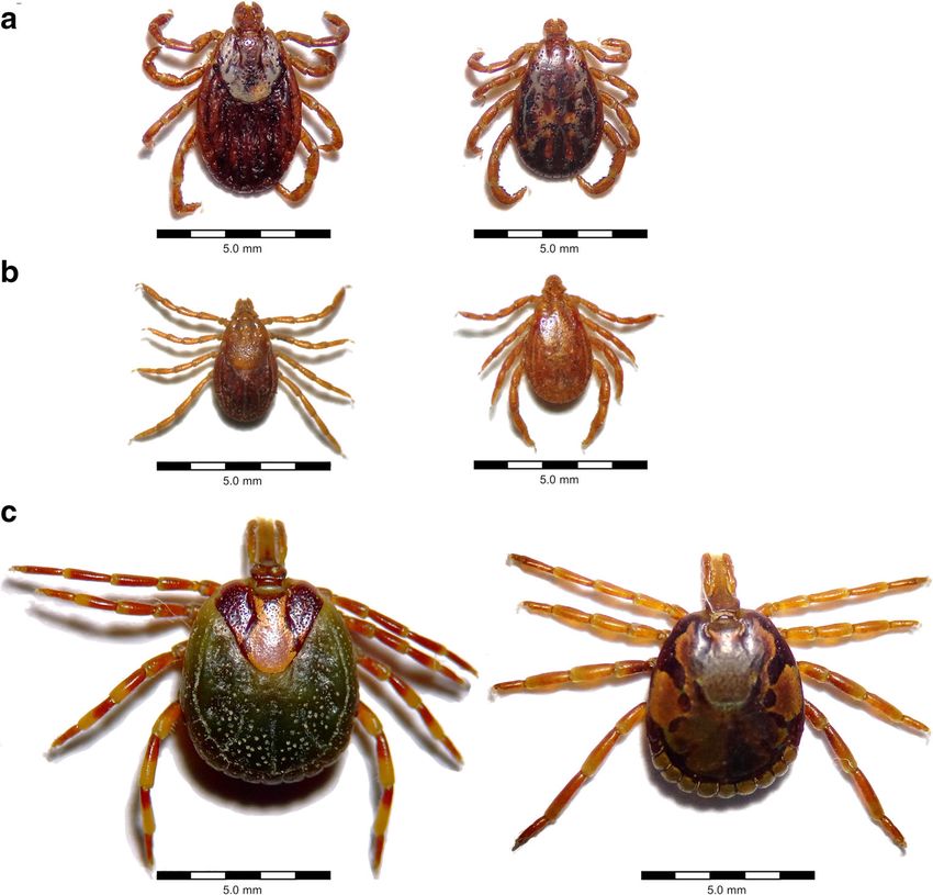
Parasitol Res

One Health significance in North America (Sonenshine 2018)                       For example, Borrelia burgdorferi sensu lato (s.l.), the causative
due to their vector function for a number of zoonotic bacterial                  agent of Lyme borreliosis, and A. phagocytophilum occur
(e.g. Rickettsia spp. and Ehrlichia spp.) and viral (e.g.                        throughout the Northern Hemisphere as both are transmitted by
Powassan and Heartland virus) pathogens.                                         ticks of the Ixodes ricinus complex. Furthermore, spotted fever
   While most zoonotic TBDs are transmitted by hard ticks,                       group rickettsiae comprise a large group of species associated
soft ticks may also play a role as vectors (Dantas-Torres et al.                 with zoonotic human disease or of unknown pathogenicity,
2012). Several Ornithodoros spp. may transmit relapsing fe-                      which are transmitted by different species (Fig. 2) of several hard
ver borreliae (Talagrand-Reboul et al. 2018), and this tick                      tick genera around the world (Parola et al. 2013). Some
genus might be implicated in the transmission of Coxiella                        rickettsioses are associated with high case fatality rates in
burnetii (Duron et al. 2015) and Alkhurma fever virus                            humans, especially Rocky Mountain spotted fever caused by
(Sawatsky et al. 2014).                                                          Rickettsia rickettsii and transmitted mainly by D. andersoni,
                                                                                 D. variabilis (Fig. 2a) and Rhipicephalus sanguineus s.l. (Fig.
Tick-borne zoonotic protozoans                                                   2b), and Mediterranean spotted fever caused by Rickettsia
                                                                                 conorii and transmitted mainly by R. sanguineus s.l. (Parola
Among tick-borne pathogens, Babesia spp. constitute the only                     et al. 2013).
zoonotic protozoans (Table 1), which are transmitted to                              In addition, ticks may play a role in the transmission of
humans by Ixodes ricinus (Fig. 1) and Ixodes scapularis and                      severe diseases such as tularemia, caused by Francisella
are thus restricted to the range of these tick species in Eurasia,               tularensis, and so-called Q fever due to C. burnetii infection.
Northern Africa and North America. Babesia spp. are usually                      Although other transmission routes are regarded as epidemio-
highly host-specific and the natural vertebrate hosts for                        logically more important, several hard tick species, including
Babesia divergens, Babesia venatorum and Babesia microti                         D. andersoni in North America and I. ricinus (Fig. 1) as well
are cattle, wild ungulates and rodents, respectively, whereas                    as Dermacentor marginatus (Fig. 3a) in Eurasia, have been
humans are mainly affected if immunocompromised (Gray                            identified as competent vectors for both of these pathogens
et al. 2010). Interestingly, although Babesia microti occurs                     (Telford III and Goethert 2020; Duron et al. 2015).
in both Europe and North America, symptomatic human in-
fections have so far only been acquired in North America                         Tick-borne zoonotic viruses
(Azagi et al. 2020).
                                                                                 Compared to bacteria, none of the tick-borne viruses are dis-
Tick-borne zoonotic bacteria                                                     tributed worldwide (the same applies to protozoans, cf.
                                                                                 Table 1), but rather often restricted to particular geographic
In contrast to protozoans, a wide variety of zoonotic bacterial                  regions (Table 3). However, many of them cause life-
pathogens are tick transmitted (Table 2). Some of these are of                   threatening disease in humans. Among tick-borne viruses,
major importance due to their wide geographic distribution and/                  the highly pathogenic Crimean-Congo haemorrhagic fever vi-
or the severity of the disease caused in humans and/or animals.                  rus (CCHFV), transmitted mainly by Hyalomma spp. (Fig.

Table 1      Tick-borne protozoan pathogens, their vectors and reservoir hosts

Pathogen           Tick vector(s)1        Geographical          Vertebrate         Cell tropism in the          Comment(s)           References
                                          distribution          reservoir(s)       vertebrate host

Babesia            Ixodes ricinus         Europe, North         Cattle             Intracellular:                                    Reviewed by Zintl
  divergens                                 Africa, Russia                            erythrocytes                                     et al.
                                                                                                                                       (2003) and
                                                                                                                                       Gray
                                                                                                                                       et al. (2019b)
Babesia            I. ricinus, Ixodes     Eurasia, North        Rodents            Intracellular:               So far, only North   Reviewed by Gray
  microti             scapularis            America                                   erythrocytes                American strains     et al.
                                                                                                                  involved in          (2019b); Azagi
                                                                                                                  human cases          et al. (2020)
Babesia            I. ricinus             Europe                Roe deer,        Intracellular:                                      Reviewed by Gray
  venatorum                                                       possibly sheep    erythrocytes                                       et al. (2019b);
                                                                                                                                       Gray et al.
                                                                                                                                       (2019a)

1
    Main tick vectors responsible for human infections; other tick vectors may be relevant in tick-reservoir cycles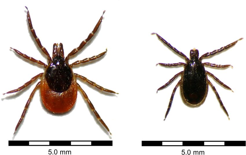
Parasitol Res

Fig. 1 Ixodes ricinus, confirmed
vector of Babesia divergens,
Babesia microti and Babesia
venatorum, among numerous
other pathogens (left: female,
right: male). Photographs were
taken under an OPTIKA SLX-2
stereomicroscope (OPTIKA
S.r.l., Ponteranica, Italy)

3c), has the widest distribution as it occurs in Africa, through-   2020). Nevertheless, cases in immunocompetent persons have
out Asia and in Eastern Europe (IZS “G. Caporale” 2009).            also recently been reported (Martinot et al. 2011). Similarly,
Likewise, TBEV has a rather wide distribution, with different       Ehrlichia canis is of major veterinary relevance as the causa-
subtypes circulating in Ixodes ticks in Europe, Siberia and far-    tive agent of canine monocytic ehrlichiosis, whereas human
eastern Asia (Dobler et al. 2012). Examples of highly patho-        ehrlichiosis cases due to this pathogen are very rare (Rar and
genic tick-borne viruses with a more restricted geographical        Golovljova 2011). Similarly, A. phagocytophilum is a fre-
distribution include Omsk haemorrhagic fever virus, transmit-       quent cause of disease in dogs, horses and ruminants in
ted by D. marginatus (Fig. 3a) and D. reticulatus (Fig. 3b)         Europe (Silaghi et al. 2011; Kohn et al. 2008), whereas human
(Růžek et al. 2010) in Russia, and Kyasanur Forest virus,           cases are rarely reported on the continent (Azagi et al. 2020).
transmitted by Haemaphysalis spinigera (Shah et al. 2018)           In North America on the other hand, human granulocytic an-
in India.                                                           aplasmosis cases are numerous but tick-borne fever in rumi-
                                                                    nants has never been confirmed (Dugat et al. 2015). These
                                                                    epidemiological differences are attributed to different circulat-
Human and veterinary relevance of tick-borne zoo-                   ing strains of A. phagocytophilum (Dugat et al. 2015).
notic pathogens                                                        Finally, Lyme borreliosis is sometimes (subjectively)
                                                                    regarded as equally important in both fields, especially by
The relevance of different tick-borne pathogens varies in the       dog owners, although pathogenicity for dogs has only been
fields of human vs. veterinary medicine. For example, tick-         proven for B. burgdorferi sensu stricto (s.s.) and remains ques-
borne encephalitis (TBE) cases occur mostly in humans and           tionable for other genospecies of the B. burgdorferi s.l. com-
only rarely in domestic animals, which are mainly regarded as       plex (Littman et al. 2018).
sentinels of virus occurrence (Imhoff et al. 2015). However,
domestic ruminants are epidemiologically important as
sources of alimentary human TBEV infections (Dobler et al.
2012) and dogs as well as horses may develop severe neuro-          Commonly used diagnostic methods for TBDs
logical signs when contracting TBE (Pfeffer and Dobler 2011;        in human and veterinary medicine
Waldvogel et al. 1981). Regarding the numerous tick-
transmitted Rickettsia spp., which are relevant globally as         The differences in clinical relevance of zoonotic TBDs are
agents of human disease (Parola et al. 2013), evidence of           reflected by the availability of commercially manufactured
pathogenicity in domestic animals is limited to Rickettsia          human vs. veterinary diagnostic tests. However, as TBDs gain
conorii and Rickettsia rickettsii in dogs (Keenan et al. 1977;      importance in both fields and rare zoonotic pathogens, such as
Solano-Gallego et al. 2006).                                        Babesia spp., are increasingly identified as causes of human
    In contrast, B. divergens is primarily a parasite of cattle,    TBDs, a One Health approach in TBD diagnostics may lead to
causing haemolytic anaemia with high case fatality rates in         synergistic benefits. In the following, commonly used diag-
naïve cattle herds (Springer et al. 2020; Zintl et al. 2003),       nostic techniques for TBDs in both fields and comparison of
whereas human B. divergens cases mainly involve                     commercial availability of tests for humans vs. domestic ani-
splenectomised or immunosuppressed patients (Azagi et al.           mals are discussed, with the aim of highlighting gaps and
Table 2 Tick-borne bacterial pathogens, their vectors and reservoir hosts
Pathogen Tick vector(s)1 Geographical Vertebrate Cell/tissue tropism in the Comment(s) References
distribution reservoir(s) vertebrate host
Genus Anaplasma
Anaplasma phagocytophilum Ixodes pacificus, Ixodes Eurasia, North Zoonotic strains: red Intracellular: neutrophilic Different strains with Jaarsma et al. (2019); Rar and
persulcatus, Ixodes ricinus, America deer, possibly and eosinophilic differing zoonotic Golovljova (2011)
Ixodes scapularis wild boar and granulocytes potential
hedgehogs
Anaplasma platys Rhipicephalus sanguineus s.s. Worldwide Dogs Intracellular: Rare human infections Arraga-Alvarado et al. (2014);
(temperate lineage) thrombocytes with unknown Snellgrove et al. (2020)
pathogenicity
Genus Bartonella
Bartonella henselae Probably2 I. ricinus and other Worldwide Cats, rabbits, Intracellular: Predominantly other Reviewed by Cheslock and
and other zoonotic ixodid ticks possibly dogs and erythrocytes transmission routes2 Embers (2019)
Bartonella spp. rodents
Genus Borrelia
Lyme borreliae: Borrelia I. pacificus, I. persulcatus, Eurasia, North Small mammals, Extracellular: skin, Tissue tropism may Reviewed by Rudenko et al.
burgdorferi sensu I. ricinus, I. scapularis America, birds, lizards joints, nervous system differ between (2011); Barbieri et al.
lato (s.l.) complex South (depending on genospecies (2013)
America genospecies)
Relapsing fever borreliae: I. ricinus, I. scapularis, Eurasia, North Small mammals Extracellular: probably Reviewed by Cutler et al.
Borrelia miyamotoi probably3 I. pacificus, America nervous system (2019)
I. persulcatus, Ixodes ovatus
Relapsing fever borreliae: Ornithodoros spp. Asia, Africa, Small mammals Extracellular: blood, Reviewed by
Borrelia duttonii, Borrelia North different organs Talagrand-Reboul et al.
hermsii, Borrelia turicatae America and (2018)
and others South
America
Relapsing fever borreliae: Amblyomma americanum North America Deer Extracellular: skin Varela-Stokes (2007)
Borrelia lonestari
Genus Coxiella
Coxiella burnetii Dermacentor andersoni, Worldwide Ruminants Intracellular: mononuclear Transmission by Reviewed by Duron et al.
Dermacentor marginatus, phagocytes, inhalation of tick (2015) and Voth and
Hyalomma asiaticum, Ixodes pneumocytes, faeces more probable Heinzen (2007); Körner
holocyclus, I. ricinus, several fibroblasts, endothelial than by tick bite et al. (2020)
Ornithodoros spp. cells
Francisella tularensis A. americanum, D. andersoni, Northern Rodents and Facultatively intracellular: Multiple transmission Reviewed by Telford III and
D. marginatus, Dermacentor Hemisphere lagomorphs macrophages, broad routes, including bites Goethert (2020) and Ozanic
variabilis, I. ricinus range of other cells of other arthropods et al. (2015); Výrosteková
(1994)
Genus Ehrlichia and
Neoehrlichia
Ehrlichia chaffeensis A. americanum, probably3 North America, Deer Intracellular: Reviewed by Yabsley (2010)
other tick species South monocytes/macrophages and Rar and Golovljova
(2011)
Parasitol ResTable 2 (continued)
Pathogen Tick vector(s)1 Geographical Vertebrate Cell/tissue tropism in the Comment(s) References
Parasitol Res
distribution reservoir(s) vertebrate host
America,
Asia, Africa
Ehrlichia canis D. variabilis, Rhipicephalus Worldwide Canids Intracellular: Rare human infections Reviewed by Rar and
sanguineus tropical lineage monocytes/macrophages Golovljova (2011);
Moraes-Filho et al. 2015
Ehrlichia ewingii A. americanum, probably3 North America, Deer Intracellular: neutrophilic Reviewed by Rar and
other tick species South and eosinophilic Golovljova (2011)
America, granulocytes
Africa
Ehrlichia muris euclairensis I. scapularis North America Rodents Intracellular: Rare human cases Pritt et al. (2017); Karpathy
(formerly E. muris–like) monocytes/macrophages et al. (2016)
Ehrlichia ruminantium Amblyomma hebraeum, Africa Domestic and wild Intracellular: endothelial Rare human cases Reviewed by Rar and
Amblyomma variegatum ruminants cells, neutrophilic Golovljova (2011)
and other Amblyomma spp. granulocyte
macrophages
Neoehrlichia mikurensis Probably3 Ixodes ricinus Europe, Asia Rodents Intracellular: endothelial Reviewed by Wennerås
and other Ixodes spp. cells, neutrophilic (2015); Wass et al. (2019)
granulocytes
Genus Rickettsia4
Rickettsia africae A. hebraeum, probably3 A. Africa, Unknown Intracellular: endothelial Reviewed by Parola et al.
variegatum and other Caribbean cells, smooth muscle (2013) and Sahni and
Amblyomma spp. cells, Rydkina (2009); Kelly and
monocytes/macrophages Mason (1991)
Rickettsia conorii R. sanguineus s.l., probably3 Europe, Africa, Possibly dogs Intracellular: endothelial Reviewed by Parola et al.
other species of the Asia cells, smooth muscle (2013) and Sahni and
Rhipicephalus sanguineus cells, Rydkina (2009)
group monocytes/macrophages
Rickettsia helvetica I. ricinus, I. persulcatus Europe, North Small mammals Intracellular: endothelial Reviewed by Parola et al.
Africa, Asia cells, smooth muscle (2013) and Sahni and
cells, Rydkina (2009)
monocytes/macrophages
Rickettsia rickettsii A. americanum, Amblyomma North America, Small mammals Intracellular: endothelial Reviewed by Parola et al.
aureolatum, Amblyomma South cells, smooth muscle (2013) and Sahni and
cajennense, D. andersoni, America cells, Rydkina (2009)
D. variabilis, R. sanguineus s.l. monocytes/macrophages
1
Main tick vectors responsible for human infections; other tick vectors may be relevant in tick-reservoir cycles
2
Vector competence of ticks experimentally proven for Bartonella birtlesii only
3
Vector competence not experimentally proven
4
Only the most prevalent and/or pathogenic tick-borne Rickettsia spp. included; for an overview of other tick-borne Rickettsia spp. (see Parola et al. 2013)Parasitol Res
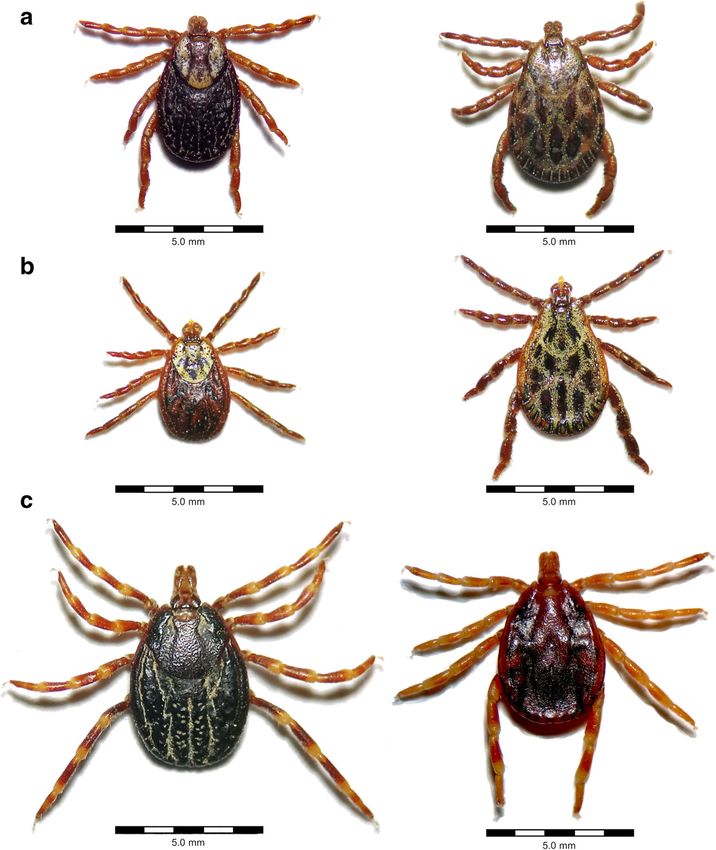
Fig. 2 Important vectors of
zoonotic tick-borne bacteria (left:
females, right: males). a
Dermacentor variabilis, con-
firmed vector of Ehrlichia canis,
Rickettsia rickettsii and
F. tularensis. b Rhipicephalus
sanguineus s.l., confirmed vector
of E. canis, Rickettsia conorii and
R. rickettsii. c Amblyomma
hebraeum, confirmed vector of
Ehrlichia ruminantium and
Rickettsia africae. Photographs
were taken under an OPTIKA
SLX-2 stereomicroscope
(OPTIKA S.r.l., Ponteranica,
Italy)
opportunities for collaboration between medical and veteri- pathogens grow slowly and require special media or cell cul-
nary scientists. tures. For example, the time to positive culture may span sev-
eral weeks for B. burgdorferi s.l. (Eldin et al. 2019) and up to
Direct detection methods 30 days for Rickettsia spp. (Portillo et al. 2017). Challenges
associated with culturing tick-borne pathogens are further il-
Traditionally, microscopy, culture of the pathogen or xenodi- lustrated by the example of Neoehrlichia mikurensis, which
agnosis was widely used for direct detection of tick-borne was only recently successfully cultured in human and tick cell
pathogens in patient samples, but nowadays, nucleic acid– lines, although the pathogen has been known since 2004
based methods are more commonly employed. Nevertheless, (Wass et al. 2019).
microscopic examination of stained blood smears is still the Nucleic acid amplification techniques are often more
method of first choice for diagnosis of acute Babesia infec- sensitive than the aforementioned methods and consider-
tions, in both human and veterinary medicine (Ord and Lobo ably faster than pathogen culture, improving diagnostic
2015; Solano-Gallego et al. 2016). Furthermore, blood smear efficiency (Korber et al. 2017). In routine diagnostic set-
analysis is helpful to demonstrate intracellular morulae during tings, real-time quantitative PCR (qPCR) is often used
anaplasmosis and ehrlichiosis (Schotthoefer et al. 2013). This due to increased sensitivity and speed as compared to
method is relatively fast and low-cost; however, sensitivity conventional PCR. Additionally, real-time qPCR allows
depends on the level of parasitaemia and pathogen species quantification by the gene copy numbers of the given
differentiation is not always possible (Ord and Lobo 2015). pathogen or cycle threshold (Ct) values and can therefore
Therefore, diagnosis should be corroborated by molecular also be useful for monitoring the course of infection (Che
techniques (Solano-Gallego et al. 2016). et al. 2019). However, it should be kept in mind that
Pathogen culture can be difficult and time consuming, may detection of DNA does not necessarily indicate that viable
require special biosafety conditions and is therefore often per- pathogens are present, and false-positive results may be
formed by specialised laboratories only. Many tick-borne obtained after successful treatment (Kuleš et al. 2017).Parasitol Res Fig. 3 Important vectors of zoonotic tick-borne viruses (left: females, right: males). a Dermacentor marginatus, con- firmed vector of Crimean-Congo haemorrhagic fever virus (CCHFV) and Omsk haemorrhagic fever virus (OHFV). b Dermacentor reticulatus, confirmed vector of OHFV and tick-borne encephali- tis virus. c Hyalomma rufipes, confirmed vector of CCHFV. Photographs were taken under an OPTIKA SLX-2 stereomicro- scope (OPTIKA S.r.l., Ponteranica, Italy) Adaptations of the real-time qPCR method include digital multiplex real-time qPCRs for simultaneous amplification of a PCR (dPCR), which allows detection and quantification of wide range of pathogens have been developed (Guido et al. rare target sequences by partitioning the sample into many 2016); however, not all of them detect tick-borne pathogens. parallel PCR reactions, thus improving test sensitivity. This Recently, multiplex PCR followed by electrospray ionisation technique has recently been successfully applied for mass spectrometry (PCR/ESI-MS) has been used to diagnose B. burgdorferi s.l. identification in patient blood, which was early B. burgdorferi s.s. (Eshoo et al. 2012), Ehrlichia spp. previously hindered by extremely low numbers of circulating and R. rickettsii (Eshoo et al. 20 10) as we ll as spirochaetes (Das et al. 2020). A. phagocytophilum (Lagler et al. 2017) infections. This tech- Aside from singleplex PCRs, multiplex assays may be used nique provides the advantage of identifying and genotyping as screening tests. For example, multiplex assays combining pathogens in a short time, but it was only adopted by a few real-time qPCR detection of A. phagocytophilum with hospitals in Europe and was discontinued by the manufacturer Ehrlichia spp. or B. burgdorferi s.l. are available (e.g. in 2017, probably due to economic reasons (Özenci et al. Courtney et al. 2004; Reller and Dumler 2018), while a 2017). broad-panel system for the simultaneous detection of nine In general, PCR requires expensive equipment, which may tick-borne pathogens is currently available for research use be a problem in less-developed countries or in field settings. only (Buchan et al. 2019). For patients suspected of sepsis, Loop-mediated isothermal amplification (LAMP) is a low-
Table 3      Tick-borne viral pathogens, their vectors and reservoir hosts

Pathogen                        Tick vector(s)1                       Geographical     Vertebrate reservoir (s)   Cell tropism in the vertebrate host Comment(s) References
                                                                      distribution

Alkhurma virus             Unknown (possibly                          Saudi Arabia     Unknown                    Unknown; probably mesangial                    Tambo and El-Dessouky (2018);
                             Hyalomma spp.                                                                          cells, mononuclear phagocytes                  Sawatsky et al. (2014)
                             or Ornithodoros spp.)
Colorado tick fever virus  Dermacentor andersoni                      North America Small mammals                 Haematopoietic cells                           Reviewed by Yukl and Wong (2016)
Crimean-Congo              Dermacentor marginatus,                    Southern       Lagomorphs, large wild       Mononuclear phagocytes,                        Reviewed by IZS “G. Caporale” (2009)
  haemorrhagic fever virus   Hyalomma                                   Europe,        and domestic                 endothelial cells, hepatocytes
                             impeltatum, Hyalomma                       Africa, Asia   mammals
                             marginatum,
                             Hyalomma truncatum, Hyalomma
                             rufipes, Rhipicephalus rossicus
Heartland virus            Amblyomma americanum                       North America Unknown                       Mononuclear phagocytes                         Reviewed by Brault et al. (2018)
Kyasanur Forest disease         Haemaphysalis spinigera               India            Small mammals            Possibly                                         Reviewed by Shah et al. (2018)
  virus                                                                                                           monocytes/macrophages and
                                                                                                                  dendritic cells
Louping ill virus               Ixodes ricinus                        British Isles,   Sheep, lagomorphs, birds Neurons, histiocytes                 Rare human Reviewed by Gilbert (2016); Sheahan
                                                                        Norway,                                                                        infections et al. (2002)
                                                                        Spain
Omsk haemorrhagic fever         Dermacentor reticulatus,              Russia           Small mammals              Haematopoietic and vascular                    Reviewed by Růžek et al. (2010)
  virus                           Dermacentor marginatus                                                            tissues
Powassan virus/deer tick        Dermacentor andersoni, Ixodes         North America, Small mammals                Neurons                                        Reviewed by Ebel (2010)
  virus                           scapularis, probably2 Ixodes          Russia
                                  cookei
Severe fever with               Haemaphysalis longicornis             East Asia        Unknown, possibly          Monocytes/macrophages,                         Reviewed by Mansfield et al. (2017);
  thrombocytopenia                                                                       domestic ruminants        dendritic cells, B cells                        Cheng et al. (2019); Suzuki et al.
  syndrome virus                                                                                                                                                   (2020)
Tick-borne encephalitis         Ixodes persulcatus, I. ricinus,       Eurasia          Small mammals              Dendritic cells, neurons, glial                Reviewed by Dobler et al. (2012); Fares
  virus                            Ixodes ovatus, D. reticulatus                                                    cells                                          et al. (2020); Ličková et al. (2020)

1
    Main tick vectors responsible for human infections; other tick vectors may be relevant in tick-reservoir cycles
2
    Vector competence not experimentally proven
                                                                                                                                                                                                           Parasitol Res
Parasitol Res
cost DNA amplification technique that works at a constant results due to cross-reactive IgM antibodies induced by other
temperature and thus does not require a thermocycler flaviviruses or in cases of atypical antibody responses, e.g.
(Becherer et al. 2020). LAMP assays to detect tick-borne path- when IgM antibodies are persistently elevated past the acute
ogens have mainly been developed not only for veterinary phase of infection (Vilibic-Cavlek et al. 2016). For Lyme
applications (e.g. Faggion et al. 2013; Singh et al. 2019; borreliosis, a recently developed IgG avidity Western blot
Wang et al. 2017) but also for detection of TBEV (Hayasaka has shown promising first results to identify disease stage
et al. 2013) and severe fever with thrombocytopenia syn- (Mavin et al. 2018).
drome virus (SFTSV) (Baek et al. 2018) in human patients The most frequently used serologic methods include the
in resource-limited settings. enzyme-linked immunosorbent assay (ELISA), immunofluo-
Mass spectrometry–based approaches, e.g. matrix-assisted rescence antibody test (IFAT) and immunoblotting. ELISA
laser desorption ionisation time-of-flight (MALDI-TOF), are tests can be performed with high sample throughput but may
routinely used to identify cultured pathogens in microbiolog- suffer from lower specificity as compared to other tests.
ical laboratories, based on comparison of protein signatures to Therefore, a two-tiered approach is often recommended,
existing databases. Although not yet routinely used for diag- confirming positive or borderline ELISA tests with more spe-
nosis of TBDs, applicability for identification and typing of cific techniques such as immunoblotting (e.g. in Lyme
cultured B. burgdorferi s.l. has recently been demonstrated borreliosis, Sanchez et al. 2016) or seroneutralisation tests
(Neumann-Cip et al. 2020). Mass spectrometry also offers (e.g. in TBE, Reusken et al. 2019).
new opportunities to identify biomarkers of specific diseases Modifications of the ELISA technique include magnetic
in patient samples, as shown, for example, for Babesia microti bead–based multianalyte assays, which are characterised by
infections in an experimental hamster model (Magni et al. high sensitivity even if antibody titres are low. Bead-based
2020). Similarly, MALDI-TOF analysis of canine serum sam- assays have been developed, for example, for the detection
ples may aid in the diagnosis of Babesia canis infections in of anti-B. burgdorferi s.l. antibodies in humans (Gerritzen
dogs (Adaszek et al. 2014). and Brandt 2012) as well as in horses and dogs (Wagner
et al. 2011a; Wagner et al. 2011b).
Indirect detection methods: detection of humoral For rickettsial diseases, the IFAT is considered the serolog-
immune response ical reference method (Portillo et al. 2017). IFATs are also
commonly employed to detect and quantify anti-Babesia
In some TBDs, direct pathogen detection is particularly diffi- (Sanchez et al. 2016; Solano-Gallego et al. 2016) as well as
cult. For example, B. burgdorferi s.l. spirochaetes are only anti-Ehrlichia antibodies (Dumler et al. 2007). However, the
present at transient and low levels in patient blood (Schutzer technique is relatively labour intensive as compared to ELISA
et al. 2018). Similarly, direct detection of TBEV is only pos- and can be somewhat subjective as it involves microscopic
sible in the early, viraemic phase of the disease (Girl et al. evaluation of antigen-coated glass slides.
2020). Therefore, serological tests are commonly employed In addition, rapid immunochromatographic tests are com-
in TBD diagnosis. However, it has to be kept in mind that mercially available for non-laboratory settings. These tests are
there is usually a time lag of several days to weeks between easy to use; however, they offer only a positive/negative re-
disease onset and development of antibody and, furthermore, sult, allowing no quantification of antibody titres.
that elevated antibody levels indicate pathogen exposure, but Furthermore, some commercially available rapid tests suffer
not necessarily current infection. Therefore, positive titres from low sensitivity, as shown e.g. for Lyme borreliosis (Liu
should always be interpreted in conjunction with the clinical et al. 2018; Smit et al. 2015).
presentation (Portillo et al. 2017; Sanchez et al. 2016). Acute Sensitivity and specificity of serologic tests greatly depend
infections may be detected by seroconversion or a rise in an- on the antigen(s) used. Use of purified or recombinant anti-
tibody titres. Therefore, testing of sequential samples taken gens as well as synthetic peptides rather than whole-cell ly-
several weeks apart is often recommended (e.g. Portillo et al. sates may improve specificity. For example, ELISA tests
2017; Solano-Gallego et al. 2016). IgM antibody titres are the based on a synthetic C6 peptide, a highly invariant region of
first to rise and may therefore be targeted during early phases the B. burgdorferi s.l. VlsE (variable major protein-like se-
of the infection. However, IgM antibody tests are particularly quence, expressed) protein, have superior specificity as op-
prone to produce false-positive results and should thus be posed to whole-cell antigen ELISAs (Waddell et al. 2016).
accompanied by other methods, e.g. direct pathogen detection However, cross-reactivity with sera from Borrelia
or documentation of IgG seroconversion (Landry 2016; miyamotoi–infected patients has recently been described
Seriburi et al. 2012). IgG avidity testing represents an addi- (Molloy et al. 2017). In dogs, for which B. burgdorferi s.s.
tional approach to determine the stage of an infection, as IgG and s.l. (Borrelia afzelii and Borrelia garinii) vaccines are
binding avidity increases as the infection progresses. For TBE, available, use of the C6 peptide in serological tests allows
IgG avidity testing may be useful to rule out false-positive discrimination between vaccinated and infected animalsParasitol Res
(Pantchev et al. 2015). In human TBDs, discrimination be- B. microti, Babesia venatorum and TBEV (Rizzoli et al.
tween infection-induced and vaccination-induced antibodies 2014). With an estimate of 60,000–100,000 total and 7500
is relevant for TBE. For this purpose, an ELISA based on hospitalised cases annually, Lyme borreliosis is regarded as
the non-structural protein 1 (NS1) of TBEV has recently been the most frequent human TBD in Germany (Lohr et al. 2015).
developed, which is exclusively indicative of natural infection However, since only certain manifestations of Lyme
and also allows significant discrimination from other flavivi- borreliosis are reportable in some, but not all, federal states,
rus infections (Girl et al. 2020). this number may be inaccurate (Lohr et al. 2015). In contrast,
Similar to direct tests, serological assays such as immuno- TBE is notifiable in all parts of Germany and annual case
blots and rapid immunochromatographic tests are also avail- numbers ranged between 195 and 584 in the period 2001–
able in multiplex formats. For example, a rapid test frequently 2019 (Robert Koch-Institut 2020). Less is known regarding
employed in veterinary medicine allows the simultaneous de- other TBDs in Germany, but human cases of neoehrlichiosis
tection of canine antibodies against B. burgdorferi s.l., (von Loewenich et al. 2010) and babesiosis due to
Ehrlichia spp. and Anaplasma spp., in addition to canine B. venatorum (Häselbarth et al. 2007) and B. microti
heartworm antigen (Chandrashekar et al. 2010). (Hildebrandt et al. 2007) have been reported during the past
decades. With regard to Rickettsia spp., R. helvetica is the
Indirect detection methods: detection of cellular predominant species, but Rickettsia monacensis, Rickettsia
immune response slovaca and Rickettsia raoultii also occur in Germany
(Dobler and Pfeffer 2012). In addition, travellers returning
Aside from antibody production, many tick-borne pathogens from other countries may be infected with non-endemic tick-
induce specific T cell responses. T cell–based assays might be borne pathogens, e.g. Rickettsia africae (Antal et al. 2013),
helpful to bridge the gap between infection and onset of anti- necessitating appropriate diagnostic possibilities.
body production or might be employed as confirmatory tests to Regarding veterinary medicine, no estimates of annual
rule out false-positive serology results (Jin et al. 2013). The TBD incidence exist. However, granulocytic anaplasmosis is
enzyme-linked immunospot assay (ELISPOT) is a sensitive regarded as the most important TBD in dogs, whereas Lyme
method to measure the cytokine response of T cells upon an- borreliosis may be overdiagnosed (Gerber et al. 2009).
tigen stimulation (Kalyuzhny 2005). ELISPOT assays have Furthermore, A. phagocytophilum is relevant as the causative
been developed for a variety of TBDs; however, their utility agent of granulocytic anaplasmosis in horses (Silaghi et al.
is controversially discussed, especially regarding Lyme 2011) and tick-borne fever in ruminants (Nieder et al. 2012).
borreliosis. ELISPOT assays developed for Lyme borreliosis, In ruminants, redwater fever due to B. divergens occurs spo-
which exclusively measure interferon-γ release, show a wide radically and may lead to significant mortality in naïve cattle
range of sensitivity and specificity and poor reproducibility herds (Springer et al. 2020). In addition, sporadic clinical
and are therefore currently not recommended for routine diag- cases of TBE have been described in German dogs (Reiner
nostic use (Raffetin et al. 2020). Similarly, lymphocyte trans- and Fischer 1998; Saenger et al. 2013).
formation tests (LTTs) assess the proliferative response of T Commercially available diagnostic kits, taking Germany as
cells upon stimulation with specific antigens. LTTs are offered an example, were identified by Google Search using combi-
by some laboratories for diagnosis of active Lyme borreliosis nations of the following keywords: Anaplasma, Babesia,
in humans; however, current guidelines do not recommend Borrelia, Rickettsia, Ehrlichia, TBE, FSME, IgG, IgM,
these tests due to low specificity (Dessau et al. 2014). PCR, ELISA, ELISPOT, IFAT, serology and kit.
Cytokines and chemokines as evidence of a cellular im- Furthermore, a list of available diagnostic tests for
mune response may also be measured directly in patient sam- B. burgdorferi s.l. and TBEV was obtained from the
ples. For example, the chemokine CXCL13 in cerebrospinal German National Reference Center for Borrelia and the
fluid constitutes a sensitive and specific marker of acute Lyme German National Consiliary Laboratory for TBEV, respec-
neuroborreliosis in humans (Raffetin et al. 2020). tively. In addition, the German Diagnostics Industry
Association contributed a list of relevant manufacturers,
whose websites were searched for relevant test kits.
Relevant zoonotic TBDs and commercial In Table 4, the relative quantities of commercially available
availability of diagnostic test kits diagnostic test kits for human vs. veterinary use for each path-
by the example of Germany ogen are shown. Only tests designed for patient samples were
included, i.e. tests for pathogen detection in ticks were not
In Germany, as in other central European countries, I. ricinus considered, since a positive result in the detached tick is not
is the most relevant vector of zoonotic tick-borne pathogens, a reliable indicator of human or animal infection. In-house
including B. burgdorferi s.l., B. miyamotoi, tests and research-use only tests were also not considered.
A. phagocytophilum, Rickettsia helvetica, B. divergens, No absolute numbers are shown, because we cannot guaranteeParasitol Res
Table 4 Relative quantity of commercially available diagnostic tests for zoonotic tick-borne pathogens in Germany
Pathogen Nucleic acid detection Antibody detection Other tests
(e.g. ELISPOT)
For For For vet. use For human use For For
veterinary human vet. human
(vet.) use use use use
Babesia divergens − + + (IgG: +, IgM: −, − − −
IgG/IgM: −)
Babesia microti − + + (IgG: +, IgM: −, + (IgG: +, IgM: −, IgG/IgM: −) − +
IgG/IgM: −)
Babesia venatorum − + − − − −
Bartonella henselae1 − + + (IgG: +, IgM: −, + (IgG: +, IgM: +, IgG/IgM: −) − +
IgG/IgM: −)
Borrelia burgdorferi s.l. + ++ ++ (IgG: ++, IgM: +, IgG/IgM: +) +++ (IgG: +++, IgM: +++, IgG/IgM: ++) + +
Borrelia miyamotoi − − − − − +
Coxiella burnetii ++ − ++ (IgG: ++, IgM: +, IgG/IgM: −) +++ (IgG: ++, IgM: ++, IgG/IgM: −) − −
Francisella tularensis − + + (IgG: +, IgM: −, ++ (IgG: ++, IgM: +, IgG/IgM: −) − −
IgG/IgM: −)
Anaplasma + + ++ (IgG: ++, IgM: −, IgG/IgM: −) ++ (IgG: ++, IgM: +, IgG/IgM: −) −
phagocytophilum
Ehrlichia spp. + − ++ (IgG: ++, IgM: −, IgG/IgM: −) + (IgG: +, IgM: +, IgG/IgM: −) − +
Neoehrlichia mikurensis − − − − − −
Rickettsia spp. − ++ ++ (IgG: ++, IgM: −, IgG/IgM: −) ++ (IgG: ++, IgM: ++, − −
IgG/IgM: −)
Tick-borne + + + (IgG: +, IgM: −, +++ (IgG: ++, IgM: ++, − −
encephalitis virus IgG/IgM: −) IgG/IgM: +)
+++, > 20 kits on the market; ++, 6–20 kits on the market; +, ≤ 5 kits on the market; −, no marketed kits found
1
Vector competence of ticks for B. henselae not proven
that the search was exhaustive and, furthermore, the market is available veterinary serology kits for Ehrlichia spp. even
subject to frequent changes. exceeded the amount available for use in human medicine, but
Results indicate that a multitude of serologic kits and, to a no direct detection kits for Ehrlichia spp. were identified for
lesser extent, nucleic acid detection kits are available for di- veterinary use. Ticks transmitting zoonotic Ehrlichia spp.
agnosis of Lyme borreliosis and TBE in humans in Germany (Rhipicephalus sanguineus s.l., A. americanum) are not endemic
(Table 4). A rather large number of kits was also retrieved for in Germany; thus, ehrlichioses are only relevant as imported
Lyme borreliosis in animals, but only few for TBE, although diseases. E. canis is a major threat to canine health worldwide
domestic animals have proven useful as sentinels of human (Rar and Golovljova 2011), including in Mediterranean Europe
disease risk (Imhoff et al. 2015). In addition, most veterinary from where many dogs are imported to Germany and other
serology kits for B. burgdorferi s.l. detect IgG antibodies only, Central or Northern European countries. In contrast, human ehr-
whereas an equal amount of IgG and IgM tests exists for lichiosis cases are rather rare, occurring mainly in North America
humans. This can be explained by the fact that animals usually (Rar and Golovljova 2011), and are thus more rarely imported to
do not develop acute disease after B. burgdorferi s.l. exposure, Germany than canine cases. Consequently, the available veteri-
and IgM testing is thus not recommended (Littman et al. nary kits were mostly designed for E. canis antibody detection.
2018). However, a positive IgG titre is not an indicator of In contrast, only few kits for the diagnosis of rickettsioses
active infection and it can be extremely difficult to determine in animals were identified, probably because it is unknown
whether clinical disease in animals is actually due to Borrelia whether Rickettsia spp. cause disease in animals, with the
infection (Divers 2013; Littman et al. 2018). To reduce un- exception of R. conorii and R. rickettsii in dogs (Keenan
necessary antibiotic use, reliable tests indicative of active in- et al. 1977; Solano-Gallego et al. 2006). Neither of these spe-
fection would be extremely helpful in both disciplines. As cies is endemic in Germany (Dobler and Pfeffer 2012).
highlighted above, IgG avidity testing or improved PCR pro- Regarding humans, several serologic as well as direct detec-
cedures, such as digital PCR, could be promising approaches. tion kits for tick-borne Rickettsia spp. were identified, mainly
Regarding A. phagocytophilum, a similar amount of serologic designed for R. rickettsii and R. conorii detection.
as well as nucleic acid detection kits was identified for the human Particularly few diagnostic kits were identified regarding in-
medical as well as the veterinary market, probably because fections with zoonotic Babesia spp., both in the human medical
A. phagocytophilum plays an important role in veterinary medi- and in the veterinary sector. This may be due to the fact that
cine, affecting several species as described above. The number of Babesia infections are often diagnosed by blood smears and/orParasitol Res
in-house PCR tests in acute cases. However, blood smears have disciplines. Recently emerged tick-borne pathogens, such as
a limited sensitivity when parasitaemia is low or limited speci- N. mikurensis and B. miyamotoi, open up further opportunities
ficity when parasite morphology has been altered due to refrig- for collaboration, since no standardised tests for these patho-
eration prior to blood smear preparation (Cursino-Santos et al. gens are yet commercially available. Test development for
2014). In addition, many human babesiosis cases in immuno- these pathogens could save substantial time and effort for
competent individuals might be overlooked when symptoms are the benefit of both human and animal health.
mild, which represents a problem regarding blood transfusions,
for example (Hildebrandt et al. 2008; Ord and Lobo 2015). In Acknowledgements The authors are grateful to Volker Fingerle (German
National Reference Center for Borrelia, Oberschleissheim, Germany) and
the veterinary field, a recent outbreak of bovine babesiosis
Gerhard Dobler (German National Consiliary Laboratory for TBEV,
(B. divergens) in Germany has shown that mortality rates and Munich, Germany) for providing information on available B. burgdorferi
the subsequent economic impact may be high if diagnosis is s.l. and TBEV diagnostic kits, respectively. Furthermore, the authors thank
delayed (Springer et al. 2020). Therefore, sensitive, easy-to- Carolin Schächterle (German Diagnostics Industry Association, Berlin,
Germany) for providing information on relevant test manufacturers. Open
use and rapid diagnostic tools for zoonotic Babesia spp. are
Access funding enabled and organized by Projekt DEAL.
needed. Recently, an immunochromatographic test based on a
recombinant B. microti surface antigen showed promising re- Authors’ contributions CS designed the study. AS, AG and JP collected
sults in experimentally infected mice (Cai et al. 2018). the data on available diagnostic tests. AS drafted the manuscript, and CS,
Regarding B. miyamotoi and N. mikurensis, which have only AG and JP reviewed the manuscript. All authors read and approved the
final manuscript.
recently been identified as human and, possibly, veterinary path-
ogens (Diniz et al. 2011; Platonov et al. 2011; Welinder-Olsson
Funding This study was supported by a grant of the European Union
et al. 2010), no commercially available kits were identified at all, through the European Regional Development Fund and the Interreg
except for one ELISPOT kit designed for B. miyamotoi. In gen- North Sea Region Programme 2014–2020 as part of the NorthTick pro-
eral, only few ELISPOT assays are currently available in ject (reference number J-No.: 38-2-7-19).
Germany, reflecting the fact that their utility is controversially
discussed. Identified tests included EPISPOTS for detecting cel- Compliance with ethical standards
lular immunity against B. burgdorferi s.l. in humans, horses and
dogs, as well as against B. miyamotoi, B. microti, Ehrlichia spp. Conflict of interest The authors declare that they have no conflict of
interest.
and Bartonella henselae in humans.
For Bartonella henselae, C. burnetii and F. tularensis, tick- Open Access This article is licensed under a Creative Commons
borne transmission plays a minor role. Several diagnostic kits Attribution 4.0 International License, which permits use, sharing, adap-
tation, distribution and reproduction in any medium or format, as long as
were identified for C. burnetii for both disciplines, as this path-
you give appropriate credit to the original author(s) and the source, pro-
ogen is economically important as a cause of abortions in rumi- vide a link to the Creative Commons licence, and indicate if changes were
nants as well as from a public health perspective (Duron et al. made. The images or other third party material in this article are included
2015). In contrast, identified diagnostic kits for F. tularensis were in the article's Creative Commons licence, unless indicated otherwise in a
credit line to the material. If material is not included in the article's
mainly for human use, as symptomatic infections in domestic
Creative Commons licence and your intended use is not permitted by
animals are limited to cats and rabbits (Telford III and Goethert statutory regulation or exceeds the permitted use, you will need to obtain
2020). permission directly from the copyright holder. To view a copy of this
licence, visit http://creativecommons.org/licenses/by/4.0/.
Conclusions
Human and animal health are closely linked by ticks acting as References
vectors for zoonotic pathogens, making tick-borne diseases
excellent examples of the One Health concept. Animals are Adaszek Ł, Banach T, Bartnicki M, Winiarczyk D, Łyp P, Winiarczyk S
either clinically affected by the same tick-borne pathogens as (2014) Application the mass spectrometry MALDI-TOF technique
humans and/or play a role in tick cycle maintenance and as for detection of Babesia canis canis infection in dogs. Parasitol Res
113:4293–4295. https://doi.org/10.1007/s00436-014-4124-1
reservoirs or sentinel pathogen hosts. Using the German mar-
Antal AS, Flaig MJ, Schneck C, Thoma B, Herzinger T (2013) Souvenir
ket as an example, several gaps in commercial availability of from South Africa. Infection 41:597–598. https://doi.org/10.1007/
diagnostic tests for zoonotic tick-borne pathogens were iden- s15010-013-0425-z
tified. Regarding B. burgdorferi s.l., sensitive tests indicative Arraga-Alvarado CM, Qurollo BA, Parra OC, Berrueta MA, Hegarty BC,
of active infection would be useful to limit unnecessary or Breitschwerdt EB (2014) Molecular evidence of Anaplasma platys
infection in two women from Venezuela. Am J Trop Med Hyg 91:
overuse of antibiotics in human as well as veterinary medi- 1161–1165. https://doi.org/10.4269/ajtmh.14-0372
cine. Furthermore, there is a need for rapid and sensitive di- Azagi T, Hoornstra D, Kremer K, Hovius JWR, Sprong H (2020)
agnostic tools for zoonotic Babesia spp. infections in both Evaluation of disease causality of rare Ixodes ricinus-borneParasitol Res
infections in Europe. Pathogens 9:150. https://doi.org/10.3390/ digital PCR. PLoS One 15:e0235372. https://doi.org/10.1101/2020.
pathogens9020150 06.16.154336
Baek YH, Cheon HS, Park SJ, Lloren KKS, Ahn SJ, Jeong JH, Choi WS, Dessau R et al (2014) The lymphocyte transformation test for the diag-
Yu MA, Kwon HI, Kwon JJ, Kim EH, Kim YI, Antigua KJC, Kim nosis of Lyme borreliosis has currently not been shown to be clin-
SY, Jeong HW, Choi YK, Song MS (2018) Simple, rapid and sen- ically useful. Clin Microbiol Infect 20:O786–O787. https://doi.org/
sitive portable molecular diagnosis of SFTS virus using reverse 10.1111/1469-0691.12583
transcriptional loop-mediated isothermal amplification (RT- Diniz PPVP, Schulz BS, Hartmann K, Breitschwerdt EB (2011)
LAMP). J Microbiol Biotechnol 28:1928–1936. https://doi.org/10. “Candidatus Neoehrlichia mikurensis” infection in a dog from
4014/jmb.1806.06016 Germany. J Clin Microbiol 49:2059–2062. https://doi.org/10.1128/
Barbieri AM, Venzal JM, Marcili A, Almeida AP, González EM, JCM.02327-10
Labruna MB (2013) Borrelia burgdorferi sensu lato infecting ticks Divers TJ (2013) Equine Lyme disease. J Equine Vet Sci 33:488–492.
of the Ixodes ricinus complex in Uruguay: first report for the https://doi.org/10.1016/j.jevs.2013.03.187
Southern Hemisphere. Vector Borne Zoonotic Dis 13:147–153. Dobler G, Pfeffer M (2012) Spotted fever rickettsiae and rickettsioses in
https://doi.org/10.1089/vbz.2012.1102 Germany. In: Arthropods as vectors of emerging diseases.
Becherer L, Borst N, Bakheit M, Frischmann S, Zengerle R, von Stetten F Parasitology research monographs 3. Springer, Berlin Heidelberg,
(2020) Loop-mediated isothermal amplification (LAMP) – review pp 361–376. https://doi.org/10.1007/978-3-642-28842-5_15
and classification of methods for sequence-specific detection. Anal Dobler G, Gniel D, Petermann R, Pfeffer M (2012) Epidemiology and
Methods 12:717–746. https://doi.org/10.1039/C9AY02246E distribution of tick-borne encephalitis. Wien Med Wochenschr 162:
Brault AC, Savage HM, Duggal NK, Eisen RJ, Staples JE (2018) 230–238. https://doi.org/10.1007/s10354-012-0100-5
Heartland virus epidemiology, vector association, and disease po- Dugat T, Lagree AC, Maillard R, Boulouis HJ, Haddad N (2015)
tential. Viruses 10:498. https://doi.org/10.3390/v10090498 Opening the black box of Anaplasma phagocytophilum diversity:
Buchan BW, Jobe DA, Mashock M, Gerstbrein D, Faron ML, Ledeboer current situation and future perspectives. Front Cell Infect Microbiol
NA, Callister SM (2019) Evaluation of a novel multiplex high- 5:61. https://doi.org/10.3389/fcimb.2015.00061
definition PCR assay for detection of tick-borne pathogens in Dumler JS, Madigan JE, Pusterla N, Bakken JS (2007) Ehrlichioses in
whole-blood specimens. J Clin Microbiol 57:e00513–e00519. humans: epidemiology, clinical presentation, diagnosis, and treat-
https://doi.org/10.1128/jcm.00513-19 ment. Clin Infect Dis 45:S45–S51. https://doi.org/10.1086/518146
Cai Y et al (2018) Molecular characterization of Babesia microti Duron O, Sidi-Boumedine K, Rousset E, Moutailler S, Jourdain E (2015)
seroreactive antigen 5-1-1 and development of rapid detection The importance of ticks in Q fever transmission: what has (and has
methods for anti-B. microti antibodies in serum. Acta Trop 185: not) been demonstrated? Trends Parasitol 31:536–552. https://doi.
371–379. https://doi.org/10.1016/j.actatropica.2018.03.020 org/10.1016/j.pt.2015.06.014
Ebel GD (2010) Update on Powassan virus: emergence of a North
Chandrashekar R, Mainville CA, Beall MJ, O'Connor T, Eberts MD,
American tick-borne flavivirus. Annu Rev Entomol 55:95–110.
Alleman AR, Gaunt SD, Breitschwerdt EB (2010) Performance of
https://doi.org/10.1146/annurev-ento-112408-085446
a commercially available in-clinic ELISA for the detection of anti-
Eldin C, Jaulhac B, Mediannikov O, Arzouni J-P, Raoult D (2019)
bodies against Anaplasma phagocytophilum, Ehrlichia canis, and
Values of diagnostic tests for the various species of spirochetes.
Borrelia burgdorferi and Dirofilaria immitis antigen in dogs. Am J
Med Mal Infect 49:102–111. https://doi.org/10.1016/j.medmal.
Vet Res 71:1443–1450. https://doi.org/10.2460/ajvr.71.12.1443
2019.01.009
Che L-h et al (2019) Monitoring the course of Brucella infection with
Eshoo MW, Crowder CD, Li H, Matthews HE, Meng S, Sefers SE,
qPCR-based detection. Int J Infect Dis 89:66–71. https://doi.org/10.
Sampath R, Stratton CW, Blyn LB, Ecker DJ, Tang YW (2010)
1016/j.ijid.2019.09.013
Detection and identification of Ehrlichia species in blood by use
Cheng J, Zhang L, Hu B, Wang Q, Wu R, Zhan F, Rong S, Zhan J (2019) of PCR and electrospray ionization mass spectrometry. J Clin
Prevalence and molecular phylogenetic analysis of severe fever with Microbiol 48:472–478. https://doi.org/10.1128/jcm.01669-09
thrombocytopenia syndrome virus in domestic animals and rodents Eshoo MW, Crowder CC, Rebman AW, Rounds MA, Matthews HE,
in Hubei Province, China. Virol Sin 34:596–600. https://doi.org/10. Picuri JM, Soloski MJ, Ecker DJ, Schutzer SE, Aucott JN (2012)
1007/s12250-019-00119-y Direct molecular detection and genotyping of Borrelia burgdorferi
Cheslock MA, Embers ME (2019) Human bartonellosis: an underappre- from whole blood of patients with early Lyme disease. PLoS One 7:
ciated public health problem? Trop Med Infect Dis 4:69. https://doi. e36825. https://doi.org/10.1371/journal.pone.0036825
org/10.3390/tropicalmed4020069 Faggion S, Salvador AR, Jacobino KL, Bortolotto LFB, Lopes MB, Silva
Courtney JW, Kostelnik LM, Zeidner NS, Massung RF (2004) Multiplex M, Santos EV, Fachin AL, França SC, Marins M (2013) Loop-
real-time PCR for detection of Anaplasma phagocytophilum and mediated isothermal amplification assay for the detection of
Borrelia burgdorferi. J Clin Microbiol 42:3164–3168. https://doi. Ehrlichia canis DNA in blood samples from dogs. Arch Med Vet
org/10.1128/jcm.42.7.3164-3168.2004 45:197–201. https://doi.org/10.4067/S0301-732X2013000200012
Cursino-Santos JR, Alhassan A, Singh M, Lobo CA (2014) Babesia: Fares M, Cochet-Bernoin M, Gonzalez G, Montero-Menei CN, Blanchet
impact of cold storage on the survival and the viability of parasites O, Benchoua A, Boissart C, Lecollinet S, Richardson J, Haddad N,
in blood bags. Transfusion 54:585–591. https://doi.org/10.1111/trf. Coulpier M (2020) Pathological modeling of TBEV infection re-
12357 veals differential innate immune responses in human neurons and
Cutler S, Vayssier-Taussat M, Estrada-Peña A, Potkonjak A, Mihalca astrocytes that correlate with their susceptibility to infection. J
AD, Zeller H (2019) A new Borrelia on the block: Borrelia Neuroinflammation 17:76. https://doi.org/10.1186/s12974-020-
miyamotoi - a human health risk? Eurosurveillance 24:1800170. 01756-x
https://doi.org/10.2807/1560-7917.ES.2019.24.18.1800170 Gerber B, Eichenberger S, Haug K, Wittenbrink MM (2009) The dilem-
Dantas-Torres F, Chomel BB, Otranto D (2012) Ticks and tick-borne ma with Lyme borreliosis in the dog with particular consideration of
diseases: a One Health perspective. Trends Parasitol 28:437–446. “Lyme nephritis”. Schweiz Arch Tierheilkd 151:479–483. https://
https://doi.org/10.1016/j.pt.2012.07.003 doi.org/10.1024/0036-7281.151.10.479
Das S, Hammond-McKibben D, Guralski D, Lobo S, Fiedler PN (2020) Gerritzen A, Brandt S (2012) Serodiagnosis of Lyme borreliosis with
Development of a sensitive molecular diagnostic assay for detecting bead based immunoassays using multiplex technology. Methods
Borrelia burgdorferi DNA from blood of Lyme disease patients by 56:477–483. https://doi.org/10.1016/j.ymeth.2012.02.007Parasitol Res
Gilbert L (2016) Louping ill virus in the UK: a review of the hosts, musculus). Vector Borne Zoonotic Dis 16:145–150. https://doi.org/
transmission and ecological consequences of control. Exp Appl 10.1089/vbz.2015.1878
Acarol 68:363–374. https://doi.org/10.1007/s10493-015-9952-x Keenan KP, Buhles WC, Huxsoll DL, Williams RG, Hildebrandt PK
Girl P, Bestehorn-Willmann M, Zange S, Borde JP, Dobler G, von Buttlar (1977) Studies on the pathogenesis of Rickettsia rickettsii in the
H (2020) Tick-borne encephalitis virus nonstructural protein 1 IgG dog: clinical and clinicopathologic changes of experimental infec-
enzyme-linked immunosorbent assay for differentiating infection tion. Am J Vet Res 38:851–856 PMID: 879582
versus vaccination antibody responses. J Clin Microbiol 58: Keirans J, Needham G, Oliver J Jr (1999) The Ixodes ricinus complex
e01783–e01719. https://doi.org/10.1128/jcm.01783-19 worldwide: diagnosis of the species in the complex, hosts and dis-
Gray J, Zintl A, Hildebrandt A, Hunfeld KP, Weiss L (2010) Zoonotic tribution. Acarology IX Proceedings 2:341–347
babesiosis: overview of the disease and novel aspects of pathogen Kelly PJ, Mason PR (1991) Transmission of a spotted fever group rick-
identity. Ticks Tick Borne Dis 1:3–10. https://doi.org/10.1016/j. ettsia by Amblyomma hebraeum (Acari: Ixodidae). J Med Entomol
ttbdis.2009.11.003 28:598–600. https://doi.org/10.1093/jmedent/28.5.598
Gray A, Capewell P, Loney C, Katzer F, Shiels BR, Weir W (2019a) Koch-Institut R (2020) FSME: Risikogebiete in Deutschland (Stand:
Sheep as host species for zoonotic Babesia venatorum, United Januar 2020). Epidemiol Bull 2020:3–19
Kingdom. Emerg Infect Dis 25:2257–2260. https://doi.org/10. Kohn B, Galke D, Beelitz P, Pfister K (2008) Clinical features of canine
3201/eid2512.190459 granulocytic anaplasmosis in 18 naturally infected dogs. J Vet Intern
Gray JS, Estrada-Peña A, Zintl A (2019b) Vectors of babesiosis. Annu Med 22:1289–1295. https://doi.org/10.1111/j.1939-1676.2008.0180.x
Rev Entomol 64:149–165. https://doi.org/10.1146/annurev-ento- Korber F, Zeller I, Grünstäudl M, Willinger B, Apfalter P, Hirschl AM,
011118-111932 Makristathis A (2017) SeptiFast versus blood culture in clinical
Guido M, Tumolo MR, De Donno A, Verri T, Serio F, Bagordo F, Zizza routine - a report on 3 years experience. Wien Klin Wochenschr
A (2016) In vitro diagnosis of sepsis: a review. Pathol Lab Med Int 129:427–434. https://doi.org/10.1007/s00508-017-1181-3
8:1–14. https://doi.org/10.2147/PLMI.S49800 Körner S, Makert GR, Mertens-Scholz K, Henning K, Pfeffer M, Starke A,
Häselbarth K, Tenter AM, Brade V, Krieger G, Hunfeld K-P (2007) First Nijhof AM, Ulbert S (2020) Uptake and fecal excretion of Coxiella
case of human babesiosis in Germany – clinical presentation and burnetii by Ixodes ricinus and Dermacentor marginatus ticks. Parasit
molecular characterisation of the pathogen. Int J Med Microbiol Vectors 13:75. https://doi.org/10.1186/s13071-020-3956-z
297:197–204. https://doi.org/10.1016/j.ijmm.2007.01.002 Kuleš J, Potocnakova L, Bhide K, Tomassone L, Fuehrer HP, Horvatić A,
Galan A, Guillemin N, Nižić P, Mrljak V, Bhide M (2017) The
Hayasaka D, Aoki K, Morita K (2013) Development of simple and rapid
challenges and advances in diagnosis of vector-borne diseases:
assay to detect viral RNA of tick-borne encephalitis virus by reverse
where do we stand? Vector Borne Zoonotic Dis 17:285–296.
transcription-loop-mediated isothermal amplification. Virol J 10:68.
https://doi.org/10.1089/vbz.2016.2074
https://doi.org/10.1186/1743-422X-10-68
Lagler H, Harrison N, Kussmann M, Obermüller M, Burgmann H,
Hildebrandt A, Hunfeld KP, Baier M, Krumbholz A, Sachse S, Lorenzen
Makristathis A, Ramharter M (2017) Direct detection of
T, Kiehntopf M, Fricke HJ, Straube E (2007) First confirmed au-
Anaplasma phagocytophilum by polymerase chain reaction follow-
tochthonous case of human Babesia microti infection in Europe. Eur
ed by electrospray ionization mass spectrometry from human blood.
J Clin Microbiol Infect Dis 26:595–601. https://doi.org/10.1007/
Int J Infect Dis 60:61–63. https://doi.org/10.1016/j.ijid.2017.05.006
s10096-007-0333-1
Landry ML (2016) Immunoglobulin M for acute infection: true or false?
Hildebrandt A, Tenter AM, Straube E, Hunfeld K-P (2008) Human babesi-
Clin Vaccine Immunol 23:540–545. https://doi.org/10.1128/CVI.
osis in Germany: just overlooked or truly new? Int J Med Microbiol
00211-16
298:336–346. https://doi.org/10.1016/j.ijmm.2007.11.001
Ličková M, Fumačová Havlíková S, Sláviková M, Slovák M, Drexler JF,
Imhoff M, Hagedorn P, Schulze Y, Hellenbrand W, Pfeffer M, Niedrig M Klempa B (2020) Dermacentor reticulatus is a vector of tick-borne
(2015) Review: Sentinels of tick-borne encephalitis risk. Ticks Tick- encephalitis virus. Ticks Tick Borne Dis 11:101414. https://doi.org/
borne Dis 6:592–600. https://doi.org/10.1016/j.ttbdis.2015.05.001 10.1016/j.ttbdis.2020.101414
IZS “G. Caporale” (2009) Scientific review on crimean-congo hemor- Littman MP, Gerber B, Goldstein RE, Labato MA, Lappin MR, Moore
rhagic fever. EFSA Suppor Publicat 6:19E. https://doi.org/10. GE (2018) ACVIM consensus update on Lyme borreliosis in dogs
2903/sp.efsa.2009.EN-19 and cats. J Vet Intern Med 32:887–903. https://doi.org/10.1111/
Jaarsma RI, Sprong H, Takumi K, Kazimirova M, Silaghi C, Mysterud A, jvim.15085
Rudolf I, Beck R, Földvári G, Tomassone L, Groenevelt M, Everts Liu J, Drexel J, Andrews B, Eberts M, Breitschwerdt E, Chandrashekar R
RR, Rijks JM, Ecke F, Hörnfeldt B, Modrý D, Majerová K, Votýpka (2018) Comparative evaluation of 2 in-clinic assays for vector-borne
J, Estrada-Peña A (2019) Anaplasma phagocytophilum evolves in disease testing in dogs. Top Companion Anim Med 33:114–118.
geographical and biotic niches of vertebrates and ticks. Parasit https://doi.org/10.1053/j.tcam.2018.09.003
Vectors 12:328. https://doi.org/10.1186/s13071-019-3583-8 Lohr B, Müller I, Mai M, Norris DE, Schöffski O, Hunfeld KP (2015)
Jahfari S, Sprong H (2016) Emerging tick-borne pathogens: ticking on Epidemiology and cost of hospital care for Lyme borreliosis in
Pandora’s box. In: MAH B, Van Wieren SE, Takken W, Sprong H Germany: lessons from a health care utilization database analysis.
(eds) Ecology and prevention of Lyme borreliosis. Ecology and Ticks Tick Borne Dis 6:56–62. https://doi.org/10.1016/j.ttbdis.
control of vector-borne diseases, vol 4. Wageningen Academic 2014.09.004
Publishers, Wageningen, pp 127–147. https://doi.org/10.3920/978- Magni R, Luchini A, Liotta L, Molestina RE (2020) Proteomic analysis
90-8686-838-4_9 reveals pathogen-derived biomarkers of acute babesiosis in erythro-
Jin C, Roen DR, Lehmann PV, Kellermann GH (2013) An enhanced cytes, plasma, and urine of infected hamsters. Parasitol Res 119:
ELISPOT assay for sensitive detection of antigen-specific T cell 2227–2235. https://doi.org/10.1007/s00436-020-06712-5
responses to Borrelia burgdorferi. Cells 2:607–620. https://doi. Mansfield KL, Jizhou L, Phipps LP, Johnson N (2017) Emerging tick-
org/10.3390/cells2030607 borne viruses in the twenty-first century. Front Cell Infect Microbiol
Kalyuzhny E (2005) Handbook of ELISPOT. Methods and protocols. 7:298. https://doi.org/10.3389/fcimb.2017.00298
Methods in molecular biology, vol 302. Humana Press Inc., Martinot M, Zadeh MM, Hansmann Y, Grawey I, Christmann D,
Totowa, pp 1–323 Aguillon S, Jouglin M, Chauvin A, de Briel D (2011) Babesiosis
Karpathy SE, Allerdice MEJ, Sheth M, Dasch GA, Levin ML (2016) Co- in immunocompetent patients, Europe. Emerg Infect Dis 17:114–
feeding transmission of the Ehrlichia muris–like agent to mice (Mus 116. https://doi.org/10.3201/eid1701.100737You can also read