Guidelines for the Evaluation of Valvular Regurgitation After Percutaneous Valve Repair or Replacement
←
→
Page content transcription
If your browser does not render page correctly, please read the page content below
GUIDELINES AND STANDARDS

                Guidelines for the Evaluation of Valvular Regurgitation
                  After Percutaneous Valve Repair or Replacement
  A Report from the American Society of Echocardiography Developed in
     Collaboration with the Society for Cardiovascular Angiography and
   Interventions, Japanese Society of Echocardiography, and Society for
                    Cardiovascular Magnetic Resonance
                   William A. Zoghbi, MD, FASE, (Chair), Federico M. Asch, MD, FASE, Charles Bruce, MBChB, FASE,
                       Linda D. Gillam, MD, MPH, FASE, Paul A. Grayburn, MD, FASE, Rebecca T. Hahn, MD, FASE,
                          Ignacio Inglessis, MD, Ashequl M. Islam, MD, MPH, FSCAI, Stamatios Lerakis, MD, FASE,
                        Stephen H. Little, MD, FASE, Robert J. Siegel, MD, FASE, Nikolaos Skubas, MD, DSc, FASE,
               Timothy C. Slesnick, MD, FASE, William J. Stewart, MD, FASE, Paaladinesh Thavendiranathan, MD, MSc, FASE,
               Neil J. Weissman, MD, FASE, Satoshi Yasukochi, MD, JCC, SJSUM, and Karen G. Zimmerman, BS, ACS, RDCS,
               RVT, FASE, Houston and Dallas, Texas; Washington, District of Columbia; Rochester, Minnesota; Morristown, New
                 Jersey; New York, New York; Boston and Springfield, Massachusetts; Los Angeles, California; Cleveland, Ohio;
                            Atlanta, Georgia; Toronto, Ontario, Canada; Nagano, Japan; Morgantown, West Virginia
               Keywords: Doppler echocardiography, Valve disease, Transaortic valve replacement, Magnetic resonance
               imaging, Aortic regurgitation, Mitral regurgitation

                    In addition to the collaborating societies listed in the title, this document is endorsed by the following American
                      Society of Echocardiography International Alliance Partners: Argentine Society of Cardiology, Argentinian
                   Federation of Cardiology, Asian-Pacific Association of Echocardiography, Australasian Sonographers Association,
                  Cardiovascular Imaging Department of the Brazilian Society of Cardiology, Canadian Society of Echocardiography,
                     Chinese Society of Echocardiography, Echocardiography Section of the Cuban Society of Cardiology, Indian
                  Academy of Echocardiography, Indian Association of Cardiovascular Thoracic Anaesthesiologists, Indonesian Society
                  of Echocardiography, InterAmerican Association of Echocardiography, Iranian Society of Echocardiography, Israeli
                  Working Group on Echocardiography, Italian Association of CardioThoracic and Vascular Anaesthesia and Intensive
                  Care, Mexican Society of Echocardiography and Cardiovascular Imaging, National Association of Cardiologists of
                  Mexico, National Society of Echocardiography of Mexico, Saudi Arabian Society of Echocardiography, Thai Society of
                                               Echocardiography, and Venezuelan Society of Cardiology.

From Houston Methodist Hospital, Houston, Texas (W.A.Z. and S.H.L.); MedStar              Medtronic, Neochord, and Boston Scientific; Rebecca T. Hahn, MD, FASE, con-
Health Research Institute, Washington, District of Columbia (F.M.A. and N.J.W.);          sulted for Abbott Vascular, Edwards Lifesciences, Medtronic, Philips Healthcare,
Mayo Clinic, Rochester, Minnesota (C.B.); Morristown Medical Center, Morristown,          Siemens Healthineers and Gore and Associates, serves on the speaker’s bureau
New Jersey (L.D.G.); Baylor University Medical Center, Dallas, Texas (P.A.G.);            for Abbott Vascular, Boston Scientific, Edwards Lifesciences, Philips Healthcare,
Columbia University Medical Center, New York, New York (R.T.H.); Massachusetts            Siemens Healthineers; Ashequl M. Islam, MD, MPH, FSCAI, consulted for Edwards
General Hospital, Boston, Massachusetts (I.I.); Baystate Medical Center,                  and Medtronic; Stamatios Lerakis, MD, FASE, consulted for Edwards Lifesciences;
Springfield, Massachusetts (A.M.I.); Icahn School of Medicine at Mount Sinai, New         Stephen H. Little, MD, FASE, received research support from Medtronic and Abbott
York, New York (S.L.); Emory University School of Medicine, Atlanta, Georgia              Vascular, and consulted for Abbott Vascular. Robert J. Siegel, MD, FASE, served on
(T.C.S.); Cedars-Sinai Medical Center, Los Angeles, California (R.J.S.); Cleveland        the speaker’s bureau for Abbott Vascular and Philips; William A. Zoghbi, MD, FASE,
Clinic, Cleveland, Ohio (N.S. and W.J.S.); University of Toronto, Toronto, Ontario,       has a licensing agreement with GE Healthcare and is on the advisory board for Ab-
Canada (P.T.); Nagano Children’s Hospital, Nagano, Japan (S.Y.); West Virginia            bott Vascular, GE Healthcare, and Siemens Healthineers.
University Heart & Vascular Institute, Morgantown, West Virginia (K.G.Z.).                Reprint requests: American Society of Echocardiography, 2530 Meridian Parkway,
The following authors reported no actual or potential conflicts of interest in relation   Suite 450, Durham, NC 27713 (Email: ase@asecho.org).
to this document: Ignacio Inglessis, MD; Nikolaos Skubas, MD, FASE, DSc; Timothy
Slesnick, MD, FASE; William J. Stewart, MD, FASE; Paaladinesh Thavendiranathan,             Attention ASE Members:
MD; Satoshi Yasukochi, MD, JCC, SJSUM; Karen G. Zimmerman, BS, ACS, RDCS,                   Visit www.aseuniversity.org to earn free continuing medical education credit
RVT, FASE. The following authors reported relationships with one or more commer-            through an online activity related to this article. Certificates are available for immedi-
cial interests: Federico M. Asch, MD, FASE and Neil J. Weissman, MD, FASE have              ate access upon successful completion of the activity. Nonmembers will need to join
been directors of an academic core lab providing services for Edwards Lifescien-            the ASE to access this great member benefit!
ces, Medtronic, Boston Scientific/Symetis, Abbott/St Jude Medical, Neovasc, Mi-
tralign, GDS, Caisson/Livanova, Biotronik, and DirectFlow. Charles Bruce,
                                                                                          0894-7317/$36.00
MBChB, FASE, consulted for Edwards Lifesciences; Linda D. Gillam, MD, MPH,
                                                                                          Copyright 2019 by the American Society of Echocardiography.
FASE provided core lab services for Edwards Lifesciences and Medtronic; Paul
A. Grayburn, MD, FASE, consulted for Abbott Vascular, Neochord, and Tendyne               https://doi.org/10.1016/j.echo.2019.01.003
and received research support from Abbott Vascular, Tendyne, Valtech, Edwards,
                                                                                                                                                                                         1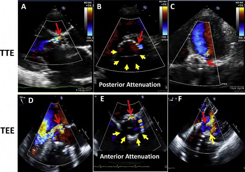
2 Zoghbi et al                                                            Journal of the American Society of Echocardiography
                                                                                                                     - 2019

                             Abbreviations          TABLE OF CONTENTS

 2D = Two-dimensional
 3D = Three-dimensional                               I. Introduction 3
                                                     II. General Principles 3
 AR = Aortic regurgitation                          III. Percutaneous Aortic Valve Interventions 4
                                                        A. Balloon-Expandable vs. Self-Expanding Valves 4
 AV = Aortic valve
                                                         B. Pre-procedural Planning for TAVR and Valve-In-Surgical Valve        5
 CD = Color Doppler                                     C. Implantation Technique for Routine TAVR 5
                                                        D. TTE vs. TEE in the Catheterization Laboratory 5
 CMR = Cardiac magnetic resonance                        E. Evaluation of Valvular Regurgitation after TAVR 6
 CS = Coronary sinus                                        1. Aortography     6
                                                          2. Hemodynamic Assessment in the Catheterization Laboratory                6
 CWD = Continuous-wave Doppler
                                                          3. Doppler Echocardiographic Assessment of AR after TAVR              10
 EROA = Effective regurgitant orifice area                   a. Color Doppler Jet Features     10
 LA = Left atrium                                            b. Continuous-Wave and Pulsed-Wave Doppler              14
                                                               c. Quantitative Doppler Assessment of PVR Severity 14
 LV = Left ventricle
                                                        F. Assessing Residual AR after Percutaneous Repair of Prosthetic Para-
 LVEDP = Left ventricular end-diastolic pressure           valvular Regurgitation 16
                                                       G. Integrative Approach to Assessment of AR 16
 LVOT = Left ventricular outflow tract                 H. Role of Cardiovascular Magnetic Resonance in Evaluating AR 17
 MDCT = Multi-detector computed tomography          IV. Percutaneous Mitral Valve Interventions 19
                                                       A. General Considerations in Evaluating Residual MR during MV Inter-
 MR = Mitral regurgitation                                 ventions 19
                                                        B. Mitral Leaflet Repair 20
 MV = Mitral valve
                                                           1. Edge-to-Edge MV Repair 20
 MVA = Mitral valve area                                   2. Evaluation of Residual MR with CD Immediately after Edge-to-Edge
 PA = Pulmonary artery                                        Repair 21
                                                           3. Interaction of Mean Transvalvular Gradient and Residual MR after
 PASP = Pulmonary artery systolic pressure                    Edge-to-Edge Repair 22
 PR = Pulmonic regurgitation                           C. Transcatheter Mitral Valve Replacement 22
                                                           1. TMVR Implantation 22
 PVR = Paravalvular regurgitation                         2. Evaluation of Residual MR Immediately after TMVR              23
 PWD = Pulsed-wave Doppler                               3. Other Considerations in TMVR 24
                                                      D. Percutaneous Mitral Annuloplasty 24
 RF = Regurgitant fraction
                                                         1. Percutaneous MV Annuloplasty Devices           24
 RV = Right ventricle                                     2. Evaluation of MR after Percutaneous Annuloplasty    25
 RVol = Regurgitant volume                             E. Transcatheter Repair of Paravalvular Prosthetic MR  25
                                                          1. Repair of Paravalvular MR     25
 RVOT = Right ventricular outflow tract                   2. Evaluation of Residual MR after Repair of Paravalvular MR   25
 SAVR = Surgical aortic valve replacement              F. Evaluation of Residual MR Outside the Catheterization Laboratory
                                                          after all MV Procedures    25
 SSFP = Steady-state free precession                      1. Color Doppler Imaging     26
 TAVR = Transcatheter aortic valve replacement            2. CW Doppler of MR Jet       28
                                                          3. Mitral Inflow Pattern and Pressure Gradient        29
 TMVR = Transcatheter mitral valve replacement
                                                          4. Pulmonary Vein Flow Pattern 29
 TPVR = Transcatheter pulmonary valve replacement         5. Pulmonary Artery Systolic Pressure      29
 THV = Transcatheter heart valve                          6. Regurgitant Volume and Fraction        29
                                                          7. An Integrative Approach to Assessing Residual MR             30
 TEE = Transesophageal echocardiography
                                                          8. Role of CMR in the Evaluation of Residual MR after Percutaneous
 TR = Tricuspid regurgitation                                MV Interventions 30
 TTE = Transthoracic echocardiography                        a. Evaluation of Residual MR 30
                                                             b. LV and LA Reverse Remodeling         32
 TV = Tricuspid valve
                                                             c. When is CMR Indicated?      32
 VCA = Vena contracta area                          V. Percutaneous Pulmonary Valve Replacement 32
                                                      A. Description of TPVR and Assessment of Acute Results 33
 VCW = Vena contracta width                            B. Evaluation of Residual Regurgitation Outside the Catheterization
 VTI = Velocity-time integral                             Laboratory 33
Journal of the American Society of Echocardiography                                                                                         Zoghbi et al 3
Volume - Number -

       1. Assessment of Pulmonary Regurgitation after TPVR with Echocardi-         depiction or anterior/posterior, medial and lateral sites in relation to
          ography 33                                                               the annulus. General principles for evaluating native valve regurgita-
       2. Role of Computed Tomography in Pulmonic Regurgitation after              tion with echocardiography, Doppler, and CMR have recently been
          TPVR 33                                                                  updated.7 The methodology of assessing regurgitation qualitatively
       3. Role of CMR in Pulmonary Regurgitation after TPVR 35                     and quantitatively with these techniques will not be reiterated in
   C. Integrative Approach to Assessing Residual Pulmonic Regurgitation            detail but summarized, with emphasis on how these parameters
       after TPVR 35                                                               may be affected in the setting of transcatheter valve replacement or
VI. Percutaneous Tricuspid Valve Interventions 36                                  repair. The committee concurs with recent ASE guidelines7 and those
   A. Tricuspid Valve Repair and Annuloplasty       36
                                                                                   of the American College of Cardiology (ACC) and American Heart
    B. Assessment of Residual TR after Tricuspid Valve Interventions 36
                                                                                   Association (AHA) on valvular heart disease8 that valvular regurgita-
   C. Role of CMR in Assessing Residual TR after Tricuspid Valve Inter-
       ventions 37                                                                 tion should be classified as mild, moderate, or severe.
   D. Integrative Approach in the Evaluation of Residual TR       38                  There are four main principles to the evaluation of valvular regur-
                                                                                   gitation with echocardiography: comprehensive imaging, integration
VII. Conclusions and Future Directions      39                                     of multiple parameters, individualization to the patient, and precise
                                                                                   language to describe the findings. Comprehensive imaging by trans-
                                                                                   thoracic echocardiography (TTE) incorporates two dimensional/
                                                                                   three-dimensional (2D/3D) structural evaluation of the implanted de-
I. INTRODUCTION                                                                    vice and surrounding structures, cardiac chamber size and function,
                                                                                   flow interrogation with pulsed-wave Doppler (PWD), continuous-
Valvular disease remains a major cause of cardiovascular morbidity                 wave Doppler (CWD), and color Doppler (CD), and volumetric
and mortality worldwide.1 Over the past decade, catheter-based in-                 quantitation as well as assessment of additional hemodynamic param-
terventions in valvular disease have evolved from balloon dilation                 eters such as pulmonary artery (PA) pressures. Each of these methods
of native stenotic valves to repair of paravalvular regurgitation                  has particular technical considerations, strengths, and limitations,
(PVR) with vascular plugs and more recently to valve replacement                   which have been described in detail.7 Unfortunately, many of these
and repair. Currently-approved interventions include transcatheter                 parameters may not be available during intra-procedural transesopha-
aortic valve replacement (TAVR), pulmonic valve replacement, and                   geal echocardiography (TEE) or TTE due to limited windows, inability
mitral valve repair, targeted to specific populations. Rapid technolog-            to align Doppler interrogation with blood flow, and foreshortening of
ical advancements in device design are likely to improve acute and                 the apex, which may preclude accurate volumetric quantitation. Thus,
long-term results and expand current indications.                                  intra-procedural echocardiography often relies heavily on CD jet
   Hemodynamics of percutaneous valves have been very favorable.2-5                characteristics, evaluating when possible its three components of
However, a challenging area has been the new or residual valve                     flow convergence, vena contracta, and jet area. CD imaging of the
regurgitation that may occur either after transcatheter valve                      jet in this setting can be impacted by hemodynamics, effects of seda-
implantation or repair of a native or prosthetic valve. This condition             tion/anesthesia, technical factors, and attenuation by the implanted
presents a diagnostic and therapeutic challenge to the interventional              device. Because CD area is mainly determined by jet momentum
and imaging cardiology team in the catheterization laboratory and to               (area  velocity2), the pressure gradient and therefore velocity driving
the clinician and imager in the outpatient setting. The current                    the jet can greatly influence jet size. For example, mitral regurgitation
document addresses the challenges of assessing residual regurgitation              (MR) jets after mitral repair or transcatheter mitral valve replacement
after percutaneous valve replacement or repair and provides a guide to             (TMVR) can be large despite a small orifice if left ventricular (LV) pres-
the cardiac team on how best to approach this condition, based on the              sure is high (e.g., hypertension or aortic stenosis). Conversely, a low
available data and a consensus of a panel of experts. This document                aortic diastolic pressure after TAVR might result in a small aortic regur-
supplements the previous American Society of Echocardiography                      gitation (AR) jet with CD despite hemodynamically significant AR. To
(ASE) guideline on the assessment of surgically implanted prosthetic               compensate for hemodynamically mediated variation inherent in CD
valves.6 It does not address flow dynamics through the percutaneous                characterization, it is common practice for the implanting physician or
prosthetic valves since, in general, the evaluation is similar to surgically im-   anesthesia team to ‘‘normalize’’ post-implant hemodynamics (increase
planted valves,6 but focuses mostly on new or residual valvular regurgita-         or decrease heart rate and systemic blood pressure) pharmacologi-
tion. In addition to the use of echocardiography and hemodynamic                   cally prior to assessing intraoperative valvular regurgitation in the pro-
assessment in the acute setting, the document incorporates the role of car-        cedure room. Moreover, valve regurgitation after percutaneous
diac magnetic resonance (CMR) imaging. This guideline is accompanied               procedures, in contrast to native or surgical prosthetic valves,
by a number of tutorials and illustrative case-studies on evaluation of            frequently arises from multiple sites with variable severity, making
valvular regurgitation after catheter-based interventions as well as native        CD assessment of regurgitation more difficult. All the above issues
valve regurgitation, posted on the following website (www.asecho.org/              highlight the need to integrate CD information with other echocar-
vrcases), which will build gradually over time.                                    diographic findings to determine overall severity of regurgitation.
                                                                                   This comprehensive evaluation may be more feasible to perform after
                                                                                   completion of the procedure, out of the catheterization laboratory
II. GENERAL PRINCIPLES                                                             setting. Intra-procedurally, the findings by echo-Doppler are comple-
                                                                                   mented with invasive hemodynamic assessment to gauge the overall
In the catheterization laboratory, members of the heart team should                results of the intervention, and cineangiography may be needed in sit-
be well versed with how to assess valve regurgitation, the language                uations where the residual regurgitation is difficult to assess, inconclu-
used to describe valve structure and position, as well as a clear, coor-           sive, or suspected to be more than mild (Figure 1). In the setting
dinated nomenclature as to the site of regurgitation, using a clock                outside the catheterization laboratory, uncertainty regarding severity
4 Zoghbi et al                                                                                   Journal of the American Society of Echocardiography
                                                                                                                                            - 2019

Figure 1 Tools for the intra-procedural assessment of paravalvular regurgitation following TAVR: color Doppler (2D/3D), pulsed-wave
Doppler of aortic flow, aortic & LV diastolic pressures and aortography. All panels are from the same patient. The valve is a self-
expanding valve. (A) Mid-esophageal TEE long-axis view showing paravalvular AR (white arrows). Off-axis imaging is frequently help-
ful. (B) Deep transgastric view showing the paravalvular jet seen in panel A. Multiple views are essential to avoid missing jets. (C)
Mid-esophageal short-axis view showing a paravalvular jet (red arrow), and a pinhole jet (arrow head). It is important to scan the valve
and image at the lower end of the valve stent to ensure that the measured jet reaches the LV. The circumferential extent of the larger jet
here is 14% but the jet is relatively wide. The pinhole jet is too small to planimeter. (D) 3D planimetry of the same paravalvular jet yields
an area of 0.22 cm2. 3D echocardiography makes it possible to precisely identify the vena contracta, something that may be chal-
lenging with 2D imaging alone. (E) Mid-esophageal TEE images of the descending thoracic aorta showing non-holodiastolic flow
reversal by pulsed-wave Doppler. Some flow reversal, usually non-holodiastolic, may be present in patients undergoing TAVR
even in the absence of aortic regurgitation; Hence it is important to establish the baseline aortic flow pattern. (F) Simultaneous LV
and aortic (Ao) pressure tracings that form the basis for the AR index. In this case, the AR index is 28%. Indices of
Journal of the American Society of Echocardiography                                                                               Zoghbi et al 5
Volume - Number -

that spontaneously expand on release from the delivery system, the         used.34-36 However, the conscious sedation approach is not
so-called self-expanding valves. Within these categories, the most         universal, and conversion to general anesthesia in 10-17% of
widely used valves have been the Edwards SAPIENÔ family of                 cases37,38 has been reported, with a more recent lower rate of
balloon-expandable valves and the Medtronic CorevalveÒ/                    conversion (5.9%) published from the National Cardiovascular
EvolutÔ self-expanding valves. Within these families of valves, im-        Data Registry (NCDR).39 TEE guidance has been associated with
provements in design have reduced the incidence and severity of            less contrast use,40 lower incidence of PVR,41 and greater procedural
PVR. The construct of these valves has been reviewed recently.23           success.39 A recent study from a high-volume site performing primar-
Although other valves have received the CE (conformite                    ily conscious sedation with TTE guidance showed that there was a
Europeenne) mark in Europe and are in varying phases of clinical trials   higher incidence of second valve implantation (7% vs. 2%;
in North America, they will not be further discussed.                      P = .026) and post-TAVR balloon dilation (38% vs 17%; P < .001)
                                                                           intra-procedurally compared to the TEE approach.42
B. Pre-procedural Planning for TAVR and Valve-In-                              Studies evaluating the clinical outcome of patients undergoing
Surgical Valve                                                             TAVR with either TEE/general anesthesia or TTE/moderate seda-
Pre-procedural planning for TAVR can be useful for predicting PVR          tion have not shown consistent results favoring one or the other
following valve implantation and allows the heart team to plan for         approach. In the Brazilian Registry,43 the use of TEE to monitor
the appropriate treatment of intra-procedural PVR. Numerous studies        the procedure compared to TTE was associated with less overall
have shown that significant under-sizing of the transcatheter heart        mortality (HR: 0.57) and late mortality (HR: 0.47). Recent observa-
valve (THV) will result in greater degrees of PVR.12,24 MDCT               tional studies support the safety of the ‘‘minimalist’’ anesthetic
reconstruction-based measurements of the aortic annulus has been           approach36,44 and the European Society of Cardiology’s
shown to reduce the incidence of greater than mild paravalvular AR         Transcatheter Valve Treatment (TCVT) Registry found that survival
compared to 2D TEE measurements.25 The accuracy of 3D TEE for              at 1 year was similar between groups.35 Lastly, the observational
annular sizing and prediction of PVR severity has also been demon-         NCDR study reported that while the conscious sedation approach
strated.26 MDCT determination of calcium location and burden is            had lower procedural success, it was associated with a reduced
another important predictor of PVR.27,28 For valve-in-surgical-valve       rate of in-hospital mortality (1.5% versus 2.4%, P < .001) and 30-
procedures, knowledge of the manufacturer’s reported internal diam-        day mortality after TAVR (2.3% versus 4.0%, P < .001), warranting
eter of the bioprosthetic surgical valve is essential for appropriate      formal study.39 This is particularly important as the field begins to
sizing. In the absence of this information, TEE or MDCT may be             address TAVR in the lower-risk patient population who are also at
used to determine the internal diameter of the valve; however, regur-      lower risk for complications during general anesthesia, and whose
gitation following this procedure is rarely more than mild.29              expected outcomes are significantly better than patients treated in
                                                                           these registries.10,45
C. Implantation Technique for Routine TAVR                                     Advantages and disadvantages of TTE and TEE as they pertain to
                                                                           assessing valvular regurgitation after TAVR are listed in Table 1. Using
Although not required, the balloon-expandable THV may be pre-              TTE, imaging from the parasternal windows requires direct place-
dilated using balloon aortic valvuloplasty, which is intended to           ment of the probe within the fluoroscopic imaging plane with
improve the positioning process but may also decrease the number           high exposure of the imager to radiation. The patient’s supine posi-
of cerebral ischemic lesions.30 The SAPIEN 3 delivery system has a         tion and operator avoidance of the sterile field may prohibit optimal
middle marker and two other markers to facilitate trans-annular posi-      transducer placement. The usual sources of ultrasound interference
tioning of the valve before deployment; because of stent cell design,      still apply, such as chest wall deformities, emphysema, obesity, etc.
the valve shortens from the ventricular end, engaging the surrounding      Intra-procedural TTE can evaluate the causes of acute hemody-
tissue early in the deployment process. After obtaining a suitable co-     namic compromise such as pericardial effusion, under-filled or
planar angle, the Edwards SAPIEN 3 valve is deployed using rapid           dysfunctional ventricles, and severe valvular regurgitation. The
ventricular pacing to reduce valve motion just before balloon expan-       assessment of PVR may be challenging unless imaging windows
sion. Medtronic’s CoreValve Evolut R and Evolut Pro have a                 are ideal. On the other hand, a major advantage of intra-
controlled and slow-release self-expanding delivery system. They do        procedural TEE guidance is continuous imaging throughout the
not require rapid pacing, although some interventionalists may use         TAVR procedure with rapid and accurate diagnosis of complications,
pacing to stabilize the valve during deployment. The valve can also        including PVR.32,46,47 Studies have shown that TEE can be safely
be repositioned and recaptured up to a point in the unsheathing pro-       performed in the setting of monitored anesthetic care.48 Since CD
cess. In addition to adequate sizing of the annulus, accurate posi-        is essential in localizing and assessing PVR severity, it is important
tioning of all valves is essential to reduce post-implantation PVR         to recognize that shadowing of the prosthetic valve may affect detec-
and reduce post-procedural pacemaker rates.31 Valve positioning            tion of paravalvular regurgitation by either TTE or TEE (TTE may not
can be imaged by TTE and TEE as well as fluoroscopy.23,32 The              optimally display posterior paravalvular regurgitation whereas TEE
amount and location of calcium within the landing zone of the              may not optimally display anteriorly located defects; Figure 2). A
valve also plays a major role in the incidence and severity of PVR         combination of both techniques may be needed in situations where
following TAVR.12,28,33                                                    an AR jet is detected in the left ventricular outflow tract (LVOT) or
                                                                           LV and the source cannot be localized by one approach. TEE must
D. TTE vs. TEE in the Catheterization Laboratory                           include standard and deep transgastric views to assess both
Echocardiography remains the primary imaging modality for assessing        valvular/LVOT hemodynamics and detect paravalvular regurgita-
PVR immediately following TAVR. Initially, the procedure was per-          tion that may be missed in esophageal views (Figure 2). Overall,
formed under general anesthesia with TEE monitoring. Performing            qualitative and quantitative echo-Doppler parameters can be ob-
TAVR under local anesthesia with conscious sedation or with moni-          tained with TTE or TEE imaging and applied to assess the severity
tored anesthesia care has been reported recently and is increasingly       of PVR either during or after the procedure.
6 Zoghbi et al                                                                                    Journal of the American Society of Echocardiography
                                                                                                                                             - 2019

 Table 1 Advantages and disadvantages of TTE versus TEE imaging for assessment of aortic regurgitation during and after TAVR

    Parameter                                         TTE                                                               TEE

  Advantages                   Standard windows for assessing valvular/cardiac                 Higher resolution with high frame rates for 2D and
  General                       structure & function                                             3D imaging, and quantitation of VCA
                               Imaging anterior paravalvular regurgitation with                Imaging posterior paravalvular regurgitation with
                                CD                                                               CD
                               Ease of CWD interrogation of AR jet
                               Ease of PWD interrogation of flow in the LVOT
                               Can provide volumetric quantitation
                               Less resource intensive
  Advantages                   Does not require intubation or general anesthesia               Continuous imaging throughout procedure,
  During TAVR                  Superior viewing of anterior paravalvular                        irrespective of TAVR access route
                                regurgitation                                                   Intra-procedural imaging may help avoid
                               May still convert to TEE approach, if needed                     complications (e.g., imaging during balloon
                                                                                                 valvuloplasty to assess risks of calcium
                                                                                                 displacement)
                                                                                                Immediate intra-procedural diagnosis of
                                                                                                 complications
                                                                                                Lower contrast use for procedure
                                                                                                Ability to hold respiration during general
                                                                                                 anesthesia for better 3D assessment of PVR, and
                                                                                                 aortic annular measurement (if needed)
  Disadvantages                Image quality dependent on patient factors (e.g.,               Image quality dependent on patient factors (e.g.,
  General                       chest morphology, lung hyperinflation,                           valve calcific acoustic shadowing, cardiac
                                suboptimal positioning, valve calcific acoustic                  position relative to esophagus and stomach)
                                shadowing)                                                      Difficulty imaging anterior paravalvular
                               Difficulty imaging posterior paravalvular                        regurgitation with CD in esophageal views
                                regurgitation with CD                                           Challenge in optimizing CWD recording of AR jet
                               Lower resolution of 2D and 3D imaging                            and CD display of AR from deep transgastric view
  Disadvantages                Procedural delay during image acquisition (to                   Requires conscious sedation or monitored
  During TAVR                   minimize radiation exposure to imager)                           anesthesia care
                               Non-continuous imaging during procedure may                     More resource intensive; may prolong
                                delay diagnosis of complications (e.g., valve mal-               hospitalization if requires intubation & there is
                                positioning, annular rupture, coronary occlusion)                difficulty extubating patient
                               Limited imaging windows for non-transfemoral                    Probe interference with fluoroscopic imaging
                                TAVR access routes                                               (minimized by articulation of probe)
                               Difficult to perform volumetric quantitation

E. Evaluation of Valvular Regurgitation after TAVR                          windows of interrogation of flow, for either TEE or TTE. Thus,
Assessment of the presence and severity of AR after TAVR is complex,        during the acute setting, hemodynamics and aortography are
requiring imaging and Doppler evaluation of the valve and annulus.          frequently used to complement CD imaging for a comprehensive
Central valve regurgitation is far less common than paravalvular            evaluation of residual PVR (Figure 1). The current document will
regurgitation. Evaluation and grading of PVR is complicated by the          therefore address aortography, hemodynamic assessment, and
complex nature of AR jets after valve implantation and the differ-          Doppler methods that can be used in the interventional arena during
ences in jet characteristics between valve designs. Regurgitant jets        TAVR (qualitative/semi-quantitative parameters) and will detail also
around a stented valve are due to mal-apposition of the stent and           other Doppler quantitative methods and CMR methods that can
the native surrounding structures. Mal-apposition may occur for a           be applied outside the interventional laboratory for a more compre-
number of reasons: 1) THV collapsible frames have empty cells be-           hensive evaluation of PVR with non-invasive techniques.
tween metal struts; 2) metallic stents have limited deformability, pre-     1. Aortography. Conventional cineangiography with an aortic root
venting complete closure of small gaps created by irregularly-calcified     injection of radiographic contrast can be used for intra-procedural
leaflets, protruding dystrophic calcium in the annulus or LVOT, or          determination of AR severity.49 This is usually performed during
triangular commissural gaps; 3) mal-positioning of the THV (too             the TAVR procedure in the catheterization laboratory when AR is de-
high or low in the annulus); and 4) under-sizing of the THV for the         tected by CD and its severity needs further evaluation. This approach,
native annular size.                                                        however, is rarely indicated in the chronic setting or for serial follow-
    Assessment of PVR relies heavily on CD imaging both during the          up. Angiographic grading, while helpful in extremes, may not corre-
procedure and in the outpatient setting, as conventional spectral           late well with quantitative assessment of AR severity, and cannot reli-
Doppler parameters may have limitations.20 CD evaluation has out-           ably distinguish central from paravalvular regurgitation.20,50,51
comes data from 2 separate trials using 2 different core labora-
tories.2,10 Spectral Doppler evaluation is particularly hampered in         2. Hemodynamic Assessment in the Catheterization
evaluating PVR during the TAVR procedure because of limited                 Laboratory. A number of investigators have attempted to use the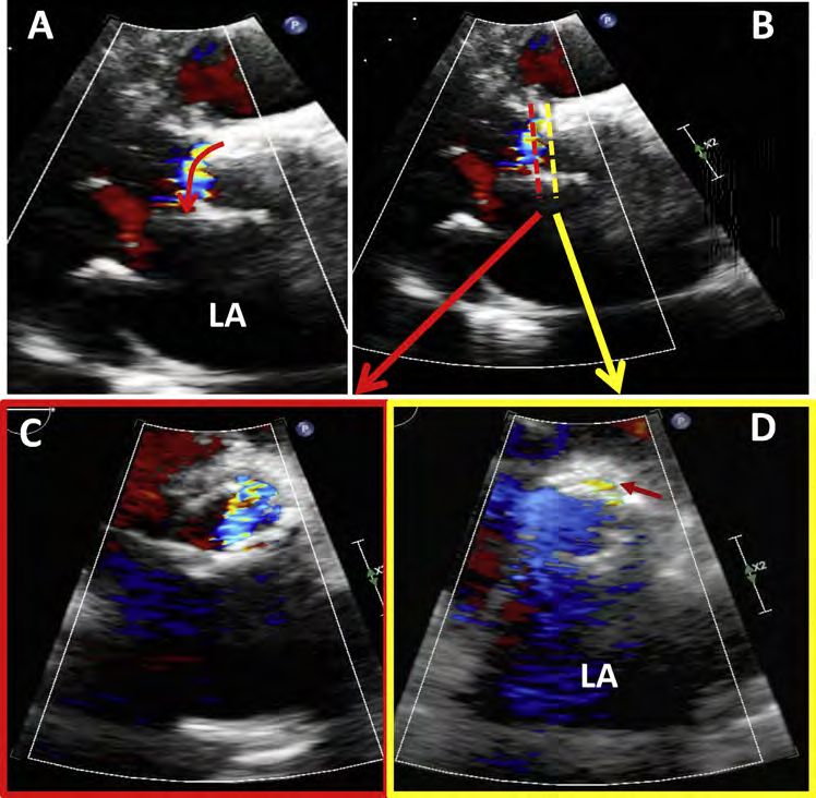
Journal of the American Society of Echocardiography                                                                                  Zoghbi et al 7
Volume - Number -

Figure 2 Demonstration of paravalvular leaks after TAVR assessed by TTE and TEE in two different patients, demonstrating ultra-
sound attenuation of the valve ring and adjacent structures by the prosthetic valve, and the importance of comprehensive color
Doppler imaging from the apical views (apical in TTE and transgastric in TEE). Panels A to C demonstrate an anterior paravalvular
leak that is identified from a parasternal long-axis view (A), short-axis view (B), and modified apical five-chamber view (C). There
is posterior attenuation (yellow arrows). Panels D-F demonstrate posterior paravalvular leak on mid-esophageal 120 degrees (D)
and short-axis views (E); the transgastric view (F) demonstrates two jets of paravalvular regurgitation, one jet is anterior and the other
is posterior. Without careful comprehensive interrogation, the anterior leak would have been potentially missed on just mid-
esophageal views because of attenuation (yellow arrows).

 Table 2 Invasive hemodynamic indexes for assessing severity of AR immediately After TAVR

       Author                Index                               Formula                                        Cutoff for significance

  Sinning et al.52
                          AR index           ([DBP– LVEDP] O SBP) x 100                            AR index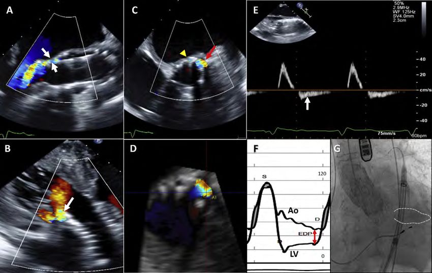
8 Zoghbi et al                                                                  Journal of the American Society of Echocardiography
                                                                                                                           - 2019

 Table 3 Echocardiographic parameters (TEE/TTE) and respective comments in determining AR severity after TAVR

      Parameter                                                                                     Comments

 Stent shape and                                                                Not accurate in predicting presence/severity of
   position                                                                      AR

 Color Doppler
   Jet number,                                                                  - Needs meticulous scanning of whole valve to
     location,                                                                    identify sites of regurgitation, which are
     direction, and                                                               frequently multiple and eccentric
     eccentricity                                                               - Essential to identify origin of jets, which may
                                                                                  not all be at the same level
                                                                                - Short-axis imaging below the valve may
                                                                                  overestimate AR severity in eccentric jets
                                                                                - Attenuation of ultrasound by prosthesis in far
                                                                                  field may hinder visualization of regurgitation
                                                                                  (anterior with TEE and posterior with TTE)
                                                                                - Deep transgastric views during TEE are
                                                                                  essential for assessment of regurgitant jets (jet
                                                                                  area and length are not used to assess
                                                                                  regurgitation severity)

   Vena contracta                                                               VCW >0.6 cm specific for severe AR; severity of
     width (VCW)                                                                multiple smaller jets more difficult to evaluate

   Vena contracta                                                               - May allow addition of multiple jets
     area (VCA)                                                                 - Prone to blooming artifacts
                                                                                - Accuracy limited by spatial resolution for small
                                                                                  jets

                                                                                                                      (Continued )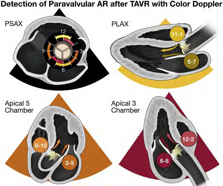
Journal of the American Society of Echocardiography                                    Zoghbi et al 9
Volume - Number -

 Table 3 (Continued )
       Parameter                                                         Comments

    Circumferential                                   - Continuous circumferential extent of AR of
      extent (%)                                        >30% indicative of severe AR
                                                      - Circumferential extent of few, smaller discrete
                                                        jets more difficult to assess

    Flow                                              Large flow convergence in aorta indicative of
      convergence                                     severe AR

  Spectral Doppler
    Flow reversal in                                  - Useful if new (relative to baseline) and
      descending                                        holodiastolic, consistent with at least
      aorta (PWD)                                       moderate AR; lesser aortic flow reversal is
                                                        non-diagnostic
                                                      - Holodiastolic flow reversal in abdominal aorta
                                                        more specific for significant AR

                                                                                          (Continued )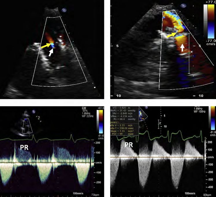
10 Zoghbi et al                                                                                         Journal of the American Society of Echocardiography
                                                                                                                                                   - 2019

 Table 3 (Continued )
       Parameter                                                                                                             Comments

    CW Doppler                                                                                          - Dense velocity waveform consistent with
     profile of AR jet                                                                                    more than mild AR
     (velocity                                                                                          - Pressure half-time in extremes (>500 or
     waveform
Journal of the American Society of Echocardiography Zoghbi et al 11
Volume - Number -
Table 4 Evaluation of severity of prosthetic aortic regurgitation after TAVR
PVR severity Mild Moderate Severe
Aortography Contrast does not fill Intermediate Contrast fills LV on
entire LV and clears first beat, ending
with each cycle with greater density
than in ascending
aorta
Invasive Hemodynamic Parameters
AR index* $2512 Zoghbi et al Journal of the American Society of Echocardiography
- 2019
Figure 3 Standard 2D echocardiographic views depicting the detection of TAVR-related paravalvular regurgitation. Color coding de-
lineates the regions around the prosthetic valve that can be visualized from the parasternal and apical views. With ultrasound plane
rotation, tilting upward or sideways, a more complete interrogation of the valve can be accomplished. Although these unconventional
planes may foreshorten cardiac structures, they are essential in detecting and localizing the residual regurgitation. The importance of
apical views lies in the fact that some jets may not be detected in the parasternal views because of either shadowing from the pros-
thesis in parasternal short axis (PSAX; lateral, medial and posterior surfaces; see Figure 2) or are located medially or laterally to be
seen in the parasternal long-axis (PLAX) view.
overestimation if assessment of the circumferential extent of the jet Importantly, the jet must enter the LV to be considered true regurgitation,
origin mistakenly includes this single short-axis view displaying jet thus imaging just below the edge of the stent will confirm the presence of
eccentricity. Because the number of jets also reflects severity, an true PVR; however, the vena contracta of the jet should be measured at its
extensive search for all jets should be performed, using multiple im- narrowest region.
4. Color flow around the THV within the sinuses of Valsalva but above the annular
aging windows, biplane imaging, and subtle manipulation of the
valve skirt should not be mistaken for PVR. Flow in the sinuses has low velocity
transducer within each window.
and does not connect with the LVOT in diastole. Scanning through the long axis
The circumferential extent of the jet is a useful parameter for as- of the valve is useful in distinguishing color flow in the sinuses from PVR.
sessing PVR severity; however, as with all parameters, it should not 5. Small jets of regurgitation are typically isolated to the open stent cells and
be used in isolation (Figure 5). The updated Valve Academic not at the ‘‘nodes’’ of the stent frame. It is important not to include the
Research Consortium (VARC-2) criteria57 used in the PARTNER I stented frame in the measurement of the circumferential extent of the
trial58 recommended the following with respect to the circumferen- regurgitation but to integrate only the regurgitant jets when determining
tial extent of paravalvular AR in short axis: trace (pinpoint jet), mild the circumferential extent (Figure 5).
(30%). Recent studies us- 6. In contrast to native valve AR, the ratio of jet width to LVoutflow tract width
ing the VARC-2 criteria, including this parameter, have shown or jet cross-sectional area to cross-sectional area of the valve or LV outflow
tract should not be used to assess the severity of AR since regurgitant jets are
increased mortality associated with worse PVR severity.10,45,59
frequently eccentric, constrained by and entrained within the LVOT, lead-
To assess residual AR with CD after TAVR, the following caveats
ing to rapid jet broadening (Figure 4).
should be considered: 7. Using a clock face to represent the short-axis view (placing the tricuspid
valve at 9 o’clock) may be helpful for recording the jet location and number,
1. The whole transcatheter valve should be scanned, from the distal (aortic) to and useful for follow-up comparisons (Figure 3).
the proximal (ventricular) end of the THV, to identify the number, loca- 8. Echo assessment of valve implant depth is important to assist the implanter
tion(s), and direction(s) of the AR jet(s). in assessing the mechanism and potential mediation (i.e post dilatation,
2. The entire short axis of the valve should be imaged in a single view, if valve repositioning, placement of a second valve or even possible plug
possible, so that the valve level imaged is the same in the far field as it is placement) of significant AR when present.
in the near field, and laterally as well as medially.
3. Central prosthetic AR jets will occur at the level of leaflet coaptation Three-dimensional echocardiography with CD can be used to
whereas PVR will be seen at the proximal (ventricular) edge of the valve. planimeter the vena contracta, an estimate of the regurgitant orificeJournal of the American Society of Echocardiography Zoghbi et al 13 Volume - Number - Figure 4 Effect of AR eccentricity on color Doppler jet recording in assessing PVR severity after TAVR. Scanning of the whole stented valve in short axis is needed to identify the vena contracta of the jet(s). Proper plane selection of the short axis is critical. In the case of an eccentric jet in the LVOT (A; curved arrow), the plane below the valve ring (B; dashed red) shows a large color jet as it spreads in the LVOT (C), overestimating AR severity. By selecting the proper short axis at the aortic annulus (D), the regurgitant orifice is best de- picted (small red arrow), more consistent with mild PVR. Similarly, a high short-axis view at the aortic root level (not shown) could be misinterpreted due to normal diastolic flow in the sinuses of Valsalva or coronary arteries; flows in these locations, however, are of lower velocity and are not aliased. Figure 5 Examples of paravalvular regurgitation of different degrees of severity using short-axis color Doppler depicting two criteria: vena contracta area (VCA), and % circumferential (Circ) extent of the jet in relation to the total circumference of the prosthetic valve ring. % Circ is calculated as the length of the jet along the valve curvature (‘‘a’’ in Panel A) divided by the total perimeter (‘‘c’’ in panel A) as: (a/c)*100. In the case of two jets (D), % Circ would be [(a + b)/c]*100. As the VCA and circumferential extent of the jet increase, AR severity is more significant. However, VCA in TAVR is affected by both circumferential extent and thickness of the PVR, i.e., separa- tion of the valve from the aortic wall. As shown in Panels A and B, the circumferential extent may at times be similar to those of mild regurgitation but the thickness of the PVR is large, leading to a larger VCA (B). Similarly, Panels B and C depict two lesions of similar moderate severity by VCA but different circumferential extent. These considerations are very important in assessing mild and mod- erate AR severity and multiple jets (A-D). Once circumferential extent exceeds 30%, PVR is usually severe.
14 Zoghbi et al                                                                                    Journal of the American Society of Echocardiography
                                                                                                                                              - 2019

Figure 6 Quantitative Doppler for assessing regurgitant volume (RVol) and regurgitant fraction (RF) in a patient with AR after TAVR.
Stroke volume (SV) is calculated at the LVOT and RVOT in this patient with mitral annular calcification. Flow quantitation complements
other assessments of AR severity.

area.60-62 Studies have shown the feasibility of measuring AR 3D             c. Quantitative Doppler Assessment of PVR Severity.–
vena contracta area (VCA) in native valves61,63-65 as well as post-          Quantitation of prosthetic AR involves the calculation of regurgitant
TAVR.62 The primary pitfalls of this technique at this time are low          volume (RVol), regurgitation fraction (RF), and less often, effective re-
frame rates and color blooming artifacts, which may be resolved              gurgitant orifice area (EROA; Figure 6). The methods for calculating
with more advanced 3D technology.                                            these parameters are well described in prior guidelines.6 However,
                                                                             it is important to highlight a few issues that may be encountered in
b. Continuous-Wave and Pulsed-Wave Doppler.–Classically, two                 the TAVR population that can modulate the approach to quantitation,
parameters from CWD recordings have been used in the evaluation              the accuracy of the derived parameters, and their clinical implications
of AR: velocity waveform density and the deceleration rate (pressure         in assessing PVR severity.
half-time). These may have limited applicability in the TAVR popula-             Methodology: RVol in aortic regurgitation is derived as total LV
tion. The common occurrence of multiple PVR jets limits the utility of       stroke volume minus systemic stroke volume. LV stroke volume is
CWD spectral density from a single jet; however, a very dense veloc-         quantitated with either pulsed-wave Doppler at the LVOT site or
ity waveform recording may signal at least moderate AR. In the cur-          with a volumetric approach (the difference between LV end-
rent elderly patient population undergoing TAVR, ventricular and             diastolic and LV end-systolic volumes). There are challenges to calcu-
aortic compliance abnormalities may limit the use of pressure half-          lation of LV stroke volume in the LVOT by PWD in the TAVR popu-
time for assessing the severity of PVR. A recent computational               lation. This stems from the measurement of LVOT diameter in the
modeling study confirmed that increasing LV and/or aortic stiffness          presence of the prosthetic valve as it protrudes into the LVOT (in
led to faster decay of the transvalvular pressure gradient and, there-       contrast to a sutured surgical valve at the aortic annulus). The core
fore, to a faster decrease of diastolic flow velocity across the aortic      lab methods for calculating LVOT stroke volume and aortic valve
valve compared with normal stiffness with the same regurgitant               area following implantation of the balloon-expandable and self-
orifice. This faster decay led to both a shorter pressure half-time (simu-   expanding valve have been recently published.71 It is important to
lating greater AR severity) and a lower regurgitant fraction (indicating     match the location of the PWD sample volume with the location of
less AR severity).66                                                         diameter measurement of the THV for accurate stroke volume calcu-
    Flow reversal in the descending aorta recorded with pulsed-wave          lations (Figure 7). The preferred approach is to measure the LVOT
Doppler may similarly be of limited value in the setting of abnormal         diameter from the outer-to-outer border of the stented valve at its
aortic or ventricular compliance. A number of studies in patients with       ventricular tip, with the corresponding PWD sample volume just api-
hypertension have shown some flow reversal in the descending aorta           cal to the valve stent (Figure 7, upper panels). In the case where the
in the absence of AR.67,68 Thus, in order to use this parameter for          prosthesis is positioned too deep into the LVOT, encroaching on
assessing post-TAVR regurgitation severity, a pre-TAVR assessment            the anterior leaflet of the mitral valve, an in-stent diameter measure-
of descending aortic flow is essential. In the absence of baseline           ment is performed at the mid-stent level (level of leaflets), with the
flow reversal, a new holodiastolic reversal of flow in the descending        PWD sample volume positioned into the stent but proximal to the
thoracic aorta is consistent with at least moderate AR,56 and likely se-     valve (Figure 7, lower panels).71 In cases of technical difficulty, the
vere if the end-diastolic velocity is >20 cm/s at a normal heart rate.69     LV volumetric method is used, provided there is no significant mitral
In severe bradycardia, holodiastolic flow reversal may not be seen,          regurgitation.
despite the presence of severe AR. Flow reversal in the abdominal                For LV volumes measurements to derive SV, avoidance of LV fore-
aorta is a more specific indication of significant regurgitation.70          shortening is essential. In general, 3D volumes are preferred to 2D
Journal of the American Society of Echocardiography Zoghbi et al 15
Volume - Number -
Figure 7 Calculation of stroke volume in the LVOT in transcatheter aortic valves. The default approach is to measure the LVOT diam-
eter using the outer edge-to-outer edge diameter at the lower (ventricular) end of the valve stent (A, arrow). The pulsed-wave Doppler
(PWD) sample volume is placed immediately proximal to the site of flow acceleration at the inlet to the stent (B). Stroke volume is then
calculated as usual, assuming a circular LVOT geometry as: 0.785*d2*VTI. In instances where a self-expanding valve is placed low in
the LV, particularly if the lower end of the stent is not in close proximity to the anterior mitral leaflet and interventricular septum, an
alternative approach is to measure the inner edge-to-inner edge diameter of the valve stent immediately proximal to the cusps (D).
The PWD sample volume should be placed just inside the stent but proximal to the site of flow acceleration at the cusps (E). Note that
with transcatheter valves there is flow acceleration at the inlet to the stent and again at the cusps. Red arrows point to the lower end of
the stent. Panels C and F show the respective PWD recordings in the LVOT.
determinations. In studies with suboptimal endocardial definition, which has been supported by larger studies.72 The usual cut-off for se-
contrast-echo enhancement is recommended to avoid underestima- vere chronic native AR (RVol of >60 mL) seems inappropriate early
tion of ventricular size and thus underestimation of all measurements after TAVR in this population with LV hypertrophy, smaller LV cavity
of total SV, RVol, and RF. Lastly, for determination of systemic stroke size, and abnormal ventricular compliance. The effect of relatively
volume, the usual mitral valve (MV) site with PWD used for mitral small RVols on patients with abnormal ventricular and aortic compli-
inflow volume may be problematic in this population, as the mitral ances could explain why even mild regurgitation may have a signifi-
annulus is frequently calcified, hindering the measurement of mitral cant impact on clinical outcomes post-TAVR.12,19
annular diameter and derivation of accurate annular area. The mitral Regurgitant fraction may be a more physiologically important
annulus site may also be problematic in the presence of more than parameter that normalizes for the lower stroke volumes seen in this
mild mitral regurgitation. In these circumstances, the pulmonic annulus population. In fact, CMR grading of PVR relies on RF and is discussed
is an alternative site to calculate systemic stroke volume (Figure 6). below. According to ASE guidelines, mild AR has a RF 50%.6,7 Data in
time integral of the AR jet, obtained by CWD. This is rarely performed the TAVR population using CMR measures of PVR severity showed a
in clinical laboratories. EROA using the proximal isovelocity surface reduced survival with a regurgitant fraction of 30%,73 supporting the
area (PISA) method is usually not feasible because the flow conver- use of this cutoff for moderate AR post-TAVR. Harmonizing grading
gence from the apical window is shadowed frequently by the pros- schemes between imaging modalities may minimize discordance be-
thesis and its shape is typically non-hemispheric. tween the two techniques.
Severity of PVR using quantitative criteria: Whereas quantitative Continued follow-up of patients following TAVR is recommen-
grading schemes have been advocated for the evaluation of prosthetic ded, particularly in the setting of recent data suggesting PVR may
valve regurgitation, there is little data to support the use of these quan- improve over time74,75 and because of the unclear long-term
titative parameters in the context of acute PVR, shortly after TAVR. durability of these valves. Patients with uncomplicated THV im-
The PARTNER IA study reported a mean LVOT Doppler stroke vol- plantation should undergo a comprehensive TTE soon after implan-
ume in patients with $mild AR following TAVR of 68 6 20 mL,14 tation to establish baseline valvular function, and subsequently at16 Zoghbi et al Journal of the American Society of Echocardiography
- 2019
Figure 8 Repair for bioprosthetic aortic paravalvular regurgitation. At baseline: 2D color Doppler depicts very eccentric paravalvular
AR evident in both short-axis (left) and long-axis (right) TEE views (arrows). An occluder device is deployed with reduction of color
Doppler intensity but with significant residual PVR noted medial to device #1 (white arrows in center image, bottom). Occluder device
#2 was deployed medial to occluder device #1 with immediate reduction in PVR severity to trace (white arrow).
1-3 months and 1 year. Studies should also be performed if unex- and the effect of high pulmonary pressures on right ventricular size
pected clinical deterioration or new murmurs occur after THV. and function.
TTE is used to identify complications of the procedure, especially
PVR, and changes in LV or RV function, assess aortic root structure G. Integrative Approach to Assessment of AR
and valvular function, and measure PA pressure. The baseline post-
The evaluation of AR by Doppler echocardiography after percuta-
TAVR TTE is integral to accurate follow-up, since changes in base-
neous interventions on the aortic valve should be a comprehensive
line hemodynamics (increase in mean gradient of $10 mm Hg or
and integrative process, based on all the information collected during
1 grade in AR) are an indication of possible valve deterioration or
the examination, since each of the parameters used in this evaluation
complication.76
has advantages and limitations. In all cases, one should routinely
perform a comprehensive sweep of the implanted valve by 2D and
F. Assessing Residual AR after Percutaneous Repair of CD echocardiography, which includes an assessment of LV size and
Prosthetic Paravalvular Regurgitation function, as well as velocity recordings in the LV outflow tract, mitral
The recently published Paravalvular Leak Academic Research and pulmonic valve annuli, and in the proximal descending aorta
Consortium Expert Statement reviews the technical aspects of per- and/or abdominal aorta. CW Doppler of the AR jet should also be
forming PVR closure after SAVR or TAVR.77 The document also de- routinely recorded but only utilized if a complete signal is obtained.
fines the clinical endpoints reflecting safety and effectiveness of Recordings of CD and pulsed-wave Doppler of prosthetic valve AR
transcatheter devices, as well as the single and composite clinical end- are more challenging compared to native AR due to the valve struc-
points for clinical trials. The therapeutic endpoint should be under- ture and mechanism of residual AR after aortic valve interventions.
stood prior to the procedure. If the procedure is being performed Based on data in the literature and a consensus of the committee
to address heart failure, any reduction in RVol is desirable. If, on the members, the Writing Group proposes a scheme for evaluation of pa-
other hand, the procedure is to address hemolysis, then complete tients with AR (Figure 9). It is in a similar format to the scheme
or near obliteration of the leak should be the objective. In deciding recently proposed for native valve AR, but differs in the incorporation
when and how to address PVR after SAVR or TAVR, it is important of VCA and % circumferential extent of VC, the removal of the con-
for the imaging physician to understand the underlying mechanism ventional AR jet width/LVOT diameter ratio because of AR jet eccen-
of the paravalvular regurgitation; this should guide the treatment tricity, and the consideration of LV size and function.7 In applying this
approach and may also predict whether a transcatheter closure device scheme, it is the consensus of the committee members that the pro-
will be successful in treating the regurgitation. cess of grading AR should be comprehensive, using a combination
Localization and grading of residual AR after repair is assessed pri- of signs and measurements obtained by Doppler echocardiography.
marily with TEE or TTE as described for acute intra-procedural TAVR If the AR is definitely determined as mild or severe using these specific
assessment (Figure 8). In addition to grading of the regurgitation, an signs, no further measurement is required, particularly for mild le-
assessment of the effects of regurgitation in the chronic setting should sions. If there are only a few parameters consistent with mild or severe
include an evaluation of change in LV size and function, PA pressures, AR, and the quality of the primary data lends itself to quantitation, it isJournal of the American Society of Echocardiography Zoghbi et al 17
Volume - Number -
Aortic Regurgitation After TAVR or Percutaneous Prosthetic Valve Repair
Yes, mild Yes, severe
* Does AR meet specific criteria of
mild or severe AR? *
Specific Criteria for Mild AR Specific Criteria for Severe AR
• VC width < 0.3 cm Intermediate values:
• VC width > 0.6 cm
• VCA < 0.10 cm2 AR probably moderate
• VCA ≥ 0.30 cm2
• Circumferential extent 500 ms Perform quantitative methods whenever possible to
• PHT < 200 ms
• No or brief diastolic flow refine assessment • Prominent holodiastolic flow
reversal in the descending aorta reversal in the descending aorta
≥ 4 criteria ≥ 4 criteria
Definitively mild Definitively severe
(quantitation not needed) RVol < 30 mL RVol 30 - 59 mL RVol ≥ 60 mL¶ (may still quantitate)
RF < 30% RF 30 - 49% RF ≥ 50%
EROA18 Zoghbi et al Journal of the American Society of Echocardiography
- 2019
Figure 10 CMR for evaluation of PVR after TAVR and subsequent valve-in-valve (ViV) implantation for treating residual AR. Three-
chamber cine view showing the PVR jet (red arrow) after TAVR (A). (B) shows the decreased severity of PVR after ViV placement.
Phase-contrast imaging of the flow through the prosthetic valve (C and D) shows the PVR jet, which has decreased significantly after
ViV. The right panels show the flow-volume curves through the aortic valve (plane of acquisition in A & B) before and after ViV place-
ment. Significant backward flow was seen due to PVR with a RF of 36% (moderate severity). After ViV placement, regurgitation was
reduced and the RF decreased to 12% (mild severity).
encoding can be used to more reliably visualize the jet. Given the 2D been shown to have prognostic value in native AR93 as well as in
nature of both SSFP and phase-contrast imaging, eccentric regurgitant post-TAVR patients.78
jets may be missed. The major advantage of CMR in the characteriza- A number of different cutoffs for defining severity of aortic regur-
tion of PVR is the ability to quantify regurgitant flow regardless of gitation after TAVR have been used, adding to the confusion in the
whether a regurgitant jet has been visually identified. This is accom- literature. RF cut-offs for severe AR after TAVR in small populations
plished via through-plane phase-contrast imaging acquired at the level ranged between >30% and >40%,85 and quantitative RF values of
of the aortic root immediately above the transcatheter heart valve. AR severity obtained by phase-contrast imaging were lower than
This allows the direct measurement of forward stroke volume and those obtained by echocardiography.94,95 However, strong
RVol, and subsequent calculation of regurgitant fraction (Figure 10). outcomes evidence from recent trials using TTE grading suggests
Contrary to TTE, this technique is not affected by potential variability that this may not be the case and that TTE can accurately assess
in the shape of the regurgitant orifice during the cardiac cycle. severity.2,10 Gelfand et al.95 have shown that the CMR cutoffs that
Furthermore, the quantitative approach by CMR has very low optimized the correlation with integrative echocardiographic grades
observer variability compared to either 2D or 3D TEE.82,85,86 were similar to those recommended for both native and surgical pros-
Preliminary data have shown that paravalvular AR severity grading thetic AR in guidelines published by the American Society of
with TTE may be underestimated compared to quantitative Echocardiography: mild 50%.6,7 Developing a unified grading scheme would help
loss due to complex flow, velocity encoding choices in stenosis/regur- determine the true differences between imaging techniques, since
gitation, temporal and spatial resolution, and that reverse flow by obtaining a routine CMR in this elderly population with high
CMR includes coronary flow in addition to AR,87 partially explaining prevalence of pacemakers is frequently not possible.
the variable cutoffs for mild AR in previous studies. Alternatively, the
indirect method can be used to calculate RVol and RF. This involves Key Points
comparison of total LV stroke volume derived in the aorta with phase Assessment of paravalvular regurgitation after
contrast or from LV volumes, to stroke volume in the pulmonary ar- aortic valve interventions
tery or from the right ventricle (in the absence of significant tricuspid Paravalvular regurgitation (PVR) is a complication of trans-
or pulmonic regurgitation), which reflect systemic stroke volume.7 catheter aortic valve replacement (TAVR). Recent observa-
Similar to native valve disease, when the clinical presentation and tions with newer valve designs suggest that the incidence of
TTE results do not concur, CMR at centers with appropriate expertise PVR is low (0-2%) and that mild or lesser PVR does not affect
should be considered for further evaluation of PVR, especially when a outcomes.
therapeutic intervention is under consideration.7 CMR-based phase- Imaging prior to TAVR (echocardiography and multi-detector
contrast imaging provides direct, accurate, and reproducible quantifi- computed tomography) and intra-procedural imaging (echo-
cation of valvular regurgitation.88-90 AR quantification is more cardiography and fusion imaging) can predict and reduce the
reproducible by CMR compared to echocardiography in native risk of PVR following TAVR. The predictors of PVR include:
valve disease.83,91,92 CMR-derived quantitative findings also haveYou can also read