Incidentally discovered enteric duplication cyst: a case report - Unime.it
←
→
Page content transcription
If your browser does not render page correctly, please read the page content below
APMB - Atti della Accademia Peloritana dei Pericolanti                                                                       ISSN 1828-6550
       Classe di Scienze Medico Biologiche
                  Vol. 106(1) 2018
                                                                                            DOI: 10.6092 / 1828-6550 / APMB.106.1.2018.A5

Clinical Case Seminar                                                                                            A5( 1-7)

Incidentally discovered enteric duplication cyst: a case
report
Flora Maria Peri, Pietro Impellizzeri, Salvatore Arena, Valeria Barresi,
Patrizia Perrone, Carmelo Romeo
Department of Human Pathology of Adult and Childhood “G.Barresi”- Unit of Pediatric Surgery -
University of Messina

Abstract
Enteric duplications are rare congenital diseases with heterogeneous clinical pictures ranging from an
asymptomatic course to life-threatening consequences, most commonly arising at the ileal and ileocecal
region. The antenatal discovery is possible when it concerns a voluminous cystic form enabling an early
management. The radiologic examinations are nonspecific and no diagnostic tools can allow a certain
diagnosis on its own. Sometimes, the diagnosis of intestinal duplication is only made during the surgical
exploration and confirmed after a histopathological examination.
We report a 4 years old girl with antenatal diagnosed ovarian cystic mass of about 4 mm. She was
admitted to our unit for abdominal pain and constipation. Abdominal ultrasonography showed a cystic
mass in the right iliac fossa. MRI revealed a well-defined cystic mass (6 x 4.2 x 5.4 cm) in the right mid
abdomen displacing the bowel to the left, likely to be strongly adherent to the last part of the ileum. 99mtc
pertechnetate scan was negative for ectopic gastric mucosa. A laparoscopic approach was eventually
necessary and the diagnosis of duplication cyst was confirmed.
Children with antenatal diagnosis of abdominal mass need a close follow up and enteric duplication should
be considered as potential diagnosis. The laparoscopic approach has an important role in differential
diagnosis between intestinal duplications and other mass.

KeyWords: enteric duplication, ileocecal resection, ileocecal
valve/junction, abdominal mass

Introducing Member: Pietro Impellizzeri
Corresponding Author: Pietro Impellizzeri - impellizzerip@unime.it

Introduction
Enteric duplications (ED) are rare congenital diseases occurring in 4500-12.500 live births (1),
more frequent in males (2).
They have heterogeneous clinical presentations ranging from an asymptomatic course to life-
threatening consequences (3, 4).
Clinical Case
A 4 years old girl was referred at our Institution with the ultrasonographic diagnosis of a 70mm
cystic mass in right iliac fossa and an history of constipation. She had an antenatal diagnosis of

              APMB - Atti della Accademia Peloritana dei Pericolanti - Classe di Scienze Medico-Biologiche (2018), 106(1):A5(1-7)
                                             DOI: 10.6092 / 1828-6550 / APMB.106.1.2018.A5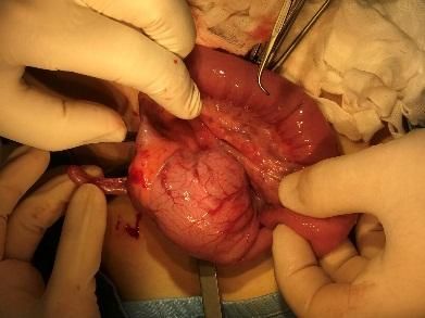
APMB - Atti della Accademia Peloritana dei Pericolanti ISSN 1828-6550
Classe di Scienze Medico Biologiche
Vol. 106(1) 2018
DOI: 10.6092 / 1828-6550 / APMB.106.1.2018.A5
ovarian cyst of about 4 mm and no other relevant symptoms in the postnatal period. Because of
referred poor weight gain, constipation and recurrent abdominal pain at age of one she was been
investigated for celiac disease and cystic fibrosis with negative results. During a routinary clinical
evaluation by General Practioner, an abdominal mass was found in the right hypochondrium and
she was referred to the Pediatric Emergency Department where she underwent an
ultrasonographic evaluation and was admitted at Pediatric Surgery Department. During the
hospitalization, additional investigations were performed.
A MRI revealed a well-defined mass (6 x 4.2 x 5.4 cm) in the right mid abdomen. The mass
displaced the bowel to the left and anteriorly and appeared to be strongly adherent to the last part
of the ileum. 99 Tcm-perechnetate scanning showed no ectopic gastric mucosa.
A diagnostic laparoscopy was planned: a large cystic mass occupied the inferior-right quadrant of
the abdomen. Pelvic exploration allowed excluding adnexal mass. The cyst was laparoscopically
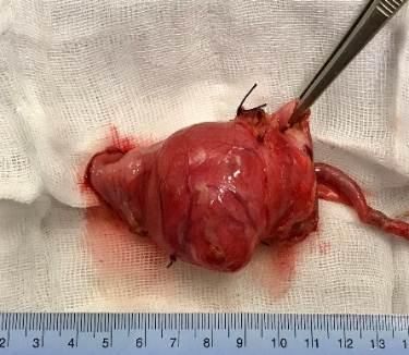
mobilized and a median sub-umbilical incision (2 cm) was made to bring out the mass. The mass
shared a common muscular wall with the terminal ileum, close to the valve and compressed the
cecum. The cecal rim was not clearly evident so a segmental bowel resection was necessary with
the loss of ileo-colic valve (Fig. 1)
Fig. 1 Intraoperative view (a and b) and view of the excised mass (c)
a b c
The histopathology examination revealed a cystic mass contained stained fluid, compatible with a
duplication cystic mass. The mucosa of the cyst included heterotopic immature gastric
mucosa(Fig. 2) better evidenced at magnification (Fig. 3).
The post-operative course was uneventful .
.
APMB - Atti della Accademia Peloritana dei Pericolanti - Classe di Scienze Medico-Biologiche (2018), 106(1):A5(1-7)
DOI: 10.6092 / 1828-6550 / APMB.106.1.2018.A5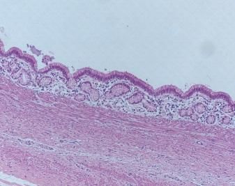
APMB - Atti della Accademia Peloritana dei Pericolanti                                                                        ISSN 1828-6550
       Classe di Scienze Medico Biologiche
                  Vol. 106(1) 2018
                                                                                             DOI: 10.6092 / 1828-6550 / APMB.106.1.2018.A5

Fig.2 Double lumen sharing a common muscular wall. Columnar
ileal epithelium with goblet cells (left), immature heterotopic gastric   Fig 3.      A higher-power magnification shows immature
mucosa (right) (H&E, 50x)                                                 heterotopic gastric mucosa (H&E, 100x)

Discussion
The term ‘‘digestive duplications’’ was firstly used by Calder in 1733s and later by Fitz in 1884
even if it was not widely used until it was popularized by Ladd in 1937 (5, 6) . It refers to cystic
or tubular formations in intimate contact with the various segments of the alimentary canal (3)
(Table 1). They can be located at any level of the gastrointestinal tract, but most commonly at the
mid or terminal ileum (2, 7). They can present at any age but about 80% of cases present within
first two years, mostly during the first three months of life (8, 9). They are thought to arise from
disturbances in embryologic development. Multiple theories have been postulated to account for
their development even if to date no single theory has explained the origin, various locations
involved, and the associated anomalies of the duplication cyst. Bentley and Smith (10) proposed
that the primary defect was the development of a split notochord that connect the yolk sac
endoderm to the ectoderm and that subsequent duplication of the gut resulted from eventration of
the yolk sac between the halves of the vertebra. Other authors suggested that a abnormal
recanalization of the gut could explain the pathogenesis of ED (11).
Parker et al described duplication of the alimentary tract to form a cystic or spherical structure
attached to a part of the bowel, sharing a wall of smooth muscle and lined by a mucous
membrane similar to some part of the alimentary canal. ED symptoms varies according to size,
location, type of duplication, and presence of heterotopic mucosa. Prenatal ultrasonography can
detect ED as early as 16 weeks of gestational age (12). In some case ED may be completely
asymptomatic and they are identified on routine physical examination or during incidental
investigations (13).

               APMB - Atti della Accademia Peloritana dei Pericolanti - Classe di Scienze Medico-Biologiche (2018), 106(1):A5(1-7)
                                              DOI: 10.6092 / 1828-6550 / APMB.106.1.2018.A5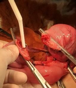
APMB - Atti della Accademia Peloritana dei Pericolanti                                                                       ISSN 1828-6550
       Classe di Scienze Medico Biologiche
                  Vol. 106(1) 2018
                                                                                              DOI: 10.6092 / 1828-6550 / APMB.106.1.2018.A5

                                Table 1. Prevalence in % of duplications for morphology, sex and localization.

 TOTAL CASES                       % CISTIC                        % TUBULAR                          Morphology Nd

 220                               80 %                            14 %                               6%

 TOTAL CASES                      F                                 M                                 Sex Nd

 220                              40 %                              52 %                              8%

 TOTAL CASES                       FOREGUT                         MIDGUT                             HINDGUT

 220                               25,45 %                         64,10 %                            10,45 %

Nd = not described

In our case the localization of ED was at IC region. ED at the IC junction is considered a
particular entity, to be distinguished by other ileal ED as such. Only few Authors described the IC
type, for which the operative approach consisted of segmental resection with primary
anastomosis (14) . In particular Puligandla et al. examined the duplications found in the IC valve
region as an independent entity from the duplications found in the ileum and described a
frequency of 30,2% and 31% respectively, reporting the largest series (15). In our case pain and
constipation were the only symptoms and the mass was incidentally found during a routine
medical examination. The preoperative diagnosis duplication of cysts are often inaccurate (16).
USG is the imaging modality of choice for the evaluation of an abdominal mass in the neonate
which can demonstrate nature and location of the mass. Using scans during pregnancy result in a
higher rate of antenatal detection of duplications which allow early treatment and avoidance of
possible complications (17). Ultrasound plays a crucial role in the diagnosis of intestinal
duplication because it identifies the cyst and its anatomical location (18). CT scans are more
useful in demonstrating the precise anatomical relationship between the cysts and surrounding
structures (19). These cysts can manifest as smooth, rounded, fluid filled cysts or tubular
structure with thin slightly enhancing wall on CT scan. Magnetic resonance imaging (MRI) and
endoscopic ultrasonography are other diagnostic modalities (20). Radioisotope scanning is useful
for evaluation of bleeding from these cysts. However, all these modalities allow us only to
suspect the presence of an abnormal lesion and diagnostic confirmation is possible only after
resection. In our case there was an antenatal diagnosis of presumptive ovarian cyst. Abdominal
ultrasound and MRI revealed the presence of the mass but the differential diagnosis between ED

              APMB - Atti della Accademia Peloritana dei Pericolanti - Classe di Scienze Medico-Biologiche (2018), 106(1):A5(1-7)
                                             DOI: 10.6092 / 1828-6550 / APMB.106.1.2018.A5
APMB - Atti della Accademia Peloritana dei Pericolanti ISSN 1828-6550
Classe di Scienze Medico Biologiche
Vol. 106(1) 2018
DOI: 10.6092 / 1828-6550 / APMB.106.1.2018.A5
and other mass was not reliable. Also Technetium-99m pertechnetate scanning was negative for
heterotopic gastric mucosa. Histopathological examination enabled us to confirm the diagnosis
and revealed the presence of immature heterotopic gastric mucosa. Usually, the treatment of
choice for cystic masses in children is complete surgical resection, through laparotomy,
laparoscopy or laparo-assisted surgery (11, 21). Total resection when possible should be the aim
of the intervention because the partial excision contains high risk of recurrence (22). Due the risk
of potential complications, ED should be always excised, even if they are asymptomatic
(22,23,24).
Complications include intussusception, intestinal obstruction and ulceration, perforation, and
hemorrhage, due to the presence of gastric mucosa.
Malignant changes can occur in the mucosa of an ED (25). In the literature, from 1955 to 2012
have been reported 67 cases of malignancies arising from ED most involving the large intestine
and rectum. The age of presentation ranged from 12 to 88 years old, but most patients were
between the ages of 40 and 60. Female predominance of 3:1 is found in the colorectum site.
Malignant transformation should be suspected if any abnormal solid component is found within
the duplication or serum CEA or CA19e9 level is elevated. Indeed, although CA19e9 is not
clearly associated with malignancy, the prognostic value of CA19e9 levels in colorectal cancer
has been reported and could be of interest in the diagnosis and management of intestinal
duplications. More authors think that serum levels of CEA may serve as a valuable index for
predicting tumour progress arising from gastrointestinal duplication (26). If malignant change is
found in small bowel duplications, the high rate of lymph node metastases should be considered.
Curative resections could hardly be performed . The prognosis is generally poor once malignant
change has occurred (27).
Conclusions
ED are rare congenital lesions, which may present with nonspecific symptoms. It is important to
make a definitive diagnosis of this rare congenital anomalies since they can lead to significant
complications as perforation, obstruction, hemorrhage and malignancy.
In children with an antenatal diagnosis of mass of unknown origin, ED should be considered.
These children should be followed-up and the persistent of the mass is indication for adjunctive
diagnostic evaluations. The radiologic examinations are nonspecific and none diagnostic tools
can allow a certain diagnosis on its own. Technetium-99m pertechnetate scanning can result
negative if heterotopic gastric mucosa shows immature features. For this reason, the utility of
APMB - Atti della Accademia Peloritana dei Pericolanti - Classe di Scienze Medico-Biologiche (2018), 106(1):A5(1-7)
DOI: 10.6092 / 1828-6550 / APMB.106.1.2018.A5APMB - Atti della Accademia Peloritana dei Pericolanti ISSN 1828-6550
Classe di Scienze Medico Biologiche
Vol. 106(1) 2018
DOI: 10.6092 / 1828-6550 / APMB.106.1.2018.A5
scintigraphic scanning should be reviewed in the diagnostic course. Surgical approach should be
achieved in order to localize the mass and to clarify the diagnosis even in asymptomatic case, to
avoid eventual future severe complications.
Conflicts of Interest: All the authors have no conflict of interest to declare.
References
1. Okur, M.H., Arslan, M.S., Arslan, S., Aydogdu, B., Turkcu, G., Goya, C., Uygun, I., Cigdem,
M.K., Önen, A., Otcu S. (2014). Gastrointestinal tract duplications in children. Eur Rev Med Pharmacol
Sciences; 18: 1507-1512. PMID:24899610
2. Rattan, K.N., Bansal, S., Dhamija, A. (2017). Gatrointestinal Duplication Presenting Neonatal
Intestinal Obstruction : an experience of 15 years at tertiary care centre. J Neonat Surg.; 6:5,. DOI:
10.21699/jns.v5i4.432.
3. Spinelli, C . (2001). Chirurgia Neonatale Delle Malformazioni Dell’intestino Primitivo Di Maggiore
Incidenza. PICCIN, Padova;
4. Gross, R.E., Holcomb, G.W., Farber,S. (1952). Duplication of the alimentary tract. Pediatrics; 9:449–
468 doi: 10.1007/s40477-015-0188-8
5. Fitz, R.H. (1884). Persistent omphalomesenteric remains: their importance in the causation of intestinal
duplication, cyst formation and obstruction. Am J Med Sci.;88:30-57.
6. Gross, R.E. (1953). Duplications of the alimentary tract. The Surgery of Infancy and Childhood.:221-45
7 .Bremer, J.L. (1944). Diverticula and duplications of the intestinal tract. Arch Pathol.; 38:132–140.
8. Lister, J. (1990). Duplications of the alimentary tract. In: Lister J, Irwing M, editors. Neonatal Surgery,
England, Butterworths.:p474-84.
9. Master, V.V., Woods, R.H., Morris, L.L., Freeman, J. (2004). Gastric duplication cyst causing gastric
outlet obstruction. Pediatr Radiol.;34:574–6.
10. Al-Zaiem, M.M. (2011). Assisted laparoscopic excision of huge abdominal cysts in newborns and
infants using the umbilical laparoscopic port incision. J Pediatr Surg;46: 1459-63. DOI:
10.1016/j.jpedsurg.2011.03.004.
11. Di Serafino. M., Mercogliano, C., Vallone, G. (2016). Ultrasound evaluation of the enteric duplication
cyst: the gut signature, J Ultrasound. Jun; 19(2): 131–133. DOI: 10.1007/s40477-015-0188-8
12. Parker, B.C., Guthrie, J., France, N.E., Atwell, J.D. (1972). Gastric duplications in infancy. J Pediatr
Surg.;7:294–298. DOI: https://doi.org/10.1016/0022-3468(72)90128-5
13. Puligandla, P.S., Nguyen, L.T., St-Vil, D., Flageole, H., Bensoussan, A.L., Nguyen, V.H., Laberge,
JM. (2003). Gastrointestinal duplications. J Pediatr Surg.;38:740–744.
DOI: https://doi.org/10.1016/jpsu.2003.50197
14. Ildstad, S.T., Tollerud, D.J., Weiss, R.G. et al. (1988). Duplications of the alimentary tract. Clinical
characteristics, preferred treatment, and associated malformations. Ann Surg.;208:184–189.Crossref
15. Berseth C.L. (1998). Avery’s Diseases of the Newborn. 7th ed. WB Saunders Company;. Disorders of
the intestine and Pancreas. In: Taeusch HW, Ballard RA editors; p. 923.
16. Cavar,S., Bogovic, M., Leutic, T., Antabak, A., Batinica, S. (2006). Intestinal duplications –
experience in 6 cases. Eur Surg Res.;38:329–32. DOI:10.1159/000094021
17. Di Serafino, M., Mercogliano, C., Vallone G. (2015) Ultrasound evaluation of the enteric duplication
cyst: the gut signature. J Ultrasound. Nov 23;19(2):131-3. doi: 10.1007/s40477-015-0188-8. eCollection
2016 Jun..
18. Lee, N.K., Kim, S., Jeon, T.Y., Kim, H.S., Kim, D.H., Seo, H.I., Park, D.Y., Jang, H.J. (2010).
Complications of congenital and developmental abnormalities of the gastrointestinal tract in
adolescents and adults: evaluation with multimodality imaging. Radiographics.;30:1489-507. DOI:
10.1148/rg.306105504,
19. Fanaroff, A.A. (2001) . Care of the High Risk Neonate. 5th ed. Harcourt (India) Pvt Ltd, WB
Saunders; 2001. Selected Disorders of the Gastrointestinal Tract. In: Klaus MH, Fanaroff AA, editor; p.
APMB - Atti della Accademia Peloritana dei Pericolanti - Classe di Scienze Medico-Biologiche (2018), 106(1):A5(1-7)
DOI: 10.6092 / 1828-6550 / APMB.106.1.2018.A5APMB - Atti della Accademia Peloritana dei Pericolanti ISSN 1828-6550
Classe di Scienze Medico Biologiche
Vol. 106(1) 2018
DOI: 10.6092 / 1828-6550 / APMB.106.1.2018.A5
179.
20. Esposito, C., Alicchio, F., Savanelli, A., Ascione, G., Settimi, A. (2009). One-trocar ileo-colic
resection in a newborn infant with a cystic lymphangioma of the small-bowel mesentery. J Laparoendosc
Adv Surg Tech A;19:447-9. DOI: 10.1089/lap.2008.0261.
21. Merrot, T., Anastasescu, R., Pankevych, T., Tercier, S., Garcia S., Alessandrini, P., Guys, J.M. (2006)
Duodenal duplications. Clinical characteristics, embryological hypotheses, histological findings,
treatment. Eur J Pediatr Surg.;16:18–23. DOI: 10.1055/s-2006-923798
22. Holcomb, G.W., Gheissari, A., O’Neill, J.A., Shorter, N.A., Bishop, H.C. (1989). Surgical
management of alimentary tract duplications. Ann Surg; 209: 167-174
23. Iyer, C.P., Mahour, G.H. (1995). Duplications of the alimentary tract in infants and children. J Pediatr
Surg.;30:1267–1270. PMID: 8523222
24. Adair, H.M., Trowell, J.E. (1981). Squamous cell carcinoma arising in a duplication of the small
bowel. J Pathol;133(1):25-31. DOI: 10.1002/path.1711330104
25. Ma, H., Xiao, W., Li, J., Li, Y.(2012). Clinical and pathological analysis of malignancies arising from
alimentary tract duplications. Surgical Oncology 21; 324-330. DOI:
https://doi.org/10.1016/j.suronc.2012.09.001
26. Blank, G., Königsrainer, A., Sipos, B., Ladurner, R. (2012). Adenocarcinoma arising in a cystic
duplication of the small bowel: case report and review of literature,. World Journal of Surgical Oncology,
10:55. DOI: 10.1186/1477-7819-10-55
©2018 by the Author(s); licensee Accademia Peloritana dei Pericolanti (Messina, Italy). This article is an open
access article distributed under the terms and conditions of the Creative Commons Attribution 4.0
International License (https://creativecommons.org/licenses/by/4.0/).
Communicated and received February 20 , 2018, revised March 16, 2018, published on line June 15, 2018
APMB - Atti della Accademia Peloritana dei Pericolanti - Classe di Scienze Medico-Biologiche (2018), 106(1):A5(1-7)
DOI: 10.6092 / 1828-6550 / APMB.106.1.2018.A5You can also read