Influence of Melatonin on Behavioral and Neurological Function of Rats with Focal Cerebral Ischemia-Reperfusion Injury via the JNK/FoxO3a/Bim ...
←
→
Page content transcription
If your browser does not render page correctly, please read the page content below
Hindawi
Computational and Mathematical Methods in Medicine
Volume 2022, Article ID 8202975, 12 pages
https://doi.org/10.1155/2022/8202975

Research Article
Influence of Melatonin on Behavioral and Neurological
Function of Rats with Focal Cerebral Ischemia-Reperfusion
Injury via the JNK/FoxO3a/Bim Pathway

 Xingwang Chen,1,2 Xueyuan Shen,3,4 Jianbo Lai,2 Zhijun Yao,2 Xian Peng,2 Long Wu,2
 Yuantong Ou,2 Huachu Wu,2 Haofeng Zhu,5 and Yiyu Deng 1,6
 1
 The Second School of Clinical Medicine, Southern Medical University, Guangzhou, 510515 Guangdong, China
 2
 Department of Critical Care and Emergency, Department of Cardiology, Shenzhen Hospital of Integrated Traditional Chinese and
 Western Medicine, Shenzhen, 518104 Guangdong, China
 3
 The Second Clinical Medical College, Guangzhou University of Chinese Medicine, Guangzhou, 510515 Guangdong, China
 4
 Department of Critical Care and Emergency, The Second Affiliated Hospital of Guangzhou University of Chinese Medicine,
 Guangdong Provincial Hospital of Chinese Medicine, Guangzhou, 510515 Guangdong, China
 5
 Furong Community Health Service Center, Shenzhen Hospital of Integrated Traditional Chinese and Western Medicine, Shenzhen,
 518104 Guangdong, China
 6
 Department of Critical Care and Emergency, Guangdong Provincial People’s Hospital, Guangzhou, 510515 Guangdong, China

 Correspondence should be addressed to Yiyu Deng; yiyudeng999@sina.com

 Received 28 October 2021; Revised 22 November 2021; Accepted 30 November 2021; Published 17 January 2022

 Academic Editor: Min Tang

 Copyright © 2022 Xingwang Chen et al. This is an open access article distributed under the Creative Commons Attribution
 License, which permits unrestricted use, distribution, and reproduction in any medium, provided the original work is
 properly cited.

 Objective. To investigate the influence of melatonin on behavioral and neurological function of rats with focal cerebral ischemia-
 reperfusion injury via the JNK/FoxO3a/Bim pathway. Methods. One hundred and twenty healthy male SD rats were randomized
 into the model group (Model: the middle cerebral artery occlusion (MCAO) model was constructed and received an equal volume
 of normal saline containing 5% DMSO), sham operation group (Sham: received no treatment except normal feeding), and low,
 medium, and high dose of melatonin group (L-MT, M-MT, and H-MT intraperitoneally injected 10, 20, and 40 mg/kg
 melatonin 30 min after IR, respectively), with 24 rats in each group. Following 24 h of reperfusion, the rats in each of the above
 groups were tested for neurological deficit symptoms and behavioral changes to screen the rats included in the study. HE and
 TUNEL stainings were performed to observe pathological changes. Levels of oxidative stress-related indexes, inflammatory
 factor-related indexes, nuclear factor-κB p65 (NF-κB p65), and interferon-γ (IFN-γ) in the rat brain were measured by ELISA.
 The JNK/FoxO3a/Bim pathway-related proteins as well as Bcl-2, Caspase-3, and Bax were examined using Western blot.
 Results. Detection of behavioral indicators showed that the MACO model was successfully constructed in rats. L-MT, M-MT,
 and L-MT groups presented reduced malondialdehyde (MDA), reactive oxygen species (ROS), tumor necrosis factor- (TNF-)
 α, interleukin- (IL-) 6, IL-1β, IFN-γ, NF-κB p65, and apoptosis compared with the Model group (P < 0:05), and the
 improvement degree was better in the M-MT group versus the L-HT group. Bcl-2 protein expression in the brain tissue of L-
 MT, M-MT, and H-MT groups increased significantly, while Bax, Caspase-3, p-JNK, p-FoxO3a, and Bim protein expression
 declined markedly, versus the Model group (P < 0:05). The changes of indexes were greater in the M-MT group compared
 with that in the L-MT group. No significant difference was observed in all the above indexes between the M-MT group and
 the H-MT group (P > 0:05). Conclusions. In the MACO rat model, melatonin can effectively reduce Bax and Caspase-3 levels
 by modulating the JNK/FoxO3a/Bim pathway, inhibit neuronal apoptosis, and alleviate neurological deficits by reducing the
 release of proinflammatory mediators, with anti-inflammatory and antioxidant effects. In addition, 20 mg/kg is the optimal
 melatonin concentration.

2 Computational and Mathematical Methods in Medicine

1. Introduction downstream target molecules, is underexpressed. In a neo-
 natal hypoxic ischemia model, Bim, a member of the Bcl-2
Cerebral stroke, also known as stroke [1], is a group of acute family, has been proved to be a critical mediator of neuronal
cerebrovascular diseases caused by sudden rupture of blood apoptosis [19]. Li et al. [20] found that the JNK/FoxO3a/Bim
vessels in the brain or inability of blood flow to the brain pathway is involved in rat neuronal apoptosis after hypoxic-
due to vascular blockage, including hemorrhagic stroke ischemic brain injury, and the regulation of FoxO3a by JNK
(HS) and ischemic stroke (IS) [2, 3]. The incidence of stroke may not depend on Akt. However, the influence of MT reg-
is increasing year by year worldwide, and the prevalence of ulation of the JNK/FoxO3a/Bim pathway on focal cerebral
IS was higher than that of HS [4]. The mortality/disability IRI remains to be clarified. The innovation and significance
rate of patients with acute IS is relatively high, which not of our study were to investigate the influence of MT regula-
only greatly increases the family financial burden and tion of the JNK/FoxO3a/Bim pathway on focal cerebral IRI,
reduces the living ability of the patients but also aggravates which might obtain some conclusions regarding the mecha-
the economic and social burden. Therefore, clarifying its nism of action of MT on focal cerebral IRI through a new
pathogenesis and carrying out targeted treatment is of pathway. This study explored the influence of MT on behav-
utmost importance. ioral and neurological functions of rats with focal cerebral
 At present, the treatment of IS is mainly aimed at reper- IRI via the JNK/FoxO3a/Bim pathway.
fusion, neuroprotection, and neurological recovery [5].
However, in the reperfusion process of recovering blood 2. Materials and Methods
flow after cerebral ischemia, it will cause more serious dam-
age to brain tissue, namely ischemia-reperfusion injury (IRI) 2.1. Experimental Animals and Groups. Adult healthy male
[6]. Because of the long-term ischemia of brain tissue, local SD rats of SFP grade (body mass: 280-320 g, average weight:
inflammation and reactive oxygen species accelerate to pro- 295:72 ± 10:23 g; Animal Laboratory, Guangzhou University
duce when the blood supply returns to normal, which, of Chinese Medicine) were assigned to the model group
instead of promoting the recovery of brain function, will (Model), sham operation group (Sham), low dose of melato-
aggravate brain injury and capillary dysfunction and lead nin group (L-MT), medium dose of melatonin group (M-
to secondary brain injury, brain edema, hemorrhagic trans- MT), and high dose of melatonin group (H-MT) according
formation, necrosis, and free radical injury with infarction to the random-number table method, with 24 rats in each
growth, resulting in strong neuroinflammatory reaction group. And the other rats serve as substitutes. The rats were
and even death in severe cases [7, 8]. The formation of focal accommodated for two weeks in the environment of room
cerebral ischemia injury is closely associated with neuronal temperature 22-23°C, relative humidity of 45-60%, and
apoptosis [9]. Melatonin (MT) is a tryptophan metabolite 12 : 12 h light-dark cycle. All animal experiments were
released from the pineal gland. As a broad-spectrum antiox- approved by the relevant ethics committee and were carried
idant and an effective free radical scavenger, it plays a neuro- out in accordance with the National Institutes of Health
protective role through antioxidant, antiapoptotic, and anti- guidelines.
inflammatory actions, in addition to the function of regulat-
ing sleep [10]. Besides, by inhibiting the Rac1/JNK/FoxO3a/ 2.2. Pretreatment of Rats and Establishment of Models. After
Bim pathway, MT can protect cardiomyocytes against successful intraperitoneal anesthesia with 1% pentobarbital
oxygen-glucose deprivation and reperfusion [11]. MT has sodium (40 mg/kg), different operations were performed in
also been correlated with brain damage. Previous literature each group. An IRI model of middle cerebral artery occlu-
has reported that the administration of MT after cerebral sion (MCAO) was constructed using the modified Longa
ischemia can significantly reduce infarct volume [12]. How- method [21]. Reperfusion was completed 6 h after ischemia
ever, the underlying mechanism and pathway of action of by directly pulling the nylon thread out of the internal
MT after cerebral ischemia remain to be further elucidated. carotid artery for about 16 mm, and the anal temperature
 As a member of the mitogen-activated protein kinase was kept at 37 ± 0:5° C until 2 h after reperfusion. Criteria
family, C-Jun amino terminal kinase (JNK) plays an impor- for the successful model establishment were as follows: after
tant role in the regulation of stress, cell differentiation, and the rats woke up, the right forelimb curled and flexed after
apoptosis [13]. In addition, it is shown to be activated in tail lifting and suspension. The rats developed left-sided
models of neuronal apoptosis under excitotoxicity, nutrient Horner syndrome and fell to the right side or turned in cir-
withdrawal, and ischemia inducements [14]. Fork-head cles while crawling. Elimination criteria are as follows: rats
transcription factor (FoxO) is the key effector of JNK- with subarachnoid hemorrhage, no pathological changes of
mediated tumor suppression [15]. Activation of FoxO regu- ischemia, or death at the observation time point were elimi-
lated by JNK enhances the ability of animals to resist oxida- nated. In the process of modeling, a total of 11 rats were
tive stress and longevity [16, 17]. It has been reported [18] eliminated, all of which were supplemented by prepared
that fork-head box O3a (FoxO3a) is involved in the myocar- substitutes. Separation of the left common carotid artery as
dial injury process caused by IRI, and the activation of the well as the internal and external carotid arteries and suture
PI3K/Akt pathway can phosphorylate FoxO3a and transfer without nylon thread insertion were performed in the Sham
it from the nucleus to the cytoplasm to participate in reduc- group. MT (Sigma) was dissolved in dimethyl sulfoxide
ing the apoptosis of cardiomyocytes. Meanwhile, the expres- (DMSO, Sigma) and diluted with normal saline to a final
sion of apoptosis regulatory protein Bim, one of FoxO3a’s 5% DMSO solution. The five groups received the following
Computational and Mathematical Methods in Medicine 3

interventions: L-MT group: intraperitoneally injected with 2.5. HE Staining. The paraffin section specimens for HE
low concentration of MT (10 mg/kg) 30 min after IR; M- staining were taken out and placed at indoor temperature
MT group: intraperitoneally injected with medium concen- for 30 min and then dewaxed in xylene for 5-10 min accord-
tration of MT (20 mg/kg) 30 min after IR; H-MT group: ing to the instructions of HE staining kit (Beyotime Biotech,
intraperitoneally injected with high concentration of MT China). Then, fresh xylene was used for another 5-10 min of
(40 mg/kg) 30 min after IR; Model group: received an equal dewaxing. After that, they were then dehydrated with anhy-
volume of normal saline containing 5% DMSO; and Sham drous ethanol, 90% ethanol, 80% ethanol, and 70% ethanol
group: received no treatment except normal feeding. for 5 min, 2 min, 2 min, and 2 min, respectively, and then
 rinsed with distilled water for 2 min. After dyeing with
 hematoxylin and eosin in turn as well as mounting with neu-
2.3. Behavioral Tests and Neurological Scores. The behavioral
 tral gum, the sections were photographed under an optical
changes of cerebral IRI rats were observed after recovery
 microscope (Olympus, Japan) for image acquisition.
from anesthesia. The maze (size: 76 cm × 72 cm × 57 cm)
was divided into 25 squares. During the test, the rats were 2.6. TUNEL Staining. The paraffin section specimens for
placed in the middle of the grid, and the number of TUNEL staining were taken out and dewaxed with xylene
squares crawled within 10 min was counted. A consistent twice (10 min/time) in strict accordance with the instruc-
lighting intensity and a quiet environment were main- tions of the TUNEL staining kit (Beyotime Biotech). The
tained during each test. The experimental observers used apoptotic cell count and the total count in 5 nonrepetitive
blind test, that is, if the rats showed proper behavior in high magnification fields (400×) were randomly selected
one evaluation but did not show it later, they were scored from each section, and the average percentage of apoptotic
according to the previous one. The more grids crawled, cells in each high magnification field was calculated
the better the neurological function of rats. The method (number of apoptotic cells/total number of cells × 100%).
of Longa et al. [21] was used to score the behavior of four
groups of rats and record the symptoms of neurological 2.7. Oxidative Stress-Related Indicators Assay. The kits used
deficit: 0, no behavioral abnormalities; 1 point, the fore- in this experiment were all purchased from Nanjing Jian-
limb flexion on the opposite side of the infarct presented cheng Bioengineering Institute, China. The freshly preserved
a clenched fist; 2 points, muscle strength and lateral thrust rat hippocampus tissues were cut into small tissue pieces
were weakened on the contralateral limb of infarction; 3 with ophthalmic scissors and added with normal saline at
points, turning to the opposite side or dumping when the ratio of 1 : 9. The treated hippocampus suspension was
walking; and 4 points, consciousness disorder, paralysis, then added into 20 mL glass tissue homogenate for grinding
or death. Rats with a score of 1-3 were considered success- up and down and centrifuged at 4°C 3000 × g for 10 min.
fully modeled and enrolled. The obtained supernatant was used as test samples. After
 incubation with the probe, the relative fluorescence intensity
 (RFI) was detected by the microplate reader (Bio-Rad, USA).
2.4. Sampling and Specimen Preparation. Rats in each group
 Reactive oxygen species (ROS) activity and malondialdehyde
were randomized into two groups with 10 rats each. Samples
 (MDA) content were detected by corresponding detection
were collected 24 h after reperfusion, some of which were
 kits in accordance with the kit instructions.
used for HE and TUNEL stainings, and the others for WB
and ELISA. 2.8. Inflammatory Factor-Related Indicators Assay. The sam-
 HE staining and TUNEL staining are as follows: 24 h ples were the same as those used in Section 2.7. The ELISA
after reperfusion, the rats were successfully anesthetized by method was used to determine the absorbance value at
routine anesthesia and fixed by paraformaldehyde perfusion. 450 nm with a microplate reader. The contents of tumor
Then, the brain was quickly severed on ice. The cerebral cor- necrosis factor- (TNF-) α, interleukin- (IL-) 6, and IL-1β
tex of the ischemic side was fixed in 4% paraformaldehyde were calculated according to the standard curve. The operat-
for 24 h. After alcohol gradient dehydration, xylene dewax- ing steps followed the kit instructions.
ing, and paraffin embedding, paraffin sections were made
with a thickness of about 5 μm. The slices were spread, dehy- 2.9. The Contents of Nuclear Factor-κB p65 (NF-κB p65) and
drated, and baked to make paraffin sections. Interferon-γ (IFN-γ) Assay. Nuclear factor kappa-B (NF-κB)
 Preparation of partial specimens of WB and ELISA is as is an important transcriptional regulator in cells, usually in
follows: After 24 h of reperfusion, the brain tissue of rats was the form of P50-P65 isodimer binding to its inhibitor
quickly and gently removed after routine anesthesia and kappa-B (IKB) in an inactive state. NF-κB P65 can release
placed in a Petri dish on ice. Then, a 1-2 mm deep incision the proinflammatory mediator IFN-γ, which participants
was made along the coronal plane of the brain tissue at a dis- in chronic inflammatory response and produces physiologi-
tance of about 5 mm along the posterior margin of the cere- cal changes through complex molecular regulation.
bral cortex of rats. The tissue near the incision was gently With ophthalmic scissors, the freshly preserved rat hip-
removed with ophthalmic forceps, and the hippocampus pocampal tissue was cut into small pieces, which were then
was gradually exposed. The complete hippocampus was added with normal saline (tissue : normal saline, 1 : 9). The
quickly and gently removed and then placed in a numbered treated hippocampal tissue suspension was added with
freezing tube, which was quickly placed in the refrigerator at 20 mL glass tissue homogenate for grinding up and down
-80°C for cryopreservation. and centrifuged (4°C, 3000 × g) for 10 min to obtain the

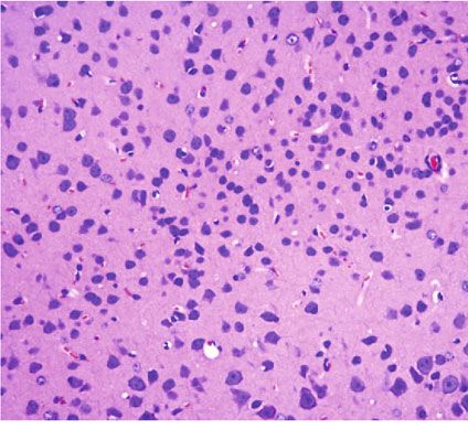
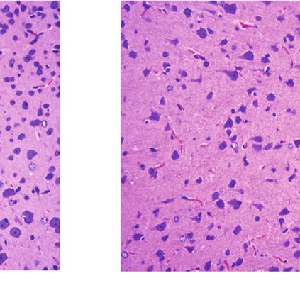
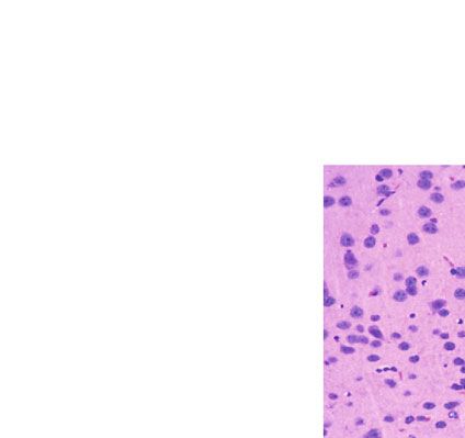
4 Computational and Mathematical Methods in Medicine

 Table 1: Behavioral indicators of rats.

 Behavioral score (n)
 Behavioral indicators (number of squares climbed)
 0 point 1 point 2 points 3 points 4 points
Sham (n = 24) 24 0 0 0 0 412:3 ± 68:4
Model (n = 24) 0 4 6 10 4 329:4 ± 45:1
L-MT (n = 24) 0 8 9 4 3 368:7 ± 42:4
M-MT (n = 24) 0 11 8 3 2 381:4 ± 51:8
H-MT (n = 24) 0 11 9 3 1 385:7 ± 54:2
Sham vs. Model P < 0:0001 P < 0:0001
Sham vs. L-MT P < 0:0001 P = 0:0127
Sham vs. M-MT P < 0:0001 P = 0:0843
Sham vs. H-MT P < 0:0001 P = 0:1422
Model vs. L-MT P = 0:1995 P = 0:0026
Model vs. M-MT P = 0:0463 P = 0:0005
Model vs. H-MT P = 0:0240 P = 0:0003
L-MT vs. M-MT P = 0:8314 P = 0:3963
L-MT vs. H-MT P = 0:6556 P = 0:2606
M-MT vs. H-MT P = 0:9419 P = 0:7799

 Sham Model L-MT

 40 m 40 m 40 m

 M-MT H-MT

 40 m 40 m

 Figure 1: Observation of pathological changes of rat brain tissue by HE staining.

homogenate as the test specimen. NF-κB P65 and IFN-γ was used to determine the protein content. Thereafter,
contents in tissues were detected by ELISA kits (Wuhan 50 μg of the protein sample was treated with routine loading,
Elabscience Biotechnology, China) strictly following the kit electrophoresis, membrane transfer, and 1 h of sealing with
instructions. 5% skim milk powder. Then, the closed membrane was
 moved into the diluted primary antibody working solution
2.10. Western Blot (WB). The cryopreserved rat hippocam- for one night of cultivation at 4°C. The primary antibodies
pal tissue was cut into fine fragments and weighed and included Bcl-2 (1 : 1000, Cell Signaling Technology, USA),
added with 200-400 μL of prepared lysate per 20 mg of tissue Bax (1 : 1000, Cell Signaling Technology, USA), Caspase-3
mass. The tissue was homogenated with a homogenizer until (1 : 1000, Cell Signaling Technology, USA), JNK (1 : 2000,
they were completely decomposed and centrifuged (12000 r/ Cell Signaling Technology, USA), phosphorylated JNK (p-
min, 15 min) to collect the supernatant. The BCA method JNK; 1 : 2000, Cell Signaling Technology, USA), p-FoxO3a
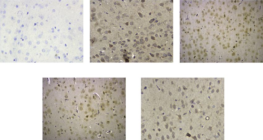
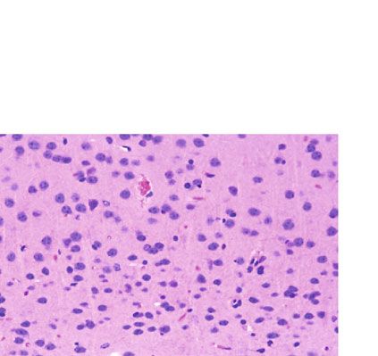
Computational and Mathematical Methods in Medicine 5

 Sham Model L-MT

 40 m 40 m 40 m

 M-MT H-MT

 40 m 40 m

 (a)
 100
 ⁎⁎⁎
 ⁎⁎⁎
 TUNEL positive cells (%)

 80
 ⁎⁎
 60

 40

 20

 0
 Sham Model L-MT M-MT H-MT
 (b)

Figure 2: Observation of the expression of apoptotic cells in rat brain tissue by TUNEL staining. (a) TUNEL staining of brain tissue of rats
in each group. (b) Expression of apoptotic cells in rat brain group in each group. ∗∗ P < 0:01, ∗∗∗ P < 0:001.

(1 : 1000, Cell Signaling Technology, USA), and Bim two groups. Differences among multiple groups were ana-
(1 : 1000, Cell Signaling Technology, USA). After eluting lyzed by one-way ANOVA followed by Bonferroni/Dunnett
with TBST buffer 3 times (5 min/time), it was immersed in post hoc tests. All the analyses with P < 0:05 were considered
horseradish peroxidase-labeled goat anti-rabbit IgG second- significant, using α = 0:05 as the test standard.
ary antibody (1 : 3000, Santa Cruz Biotechnology) and incu-
bated in a shaking table at ambient temperature for 2 h,
followed by TBST elution 3 times (5 min/time). ECL lumi- 3. Results
nescent solution was dripped on the front of the membrane
in a dark room, for detection with an automatic chemilumi- 3.1. Behavioral Indicators. All the 24 rats in the Sham
nescence analyzer. The ImageJ software (version 1.31; group scored 0. The number of rats with a score of 0, 1,
National Institutes of Health, USA) was used to analyze 2, 3, and 4 points was 0, 4, 6, 10, and 4, respectively, in
the gray value of the band relative to internal reference the Model group, while it was 0, 8, 9, 4, and 3, respec-
GAPDH (Cell Signaling, 1 : 1000) for semiquantitative tively, in the L-MT group, 0, 11, 8, 3, and 2, respectively,
analysis. in the M-MT group, and 0, 11, 9, 3, and 1, respectively, in
 the H-MT group. The behavioral scores of rats were lower
2.11. Statistical Processing. SPSS20.0 (SPSS Inc., USA) was in the L-MT and M-MT groups compared with the Model
used for the statistical analysis method. Mean ± standard group (P < 0:05), while the behavioral indexes were not
deviation was used to describe the quantitative data. Two- significantly different between the H-MT group and Sham
tailed t-test was adopted to analyze the differences between group (P > 0:05). Twenty rats from the five groups were
6 Computational and Mathematical Methods in Medicine

 Caspase-3
 2.0

 Protein expression level of Bcl-2
 ⁎⁎⁎
 ⁎⁎⁎
 Bax 1.5 ⁎⁎
 ⁎⁎
 Bcl-2 1.0

 0.5
 GAPDH

 Model 0.0

 L-MT

 M-MT

 H-MT
 Sham

 Sham Model L-MT M-MT H-MT

 (a) (b)

 Protein expression level of caspase-3
 2.5 3
 ⁎⁎⁎ ⁎⁎⁎
 Protein expression level of Bax

 ⁎⁎⁎ ⁎⁎⁎
 2.0 ⁎⁎⁎ ⁎⁎⁎
 ⁎⁎⁎ 2 ⁎⁎⁎
 1.5

 1.0
 1
 0.5

 0.0 0
 Sham Model L-MT M-MT H-MT Sham Model L-MT M-MT H-MT
 (c) (d)

Figure 3: Expression of apoptosis-related proteins in rat brain tissue. (a) Western blot. (b) Bcl-2 protein expression in brain tissue of rats in
each group. (c) Bax protein expression in brain tissue of rats in each group. (d) Caspase-3 protein expression in brain tissue of rats in each
group. ∗∗ P < 0:01, ∗∗∗ P < 0:001.

included, respectively, in the experimental study, as shown (P < 0:05); the H-MT group and the M-MT group had a
in Table 1. similar apoptotic cell count (P > 0:05) (Figure 2).

3.2. HE Staining. The results of HE staining showed that in 3.4. Expression of Apoptosis-Related Proteins. Compared
the central area of ischemic focus, the neuronal gap and with the Sham group, Bcl-2 decreased while Bax and
morphology of rats in Sham group were normal, the nuclei Caspase-3 increased in the Model group (P < 0:05); the L-
were clear, large, and round, and the cytoplasm was light MT, M-MT, and H-MT groups had higher Bcl-2 while lower
and uniform. The cells in the Model group showed a disap- Bax and Caspase-3 in the rat brain tissue than the Model
pearing normal tissue structure, most of which had reduced group (P < 0:05). No significant difference was observed in
size, with loose cytoplasm and pyknotic and deeply stained the expression of apoptosis-related proteins between the
nuclei, and some presented vacuolar necrosis. The ischemic M-MT and H-MT groups (P > 0:05) (Figure 3).
damage of cells in the L-MT, M-MT, and H-MT groups 3.5. Content of Oxidative Stress-Related Indexes in Rat Brain
was notably reduced compared with that in the Model Tissue. The results showed that compared with the Sham
group, with slight edema of cell interstitium, intact cell group, the SOD activity in the Model group decreased, while
membrane, and markedly decreased necrotic area. The the MDA content and ROS level increased, with statistically
improvement degree was greater in the M-MT group as significant differences between the two groups (P < 0:05).
compared to the L-MT group, and the improvement degree Compared with the Model group, SOD activity in L-MT,
of M-MT group and H-MT group was similar (Figure 1). M-MT, and H-MT groups increased, while the MDA con-
 tent and ROS level decreased, and the improvement degree
3.3. TUNEL Staining. TUNEL staining identified no obvious in M-MT group was better than that in the L-MT group,
expression of apoptotic protein in the rat brain tissue in the with statistically significant differences (all P < 0:05). There
Sham group. The expression of apoptotic cells increased in was no significant difference between the M-MT group and
the Model group with different volumes and morphology H-MT group (P > 0:05) (Figure 4).
as well as nucleus pyknosis and agglutination, while the apo-
ptotic cells in brain tissue of the L-MT, M-MT, and H-MT 3.6. Content of Inflammatory Factors in Rat Brain Tissue.
groups decreased as compared to the Model group The TNF-α, IL-1β, and IL-6 levels in the Model group were
Computational and Mathematical Methods in Medicine 7

 300 ⁎⁎⁎
 ⁎⁎⁎ 10
 ⁎⁎ ⁎⁎⁎
 ⁎⁎ 8 ⁎⁎⁎
 ⁎⁎⁎

 MDA (mmol/mL)
 200
 SOD (U/mg)

 6 ⁎⁎

 100 4

 2

 0 0
 Sham Model L-MT M-MT H-MT Sham Model L-MT M-MT H-MT
 (a) (b)
 300 ⁎⁎⁎
 ⁎⁎⁎
 ⁎⁎
 ⁎
 200
 ROS

 100

 0
 Sham Model L-MT M-MT H-MT
 (c)

Figure 4: Content of oxidative stress related indexes in rat brain tissue. (a) SOD activity. (b) MDA content. (c) ROS level. ∗ P < 0:05, ∗∗
 P
< 0:01, ∗∗∗ P < 0:001.

significantly higher than those in the Sham group (P < 0:05). Model group versus the Sham group (P < 0:05). Compared
However, in comparison with the Model group, the above with the Model group, p-FoxO3a and Bim in rat brain tis-
inflammatory factors decreased evidently in the L-MT, M- sues in L-MT, M-MT, and H-MT groups decreased, while
MT, and H-MT groups and were lower in the M-MT group the M-MT group and H-MT group showed similar FoxO3a
compared with the L-MT group (all P < 0:05). No statistical and Bim expression (P > 0:05). Nevertheless, the levels were
difference was found between the M-MT and H-MT groups lower in the M-MT group when compared to the L-MT
(P > 0:05) (Figure 5). group (P < 0:05) (Figure 8).

3.7. Contents of NF-κB P65 and IFN-γ in the Rat Brain. 4. Discussion
ELISA results identified that NF-κB P65 and IFN-γ levels
in the Model, L-MT, M-HT, and H-MT groups were higher In this study, a rat MCAO model, which has a relatively high
than those in the Sham group (all P < 0:05). However, NF- success rate with highly similar clinical symptoms, was
κB P65 and IFN-γ in the L-MT, M-MT, and H-MT groups established by the suture-occluded method [22]. There are
were significantly lower versus the Model group (all P < many causes of focal cerebral ischemia injury, such as com-
0:05), and their levels in the M-MT group was lower when plex cascade injury responses composed of cell apoptosis,
compared to the L-MT group (P < 0:05) (Figure 6). free radical damage, oxidative stress, and inflammation [23,
 24]. In the state of acute stress, the body’s MT concentration
3.8. Comparison of JNK Expression. WB results revealed no decreases, and the function is disorganized. MT has antia-
significant difference in the total protein expression of JNK poptosis, anti-inflammation, and antioxidation effects in
in rat brain tissues among the five groups. The Model group hypoxic-ischemic brain damage [25]. By observing the path-
had higher p-JNK contents than the Sham group (P < 0:05). ological changes of brain tissue in rats with focal cerebral
p-JNK in the L-MT, M-MT, and H-MT groups decreased ischemia, we found that the nerve cells in the Model group
significantly versus the Model group. The level of p-JNK were characterized by the loss of cell structure, hyperchro-
was significantly lower in the M-MT group than that in matism, and cytoplasmic laxity, with vacuolar necrosis in
the L-MT group (P < 0:05) and was similar in the M-MT some cells, which indicated the presence of irreversible
and H-MT groups (P > 0:05) (Figure 7). ischemia and hypoxia injury in the brain tissue during reper-
 fusion injury. However, the brain tissue of rats treated with
3.9. Comparison of FoxO3a and Bim Expression. WB results MT mainly showed swelling of nerve cells, which suggested
determined obviously higher p-FoxO3a and Bim in the that the MT can slows down nerve cell necrosis after
8 Computational and Mathematical Methods in Medicine

 500 ⁎⁎⁎ ⁎⁎⁎
 300
 ⁎⁎⁎ ⁎⁎⁎
 400 ⁎⁎⁎ ⁎⁎⁎
 ⁎⁎
 ⁎⁎
 TNF- (pg/mL)

 IL-1 (pg/mL)
 200
 300

 200
 100
 100

 0 0
 Sham Model L-MT M-MT H-MT Sham Model L-MT M-MT H-MT
 (a) (b)

 200 ⁎⁎⁎
 ⁎⁎⁎
 ⁎⁎⁎
 150 ⁎⁎
 IL-6 (pg/mL)

 100

 50

 0
 Sham Model L-MT M-MT H-MT
 (c)
 ∗∗ ∗∗∗
Figure 5: Content of inflammatory factors in rat brain tissue. (a) Level of TNF-α. (b) Level of IL-1β. (c) Level of IL-6. P < 0:01, P
< 0:001.

 ⁎⁎⁎
 ⁎⁎⁎
 30 12 ⁎⁎⁎
 ⁎⁎⁎ ⁎⁎⁎
 ⁎⁎⁎
 ⁎⁎⁎
 ⁎⁎⁎
 NF- B p65 (pg/mg)

 9
 IFN- (pg/mg)

 20

 6

 10
 3

 0 0
 Sham Model L-MT M-MT H-MT Sham Model L-MT M-MT H-MT
 (a) (b)

Figure 6: Contents of NF-κB p65 and IFN-γ in the brain tissue of rats in each group. (a) NF-κB p65 content. (b) IFN-γ content. ∗∗∗ P <
0:001.

reperfusion injury to a certain extent. Similar findings have transcription and promotes gene transcription and expres-
been found by Gou et al. [26], who reported that exogenous sion in other cells, which is closely related to immune
MT reduced the pathological damage of brain tissue and response, inflammatory response, cellular differentiation,
suppressed neuronal apoptosis in hypoxic-ischemic rats via proliferation, and apoptosis [28]. NF-κB p65 is shown to
the Akt/Nrf2/Gpx4 pathway. activate P38 kinase by releasing the proinflammatory
 This research also showed that MT can effectively medium IFN-γ, which induces cytokine expression and
decrease the expression of Bax and Caspase-3, downregulate major histocompatibility complexes, inducing apoptosis
the levels of NF-κB p65 and IFN-γ in brain tissue, and [29]. The results observed in this study also reflect this
reduce the release of proinflammatory mediators. The acti- mechanism. NF-κB P65 and IFN-γ in the Model group, sig-
vation of inflammatory cells and cascade reaction of inflam- nificantly elevated compared with the Sham group, were
matory factors play a dominant role in reperfusion injury effectively downregulated by MT. The downregulation level
[27]. NF-κB p65 contains a processing region that activates increased with the increase of MT concentration; but the
Computational and Mathematical Methods in Medicine 9

 p-JNK

 1.2

 Protein expression level of JNK
 0.9
 JNK

 0.6

 GAPDH 0.3

 0.0
 Model

 L-MT

 M-MT

 H-MT
 Sham

 Sham Model L-MT M-MT H-MT

 (a) (b)
 ⁎⁎⁎
 1.2 ⁎⁎⁎
 Protein expression level of p-JNK

 ⁎⁎⁎
 ⁎⁎
 0.9

 0.6

 0.3

 0.0
 Sham Model L-MT M-MT H-MT
 (c)
 ∗∗
Figure 7: JNK expression in brain tissue of rats in each group. (a) Western blot. (b) JNK expression. (c) p-JNK expression. P < 0:01,
∗∗∗
 P < 0:001.

changes that resulted from 40 mg/kg of MT were similar to tors including various cytokines, and production of excessive
those caused by medium concentration of MT. Therefore, ROS, resulting in tissue damage [33]. Reducing inflamma-
the optimal MT concentration in this study was 20 mg/kg. tory mediators and releasing and controlling oxidative stress
In addition, the levels of TNF-α, IL-1β, and IL-6 in the rat through drug action are commonly used to alleviate focal
brain tissue in the Model group were significantly higher cerebral ischemia. Our results found that after MT interven-
than those in the Sham group. However, these inflammatory tion, SOD activity increased, MDA content decreased, and
cytokines decreased after intervention with different concen- ROS level decreased in the Model group. SOD is the main
trations of MT, and the decreased levels increased as the enzyme for removing ROS and other components in the
concentration increased, which also indicated that MT has body. When oxidative stress occurs, SOD is constantly con-
an anti-inflammatory effect. Furthermore, we found that sumed, and ROS accumulates in large quantities, resulting in
MT affected neuronal apoptosis by regulating the JNK/ cell damage. Meanwhile, MDA is produced during lipid per-
FoxO3a/Bim pathway. As an important stress kinase, JNK oxidation, which reflects the degree of oxidative stress. It can
can be activated by brain injury in various forms [30]. p- be concluded that MT plays a certain role in antioxidant
JNK is the activated form of JNK. Phosphorylation of acti- stress, which may be accomplished through the regulation
vated JNK stimulates its downstream transcription factors, of oxidative stress produced by the JNK/FoxO3a/Bim signal-
which triggers Bim of the proapoptotic target gene, thus ing pathway. Phosphorylation of FoxO3a can inhibit the
inducing apoptosis. JNK has been shown to modulate activity of FoxO3a and regulate the target gene. Bim, a mem-
FoxO3a activity in many ways [31], while FoxO3a, a sub- ber of the BH3-only subgroup of the Bcl-2 family, is a vital
family of the FoxO family discovered in recent years, partic- proapoptotic protein [34]. It has been confirmed that Bim
ipates in many pathophysiological processes in vivo, is the downstream gene of FoxO3a, and FoxO3a can bind
including regulating apoptosis, oxidative stress, and energy to Bim promoters to enhance Bim protein expression,
metabolism [32]. When focal cerebral ischemia occurs, there thereby initiating the apoptotic process of the mitochondrial
will be infiltration of the white blood cells and brain cells pathway [35]. Of note, FoxO3a is identified as a crucial
(neurons and glia), release of various inflammatory media- downstream target of MT, mediating the transcriptional
10 Computational and Mathematical Methods in Medicine

 ⁎⁎⁎

 Protein expression level of p-Foxo3a
 2.5
 ⁎⁎⁎
 Bim ⁎⁎⁎
 2.0
 ⁎
 1.5
 p-Foxo3a
 1.0

 0.5
 GAPDH
 0.0
 Model

 L-MT

 M-MT

 H-MT
 Sham

 Sham Model L-MT M-MT H-MT

 (a) (b)
 ⁎⁎⁎
 2.5 ⁎
 Protein expression level of Bim

 ⁎⁎⁎
 2.0 ⁎⁎

 1.5

 1.0

 0.5

 0.0
 Sham Model L-MT M-MT H-MT
 (c)

Figure 8: Comparison of p-FoxO3a and Bim expression in the brain tissue of rats in each group. (a) Western blot. (b) p-FoxO3a expression.
(c) Bim expression. ∗ P < 0:05, ∗∗ P < 0:01, ∗∗∗ P < 0:001.

regulation of MT-induced proapoptotic Bim [36]. The of this study was to provide a theoretical basis for the appli-
results of this study also showed that the brain tissue p- cation of MT in clinical work and hope to obtain the clinical
JNK, p-FoxO3a, and Bim levels increased significantly in significance of MT In focal cerebral IRI.
the Model group as compared to the Sham group, while all
these indexes in MT-intervened rat brain tissue reduced. Data Availability
Moreover, MT can effectively reduce the expression of
Caspase-3 in the rat brain tissue in the Model group. Exist- The labeled dataset used to support the findings of this study
ing studies have shown that Caspase-3 activation will pro- is available from the corresponding author upon request.
duce a series of injury reactions, resulting in a large
number of delayed apoptosis in infarcts and ischemic pen- Conflicts of Interest
umbra [37]. In the process of ischemic nerve injury, inhibi-
tion of Caspase-3 activation can produce neuroprotection, The authors declare no competing interests.
which is also proved by the results of this study.
 Authors’ Contributions
5. Conclusion
 Xingwang Chen and Haofeng Zhu contributed equally to
To sum up, MT can reduce the release of NF-κB p65 and this work.
IFN-γ in rats with focal cerebral IRI via the JNK/FoxO3a/
Bim pathway, inhibit neuronal apoptosis, and protect the Acknowledgments
brain against IRI, with anti-inflammatory and antioxidant
effects, which has huge implications for clinical prevention This study was funded by the Shenzhen Baoan District
and treatment of cerebral IRI. In addition, 20 mg/kg is the Science and Technology Planning Project (Project No.
optimal melatonin concentration. However, there are still 2019JD240).
some limitations in this study. Although our finding suggest
that the JNK/FoxO3a/Bim pathway might be a key signaling References
pathways for MT in behavioral and neural regulation, fur-
ther research is needed on the mode of administration of [1] A. Knight-Greenfield, J. J. Q. Nario, and A. Gupta, “Causes of
MT. In addition, this is a preliminary study, and further acute stroke: a patterned approach,” Radiologic Clinics of
studies are required to confirm its findings. The prospect North America, vol. 57, no. 6, pp. 1093–1108, 2019.
Computational and Mathematical Methods in Medicine 11

 [2] S. Paul and E. Candelario-Jalil, “Emerging neuroprotective [18] Y.-H. Sun, R. Bu, Y.-W. Wang et al., “Author correction: vali-
 strategies for the treatment of ischemic stroke: an overview dation of efficacy and mechanism of Sanwei-Tanxiang powder
 of clinical and preclinical studies,” Experimental Neurology, in improving myocardial ischemia reperfusion injuries,” Scien-
 vol. 335, article 113518, 2021. tific Reports, vol. 11, no. 1, pp. 1–12, 2021.
 [3] F. Herpich and F. Rincon, “Management of acute ischemic [19] X. Zhao, Y. Liu, G. Zhu et al., “Sirt1 downregulation mediated
 stroke,” Critical Care Medicine, vol. 48, no. 11, pp. 1654– manganese-induced neuronal apoptosis through activation of
 1663, 2020. foxo3a-bim/puma axis,” Science of the Total Environment,
 [4] C. A. Stack and J. W. Cole, “Ischemic stroke in young adults,” vol. 646, pp. 1047–1055, 2019.
 Current Opinion in Cardiology, vol. 33, no. 6, pp. 594–604, [20] D. Li, X. Li, J. Wu et al., “Involvement of the jnk/foxo3a/bim
 2018. pathway in neuronal apoptosis after hypoxic–ischemic brain
 [5] S. Bhaskar, P. Stanwell, D. Cordato, J. Attia, and C. Levi, damage in neonatal rats,” PLoS One, vol. 10, no. 7, article
 “Reperfusion therapy in acute ischemic stroke: dawn of a e0132998, 2015.
 new era?,” BMC Neurology, vol. 18, no. 1, pp. 1–26, 2018. [21] E. Z. Longa, P. R. Weinstein, S. Carlson, and R. Cummins,
 [6] E. Golts and M. Onaitis, “Commentary: ischemia reperfusion– “Reversible middle cerebral artery occlusion without craniec-
 looking ahead,” The Journal of Thoracic and Cardiovascular tomy in rats,” Stroke, vol. 20, no. 1, pp. 84–91, 1989.
 Surgery, vol. 161, no. 2, pp. e124–e125, 2021. [22] D.-M. Cao, Q.-X. Guan, Y.-L. Liu, and S.-M. Wang, “Effect of
 [7] T. Nakagomi, Y. Tanaka, N. Nakagomi, T. Matsuyama, and ginsenosides on serous metabonomic profiles in cerebral
 S. Yoshimura, “How long are reperfusion therapies beneficial ischemia-reperfusion rats based on~1h-nmr,” Zhongguo
 for patients after stroke onset? Lessons from lethal ischemia Zhong yao za zhi= Zhongguo zhongyao zazhi=China journal
 following early reperfusion in a mouse model of stroke,” Inter- of Chinese materia medica, vol. 45, pp. 1142–1148, 2020.
 national Journal of Molecular Sciences, vol. 21, no. 17, p. 6360, [23] A. Popa-Wagner, E. Petcu, B. Capitanescu, D. Hermann,
 2020. E. Radu, and A. Gresita, “Ageing as a risk factor for cerebral
 [8] T. Imai, H. Matsubara, S. Nakamura, H. Hara, and ischemia. Underlying mechanisms and therapy in animal
 M. Shimazawa, “The mitochondria-targeted peptide, Benda- models and in the clinic,” Mechanisms of Ageing and Develop-
 via, attenuated ischemia/reperfusion- induced stroke damage,” ment, vol. 190, article 111312, 2020.
 Neuroscience, vol. 443, pp. 110–119, 2020. [24] H. Liu, X. Wu, J. Luo et al., “Adiponectin peptide alleviates oxi-
 [9] Y. Naderi, Y. Panahi, G. E. Barreto, and A. Sahebkar, “Neuro- dative stress and nlrp3 inflammasome activation after cerebral
 protective effects of minocycline on focal cerebral ischemia ischemia-reperfusion injury by regulating ampk/gsk-3β,”
 injury: a systematic review,” Neural Regeneration Research, Experimental Neurology, vol. 329, article 113302, 2020.
 vol. 15, no. 5, p. 773, 2020. [25] D. P. Cardinali, “An assessment of melatonin’s therapeutic
[10] J. Cipolla-Neto and F. G. . Amaral, “Melatonin as a hormone: value in the hypoxic-ischemic encephalopathy of the new-
 new physiological and clinical insights,” Endocrine Reviews, born,” Frontiers in Synaptic Neuroscience, vol. 11, p. 34, 2019.
 vol. 39, no. 6, pp. 990–1028, 2018. [26] Z. Gou, X. Su, X. Hu et al., “Melatonin improves hypoxic-
[11] Y. Wang, Y. Jian, X. Zhang, B. Ni, M. Wang, and C. Pan, Mel- ischemic brain damage through the akt/nrf2/gpx4 signaling
 atonin Protects Cardiomyocytes from Oxygen Glucose Depriva- pathway,” Brain Research Bulletin, vol. 163, pp. 40–48, 2020.
 tion and Reperfusion-Induced Injury by Inhibiting rac1/JNK/ [27] H. Liu, X. Wu, J. Luo et al., “Pterostilbene attenuates astrocytic
 FoxO3a/BIM Signaling Pathway, Research Square, 2021. inflammation and neuronal oxidative injury after ischemia-
[12] M. Zang, Y. Zhao, L. Gao et al., “The circadian nuclear recep- reperfusion by inhibiting NF-κB phosphorylation,” Frontiers
 tor RORα negatively regulates cerebral ischemia- reperfusion in Immunology, vol. 10, p. 2408, 2019.
 injury and mediates the neuroprotective effects of melatonin,” [28] S. Giridharan and M. Srinivasan, “Mechanisms of NF-
 Biochimica et Biophysica Acta (BBA)-Molecular Basis of Dis- κB p65 and strategies for therapeutic manipulation,”
 ease, vol. 1866, no. 11, article 165890, 2020. Journal of Inflammation Research, vol. Volume 11, pp. 407–
[13] M. Irwin, M. Tare, A. Singh et al., “A positive feedback loop of 419, 2018.
 Hippo- and c-Jun-amino-terminal kinase signaling pathways [29] Q. Wang, X. Zhou, Y. Zhao et al., “Polyphyllin i ameliorates
 regulates amyloid-beta-mediated neurodegeneration,” Fron- collagen-induced arthritis by suppressing the inflammation
 tiers in Cell and Developmental Biology, vol. 8, p. 117, 2020. response in macrophages through the nf-κb pathway,” Fron-
[14] Z. Vahidinia, A. Azami Tameh, M. Nejati et al., “The protec- tiers in Immunology, vol. 9, p. 2091, 2018.
 tive effect of bone marrow mesenchymal stem cells in a rat [30] S. Bhowmick, V. D’Mello, and P. Abdul-Muneer, “Synergistic
 model of ischemic stroke via reducing the c-Jun n-terminal inhibition of erk1/2 and jnk, not p38, phosphorylation amelio-
 kinase expression,” Pathology-Research and Practice, vol. 215, rates neuronal damages after traumatic brain injury,” Molecu-
 no. 9, article 152519, 2019. lar Neurobiology, vol. 56, no. 2, pp. 1124–1136, 2019.
[15] Z. Liu, C. Li, G. Wu et al., “Involvement of jnk/foxo1 pathway [31] W. Kong, C. Li, Q. Qi, J. Shen, and K. Chang, “Cardamonin
 in apoptosis induced by severe hypoxia in porcine granulosa induces G2/M arrest and apoptosis via activation of the
 cells,” Theriogenology, vol. 154, pp. 120–127, 2020. JNK–FoxO3a pathway in breast cancer cells,” Cell Biology
[16] B. Bourgeois and T. Madl, “Regulation of cellular senescence- International, vol. 44, no. 1, pp. 177–188, 2020.
 viathe FOXO4-p53 axis,” FEBS Letters, vol. 592, no. 12, [32] C. Fasano, V. Disciglio, S. Bertora, M. Lepore Signorile, and
 pp. 2083–2097, 2018. C. Simone, “Foxo3a from the nucleus to the mitochondria: a
[17] M. Cheng, X. Wu, F. Wang, B. Tan, and J. Hu, “Electro-acu- round trip in cellular stress response,” Cell, vol. 8, no. 9,
 puncture inhibits p66shc-mediated oxidative stress to facilitate p. 1110, 2019.
 functional recovery after spinal cord injury,” Journal of Molec- [33] T. Zhang, Z. Li, Z. Qin et al., “Neuroprotection of chikusetsu
 ular Neuroscience, vol. 70, no. 12, pp. 2031–2040, 2020. saponin v on transient focal cerebral ischemia/reperfusion
12 Computational and Mathematical Methods in Medicine

 and the underlying mechanism,” Phytomedicine, vol. 84, arti-
 cle 153516, 2021.
[34] P. K. Singh, A. Roukounakis, A. Weber et al., “Dynein light
 chain binding determines complex formation and posttransla-
 tional stability of the bcl-2 family members BMF and BIM,”
 Cell Death & Differentiation, vol. 27, no. 2, pp. 434–450, 2020.
[35] W. Liao, C. Zhang, F. Liu, and W. Wang, “Effects of mir-155
 on proliferation and apoptosis by regulating FoxO3a/BIM in
 liver cancer cell line HCCLM3,” European Review for Medical
 and Pharmacological Sciences, vol. 22, pp. 1277–1285, 2018.
[36] T. Ali, S. U. Rahman, Q. Hao et al., “Melatonin prevents neu-
 roinflammation and relieves depression by attenuating
 autophagy impairment through foxo3a regulation,” Journal
 of Pineal Research, vol. 69, no. 2, article e12667, 2020.
[37] M. Wu, H. Zhang, J. Kai et al., “Rapamycin prevents cerebral
 stroke by modulating apoptosis and autophagy in penumbra
 in rats,” Annals of Clinical and Translational Neurology,
 vol. 5, no. 2, pp. 138–146, 2018.
You can also read