Injectable nanocurcumin-dispersed gelatin-pluronic nanocomposite hydrogel platform for burn wound treatment
←
→
Page content transcription
If your browser does not render page correctly, please read the page content below
Bull. Mater. Sci. (2019) 42:71                                                                             © Indian Academy of Sciences
https://doi.org/10.1007/s12034-019-1745-0

Injectable nanocurcumin-dispersed gelatin–pluronic
nanocomposite hydrogel platform for burn wound treatment

              LE HANG DANG1,2,† , NGOC TRINH HUYNH3,4,† , NGOC OANH PHAM1,3 ,
              CONG TRUC NGUYEN3 , MINH THANH VU5 , VAN THOAI DINH2,6 , VAN THU LE2,3
              and NGOC QUYEN TRAN2,3,6,∗
              1 School  of Biotechnology, International University, National Universities in HCMC, 70000 Ho Chi Minh City, Viet Nam
              2 Graduate   University of Science and Technology, Ho Chi Minh City 700000, Viet Nam
              3 Institute of Applied Materials Science, Viet Nam Academy of Science and Technology, Ho Chi Minh City 700000,

              Viet Nam
              4 Faculty of Chemistry, Tra Vinh University, Tra Vinh Province 940000, Viet Nam
              5 Institute of Chemistry and Materials, 17 Hoang Sam 100000, Cau Giay, Ha Noi, Viet Nam
              6 NTT Hi-Tech Institute, Nguyen Tat Thanh University, District 4, Ho Chi Minh City 700000, Viet Nam
              ∗ Author for correspondence (tnquyen@iams.vast.vn)
              † First two authors contributed equally to this study.

              MS received 13 May 2018; accepted 16 August 2018; published online 6 March 2019

              Abstract. To utilize the potent pharmaceutical properties of curcumin (Cur) and gelatin-based materials in tissue regen-
              eration, we fabricated a thermosensitive nanocomposite hydrogel based on pluronic-grafted gelatin (PG) and nanocurcumin
              (nCur) to enhance burn healing. In this method, the amphiphilic PG played a role as a surfactant to prepare and protect nano-
              sized Cur particles, which could overcome the poor dissolution of the phytochemical. The synthesized PG was identified by
              1 H nuclear magnetic resonance. Depending on the amount of Cur, size distribution of the dispersed nCur ranged from 1.5 ± 0.5

              to 16 ± 3.2 nm as observed using transmission electron microscopy and dynamic light scattering. The nCur-dispersed PG
              solution formed nCur–PG nanocomposite hydrogel on warming up to 35◦ C. Release profile indicated sustainable release
              of Cur from the injectable platform. Fibroblast cells were well proliferated on the nanocomposite hydrogel. The nCur–PG
              enhanced the healing process of second-degree burn wound. These results showed potential applications of the biomaterial
              in tissue regeneration.

              Keywords.     Nanocurcumin; gelatin; pluronic F127; nanocomposite hydrogel; wound healing.

1. Introduction                                                       and bioavailability, various new Cur-dispersed formulations
                                                                      in the polymeric or hydrogel platforms and its conjugated
Nowadays, wound and burn healing fields are gaining sig-              derivatives have been developed, for example, Cur–chitosan–
nificant attention in multidisciplinary studies expanding from        alginate blend [8], Cur-loaded poly(ε-caprolactone)-poly
traditional herb to advanced biomaterials or their formula-           (ethylene glycol)-poly(ε-caprolactone) hydrogel [9], Cur-
tions [1,2]. Several kinds of phytochemicals have recently            conjugated hyaluronic acid [10], etc. All these formulations
received much attention in the field due to their broad-              showed its healing ability and its potential in biomedical
spectrum bioactivities [1,2]. Among them, curcumin (Cur),             applications. However, it is difficult to obtain homogeneous
an active substance in turmeric, exhibits multiple pharmaco-          materials due to low Cur dispersion. Regarding this, there is
logical properties such as anti-inflammatory, anti-infectious,        some evidence indicating that Cur-encapsulated platforms for
anti-tumoural and anti-oxidation activities as well as positive       topical applications exhibit a higher effect on wound healing
effects in wound or burn healing [3–5]. In wound healing              than its oral administration [11,12].
applications, several reports have indicated that Cur treat-             Interestingly, several reports have indicated that using
ment reduces healing time in puncture wound models by                 nano-sized Cur improved Cur bioavailability and dispersion.
improving the restoration of structural epidermis and enhanc-         Following this approach, many methods were used to fabri-
ing deposition of collagen as well as vascular density in wound       cate Cur nanoparticles, such as low flow injection, evapora-
sites leading to increased healing effects [4,6]. However, free       tion, precipitation or nanosuspension [13]. Various surfactants
Cur is highly hydrophobic and is poorly absorbed leading to           were exploited to fabricate nanocurcumin (nCur) [14–16].
low bioavailability within the body that partially limits its         The surfactant interactions may cause dramatic changes in
biomedical applications [1,7]. To improve its dispersion              the solubilizing capacity of hydrophobic drugs, rheological

                                                                                                                                         1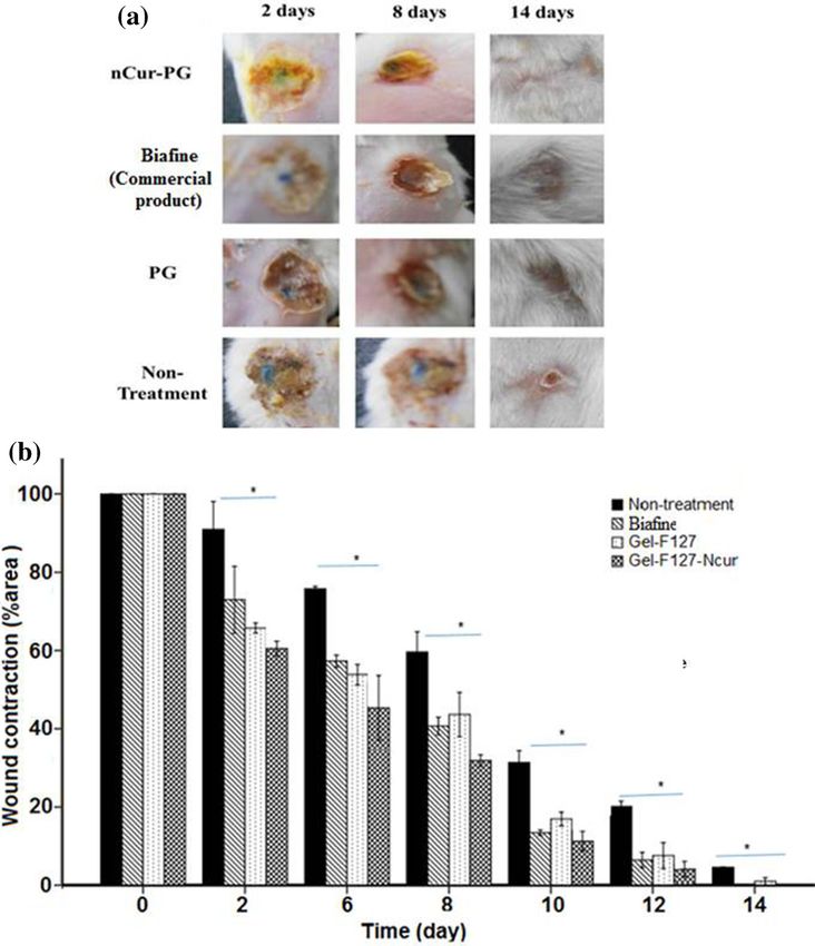
71   Page 2 of 10                                                                                Bull. Mater. Sci. (2019) 42:71

properties of polymer aqueous dispersions and in drug             wt/wt. Briefly, in a round flask, gelatin (1 g) was dissolved
diffusion and penetration through the skin and mucous.            in deionized (DI) water. An aqueous NPC-P-OH (15 g) solu-
Consequently, incorporation of the polymeric surfactant           tion was added drop-wise into the flask at 20◦ C under stirring
opens a wide range of possibilities for developing drug-          overnight. Then, the mixture was dialysed against distilled
delivery systems [16,17]. Up to now, pluronic or poloxamer        water for 3 days using a cellulose membrane (MWCO 14 kDa)
has been one of the best surfactants. Pluronic consists of        and lyophilized to obtain the powder as a thermo-sensitive
hydrophilic poly(ethylene oxide) (PEO) and hydrophobic            copolymer platform for further study as seen in figure 1.
poly(propylene oxide) (PPO) blocks arranged in an A–B–            Grafting yield of samples obtained around 75–80% wt/wt.
A tri-block structure (PEO–PPO–PEO) that is well-known            The copolymer was characterized using 1 H NMR spectrum
for its fast thermally reversible property and being an           and Fourier-transform infrared (FT–IR) spectrum.
Food and Drug Administration (FDA)-approved copolymer
[18]. Because of having both hydrophobic and hydrophilic          2.3 Sol–gel transition behaviour
domains, pluronic displays surfactant properties in interac-
tions with hydrophobic drugs and cellular membranes that          Aqueous copolymer solutions of 0.5 ml were prepared from
play a vital role in drug-delivery platforms. Notwithstand-       varying the PG samples (ratio of G:P = 1:10, 1:15 and 1:20
ing the evidence, some drawbacks of Pluronic F127-based           wt/wt) at 20◦ C. The designated range temperature was set
hydrogels include their weak mechanical strength, rapid ero-      up at 4, 25, 30, 37, 40 and 50◦ C to determine the sol–gel
sion (dissolution of the surface), non-biodegradability at high   transition behaviour of nanocomposite hydrogel using the test
concentrations and limited bio-compatibility [19]. Therefore,     tube inversion method which could observe the ‘flow as the
a recent approach has utilized the pluronic-grafted copoly-       liquid solution’ or ‘no flow as the gel formation’. Sol–gel
mers to overcome the mentioned drawbacks [20,21].                 phase diagram was built using the recorded data.
   In this study, we prepared a thermo-responsive pluronic-
grafted gelatin (PG) copolymer as a dispersant platform for
fabricating nCur under assisted sonication. The colloidal         2.4 Biodegradation test
PG copolymer solution could form an injectable nanocom-
                                                                  To characterize the degradation, 1 ml of 20 w/v% copolymer
posite hydrogel at body temperature that may be useful in
                                                                  samples were dissolved in phosphate-buffered saline (PBS)
tissue regeneration due to beneficial properties of Cur and
                                                                  at 20◦ C and poured into test tubes. The samples were equili-
gelatin-based materials. Gelatin has gained much attention
                                                                  brated in a water bath at 37◦ C and then 5 ml was added into
in tissue engineering because of its high biocompatibility
                                                                  the gel-containing test tubes. At pre-determined time inter-
and biodegradability as it contains Arg–Gly–Asp (RGD)
                                                                  vals, the samples were removed from the buffer, dried and
sequences that promote cell adhesion and migration [22,23].
                                                                  weighed and a fresh PBS solution was added into the tubes
These factors could promote the wound-healing process.
                                                                  with the same volume. Degradation rate was recorded via a
Gelatin-based hydrogel indicated a higher wound contrac-
                                                                  mass difference between each time point, computed by using
tion and re-epithelialization [24]. Therefore, a combination of
                                                                  equation (1), in which Wi is initial dry weight and Wt is dry
Cur nanoparticles and the injectable gelatin-based hydrogels
                                                                  weight at each time point. Data point was performed three
could offer multifunctional biomaterials for second-degree
                                                                  times and expressed as mean ± SE:
burn treatment.
                                                                                         Wi − Wt
                                                                    Weight loss (%) =            × 100.                      (1)
2. Materials and methods                                                                    Wi

2.1 Materials                                                        Fabrication of nCur-dispersed PG copolymer and its nCur–
                                                                  PG form 2.5 mg Cur which was dissolved in 5 ml absolute
Porcine gelatin (bloom 300), Pluronic F127 and Cur were           ethanol under sonication. The suspension was added drop-
purchased from Sigma Aldrich (St. Louis, USA). Mono               wise to the PG copolymer solution (500 mg PG in 2.5 ml DI
p-nitrophenyl chloroformate-activated pluronic (NPC-P-OH)         water and 5 ml ethanol). Then, ethanol solvent was evaporated
was prepared in our previous study [25]. Diethyl ether was        by the rotary evaporator to obtain a homogeneous nCur-
obtained from Scharlau’s Chemicals (Spain), tetrahydrofuran       loaded PG paste form and cold DI water was added to obtain
was purchased from Merck (Germany) and dialysis mem-              thermosensitive nCur-dispersed PG copolymer solution that
branes (MWCO 14 kDa and MWCO 3.5 kDa cut-off) were                could transfer into nCur–PG on warming. Morphology of
supplied from Spectrum Labs (USA).                                nCur was observed using transmission electron microscopy
                                                                  (TEM) (JEM-1400 JEOL) at 25◦ C. Spectral analysis was
2.2 Synthesis of PG copolymers                                    observed by using UV–Vis spectroscopy (Agilent 8453 UV–
                                                                  Vis Spectrophotometer) at 420 nm wavelength. Particle size
In this study, four GP copolymers were prepared at differ-        distribution was determined using dynamic light scattering
ent ratios of gelatin and pluronic 1:05, 1:10, 1:15 and 1:20      (DLS).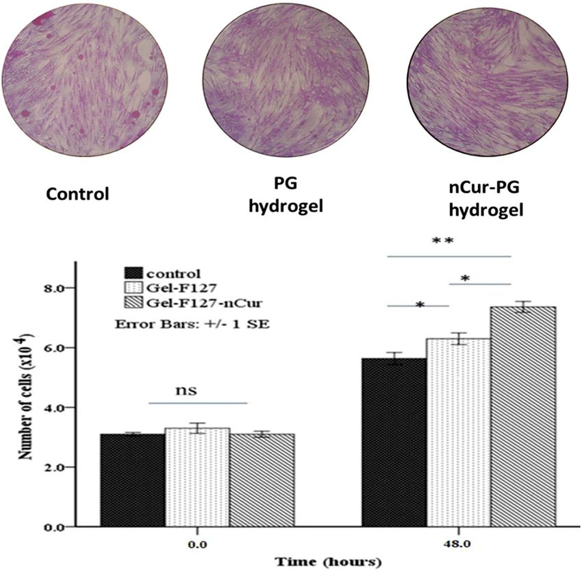
Bull. Mater. Sci. (2019) 42:71                                                                               Page 3 of 10    71

                            Figure 1. Synthetic scheme of PG copolymers.

2.5 Release study                                                Dulbecco’s Modified Eagle Medium and incubated at 37◦ C
                                                                 for 24 h. Then, ∼3 × 10⁴ fibroblast cells were seeded per
In this study, a diffusion method with a dialysis membrane was   well of a 24-well plate with overnight incubation before being
used to investigate the in vitro release of Cur from the nCur-   incubated with these prepared materials similar to the previ-
loaded composite hydrogel that was prepared from 1 ml of         ous procedure for 48 h. Treated cells were fixed with cold 50%
copolymer (20 w/v%) containing 2.5 mg nCur. The dialysis         (w/v) trichloroacetic acid solution for 2 h, washed and stained
bag (MWCO 3.5 kDa) containing 2 ml sample was immersed           with 0.2% (w/v) sulphorhodamine B (SRB) for 20 min. After
in 10 ml PBS at 37 ± 0.5◦ C in a water bath. At selected time    five washes with 1% acetic acid, protein-bound dye was sol-
intervals, 1 ml of sample was collected and replaced by an       ubilized in 10 mM Tris base solution and the absorption at
equal volume of fresh medium. The Cur content was quan-          620 nm on a microplate reader was recorded. Based on the
tified by using an Agilent 8453 UV–Vis Spectrophotometer.        standard curve which was obtained by various amounts of
The release experiments were performed in triplicate with        fibroblasts, we calculated the amount of fibroblast cells on
95% confidence interval. The cumulative release of drug was      the samples.
obtained from the below equation [26]:
                                                                2.7 Wound-healing testing on animal model
   Q = Cn Vt + Vs       Cn−1 ,                            (2)
                                                                 2.7a Animals: Healthy adult male Mus musculus var.
where Cn represents the concentration of drug in sample, Cn−1    Albino mice (33–42 g, n = 6) were procured from the Pasteur
is release concentration at t, Vt the incubated medium and Vs    Institute in Ho Chi Minh city, Viet Nam. Mice were main-
the volume of replaced medium.                                   tained in standard laboratory conditions with ad libitum access
                                                                 to feed and water, light–dark cycles and adequate ventilation.
2.6 Biocompatibility test
                                                                 2.7b Wound creation: The experiment was conducted
According to our screening experiments on behaviour of           at Laboratory of Department of Physiology and Animal
fibroblast with different Cur concentrations as well as appli-   Biotechnology under permission of the Animal Care and Use
cation of the nCur-loaded PG in tissue regeneration, nCur was    Committee of the University of Science, Vietnam National
loaded in the PG hydrogel at low concentration. Two kinds of     University at Ho Chi Minh City (registration no. 10/16-010-
freeze-dried PG hydrogel and nanocomposite hydrogel con-         00), Viet Nam. The mice were anesthetized by intraperitoneal
taining 0.5 wt/wt% of nCur were soaked in 1 ml (15%) of          ketamine (100 mg ml−1 ) and xylazine (20 mg ml−1 ) injection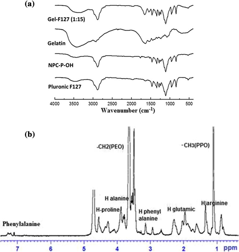
71     Page 4 of 10                                                                                Bull. Mater. Sci. (2019) 42:71

                  Figure 2. (a) FT-IR spectra and (b) 1 H NMR spectrum of PG copolymer compared with the original
                  material.

with a dosage of 0.2 ml×3 100 g×3 body weight. The dor-            2.7c Haematoxylin and eosin (H&E) staining: On 14th
sal skin of the animals was shaved and cleaned with ethanol        day, animals were anaesthetized for tissue sample collec-
(70%) and polyvinylpyrrolidone iodine (1%). A second-              tion. Tissue samples were immediately fixed by immersion in
degree burn was created by a cylindrical stainless steel rod       10% formaldehyde solution, followed by routine histological
of 1 cm diameter which is heated in boiling water at 100◦ C.       processing with paraffin embedding. Histological study was
The rod is maintained in contact with the animal skin on the       performed at the Department of Anapathological Children’s
dorsal proximal region for 5 s. Thereafter, medication was         Hospital 1, Ho Chi Minh City, Viet Nam.
initiated for these four groups (non-treatment, dressing PG,
nCur–PG copolymer (20 w/v%) containing 2.5 mg nCur and             2.7d Statistical analysis: Data are represented as means
commercial product/Biafine). Dressings were performed for          ±standard error (n = 3). Two way analysis of variance (SPPS
every 2 days and completed on day 14. Each mouse contained         software) was used for the analysis of cytotoxicity on fibrob-
two wounds with random treatments. Wound was examined              last cells and wound contraction. A p-value of < 0.05 was
on days 0, 2, 6, 8, 12 and 14. Wound size was measured using       accepted as a statistically significant difference.
a Caliper (0–200 mm Mitutoyo 530-114). The area of wound
contraction was calculated following the equation [27]:            3. Results and discussion
                     π
     Area of wound =    × l i × wi ,                               3.1 Characterization of copolymers
                      4
where li and wi represent length and width of wound surface        Despite an attractive biomaterial for not only tissue regener-
at ith day post-wounding.                                          ation, but also drug-delivery system, raw gelatin shows the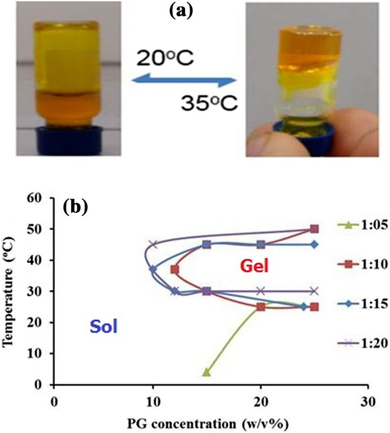
Bull. Mater. Sci. (2019) 42:71                                                                                   Page 5 of 10     71

stiffness in terms of hydrogel due to low mechanical strength.
The most common way of approaching this problem is
modification of gelatin backbone through a grafting method.
In this study, gelatin was modified with Pluronic F127 to
prepare hydrogels with good biodegradation and biocompati-
bility for wound dressing. Pluronic F127 has hydroxyl groups,
was activated with p-NPC (4-nitrophenyl chloroformate) with
two steps as in the previous report [21] resulting in the for-
mation of a NPC-remaining moiety of NPC-P-OH, which
reacted with the amino group on gelatin; consequently, PG
was obtained.
   The structure of the grafted polymer was verified via FT-
IR spectroscopy (figure 2a) by a comparison of absorption
peaks in the infrared spectrum between raw gelatin, NPC-
P-OH as well as PG copolymer. Obviously, a wide peak in
the range of 3500–3100 cm−1 , respectively attributed to the
stretching vibrations of N–H and O–H, shows a strong inten-
sity in gelatin whereas the intensity is lower in Pluronic F127
and NPC-P-OH. Compared to pure gelatin and NPC-P-OH,
the stretching vibration peak of PG copolymer in the range
of 3500–3100 cm−1 shifted to the lower wavenumber from
∼3400 to ∼3350 cm−1 with the increase of Pluronic F127 in
the grafting reaction. In addition, the C = O stretching vibra-
tion peak of the amide in gelatin shifted from 1647 to above       Figure 3. (a) The visual observation of mobility of nCur-GP 1:15
1652 cm−1 in the PG sample. All these changes indicated that       (15% wt/v) at different temperatures, left side shows sol phase
new bonds were formed between gelatin and Pluronic F127.           whereas gel-like phase is seen on the right. (b) Phase diagram of
                                                                   sol–gel transition behaviour of PG copolymer solution built by the
   To provide a strong evidence for the formation of PG
                                                                   inverted tube method.
copolymer, 1 H-NMR spectrum of PG was obtained. In the
spectrum, the resonance peak at 7.23–7.29 ppm indicated aro-
                                                                   On increasing the content of pluronic in the grafted copolymer
matic protons of phenylalanine and other typical protons of
                                                                   (PG 1:10, PG 1:15 and PG 1:20), samples were in the solution
amino acids in gelatin as noted in figure 2b. Moreover, the sig-
                                                                   phase at lower temperatures, but formed transparent hydro-
nal at 3.0 ppm assigned to the primary amino group shifted
                                                                   gels at higher temperatures following the thermal property
from its original position to 2.8 ppm in the PG copolymer
                                                                   of pluronic. For PG 1:10 sample, the gelation occurred when
indicating the presence of the urethane bond. Furthermore,
                                                                   its concentration was higher 12.5 wt/v% at 30◦ C, however,
the exhibiting proton signals of the pluronic (–CH3 of PPO at
                                                                   its physical property was weak. At the same temperature, PG
1.08 ppm and –CH2 of PEO at 3.6 ppm) confirmed that PG
                                                                   1:15 and PG 1:20 showed gelation at lower concentration of
copolymer was successfully prepared.
                                                                   PG copolymer (around 10 w/v%) and the formed gels were
                                                                   highly stable at 15 w/v% of PG which could be used for further
3.2 Thermo-reversible behaviour                                    studies. Furthermore, the thermo-reversible characteristic of
                                                                   PG hydrogel can be applied easily to dissolve for the use in
Thermoreversible PG copolymer for topical delivery of Cur
                                                                   therapeutic agents by cooling the composite hydrogels below
should be gel at skin and body temperatures (32–36◦ C), while
                                                                   their gelation temperature, which is attractive for fundamental
existing as a solution at room temperature. The thermosen-
                                                                   tissue regeneration.
sitive behaviour of PG copolymer with various amounts of
pluronic used in the grafted reaction (PG 1:05, PG 1:10, PG
1:15, PG 1:20) and concentration was investigated by the           3.3 Biodegradation test
inverted test tube method (visual observation of mobility) fol-
lowing the increase of temperature in the range of 4–50◦ C in      To characterize water absorption and stability of PG copoly-
the same manner as in the previous study [21]; presented in        mer, behaviour of the three samples PG 1:15, PG 1:20 and
figure 3a. Phase diagram of sol–gel transition behaviour in        pluronic was tested in PBS buffer of pH 7.4 at 37◦ C as
figure 3b indicates that increasing the F127 concentration led     a function of time. As shown in figure 4, it was found
to the sol–gel transforming temperature following the prop-        that PG 1:15 attained equilibrium swelling by 7 days while
erties of pluronic rather than gelatin properties, which was       pluronic and PG 1:20 as the same concentration (20 wt/v %)
in agreement with previous reports [21]. PG at ratio 1:5 was       showed dramatically different swelling behaviours without
gel-like phase at 4◦ C while in solution phases at higher tem-     the equilibrium. This trend might be due to the enhance-
perature (>30◦ C), corresponding to the property of gelatin.       ment of hydrogen bonding interactions between F127 and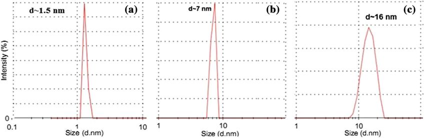
71   Page 6 of 10                                                                                       Bull. Mater. Sci. (2019) 42:71

                                                                      immersing in PBS (pH = 7.4) at 37◦ C, PG 1:15 absorbed
                                                                      much more PBS resulting in dry weight increase in the first 7
                                                                      days and maintained their dry weight in the following 5 days.
                                                                      At day 15, the network of PG 1:15 was broken and the liquid-
                                                                      like content flowed out, causing a dramatically decreased dry
                                                                      weight. Pluronic and PG 1:20 exhibited the weight loss from
                                                                      the initial experiment time. However, pluronic gel was rapidly
                                                                      degraded within 2 days whereas PG 1:20 required 12 days to
                                                                      dissolve completely. This behaviour may be because of two
                                                                      reasons. The conjugation of gelatin molecule with pluronic
                                                                      increased many side chains of the grafted copolymers and
                                                                      resulted in entangled polymer chains enhancing the stability
                                                                      of the hydrogel against degradation and higher swelling lead
                                                                      to a slow mass erosion. These results demonstrated that the
                                                                      PG 1:15 gel had excellent stability in physiologically relevant
                                                                      conditions.
Figure 4. Biodegradation behaviour of the sample PG 1:15, PG
1:20 and Pluronic F127.

                                                                      3.4 Characterization of nCur-loaded thermogel
gelatin in PG 1:15 compared to PG 1:20 due to adjusting
of the hydrophobic–hydrophilic balance in the system [28].            Several reports indicated that nano-scaled Cur could enhance
In the case of examination of sample weight lost during               cellular absorption and biodistribution of the hydrophobic

 Figure 5. TEM image of nCur dispersed in PG 1:15 with (a) 0.5, (b) 5 and (c) 10% (wt/wt) Cur.

 Figure 6. Particle size distribution of nCur dispersed in PG 1:15 with (a) 0.5, (b) 5 and (c) 10% (wt/wt) Cur.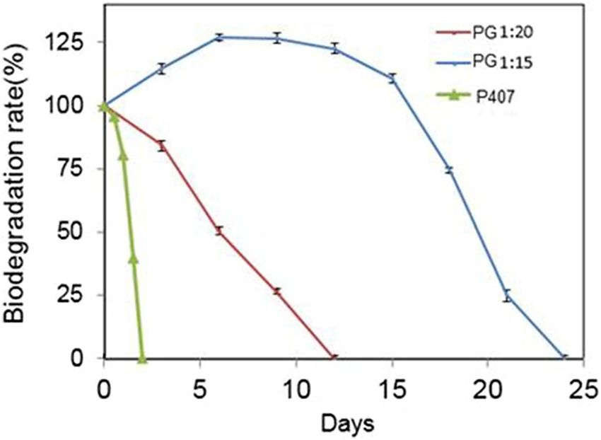
Bull. Mater. Sci. (2019) 42:71                                                                                   Page 7 of 10     71

                                                                    to stability of the nCur in the hydrophobic domain of PG
                                                                    [30]. Moreover, zeta potential measurements showed the pos-
                                                                    itively charged PG copolymer and the negatively charged
                                                                    nCur (data not shown here) which played a significant role in
                                                                    gelatin for enhancing the stability of nCur due its electrostatic
                                                                    interaction. To minimize the Cur/PG complex particle size,
                                                                    the size distribution and morphology of the Cur-loaded PG
                                                                    copolymer, various initial Cur concentration were obtained
                                                                    by TEM (figure 5) and DLS (figure 6), respectively. The size
                                                                    of the round-shaped nCur significantly increases correspond-
                                                                    ing to an increase in Cur concentration at the initial solution.
                                                                    DLS reveals hydrodynamic diameter of nanoparticles as the
                                                                    function of concentration, which is a higher concentration
                                                                    of Cur loaded in the same PG in copolymer solution and a
Figure 7. Release profile of nCur in PG gel in PBS (pH = 7.4) at
                                                                    larger size diameter of formed nanoparticles obtained such
35◦ C.
                                                                    as 1.5±0.5 nm (0.5 wt/wt%), 7±0.5 nm (5 wt/wt%) and
molecule [29]. Therefore, ultrasonication, milling, using sur-      16±3.2 nm (10 wt/wt%). However, all the TEM images show
factant, etc. are attractive methods for nCur processing. In this   the nCur–PG morphological appearance of these nanopar-
study, nCur was formulated in the PG copolymer solution             ticles which are relatively uniform and spherical in shape
along with assisted ultrasonication. Cur powder was dis-            despite the changes in the concentration of the loaded Cur.
solved in ethanol and then added drop-wise into PG solution            The drug release profile is of great importance for practical
and treated in an Ultrasonic device UP200Ht. After soni-            drug-delivery applications of the proposed hydrogel dress-
cation, Cur nanocrystals were separated from solution by            ing. The aim of this study is to investigate whether nCur–PG
centrifugation and re-suspended in DI water for further char-       hydrogel could be used in wound dressing; thus, in vitro
acterization.                                                       drug release studies were conducted via the direct dispersion
   It is more interesting that the nanosuspension solution          method at pH 7.4 in PBS buffer and the release pattern as a
could form nanocomposite hydrogel on being warmed up                function of time is shown in figure 7. The graph elucidates the
(figure 3a). The nCur could form in the PG copolymer                mediated nCur release trend over time, providing the potential
solution as concentrated and the PG copolymer contributes           matrix for drug delivery at the site administration.

                            Figure 8. Cell density in the incubated samples.
71 Page 8 of 10 Bull. Mater. Sci. (2019) 42:71
Figure 9. Macroscopic image of wound surface in animal model at 2, 8 and 14 days post treatment
(a) and the wound contracted area over treatment time (b). The error bar was presented by ±SE.
* is assigned to the statistic difference with p < 0.05) while ns is seen for non-significant difference
( p > 0.05).
3.5 Biocompatibility of nCur-PG the increase of culture time, indicating that the two kinds
of PG hydrogels were able to support cell proliferation.
To evaluate the merits of hydrogel, cytotoxicity test The highest cell density was in PG containing nCur (n=3,
was performed to determine in vitro biocompatibility pBull. Mater. Sci. (2019) 42:71 Page 9 of 10 71
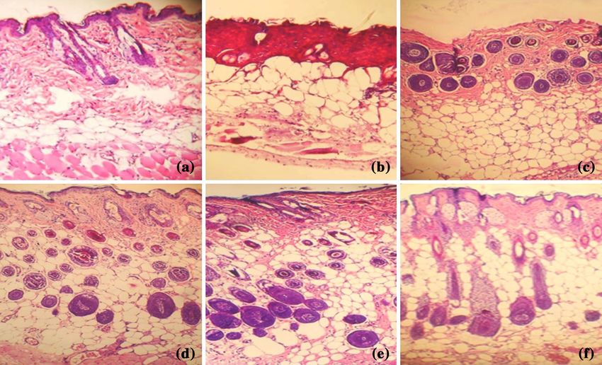
Figure 10. Histology of (a) normal tissue, (b) burn-damaged tissue and wounds at 14 days postwounding: (c) non-treatment, (d) Biafine,
(e) covered with PG gel and (f) nCur–PG.
3.6 Burn healing evaluation ability of scar formation reduced significantly. It was marked
by wound area reduction and wound recovery as seen in
Figure 9 indicates that the PG-treated wound exhibited a figure 9.
faster wound healing rate than that of control, while slower Microscopic images of H&E stained tissue sections are dis-
than the rates observed in nCur–PG and commercial dress- played in figure 10. In comparison with normal tissue, tissue
ings. The nCur–PG model described that wound recovery was sample with a second, degree burn was observed deep into
faster than other groups. Macroscopically, the wounds were dermis with destroyed cellular structure and tissue structure
almost healed for 10 days. On days 2–8, wound treated with as seen in figure 10a and b. After 14 days of healing process,
Gel-F127-nCur exhibited a significant difference ( p71 Page 10 of 10 Bull. Mater. Sci. (2019) 42:71
Based on H&E and Tri results, we suggest that by using [9] Gong C Y, Wu Q J, Wang Y J, Zhang D D, Luo F, Zhao X, Wei
nCur–PG, second-degree burn wounds can be healed not only Y Q and Qian Z Y 2013 Biomaterials 34 6377
at surface structures, but also in the critical barrier function [10] Sharma M, Sahu K, Singh S P and Jain B 2018 Artif. Cells
of skin. Nanomed. Biotechnol. 28 1009
The positive efficacy of the nCur–PG on burn-healing [11] Akbik D, Ghadiri M, Chrzanowski W and Rohanizadeh R 2014
Life Sci. 116 1
process and regeneration of its functional tissue could be
[12] Merrell J G, McLaughlin S W, Tie L, Laurencin C T, Chen
contributed synergistically by gelatin-based hydrogel and the
A F and Nair L S 2009 Clin. Exp. Pharmacol. Physiol. 36
encapsulated nCur in the nCur–PG [34–36]. 1149
[13] Carvalho D M, Takeuchi K P and Geraldine R M and Moura
C J 2015 Food Sci. Technol. 35 115
4. Conclusions [14] Lin C C, Lin H Y, Chen H C, Yu M W and Lee M H 2009 Food
Chem. 116 923
We successfully synthesized the thermosensitive PG copoly- [15] Kakkar V, Singh S, Singla D and Kaur I P 2011 Mol. Nutr. Food
mer which served as a dispersant to produce small size and Res. 55 495
high content of nCur (You can also read