Liraglutide Activates Type 2 Deiodinase and Enhances b3- Adrenergic-Induced Thermogenesis in Mouse Adipose Tissue
←
→
Page content transcription
If your browser does not render page correctly, please read the page content below
BRIEF RESEARCH REPORT
                                                                                                                                               published: 04 January 2022
                                                                                                                                         doi: 10.3389/fendo.2021.803363

                                           Liraglutide Activates Type 2
                                           Deiodinase and Enhances b3-
                                           Adrenergic-Induced Thermogenesis
                                           in Mouse Adipose Tissue
                                           Fernanda C. B. Oliveira 1, Eduarda J. Bauer 1, Carolina M. Ribeiro 1, Sidney A. Pereira 1,
                                           Bruna T. S. Beserra 1, Simone M. Wajner 2, Ana L. Maia 2, Francisco A. R. Neves 1,
                                           Michella S. Coelho 1 and Angelica A. Amato 1*
                                           1 Laboratory of Molecular Pharmacology, School of Health Sciences, University of Brasilia, Brasilia, Brazil, 2 Endocrine

                          Edited by:       Division, Hospital de Clinicas de Porto Alegre, Federal University of Rio Grande do Sul, Porto Alegre, Brazil
         Magdalene K. Montgomery,
The University of Melbourne, Australia

                        Reviewed by:
                                           Aims: Liraglutide is a long-acting glucagon-like peptide 1 (GLP-1) receptor agonist used
                     Dana Hutchinson,      as an anti-hyperglycemic agent in type 2 diabetes treatment and recently approved for
           Monash University, Australia    obesity management. Weight loss is attributed to appetite suppression, but therapy may
                         Dehua Wang,
     Institute of Zoology (CAS), China     also increase energy expenditure. To further investigate the effect of GLP-1 signaling in
                      Kazuhiro Kimura,     thermogenic fat, we assessed adipose tissue oxygen consumption and type 2 deiodinase
            Hokkaido University, Japan
                                           (D2) activity in mice treated with liraglutide, both basally and after b3-adrenergic treatment.
                 *Correspondence:
                   Angelica A. Amato       Methods: Male C57BL/6J mice were randomly assigned to receive liraglutide (400 mg/kg,
                angelicamato@unb.br        n=12) or vehicle (n=12). After 16 days, mice in each group were co-treated with the
     orcid.org/0000-0002-8454-5504
                                           selective b3-adrenergic agonist CL316,243 (1 mg/kg, n=6) or vehicle (n=6) for 5 days.
                   Specialty section:      Adipose tissue depots were assessed for gene and protein expression, oxygen
         This article was submitted to     consumption, and D2 activity.
                              Obesity,
               a section of the journal    Results: Liraglutide increased interscapular brown adipose tissue (iBAT) oxygen
            Frontiers in Endocrinology     consumption and enhanced b3-adrenergic-induced oxygen consumption in iBAT and
        Received: 27 October 2021          inguinal white adipose tissue (ingWAT). These effects were accompanied by upregulation
      Accepted: 13 December 2021
       Published: 04 January 2022          of UCP-1 protein levels in iBAT and ingWAT. Notably, liraglutide increased D2 activity
                             Citation:     without significantly upregulating its mRNA levels in iBAT and exhibited additive effects to
 Oliveira FCB, Bauer EJ, Ribeiro CM,       b3-adrenergic stimulation in inducing D2 activity in ingWAT.
Pereira SA, Beserra BTS, Wajner SM,
Maia AL, Neves FAR, Coelho MS and          Conclusions: Liraglutide exhibits additive effects to those of b3-adrenergic stimulation in
Amato AA (2022) Liraglutide Activates      thermogenic fat and increases D2 activity in BAT, implying that it may activate this adipose
Type 2 Deiodinase and Enhances b3-
Adrenergic-Induced Thermogenesis in
                                           tissue depot by increasing intracellular thyroid activation, adding to the currently known
               Mouse Adipose Tissue.       mechanisms of GLP-1A-induced weight loss.
        Front. Endocrinol. 12:803363.
    doi: 10.3389/fendo.2021.803363         Keywords: GLP-1 receptor agonist, liraglutide, adipose tissue, b3-adrenergic stimulation, type 2 deiodinase

Frontiers in Endocrinology | www.frontiersin.org                                    1                                        January 2022 | Volume 12 | Article 803363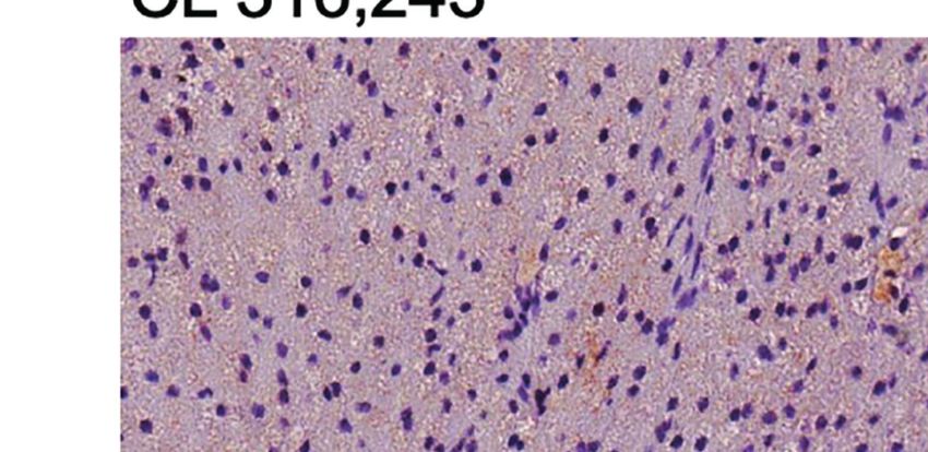
Oliveira et al.                                                                                Liraglutide and b3-Adrenergic Action in Thermogenic Fat

INTRODUCTION                                                              to obese mice on D2 activity in interscapular BAT and inguinal
                                                                          WAT, and on b3-adrenergic-induced UCP-1 expression and
Glucagon-like peptide 1 (GLP-1) is an incretin hormone with               oxygen consumption in both adipose tissue depots.
pleiotropic physiological effects, many of which occur at key sites
of energy balance control and favorably affect metabolic
homeostasis. Supraphysiological GLP-1 signaling by GLP-1A
(GLP-1A) is exploited in the treatment of several metabolic               METHODS
disorders, such as type 2 diabetes, obesity, non-alcoholic fatty
                                                                          Animal Care and Maintenance
liver disease, and cardiovascular disease (1). GLP-1A are
                                                                          The number of animals required for the study was calculated
currently approved for hyperglycemia treatment in type 2
                                                                          using a priori power analysis, considering weight loss as the main
diabetes due to their action to stimulate glucose-dependent
                                                                          outcome (G*Power 3.1.9.4). In a pilot study, liraglutide (200 or
insulin release from pancreatic beta-cells (2). More recently,
                                                                          400 mg/kg body weight/d) or vehicle were administered to 16-
liraglutide, a GLP-1A sharing 97% homology with native GLP-
                                                                          week-old male C57BL/6J mice fed a high-fat diet since the age of
1, was approved as an adjunct to lifestyle therapy in obesity
                                                                          5 weeks (n = 5 mice/group), for 14 days (Supplementary
management in adults (3).
                                                                          Figure 1). As expected, liraglutide treatment at both doses
    Weight loss induced by GLP-1A treatment is largely
                                                                          reduced fasting blood glucose levels and energy intake, in
attributed to reduced appetite and energy intake through GLP-
                                                                          addition to inducing weight loss and diminishing hepatic fat
1 signaling in the hypothalamus. Data from recent rodent and
                                                                          accumulation. There were no signs of toxicity as indicated by
human studies indicate GLP-1 receptors are expressed in adipose
                                                                          animal observation and hepatic histologic features.
tissue and adipocytes (4–6). Moreover, there is evidence that
                                                                             Mice treated with liraglutide at 400 mg/kg body weight/d
GLP-1A may also affect energy expenditure by activating brown
                                                                          exhibited a mean loss of 19.2% of initial body weight (-7.10 ±
adipose tissue (BAT) and recruiting beige/brite adipocytes in
                                                                          1.2 g), whereas vehicle-treated mice gained a mean of mean 2.1%
white adipose tissue (WAT), a process so-called browning of
                                                                          gain of initial body weight (+ 0.79 ± 1.17 g),. Setting type I and II
WAT, through several mechanisms. Central administration of
                                                                          errors at 0.05 and the effect size Cohen’s d = 7.6, a sample size of 5
liraglutide to mice was shown to increase sympathetic nervous
                                                                          mice per group would provide a power of over 95%.
system (SNS) activity, leading to activation BAT thermogenesis
                                                                             Male C57BL/6J mice were purchased from the University of
(7) and browning of epididymal WAT (7) by decreasing
                                                                          Sao Paulo, Brazil, at the age of 4 weeks, and allowed to acclimate
hypothalamic AMPK activity (7). Moreover, peripheral
                                                                          to the new environment for 1 week. They were housed under
administration of exenatide induced browning of epididymal
                                                                          specific pathogen-free conditions, in individually ventilated cages
WAT in mice by upregulating sirtuin 1 expression in adipocytes
                                                                          with 5 to 6 mice per cage until the age of 10 weeks, and 1 mouse
(8), and peripheral administration of liraglutide activated
                                                                          per cage thereafter. Mice were maintained at controlled
invariant natural killer (iNKT) cells resident in WAT to
                                                                          temperature (25°C) and illumination (12-h light/dark cycle),
increase fibroblast growth factor 21 (FGF21) production,
                                                                          with ad libitum access to water/food.
leading to browning of inguinal white fat in mice.
                                                                             At the end of the experiment, mice were anesthetized with
Interestingly, in the latter study it was shown that increased
                                                                          inhalant 5% isoflurane and euthanized by decapitation.
peripheral levels of iNKT cells and FGF-21 were observed in
                                                                          Interscapular BAT (iBAT) and inguinal WAT (ingWAT) were
humans treated with liraglutide and were positively correlated
                                                                          removed, and immediately assessed for oxygen consumption or
with the degree of weight loss induced by therapy (9).
                                                                          frozen in liquid nitrogen and kept at -80°C until analysis.
    The SNS and thyroid hormone (TH) are critical regulators of
                                                                          Experiments were approved by the Ethics Committee of the
BAT and beige/brite adipocyte activity and crosstalk to promote
                                                                          University of Brasilia.
an appropriate thermogenic response (10). The interaction
between SNS and TH has been best characterized in BAT. In
mature brown adipocytes, catecholamines signal through the b3-            Animal Study
adrenergic receptor to activate the thermogenic machinery (11)            After the age of 5 weeks, mice were fed a high-fat diet (60% kcal
and through the a1-adrenergic receptor to promote intracellular           from fat, 20% kcal from protein, and 20% kcal from
TH activation by increasing type 2 deiodinase (D2) activity (12).         carbohydrate; D12492, Research Diets, New Brunswik, NK). At
On the other hand, TH enhances adrenergic signaling in                    17 weeks, they were randomly assigned to receive intraperitoneal
adipocytes through TH receptor alpha activation (13) and                  liraglutide (400 mg/kg/d, Novo Nordisk; n = 11) or vehicle
directly increases the expression of thermogenesis-related genes          (saline; n = 11), for 3 weeks. After 16 days of treatment, five
by TH receptor beta-mediated signaling (10). Despite previous             mice in each group were randomly assigned to receive the b3-
evidence indicating that GLP-1A activate thermogenic fat, at              adrenergic selective agonist CL316,243 (1 mg/kg body weight/d,
least in part, by increasing SNS activity (7), it is unknown              the last dose administered 2 h before euthanasia; Sigma-Aldrich)
whether GLP-1 signaling could affect D2 activity or adrenergic            or vehicle (saline, Figure 1A), using the intraperitoneal route.
action in adipose tissue.                                                 Both randomizations were stratified according to weight and
    To further investigate the effects of GLP-1A on the main              conducted using Microsoft Excel. Body weight and food intake
regulators of the thermogenic response in adipose tissue, we              were measured weekly until 17 weeks, and daily during
examined the impact of prolonged administration of liraglutide            treatment. Caloric or feeding efficiency was calculated by

Frontiers in Endocrinology | www.frontiersin.org                      2                                   January 2022 | Volume 12 | Article 803363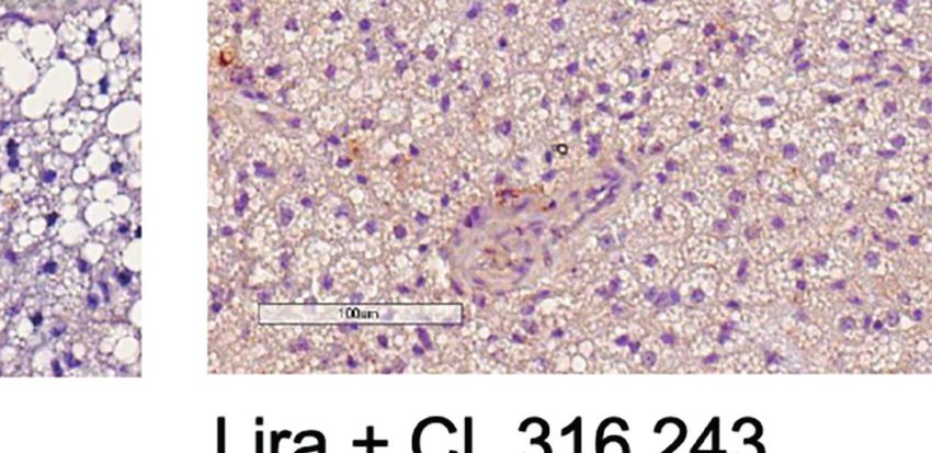
Oliveira et al.                                                                                                   Liraglutide and b3-Adrenergic Action in Thermogenic Fat

                              A                                                                       B

                                      C                                                               D

                                  E                                                                   F

                                      G                                                               H

                                          I                                                       J

  FIGURE 1 | Effect of liraglutide and b3-AR stimulation on fasting blood glucose, body weight, fat mass, and food intake. (A) Study design: male C57BL/6J received
  liraglutide (400 mg/kg/d) or control for 21 days, ± the b3-adrenergic agonist CL 316,243 (CL) during the last 5 days of treatment. (B) Fasting blood glucose levels
  after liraglutide and CL treatment, (C) body weight before and during liraglutide and CL treatment, (D) body weight change after liraglutide and CL treatment,
  (E) energy intake before and during liraglutide and CL treatment, (F) mean daily energy intake during liraglutide and CL treatment, (G) caloric efficiency, and (H) iBAT,
  (I) ingWAT, and (J) epiWAT mass. #p < 0.05 liraglutide-treated mice (Lira and Lira + CL) vs vehicle by Two-way ANOVA followed by Tukey’s multiple comparison
  test. *p < 0.05, **p < 0.01, ***p < 0.001, ****p < 0.0001 vs control or indicated group, by One-way ANOVA followed by Tukey’s multiple comparison test. Data
  presented as mean ± SEM, n=5-6 for each experimental group. CL, CL316,243; epiWAT, epidydimal white adipose tissue; iBAT, interscapular brown adipose tissue;
  ingWAT, inguinal white adipose tissue; Lira, liraglutide.

Frontiers in Endocrinology | www.frontiersin.org                                     3                                        January 2022 | Volume 12 | Article 803363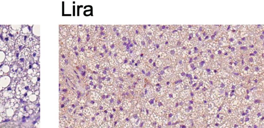
Oliveira et al.                                                                               Liraglutide and b3-Adrenergic Action in Thermogenic Fat

dividing weight gain by energy intake during treatment.                   All reactions were performed in duplicates or triplicates, and the
Treatments and outcome assessment were conducted in the                   experiments were repeated twice for each sample from each
morning (8 to 10 am). The individual mouse was considered                 mouse. Liver samples were assessed as a negative control (16),
the experimental unit. Investigators administering treatments             and no D2 activity was detected (data not shown).
(liraglutide, CL316,243, saline) and assessing outcomes were
aware of the treatment group allocation, whereas data analysis            Data Report and Analysis
was conducted by a different researcher in a blinded manner.              Results were reported using the Animal Research: Reporting In
                                                                          Vivo Experiments guidelines (17), and data from all mice was
Immunohistochemistry                                                      included in data presentation and analysis, since there were no
BAT and WAT immunostaining for UCP-1 was assessed using                   losses. Data were presented as mean ± standard error of mean
polyclonal rabbit anti-UCP-1 antibody (1:100, Santa Cruz                  (SEM), and analyses were performed using GraphPad Prism 7.0
Biotechnology, sc-6528) and R.T.U Vectastain Universal Quick Kit          (GraphPad). Statistical significance was considered at p < 0.05,
(Vector Laboratories, PK-7800). Sections were imaged with a digital       by specific tests described in the figure legends.
slide scanner (Leica Biosystems). ingWAT adipocyte diameter was
determined using ImageJ, as previously described (14).
                                                                          RESULTS
RNA Isolation and Real-Time PCR
Total RNA was isolated from BAT, ingWAT, and epiWAT using                 Liraglutide Decreases Body Weight, Body
Trizol (Invitrogen) and chloroform-isopropanol (Sigma-                    Fat, and Caloric Efficiency
Aldrich), treated with RNase-free DNase I (Sigma-Aldrich)                 Daily peripheral administration of liraglutide lowered fasting
and used for real-time quantitative PCR with Power SYBR®                  blood glucose levels (Figure 1B) and led to persistent weight
Green RNA-to-CT™ 1-Step kit (Applied Biosystems). Relative                decrease after eight days (Figure 1C), inducing significant weight
mRNA expression was calculated by the 2-DDCq method using                 loss at the end of treatment (Figure 1D). Liraglutide treatment
Gapdh as the reference gene (Supplementary Figure 2). Due to              also diminished energy intake (Figures 1E, F). The reduction
low expression levels in WAT, relative Ucp1 mRNA expression               was most pronounced acutely after treatment initiation and was
was calculated by the 2-DCq method (15). Primer sequences are             partially reversed after six days (Figure 1E). Liraglutide also
described in Supplementary Table 1, and MIQE checklist is                 reduced caloric efficiency (Figure 1G), in addition to ingWAT
presented in Supplementary Table 2.                                       and epiWAT mass (Figures 1H–J). Body weight, energy intake,
                                                                          caloric efficiency, and epiWAT mass were further reduced by
Tissue Oxygen Consumption                                                 short-term treatment with CL316,243 in liraglutide-treated mice
Total oxygen consumption was measured in fresh iBAT (20-50                (Figures 1D–H). GLP-1 receptor mRNA was not detected in any
mg sample/mouse) and ingWAT (50-100 mg sample/mouse)                      of the adipose depots assessed (iBAT, ingWAT, and epiWAT;
using a Clark-type electrode (Strathkelvin). Briefly, immediately          data not shown). Arcuate nucleus samples were used as positive
after euthanasia, fat depots were dissected and placed in sterile         controls and exhibited detectable levels of GLP-1 receptor
saline on ice. Samples were cut into pieces weighed (20 to 50 mg          mRNA (mean Cq value of 25.7).
for iBAT and 50-100 mg for ingWAT, from each mouse). The
samples were then placed in empty 1.5 mL tubes and thoroughly             Liraglutide Activates BAT, Induces
minced using a spring scissors. Minced samples were then placed           Browning of WAT, and Enhances
into the respirometer chamber containing respiration buffer (2%           b3-Adrenergic-Induced Adipose
albumin (w/v), 25 mM glucose and 1.1 mM sodium pyruvate in                Tissue Oxygen Consumption
DPBS), at 37°C. The chamber was closed, and the rate of total             Prolonged peripheral administration of liraglutide reduced lipid
oxygen consumption was measured for 15 sec. Total oxygen                  droplet size and upregulated UCP-1 protein expression in iBAT
consumption was normalized to tissue weight and expressed as              (Figure 2A). However, UCP-1 mRNA levels assessed at the end
ug O2/min/mg tissue.                                                      of treatment were unchanged in iBAT of liraglutide-treated
                                                                          mice compared with control (Figure 2B). Accordingly,
Type 2 Deiodinase Activity                                                iBAT from liraglutide-mice exhibited increased oxygen
Tissue samples were homogenized and sonicated on ice in PE                consumption (Figure 2C).
buffer (0.1 M potassium phosphate, 1 mM EDTA) containing                      b3-adrenergic stimulation by CL316,243 treatment for five
0.25 M sucrose, and 10 mM dithiothreitol, pH 6.9. The reaction            days upregulated UCP-1 mRNA and protein expression in iBAT
mixtures containing 100-250 mg tissue proteins were incubated             (Figures 2A, B), in addition to increasing oxygen consumption at
with 100,000 c.p.m. of L-[125I]T4 (Amersham), 1 nM unlabeled              this depot (Figure 2C). Interestingly, liraglutide-induced UCP-1
T4, 20 mM DTT, and 1 mM propylthiouracil (Sigma-Aldrich) in               protein expression and oxygen consumption were enhanced by
PE buffer at 37°C for 2-3 h. Reactions were terminated by the             short-term b3-adrenergic stimulation (Figures 2A, C).
addition of 200 mL horse serum and 100 mL 50% trichloroacetic                 We also assessed the effect of prolonged liraglutide treatment
acid. After centrifugation at 12,000 g for 3 min, free 125I in the        on ingWAT. Liraglutide reduced lipid droplet size and induced
supernatant was counted using a gamma counter. Activity was               the appearance of multilocular adipocytes in ingWAT
expressed as femtomoles iodide generated/min per mg protein.              (Figures 3A, B), a feature of beige/brite adipocytes (18).

Frontiers in Endocrinology | www.frontiersin.org                      4                                  January 2022 | Volume 12 | Article 803363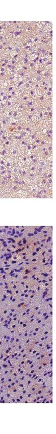
Oliveira et al. Liraglutide and b3-Adrenergic Action in Thermogenic Fat
A
B C
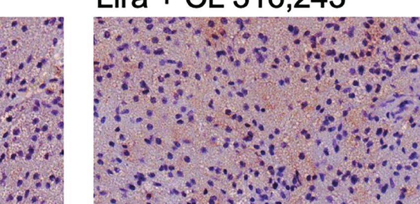
FIGURE 2 | Additive effects of liraglutide and b3-AR stimulation in activating iBAT. (A) iBAT immunostaining for UCP-1 (magnification: X10; scale bar: 100 mm),
(B) iBAT UCP-1 mRNA levels, (C) iBAT oxygen consumption. *p < 0.05, ***p < 0.001, ****p < 0.0001 vs control or indicated group, by One-way ANOVA followed
by Tukey’s multiple comparison test. Data presented as mean ± SEM, n=5-6 for each experimental group. CL, CL316,243; iBAT, interscapular brown adipose
tissue; Lira, liraglutide.
Moreover, liraglutide upregulated UCP-1 protein (Figure 3A) significantly upregulated UCP-1 protein (Figure 3A) and
but not UCP-1 mRNA (Figure 3C) expression in ingWAT, mRNA expression (Figure 3B) compared with either treatment
assessed at the end of treatment. Oxygen consumption was alone. Moreover, oxygen consumption was significantly
unchanged in ingWAT by treatment with liraglutide alone increased by co-treatment with liraglutide and CL316,243,
compared with control (Figure 3D). compared with CL316,243 alone (Figure 3C).
Short-term administration of the b3-adrenergic agonist Prolonged liraglutide treatment did not change UCP-1
CL316,243 did not affect ingWAT adipocyte size (Figure 3A) mRNA levels in epiWAT, but increased UCP-1 protein
but upregulated UCP-1 mRNA expression (Figure 3B) and content. Short term administration of CL316,243 increased
increased oxygen consumption (Figure 3C). In mice treated UCP-1 mRNA and protein levels in epiWAT. In mice
with liraglutide, short-term b3-adrenergic stimulation treated with liraglutide, CL316,243 enhanced UCP-1 protein
Frontiers in Endocrinology | www.frontiersin.org 5 January 2022 | Volume 12 | Article 803363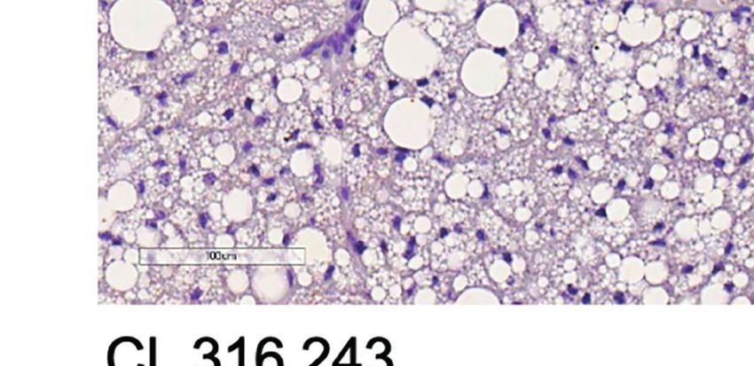
Oliveira et al.                                                                                              Liraglutide and b3-Adrenergic Action in Thermogenic Fat

                                 A

                                 B

                                 C                                                     D

  FIGURE 3 | Additive effects of liraglutide and b3-AR stimulation in inducing browning of ingWAT. (A) ingWAT immunostaining for UCP-1 (magnification: X10; scale
  bar: 200 mm), (B) adipocyte diameter, (C) ingWAT UCP-1 mRNA levels, (D) ingWAT oxygen consumption. *p < 0.05, **p < 0.001, ***p < 0.001, vs control or
  indicated group, by One-way ANOVA followed by Tukey’s multiple comparison test. Data presented as mean ± SEM, n=5-6 for each experimental group. CL,
  CL316,243; ingWAT, inguinal white adipose tissue; Lira, liraglutide.

Frontiers in Endocrinology | www.frontiersin.org                                 6                                      January 2022 | Volume 12 | Article 803363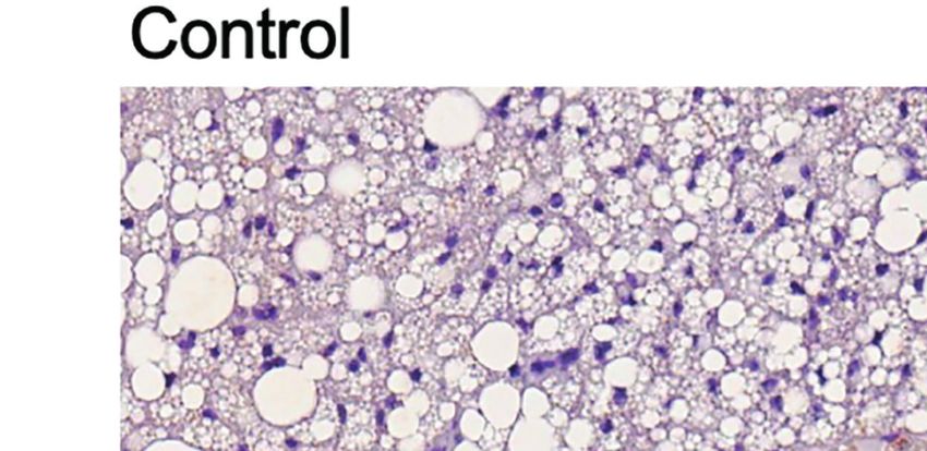
Oliveira et al.                                                                                                   Liraglutide and b3-Adrenergic Action in Thermogenic Fat

expression compared with liraglutide treatment alone                                           Liraglutide did not change D2 activity in ingWAT
(Supplementary Figure 4).                                                                   (Figure 4C), despite significantly inducing D2 mRNA levels
                                                                                            (Figure 4D). However, liraglutide treatment enhanced b3-
Liraglutide Increases Type 2 Deiodinase                                                     adrenergic signaling-induced upregulation of D2 activity
Activity in iBAT                                                                            (Figure 4C). Moreover, liraglutide induced SLC16A2 mRNA
Given the critical role of intracellular TH activation by D2 in                             expression in ingWAT (Figure 4D).
thermogenic adipocytes (19), we investigated whether liraglutide                               In epiWAT, liraglutide treatment did not modify D2 mRNA
treatment affected D2 mRNA expression and activity. D2 activity                             levels or D2 activity, but upregulated SLC16A2 mRNA
was significantly higher in iBAT of vehicle-treated mice when                                expression. Short-term b3-adrenergic agonist treatment
compared with WAT (Supplementary Figure 3). Prolonged                                       increased D2 mRNA levels and did not affect D2 activity
liraglutide administration significantly upregulated D2 activity                             (Supplementary Figure 4).
in iBAT (Figure 4A), despite not changing D2 mRNA levels
(Figure 4B). Short-term b3-adrenergic stimulation with
CL316,243 did not change D2 activity (Figure 4A), despite                                   DISCUSSION
inducing D2 mRNA expression in iBAT (Figure 4B).
Moreover, short-term b3-adrenergic stimulation did not affect                               We report that prolonged peripheral administration of
liraglutide-induced upregulation of D2 activity (Figure 4A). We                             liraglutide increases D2 activity in BAT of obese male mice to
also found that both liraglutide and b3-adrenergic activation                               a greater extent than b3-adrenergic stimulation. To our
increased SLC16A2 mRNA levels, and liraglutide slightly                                     knowledge, this is the first evidence suggesting that liraglutide
induced THRB mRNA levels (Figure 4B).                                                       may modulate brown fat function by inducing D2 activity, which

                               A                                                B

                               C                                                    D

  FIGURE 4 | Effect of liraglutide treatment on D2 activity in adipose tissue. (A) D2 activity in iBAT, (B) mRNA levels of D2, thyroid hormone transporters and thyroid
  hormone receptors in iBAT, (C) D2 activity in ingWAT, (D) mRNA levels of D2, thyroid hormone transporters and thyroid hormone receptors in ingWAT. *p < 0.05,
  **p < 0.01 vs control by One-way ANOVA followed by Tukey’s multiple comparison test. Data presented as mean ± SEM, n=5-6 for each experimental group. CL,
  CL316,243; iBAT, interscapular brown adipose tissue; ingWAT, inguinal white adipose tissue; Lira, liraglutide.

Frontiers in Endocrinology | www.frontiersin.org                                        7                                    January 2022 | Volume 12 | Article 803363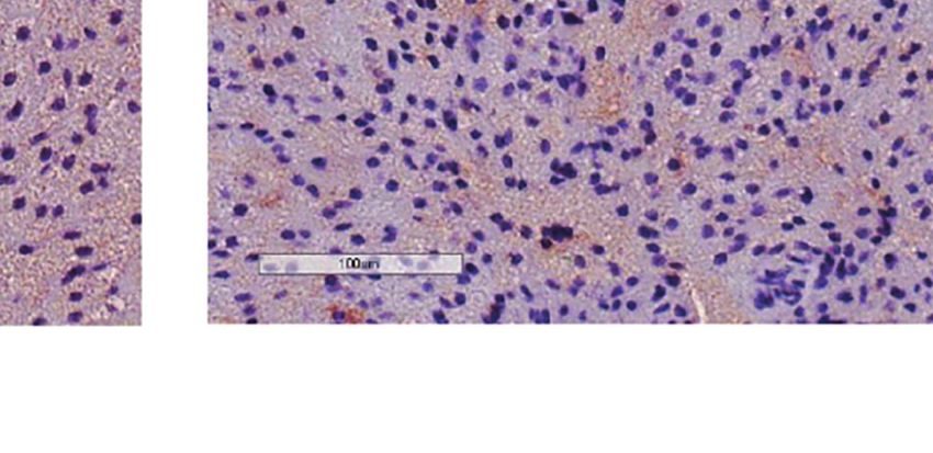
Oliveira et al.                                                                               Liraglutide and b3-Adrenergic Action in Thermogenic Fat

acts to promote intracellular TH activation. In addition, we              decrease caloric output in feces (27). Therefore, it is most likely
observed that liraglutide enhanced UCP-1 upregulation and                 that we underestimated caloric input and the reduction in
oxygen consumption induced by b3-adrenergic signaling in                  caloric efficiency.
both BAT and subcutaneous WAT, a finding that could                            Unlike previous studies that assessed whole-body energy
suggest that liraglutide affects thermogenic fat by mechanisms            expenditure (7, 21), we examined oxygen consumption directly
other than SNS activation.                                                in adipose tissue and found that liraglutide increased oxygen
    Liraglutide treatment reduced body weight, body fat, and              consumption to levels comparable to those induced by b3-
energy intake of male obese mice, in agreement with previous              adrenergic signaling. Increased total oxygen consumption in
data from human (20) and rodent (21) studies. In addition,                BAT and WAT, in the setting of weight loss and reduced
liraglutide-treated mice showed persistent weight loss despite            caloric efficiency, most likely reflects increased thermogenesis.
reduced suppression of food intake after the first week of                 Indeed, weight reduction solely due to diminished energy intake
treatment, suggesting that long-term weight loss induced by               reduces energy expenditure in animal models (28) and humans
GLP-1A is not fully explained by reduced energy intake.                   (29, 30). Therefore, it would be expected to diminish adipose
Alternative explanations for continued weight loss in this                tissue oxygen consumption. Moreover, the increased adipose
setting are fecal energy loss or increased energy expenditure.            tissue oxygen consumption found herein was accompanied by
    Little is known about how GLP-1 signaling affects fecal energy        upregulation of UCP-1 protein levels in iBAT and ingWAT,
loss. In humans, it was reported that GLP-1 (22) or liraglutide           indicating BAT activation and browning of WAT (18). The latter
(23) administration diminished small intestine motility leading           findings are also in agreement with increased thermogenesis.
to increased transit time in subjects with normal glucose                     It is important to point that liraglutide did not significantly
tolerance (22) or type 2 diabetes (23). Moreover, GLP-1                   change iBAT UCP-1 mRNA levels, despite increasing UCP-1
receptor signaling by endogenous GLP-1 or exogenous agonists              protein expression and oxygen consumption in this adipose
exhibited intestinotrophic actions in mice, mediated by fibroblast         depot. This is consistent with previous data indicating that UCP-
growth factor 7 (24). Despite potentially augmenting nutrient             1 mRNA expression does not parallel BAT thermogenic activity (31,
absorption, whether GLP-1 receptor-mediated actions to                    32). Moreover, the higher fold-induction of UCP-1 mRNA in
increase small intestine transit time or promote intestinal               ingWAT of liraglutide- and CL316,243-treated mice, compared
growth affect fecal energy loss remains unexplored.                       with UCP-1 protein and oxygen consumption induction, is also in
Additionally, in mouse models of obesity, liraglutide treatment           accordance with previous findings and most likely stems from lower
induced significant gut microbiome changes (25). Although it is            expression levels in unstimulated WAT (32).
not possible to rule out that the latter changes were secondary to            Interestingly, liraglutide enhanced b3-adrenergic-induced
liraglutide-induced weight loss per se, it is reasonable to               oxygen consumption in both iBAT and ingWAT. UCP-1
hypothesize that gut microbiome composition modifications                  protein levels in iBAT and ingWAT were also additively
could affect nutrient absorption and, therefore, fecal energy loss.       upregulated by liraglutide and b3-adrenergic stimulation. This
    An alternative explanation for continued weight loss induced          finding may reflect nonsympathetically-mediated action of
by liraglutide despite a less pronounced reduction in food intake         liraglutide on thermogenic fat since additive effects to b3-
with prolonged treatment is increased energy expenditure. The             adrenergic stimulation would not be expected if liraglutide
latter is supported by evidence indicating that GLP-1 signaling           promoted oxygen consumption exclusively by SNS activation.
affects energy expenditure in animal models (7, 21) by increasing         The dose of the b3-adrenergic agonist used herein, CL316,243, is
the sympathetic output to thermogenic fat (7). Notably, energy            presumably a saturating one (33). Hence, it would be unlikely to
expenditure changes in response to GLP-1A treatment were also             observe the effect of sympathetic activation in the setting of
assessed in human studies. It was reported that obese subjects            maximal b3-adrenergic activation.
with type 2 diabetes treated liraglutide or exenatide for 52 weeks            Nonsympathetically-mediated actions of liraglutide on
showed a significant decrease in body mass index and total fat             thermogenic fat could be explained by direct signaling on
mass and increase in fat-free mass (7). These findings were                adipose tissue. Indeed, data from cell-based studies indicate
accompanied by increased resting energy expenditure adjusted              direct effects of GLP-1 on adipocytes, such as adipogenesis
for fat-free mass (7). Accordingly, a longitudinal study revealed         induction (5) and activation of both lipogenesis and lipolysis
that 12-week exenatide treatment increased BAT activity                   dependent upon GLP-1/GLP-1A concentration (34, 35).
measured by [ 18 F]fluorodeoxyglucose positron emission                    Importantly, 3T3-L1 adipocyte treatment with exendin-4
tomography (26).                                                          increased mitochondrial biogenesis and function (8). Similarly,
    Although we did not assess energy expenditure, we observed a          liraglutide-treated 3T3-L1 adipocytes exhibited a thermogenic
significant decrease in the weight gain-caloric intake ratio               phenotype, indicated by increased expression of thermogenesis-
(caloric efficiency) of liraglutide-treated mice. This finding              related genes and proteins, in addition to increased
would not be expected in the setting of weight loss induced               mitochondrial biogenesis (36). Although our study design
solely by diminished caloric intake. It should be pointed out that        precludes defining direct actions liraglutide on adipose tissue,
caloric efficiency is most accurately assessed when caloric output         the latter findings from cell-based studies support our hypothesis
in feces is taken into account to determine the net energy input.         that additive effects of liraglutide and maximal b3-adrenergic
However, weight loss due to caloric restriction is expected to            activation on iBAT and ingWAT could suggest that liraglutide

Frontiers in Endocrinology | www.frontiersin.org                      8                                  January 2022 | Volume 12 | Article 803363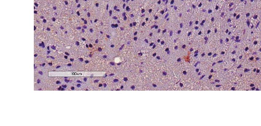
Oliveira et al.                                                                                     Liraglutide and b3-Adrenergic Action in Thermogenic Fat

can affect adipose tissue by mechanisms other than sympathetic                  therefore, activate BAT by promoting intracellular TH activation.
activation, such as direct signaling in adipose tissue. An                      Moreover, liraglutide enhances the effect of b3-adrenergic
additional line of evidence supporting the possibility of a                     stimulation to increase oxygen consumption and UCP-1
nonsympathetically-mediated action of liraglutide on adipose                    expression in both ingWAT and BAT, suggesting it may affect
tissue found herein is the previous report that GLP-1-induced                   thermogenic fat by mechanisms other than SNS activation. These
activation of SNS is blunted in obese mice (37).                                findings may be clinically relevant and add to the currently known
    GLP-1 mRNA was not detected in any of the adipose tissue                    mechanisms of GLP-1A-induced weight loss.
depots assessed herein, although GLP-1 receptor protein expression
was previously shown in both mouse and human adipose tissue/
adipocytes (4–6), as indicated by antibody-based detection                      DATA AVAILABILITY STATEMENT
methods. However, it is acknowledged that available antibodies
against GLP-1 receptor have several limitations with respect to                 The original contributions presented in the study are included in
accuracy, reliability, and reproducibility (38–40). Moreover, it is still       the article/Supplementary Material. Further inquiries can be
unclear whether GLP-1 exerts its direct actions on adipose tissue               directed to the corresponding author.
through the ‘classical’ GLP-1 receptor, which signal through the
adenylate cyclase/cAMP pathway, or ‘alternative’ GLP-1 receptors,
which signal through other pathways (41). It was previously                     ETHICS STATEMENT
reported that the lipolytic action of GLP-1 in 3T3-L1 adipocytes                The animal study was reviewed and approved by Ethics
was accompanied by increased intracellular cAMP levels, implying                Committee of the University of Brasilia.
that the classical receptor was activated by GLP-1 signaling in
adipocytes (4). Accordingly, it was shown that the GLP-1A
exendin-4 activated adenylate cyclase/cAMP/protein kinase A                     AUTHOR CONTRIBUTIONS
pathway to induce the expression of adiponectin and suppress
inflammatory cytokine expression in 3T3-L1 adipocytes (42).                      CR, EB, and SP contributed to data acquisition. FO, SW, and AM
    The possibility of direct GLP-1 actions in adipose tissue                   contributed to data acquisition, analysis, and interpretation, and
mediated by the classical GLP-1 receptor remains elusive.                       writing of the manuscript. MC, FN, and AA contributed to the
However, it should be pointed that the adenylate cyclase/cAMP/                  study concept and design, data analysis and interpretation, and
protein kinase A mediates the action of adrenergic signaling and                writing of the manuscript. FO and AA are the guarantors of this
also of several other activators of thermogenic adipocytes, such as             work and, as such, had full access to all the data in the study and
adenosine (43), the extracellular matrix protein Slit2 (44), transient          take responsibility for the integrity of the data and the accuracy
receptor potential melastatin 8 (45), and bile acids. The latter were           of the analysis. All authors contributed to the article and
reported to increase energy expenditure by activating G-protein-                approved the submitted version.
coupled receptor TGR5/adenylate cyclase/cAMP/protein kinase A
signaling pathway on brown adipocytes, leading to increased D2
activity and intracellular TH activation (46). TH is a critical                 FUNDING
regulator of thermogenic fat activity (19, 47), acting
synergistically with the SNS (48). Intracellular TH action in                   This work was founded by the National Council on Scientific and
brown adipocytes is determined by the availability of cellular T3,              Technological Development (grant 420562/2016-8), and by the
which depends on D2 activity (19).                                              University of Brasilia (grant DPI 01/2021 and DPI 03/2021). The
    Given the abovementioned data indicating that the effects of                study sponsor/funder was not involved in the design of the study;
GLP-1 in adipocytes are mediated by the adenylate cyclase/cAMP/                 the collection, analysis, and interpretation of data; writing the
protein kinase A signaling pathway activated by G-protein coupled               report; and did not impose any restrictions regarding the
receptors, we investigated whether liraglutide action in adipose                publication of the report.
tissue to promote increased oxygen consumption could involve TH
activation by affecting D2 activity. We found that liraglutide, but not
b3-adrenergic stimulation, increased D2 activity in iBAT, possibly              ACKNOWLEDGMENTS
through a nontranscriptional mechanism, since D2 mRNA levels
were not significantly changed. Interestingly, liraglutide treatment             The authors thank Wagner Vasques Dominguez with the
did not affect D2 activity in ingWAT, implying that it may promote              assistance with adipocyte diameter measurement.
TH activation in thermogenic fat in a depot-specific manner.
Liraglutide also increased the transcription of the gene encoding
the TH transporter SLC16A2 in adipose tissue. Given that the latter             SUPPLEMENTARY MATERIAL
mediates TH entry into cells (49), this finding suggests another
possible effect to increase intracellular TH action.                            The Supplementary Material for this article can be found online at:
    In summary, our findings indicate that liraglutide increases D2              https://www.frontiersin.org/articles/10.3389/fendo.2021.803363/
activity and oxygen consumption in BAT from obese mice and may,                 full#supplementary-material

Frontiers in Endocrinology | www.frontiersin.org                            9                                  January 2022 | Volume 12 | Article 803363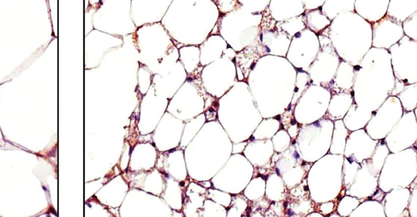
Oliveira et al.                                                                                                         Liraglutide and b3-Adrenergic Action in Thermogenic Fat

REFERENCES                                                                                          Management. N Engl J Med (2015) 373(1):11–22. doi: 10.1056/
                                                                                                    NEJMoa1411892
 1. Andersen A, Lund A, Knop FK, Vilsbøll T. Glucagon-Like Peptide 1 in Health                21.   Raun K, von Voss P, Gotfredsen CF, Golozoubova V, Rolin B, Knudsen LB.
    and Disease. Nat Rev Endocrinol (2018) 14(7):390–403. doi: 10.1038/s41574-                      Liraglutide, a Long-Acting Glucagon-Like Peptide-1 Analog, Reduces Body
    018-0016-2                                                                                      Weight and Food Intake in Obese Candy-Fed Rats, Whereas a Dipeptidyl
 2. Knudsen LB, Lau J. The Discovery and Development of Liraglutide and                             Peptidase-IV Inhibitor, Vildagliptin, Does Not. Diabetes (2007) 56(1):8–15.
    Semaglutide. Front Endocrinol (Lausanne) (2019) 10:155. doi: 10.3389/                           doi: 10.2337/db06-0565
    fendo.2019.00155                                                                          22.   Halim MA, Degerblad M, Sundbom M, Karlbom U, Holst JJ, Webb DL, et al.
 3. FDA. Saxenda (Liraglutide 3.0 Mg) Prescribing Information. Springer Field,                      Glucagon-Like Peptide-1 Inhibits Prandial Gastrointestinal Motility Through
    Maryland: Food and Drug Administration (2014).                                                  Myenteric Neuronal Mechanisms in Humans. J Clin Endocrinol Metab (2018)
 4. Vendrell J, El Bekay R, Peral B, Garcı́a-Fuentes E, Megia A, Macias-Gonzalez                    103(2):575–85. doi: 10.1210/jc.2017-02006
    M, et al. Study of the Potential Association of Adipose Tissue GLP-1 Receptor             23.   Nakatani Y, Maeda M, Matsumura M, Shimizu R, Banba N, Aso Y, et al. Effect
    With Obesity and Insulin Resistance. Endocrinology (2011) 152(11):4072–9.                       of GLP-1 Receptor Agonist on Gastrointestinal Tract Motility and Residue
    doi: 10.1210/en.2011-1070                                                                       Rates as Evaluated by Capsule Endoscopy. Diabetes Metab (2017) 43(5):430–
 5. Challa TD, Beaton N, Arnold M, Rudofsky G, Langhans W, Wolfrum C.                               7. doi: 10.1016/j.diabet.2017.05.009
    Regulation of Adipocyte Formation by GLP-1/GLP-1R Signaling. J Biol Chem                  24.   Koehler JA, Baggio LL, Yusta B, Longuet C, Rowland KJ, Cao X, et al. GLP-1R
    (2012) 287(9):6421–30. doi: 10.1074/jbc.M111.310342                                             Agonists Promote Normal and Neoplastic Intestinal Growth Through
 6. Ejarque M, Guerrero-Pé rez F, de la Morena N, Casajoana A, Virgili N, Ló pez-                 Mechanisms Requiring Fgf7. Cell Metab (2015) 21(3):379–91. doi: 10.1016/
    Urdiales R, et al. Role of Adipose Tissue GLP-1R Expression in Metabolic                        j.cmet.2015.02.005
    Improvement After Bariatric Surgery in Patients With Type 2 Diabetes. Sci                 25.   Madsen MSA, Holm JB, Pallejà A, Wismann P, Fabricius K, Rigbolt K, et al.
    Rep (2019) 9(1):6274. doi: 10.1038/s41598-019-42770-1                                           Metabolic and Gut Microbiome Changes Following GLP-1 or Dual GLP-1/
 7. Beiroa D, Imbernon M, Gallego R, Senra A, Herranz D, Villarroya F, et al.                       GLP-2 Receptor Agonist Treatment in Diet-Induced Obese Mice. Sci Rep
    GLP-1 Agonism Stimulates Brown Adipose Tissue Thermogenesis and                                 (2019) 9(1):15582. doi: 10.1038/s41598-019-52103-x
    Browning Through Hypothalamic AMPK. Diabetes (2014) 63(10):3346–58.                       26.   Janssen LGM, Nahon KJ, Bracké KFM, van den Broek D, Smit R, Sardjoe
    doi: 10.2337/db14-0302                                                                          Mishre ASD, et al. Twelve Weeks of Exenatide Treatment Increases [(18)F]
 8. Xu F, Lin B, Zheng X, Chen Z, Cao H, Xu H, et al. GLP-1 Receptor Agonist                        fluorodeoxyglucose Uptake by Brown Adipose Tissue Without Affecting
    Promotes Brown Remodelling in Mouse White Adipose Tissue Through                                Oxidative Resting Energy Expenditure in Nondiabetic Males. Metabolism
    SIRT1. Diabetologia (2016) 59(5):1059–69. doi: 10.1007/s00125-016-3896-5                        (2020) 106:154167. doi: 10.1016/j.metabol.2020.154167
 9. Lynch L, Hogan AE, Duquette D, Lester C, Banks A, LeClair K, et al. iNKT                  27.   Nilaweera KN, Speakman JR. Regulation of Intestinal Growth in Response to
    Cells Induce FGF21 for Thermogenesis and Are Required for Maximal                               Variations in Energy Supply and Demand. Obes Rev (2018) 19 Suppl;1:61–72.
    Weight Loss in GLP1 Therapy. Cell Metab (2016) 24(3):510–9.                                     doi: 10.1111/obr.12780
    doi: 10.1016/j.cmet.2016.08.003                                                           28.   Redman LM, Smith SR, Burton JH, Martin CK, Il’yasova D, Ravussin E.
10. Ribeiro MO, Carvalho SD, Schultz JJ, Chiellini G, Scanlan TS, Bianco AC,                        Metabolic Slowing and Reduced Oxidative Damage With Sustained Caloric
    et al. Thyroid Hormone–Sympathetic Interaction and Adaptive                                     Restriction Support the Rate of Living and Oxidative Damage Theories of
    Thermogenesis are Thyroid Hormone Receptor Isoform–Specific. J Clin                              Aging. Cell Metab (2018) 27(4):805–15.e804. doi: 10.1016/j.cmet.2018.02.019
    Invest (2001) 108(1):97–105. doi: 10.1172/jci12584                                        29.   Leibel RL, Hirsch J. Diminished Energy Requirements in Reduced-Obese
11. Kooijman S, van den Heuvel JK, Rensen PCN. Neuronal Control of Brown Fat                        Patients. Metabolism (1984) 33(2):164–70. doi: 10.1016/0026-0495(84)90130-6
    Activity. Trends Endocrinol Metab (2015) 26(11):657–68. doi: 10.1016/                     30.   Ebbeling CB, Swain JF, Feldman HA, Wong WW, Hachey DL, Garcia-Lago E,
    j.tem.2015.09.008                                                                               et al. Effects of Dietary Composition on Energy Expenditure During Weight-
12. Silva JE, Larsen PR. Adrenergic Activation of Triiodothyronine Production in                    Loss Maintenance. Jama (2012) 307(24):2627–34. doi: 10.1001/jama.
    Brown Adipose Tissue. Nature (1983) 305(5936):712–3. doi: 10.1038/                              2012.6607
    305712a0                                                                                  31.   Jacobsson A, Mühleisen M, Cannon B, Nedergaard J. The Uncoupling Protein
13. Pelletier P, Gauthier K, Sideleva O, Samarut J, Silva JE. Mice Lacking the Thyroid              Thermogenin During Acclimation: Indications for Pretranslational Control.
    Hormone Receptor-Alpha Gene Spend More Energy in Thermogenesis, Burn                            Am J Physiol (1994) 267(4 Pt 2):R999–1007. doi: 10.1152/ajpregu.1994.
    More Fat, and are Less Sensitive to High-Fat Diet-Induced Obesity.                              267.4.R999
    Endocrinology (2008) 149(12):6471–86. doi: 10.1210/en.2008-0718                           32.   Nedergaard J, Cannon B. UCP1 mRNA Does Not Produce Heat. Biochim
14. Hu Y, Yu J, Cui X, Zhang Z, Li Q, Guo W, et al. Combination Usage of                            Biophys Acta (2013) 1831(5):943–9. doi: 10.1016/j.bbalip.2013.01.009
    AdipoCount and Image-Pro Plus/ImageJ Software for Quantification of                        33.   Xiao C, Goldgof M, Gavrilova O, Reitman ML. Anti-Obesity and Metabolic
    Adipocyte Sizes. Front Endocrinol (Lausanne) (2021) 12:642000.                                  Efficacy of the Beta3-Adrenergic Agonist, CL316243, in Mice at
    doi: 10.3389/fendo.2021.642000                                                                  Thermoneutrality Compared to 22 Degrees C. Obes (Silver Spring) (2015)
15. Schmittgen TD, Livak KJ. Analyzing Real-Time PCR Data by the Comparative                        23(7):1450–9. doi: 10.1002/oby.21124
    C(T) Method. Nat Protoc (2008) 3(6):1101–8. doi: 10.1038/nprot.2008.73                    34.   Villanueva-Penacarrillo ML, Marquez L, Gonzalez N, Diaz-Miguel M,
16. Luongo C, Dentice M, Salvatore D. Deiodinases and Their Intricate Role in                       Valverde I. Effect of GLP-1 on Lipid Metabolism in Human Adipocytes.
    Thyroid Hormone Homeostasis. Nat Rev Endocrinol (2019) 15(8):479–88.                            Horm Metab Res (2001) 33(2):73–7. doi: 10.1055/s-2001-12428
    doi: 10.1038/s41574-019-0218-2                                                            35.   El Bekay R, Coin-Araguez L, Fernandez-Garcia D, Oliva-Olivera W, Bernal-
17. Percie du Sert N, Ahluwalia A, Alam S, Avey MT, Baker M, Browne WJ, et al.                      Lopez R, Clemente-Postigo M, et al. Effects of Glucagon-Like Peptide-1 on the
    Reporting Animal Research: Explanation and Elaboration for the ARRIVE                           Differentiation and Metabolism of Human Adipocytes. Br J Pharmacol (2016)
    Guidelines 2.0. PloS Biol (2020) 18(7):e3000411. doi: 10.1371/                                  173(11):1820–34. doi: 10.1111/bph.13481
    journal.pbio.3000411                                                                      36.   Zhu E, Yang Y, Zhang J, Li Y, Li C, Chen L, et al. Liraglutide Suppresses
18. Ikeda K, Maretich P, Kajimura S. The Common and Distinct Features of                            Obesity and Induces Brown Fat-Like Phenotype via Soluble Guanylyl Cyclase
    Brown and Beige Adipocytes. Trends Endocrinol Metab (2018) 29(3):191–200.                       Mediated Pathway In Vivo and In Vitro. Oncotarget (2016) 7(49):81077–89.
    doi: 10.1016/j.tem.2018.01.001                                                                  doi: 10.18632/oncotarget.13189
19. de Jesus LA, Carvalho SD, Ribeiro MO, Schneider M, Kim S-W, Harney JW,                    37.   Kooijman S, Wang Y, Parlevliet ET, Boon MR, Edelschaap D, Snaterse G, et al.
    et al. The Type 2 Iodothyronine Deiodinase is Essential for Adaptive                            Central GLP-1 Receptor Signalling Accelerates Plasma Clearance of
    Thermogenesis in Brown Adipose Tissue. J Clin Invest (2001) 108(9):1379–                        Triacylglycerol and Glucose by Activating Brown Adipose Tissue in Mice.
    85. doi: 10.1172/JCI13803                                                                       Diabetologia (2015) 58(11):2637–46. doi: 10.1007/s00125-015-3727-0
20. Pi-Sunyer X, Astrup A, Fujioka K, Greenway F, Halpern A, Krempf M, et al. A               38.   Pyke C, Knudsen LB. The Glucagon-Like Peptide-1 Receptor–or Not?
    Randomized, Controlled Trial of 3.0 Mg of Liraglutide in Weight                                 Endocrinology (2013) 154(1):4–8. doi: 10.1210/en.2012-2124

Frontiers in Endocrinology | www.frontiersin.org                                         10                                         January 2022 | Volume 12 | Article 803363
Oliveira et al. Liraglutide and b3-Adrenergic Action in Thermogenic Fat
39. Aroor A, Nistala R. Tissue-Specific Expression of GLP1R in Mice: Is the Thyroid Hormone Activation. Nature (2006) 439(7075):484–9.
Problem of Antibody Nonspecificity Solved? Diabetes (2014) 63(4):1182–4. doi: 10.1038/nature04330
doi: 10.2337/db13-1937 47. Calvo RM, Obregon MJ. Presence and Regulation of D1 and D2 Deiodinases
40. Ast J, Arvaniti A, Fine NHF, Nasteska D, Ashford FB, Stamataki Z, et al. in Rat White Adipose Tissue. Metabolism (2011) 60(9):1207–10. doi: 10.1016/
Super-Resolution Microscopy Compatible Fluorescent Probes Reveal j.metabol.2011.01.014
Endogenous Glucagon-Like Peptide-1 Receptor Distribution and Dynamics. 48. Mullur R, Liu YY, Brent GA. Thyroid Hormone Regulation of Metabolism.
Nat Commun (2020) 11(1):467. doi: 10.1038/s41467-020-14309-w Physiol Rev (2014) 94(2):355–82. doi: 10.1152/physrev.00030.2013
41. Cantini G, Di Franco A, Samavat J, Forti G, Mannucci E, Luconi M. Effect of 49. Bernal J, Guadaño-Ferraz A, Morte B. Thyroid Hormone Transporters–
Liraglutide on Proliferation and Differentiation of Human Adipose Stem Functions and Clinical Implications. Nat Rev Endocrinol (2015) 11(7):406–
Cells. Mol Cell Endocrinol (2015) 402:43–50. doi: 10.1016/j.mce.2014.12.021 17. doi: 10.1038/nrendo.2015.66
42. Kim Chung le T, Hosaka T, Yoshida M, Harada N, Sakaue H, Sakai T, et al.
Exendin-4, a GLP-1 Receptor Agonist, Directly Induces Adiponectin Conflict of Interest: The authors declare that the research was conducted in the
Expression Through Protein Kinase A Pathway and Prevents Inflammatory absence of any commercial or financial relationships that could be construed as a
Adipokine Expression. Biochem Biophys Res Commun (2009) 390(3):613–8. potential conflict of interest.
doi: 10.1016/j.bbrc.2009.10.015
43. Gnad T, Scheibler S, von Kügelgen I, Scheele C, Kilić A, Glöde A, et al. Publisher’s Note: All claims expressed in this article are solely those of the authors
Adenosine Activates Brown Adipose Tissue and Recruits Beige Adipocytes via and do not necessarily represent those of their affiliated organizations, or those of
A2A Receptors. Nature (2014) 516(7531):395–9. doi: 10.1038/nature13816 the publisher, the editors and the reviewers. Any product that may be evaluated in
44. Svensson KJ, Long JZ, Jedrychowski MP, Cohen P, Lo JC, Serag S, et al. A this article, or claim that may be made by its manufacturer, is not guaranteed or
Secreted Slit2 Fragment Regulates Adipose Tissue Thermogenesis and endorsed by the publisher.
Metabolic Function. Cell Metab (2016) 23(3):454–66. doi: 10.1016/j.cmet.
2016.01.008 Copyright © 2022 Oliveira, Bauer, Ribeiro, Pereira, Beserra, Wajner, Maia, Neves,
45. Jiang C, Zhai M, Yan D, Li D, Li C, Zhang Y, et al. Dietary Menthol-Induced Coelho and Amato. This is an open-access article distributed under the terms of the
TRPM8 Activation Enhances WAT “Browning” and Ameliorates Diet- Creative Commons Attribution License (CC BY). The use, distribution or
Induced Obesity. Oncotarget (2017) 8(43):75114–26. doi: 10.18632/ reproduction in other forums is permitted, provided the original author(s) and the
oncotarget.20540 copyright owner(s) are credited and that the original publication in this journal is
46. Watanabe M, Houten SM, Mataki C, Christoffolete MA, Kim BW, Sato H, cited, in accordance with accepted academic practice. No use, distribution or
et al. Bile Acids Induce Energy Expenditure by Promoting Intracellular reproduction is permitted which does not comply with these terms.
Frontiers in Endocrinology | www.frontiersin.org 11 January 2022 | Volume 12 | Article 803363You can also read