Magnetic resonance imaging and genetic investigation of a case of rottweiler leukoencephalomyelopathy
←
→
Page content transcription
If your browser does not render page correctly, please read the page content below
Hirschvogel et al. BMC Veterinary Research 2013, 9:57
http://www.biomedcentral.com/1746-6148/9/57

 CASE REPORT                                                                                                                                     Open Access

Magnetic resonance imaging and genetic
investigation of a case of rottweiler
leukoencephalomyelopathy
Katrin Hirschvogel1, Kaspar Matiasek2, Katharina Flatz3, Michaela Drögemüller4, Cord Drögemüller4, Bärbel Reiner2
and Andrea Fischer1*

  Abstract
  Background: Leukoencephalomyelopathy is an inherited neurodegenerative disorder that affects the white matter
  of the spinal cord and brain and is known to occur in the Rottweiler breed. Due to the lack of a genetic test for
  this disorder, post mortem neuropathological examinations are required to confirm the diagnosis.
  Leukoencephalopathy with brain stem and spinal cord involvement and elevated lactate levels is a rare, autosomal
  recessive disorder in humans that was recently described to have clinical features and magnetic resonance imaging
  (MRI) findings that are similar to the histopathologic lesions that define leukoencephalomyelopathy in Rottweilers.
  Leukoencephalopathy with brain stem and spinal cord involvement is caused by mutations in the DARS2 gene,
  which encodes a mitochondrial aspartyl-tRNA synthetase. The objective of this case report is to present the results
  of MRI and candidate gene analysis of a case of Rottweiler leukoencephalomyelopathy to investigate the
  hypothesis that leukoencephalomyelopathy in Rottweilers could serve as an animal model of human
  leukoencephalopathy with brain stem and spinal cord involvement.
  Case presentation: A two-and-a-half-year-old male purebred Rottweiler was evaluated for generalised progressive
  ataxia with hypermetria that was most evident in the thoracic limbs. MRI (T2-weighted) demonstrated well-
  circumscribed hyperintense signals within both lateral funiculi that extended from the level of the first to the sixth
  cervical vertebral body. A neurodegenerative disorder was suspected based on the progressive clinical course and
  MRI findings, and Rottweiler leukoencephalomyelopathy was subsequently confirmed via histopathology. The
  DARS2 gene was investigated as a causative candidate, but a sequence analysis failed to identify any disease-
  associated variants in the DNA sequence.
  Conclusion: It was concluded that MRI may aid in the pre-mortem diagnosis of suspected cases of
  leukoencephalomyelopathy. Genes other than DARS2 may be involved in Rottweiler leukoencephalomyelopathy
  and may also be relevant in human leukoencephalopathy with brain stem and spinal cord involvement.
  Keywords: Rottweiler, DARS2, LBSL, White matter disease, Progressive ataxia

* Correspondence: a.fischer@medizinische-kleintierklinik.de
1
 Department of Veterinary Clinical Sciences Ludwig-Maximilians-Universitaet,
Neurology Service, Clinic of Small Animal Medicine, Munich, Germany
Full list of author information is available at the end of the article

                                        © 2013 Hirschvogel et al.; licensee BioMed Central Ltd. This is an Open Access article distributed under the terms of the
                                        Creative Commons Attribution License (http://creativecommons.org/licenses/by/2.0), which permits unrestricted use,
                                        distribution, and reproduction in any medium, provided the original work is properly cited.
Hirschvogel et al. BMC Veterinary Research 2013, 9:57 Page 2 of 8 http://www.biomedcentral.com/1746-6148/9/57 Background Case presentation Rottweiler leukoencephalomyelopathy (LEM) was initially A two-and-a-half-year-old male purebred Rottweiler was recognised in the US as a cause of chronic progressive referred for further investigation of progressive ataxia. The ataxia with insidious onset in Rottweilers between 1.5 and dog had been placed in an animal shelter 8 weeks prior to 4 years of age [1]. The clinical and pathological character- the study. Unfortunately, no pedigree data were available, istics of this disease entity were further defined in subse- and we were unable to ascertain whether inbreeding had quent reports originating from Australia, the Netherlands occurred. At the time of shelter placement, the dog had and the UK, which described 16 pathologically confirmed already been ataxic, and the ataxia progressed during the cases (of 22 total cases described in the literature) and subsequent 8 weeks. Haematologic and serum biochem- suggested an autosomal recessive pattern of inheritance ical analyses, thoracic and abdominal radiographs, and [2-6]. In these reports, Rottweiler LEM presented as a dis- echocardiography had been performed prior to referral, tinctive neurodegenerative disorder restricted to the lat- and the findings were unremarkable. eral and dorsal funiculi of the cervical spinal cord and Physical examination showed excessive wearing of the spinal tracts of the trigeminal nerve, pyramids, caudal nails on all four limbs, particularly of the thoracic limbs. cerebellar peduncles, cerebellar medulla and optic tracts A neurological examination showed severe generalised that showed a sharp demarcation between abnormal and ataxia with hypermetria of the thoracic (prolonged pro- normal white matter and occasional microcavitation in traction and overreaching action with limb extension) the centre of the lesion. Clinically, affected dogs exhibit and pelvic limbs. Additionally, difficulties in rising, inter- progressive ataxia with hypermetria and subtle postural mittent crossing of the thoracic limbs, and a wide-based reaction deficits. Thus far, the ante mortem diagnosis of stance of all limbs were observed. The postural reactions LEM in Rottweilers has been based on clinical suspicion (wheelbarrowing with and without neck extension, hop- and the exclusion of other diseases of the cervical spinal ping, and proprioceptive positioning) were delayed, and cord, e.g., compression/instability, neoplasia and inflam- the thoracic limbs were more severely affected than the mation. To date, there have been no magnetic resonance pelvic limbs. A supplemental movie file shows these imaging (MRI) studies or genetic investigations of this dis- findings in more detail (see Additional file 1). The spinal ease entity. reflexes (extensor carpi radialis, thoracic and pelvic limb Leukoencephalopathy with brain stem and spinal cord flexor, patellar, cranial tibial, gastrocnemius, cutaneous involvement and lactate elevation (LBSL) is a neurodegen- trunci, and perineal) were all normal. The mentation erative disease in humans with clinical features and MRI and cranial nerve function, including vision, were unim- findings that are surprisingly similar to the histopathologic paired, but an inconsistent menace response was ob- lesions of LEM in Rottweilers. Specifically, these patients served; this was attributed to the lack of cooperation by exhibit slow progressive spasticity and ataxia, MRI find- the dog but could also indicate a cerebellar lesion. Palpa- ings of selective involvement of the brain stem and spinal tion of the head and spine and neck flexion and exten- tracts in both lateral funiculi and dorsal columns and sion did not elicit any signs of pain. There was no changes in the cerebral and cerebellar white matter. Spinal evidence of tremor or uncoordinated movements of the cord involvement with MR signal intensity changes has head. The findings of the neurological examination were also been reported in other leukodystrophies in humans, most consistent with a cervical myelopathy (C1-C5 e.g., adult onset autosomal dominant leukodystrophy with spinal cord segments) involving the spinocerebellar autonomic features, Alexander’s disease, vitamin B12 defi- tracts, although a cerebellar lesion could not be ruled ciency myelopathy and sporadic cases of adult onset lyso- out completely. The differential diagnoses included several somal leukodystrophies [7-11]; however, a very distinct breed-related neurodegenerative disorders: neuronal vacu- and well-demarcated pattern of signal intensity change is olation and spinocerebellar degeneration, neuroaxonal dys- considered to be most characteristic of LBSL. In LBSL, trophy, LEM, cervical spondylomyelopathy and arachnoid high levels of lactate are frequently demonstrated in brain diverticula [16-20]. lesions using magnetic resonance (MR) spectroscopy; this A follow-up laboratory examination revealed mild eo- finding suggests a respiratory chain defect, but lactate is sinophilia (1.51 × 10³ eosinophilic granulocytes/μl; reference rarely elevated in the blood or cerebrospinal fluid (CSF) range: 0.04 - 0.6 × 10³/μl) and unremarkable serum bio- [12,13]. To date, all human cases of LBSL have been found chemical results. The dog was subsequently anesthetised to be caused by mutations in the DARS2 gene, which en- for further examination of the cervical spine and brain codes mitochondrial aspartyl-tRNA synthetase [14,15]. using MRI and CSF analysis. Electrodiagnostic examination To investigate the hypothesis that LEM in Rottweilers was scheduled as a supplemental examination to investigate could represent a possible animal model of LBSL, MRI the presence of additional lesions in the peripheral nerves. results and DARS2 gene integrity were investigated in a Magnetic resonance imaging was performed using a 1.5 T single, affected dog. magnetic resonance unit. The brain imaging protocol
Hirschvogel et al. BMC Veterinary Research 2013, 9:57 Page 3 of 8
http://www.biomedcentral.com/1746-6148/9/57
utilised sagittal, dorsal and transverse T2-weighted (TR/TE
5190/108 ms) and T1-weighted (TR/TE 386/13 ms) se-
quences and transverse FLAIR (TR/TE/TI 9110/122/
2500 ms) and gradient echo (TR/TE 1000/28 ms) se-
quences. The spinal imaging protocol included sagittal and
dorsal T2-weighted (TR/TE 2880/111 ms) and T1-
weighted (TR/TE 623/1 ms), transverse T2-weighted (TR/
TE 3290/99 ms) and T1-weighted (TR/TE 651/12 ms) and
sagittal STIR (TR/TE/TI 3310/61/140 ms) sequences. The
sagittal and dorsal spinal sequences were performed from
C1 to T3 (vertebral body), and the transverse sequences
used C1 to C7 (vertebral body). Gadolinium (0.1 mmol/kg;
0.045 mmol/lb) was administered intravenously, and post-
contrast transverse T1-weighted sequences of the brain and
dorsal and sagittal T1-weighted sequences of the spine were
acquired. Descriptions of intensity referred to normal ap-
pearance of grey matter. The spinal MRI studies showed bi-
lateral symmetrical hyperintensities in the region of both
lateral funiculi on transverse T2-weighted images (Figure 1).
The lesions were most visible on the transverse sections;
they appeared well demarcated and ovoid and extended
from the level of the first to the sixth cervical vertebral body
(Figure 2). In T1-weighted plain images, the lesions were
isointense, and no contrast enhancement was observed.
MRI studies of the brain failed to reveal any abnormalities.
Routine CSF analysis (cisterna cerebellomedullaris)
with leukocyte (0/μl; reference range 0- 5/μl) and
erythrocyte counts (4/μl), CSF cytology and protein mea-
surements (0.18 g/l; reference range 0–0.3 g/l) were un-
remarkable, as were the lactate concentrations in the
CSF (1.6 mmol/l; reference range 0.2-3.1 mmol/l
[21,22]) and serum (1.0 mmol/l; reference range 1.1-
Figure 2 Dorsal T2-weighted MR images of the cervical spinal
Figure 1 Transverse T2-weighted MR images of the cervical cord. The image shows linear, hyperintense signals (arrow)
spinal cord at the level of the C4-C5 intervertebral disc space. corresponding to the lesions in Figure 1 that extend from the level
The images show well-demarcated, ovoid, hyperintense signals with of the first to the sixth cervical vertebral body in a bilateral,
a bilateral, symmetrical appearance in the region of the lateral symmetrical fashion. The line denotes the C4-C5 intervertebral
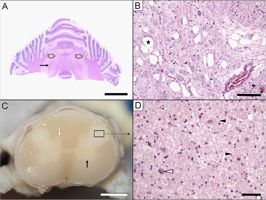
funiculi (arrow). disk space.Hirschvogel et al. BMC Veterinary Research 2013, 9:57 Page 4 of 8 http://www.biomedcentral.com/1746-6148/9/57 3.3 mmol/l [22]). No abnormal spontaneous activity pat- examination. Histologically, the cervical spinal cord (from tern was observed during electromyographic recordings C2 to C6 (vertebral body)) exhibited severe, bilaterally sym- using a concentric needle electrode in the anesthetised metrical funicular disruption of the inner dorsal part of the dog. The tibial and ulnar motor nerve conduction vel- lateral funiculus, including the rubrospinal tract, the inner- ocity, tibial nerve F-waves and repetitive nerve stimula- most layer of the dorsal spinocerebellar tract and the dorsal tion were within established laboratory reference ranges. aspects of the lateral fasciculus proprius. Upon low-power Considering the progressive clinical course and the inspection, the lesion was characterised by a severe loss of MRI lesion pattern, a neurodegenerative disorder pre- myelin staining; at high-power, the lesion resembled a dominantly involving the cervical spinal cord white mat- dense core of non-myelinated white matter with extensive ter with a bilateral and symmetrical distribution was astrocytosis and astrogliosis with the occasional observa- suspected. Due to the existing severe neurological signs, tion of bizarre cells surrounded by a rim of spongiotic the progressive deterioration and the poor prognosis, the white matter with fibre degeneration, resorptive lesions, dog was euthanised. vascular prominence and mild-to-marked angiocentric A complete necropsy was performed, and it confirmed lymphohistiocytic infiltration. The adjacent cervical grey Rottweiler LEM. Significant lesions were confined to the matter appeared hypoplastic in both the ventral and dorsal central nervous system. Macroscopic examination revealed horns, but there were no further histomorphological bilateral, symmetrical lesions restricted to the dorsal aspect changes. Another severe white matter lesion identified in of the lateral funiculi of the cervical spinal cord segments. the cerebellar roof showed focal, bilaterally symmetric tis- In transverse sections, these lesions appeared as well- sue necrosis, macrospongiosis due to interlamellar myelin demarcated, whitish, opaque discoloured areas (Figure 3). sheath oedema (ballooning) and severe intralesional No gross changes were observed in the brain. Formalin- astrogliosis and astrocytosis accompanied by fibrillary fixed and paraffin-embedded tissue samples of the brain astrogliosis and gemistocytes at the margins. A moderate and spinal cord were sectioned at 5 μm and stained using vascular prominence with endothelial hyperplasia was haematoxylin-eosin and Luxol Fast Blue for histological again observed both in the intra- and perilesional areas. Figure 3 Pathological lesions in the brain (A, B) & spinal cord (C, D). The most severe white matter lesions were observed in the cerebellum (A: asterisk) and cervical spinal cord (C: framed area). Macroscopic examination revealed bilateral, symmetrical lesions in the lateral funiculi of the cervical cord segments only. In transverse sections, these lesions appeared as well-demarcated, whitish, opaque discoloured areas (C: framed area). The cerebellar lesions spared the fibres adjacent to the roof nuclei (A: arrow). Nuclear degeneration was most severe in the raphe nuclei (B) and medial vestibular nuclei (not shown). Note the extensive juxtaneuronal vacuolisation (B: asterisk). The affected spinal cord segments show demyelination, astrogliosis and astrocytosis (D: white arrowhead) with gemistocytes (D: black arrowheads). Within the grey matter, hypoplasia of the dorsal and ventral horn (C: black arrow) is evident. Scale bars: A: 1.5 cm; B: 100 μm; C: 2 mm; D: 35 μm.
Hirschvogel et al. BMC Veterinary Research 2013, 9:57                                                            Page 5 of 8
http://www.biomedcentral.com/1746-6148/9/57

Necrotic areas exhibited macrophage-mediated resorption        changes is very helpful in defining disease because it re-
(Figure 3).                                                    veals the distribution of histopathologic changes [24,25].
   Similar demyelinating lesions were observed in the pyra-       At present, there are few case reports describing the use
mids and caudal cerebellar peduncles and – to a lesser         of MRI for the diagnosis of canine and feline neurodegen-
extent – in the medial lemniscus, optic tracts, crura          erative diseases. T2-weighted hyperintensities of white
cerebri and subcortical white matter. Lesions in the cen-      brain matter were evident in cats with GM2 gangliosidosis
tral visual pathways projected to the optic nerves and         [26,27] and in a West Highland white terrier with globoid
manifested as the degeneration of multiple fibres. Further     cell leukodystrophy [28]. Increased signal was also evident
brain stem changes included macrovacuolar degeneration         in T2-weighted images of the spinal cord of Leonberger
of the raphe nuclei and medial vestibular nuclei associated    dogs with leukoencephalomyelopathy [29]. Dogs with
with mild gliosis and axonal spheroids. Immunohisto-           GM2 gangliosidosis displayed T2-weighted hyperintensities
chemical staining for canine distemper virus was negative.     in the region of the caudate nucleus and atrophy of the
A mild diffuse endoneurial hypercellularity was observed       cerebrum and cerebellum [30,31]. MRI of Papillon dogs
in the preganglionic aspects of the dorsal roots of the cer-   with neuroaxonal dystrophy [32] and Scottish Terriers with
vical spine. Both the radial and common peroneal nerve         hereditary cerebellar degeneration demonstrated atrophy
presented with a mild dropout of myelinated fibres, as de-     only and failed to detect changes in white matter [33].
noted by enlarged subperineurial spaces with myxoid re-           MRI of the cervical spine may be used to support the
placement oedema, reduced endoneurial area and                 clinical diagnosis of LEM in Rottweilers. A similar MRI
decreased myelinated nerve fibre density that was associ-      pattern has been described in Leonberger dogs with
ated with a mild expansion of the endoneurial collagenous      LEM [29]. Interestingly, however, brain lesions were not
matrix. Residual large A (alpha)-type myelinated fibres        detected using MRI in the Leonberger dogs or in the
showed myelin ovoids, consistent with stage II – III           case reported here, although histological analyses
Wallerian degeneration, and abundant internodal and            showed that the optic tracts and particularly the cerebel-
paranodal inner and outer myelin loops due to the moder-       lar medulla were significantly affected in both breeds
ate axonal atrophy of the respective fibres.                   [2,29]. It is possible that the white matter lesions in the
   Due to the phenotypic similarities between human            brain were less advanced than those in the spinal cord at
LBSL patients and LEM-affected Rottweilers, the DARS2          the time of imaging, and improved imaging protocols
gene was investigated as a candidate for canine LEM.           may be required for the visualisation of brain lesions.
Genomic DNA was extracted from blood collected in              These protocols may include smaller slice thicknesses
tubes containing EDTA using DNeasy blood spin col-             and the application of sequences other than conven-
umns (Qiagen). For the DARS2 mutation analysis, suit-          tional T1- and T2-weighted imaging, e.g., diffusion ten-
able PCR products were amplified using AmpliTaq Gold           sor imaging, magnetisation transfer imaging or MR
360 (Life Technologies). The PCR products were                 spectroscopy [34]. It is also questionable whether the
resequenced after rAPid alkaline phosphatase (Roche)           pathological changes in these regions have sufficiently al-
and exonuclease I (New England Biolabs) treatment              tered the physics of the tissue to induce changes visible
using both PCR primers and the ABI BigDye Terminator           with a 1.5 T clinical scanner.
Sequencing Kit 3.1 (Life Technologies) in an ABI 3730             Many inherited white matter diseases and associated
sequencer (see Additional file 2: Table S1). The sequence      genetic defects have been described in humans [35].
data were analysed using Sequencer 4.9 software                Leukoencephalopathies may be characterised as lysosomal
(GeneCodes). The sequences of all 17 coding exons and          or peroxisomal disorders, mitochondrial disorders, methy-
flanking intron sequences of the DARS2 gene from the           lation cycle disorders, organic acidaemias or amino acid
affected Rottweiler were identical to a canine reference       disorders or as leukoencephalopathy associated with calci-
genome sequence (CanFam3 assembly; http://genome.              fication, hypomyelination, abnormal lipid metabolism,
ucsc.edu).                                                     vasculopathy or muscular dystrophy. Many distinct en-
                                                               tities, e.g., Alexander’s disease, adult onset autosomal dom-
Conclusion                                                     inant leukoencephalopathy, vanishing white matter disease
Magnetic resonance imaging has become the primary              and adult polyglucosan encephalopathy, have also been
tool for the ante mortem diagnosis of white matter dis-        recognised. A vast number of genetic defects are currently
ease in humans due to its high sensitivity for detecting       associated with these conditions, and many more remain
changes in white matter. Decreased myelin and elevated         to be elucidated; the molecular cause remains unknown
water content is revealed by increased T1 and T2 relax-        in ~50% of affected humans [35]. The lesion distribution
ation times, with a consequent reduction in signal inten-      and MRI appearance of LBSL are considered unique and
sity in T1-weighted images and increased signal intensity      diagnostic in humans; consequently, only a single candi-
in T2-weighted images [23]. Thus, the pattern of MRI           date gene was examined in the present study [36].
Hirschvogel et al. BMC Veterinary Research 2013, 9:57 Page 6 of 8 http://www.biomedcentral.com/1746-6148/9/57 Leukoencephalopathy with brain stem and spinal cord Further limitations of our case report include the lack involvement is a rare, autosomal recessive disorder that of pedigree analysis and brain lactate MR spectroscopy typically manifests in childhood or adolescence. The measurements and the failure of MR to demonstrate the diagnosis of LBSL in humans is based on clinical presen- involvement of the cerebrum despite the pathology ob- tation and is characterised by a slowly progressive cere- served in histological sections. Another limitation is that bellar ataxia, spasticity, dorsal column dysfunction and a the comparison of the pathologies of these diseases in highly characteristic pattern of abnormalities observed dogs and humans is limited by the paucity of case data using MRI and spectroscopy. Typical MRI findings in- from both. To date, there is only one short description clude a combination of high T2-weighted signal changes of the pathology of LBSL in humans which has shown in the cerebral white matter accompanied by the select- spongy white matter degeneration, rarefaction of the ive involvement of the brain stem and spinal cord tracts neuropil, macrophage infiltration and an increased num- (the entire length of the pyramidal tracts with the add- ber of astrocytes in the white matter of the brain and itional involvement of cerebellar connections and the axonal degeneration of the peripheral nerves [25]. Spinal intraparenchymal and mesencephalic parts of the tri- cord changes have not been noted, but it is unknown geminal nerve) [13,37]. MR spectroscopy demonstrates whether this part of the CNS was sampled and investi- an elevation in lactate levels in the abnormal white mat- gated. In dogs with LBSL-like changes, the neuroana- ter of almost all of the affected human patients. These tomical mapping of CNS lesions is more precise [2,29]. findings led researchers to assume that the disease was a Clinical and pathological findings emphasise cerebellar mitochondrial disorder, which was subsequently con- and spinal changes, although the microscopic white mat- firmed by the discovery of various mutations in the ter damage is far more widespread and extends from the DARS2 gene, which encodes mitochondrial aspartyl- lower brain stem to the subcortical white matter. Con- tRNA synthetase [14,38]. As demonstrated by multiple sistent with the fibres affected, Gamble et al. discovered case reports of LBSL in humans, normal CSF and blood secondary grey matter changes in connected brain stem lactate concentrations, as were noted in the case nuclei, such as the accessory cuneate nucleus, nucleus reported herein, do not exclude a mitochondrial disorder gracilis, nucleus cuneatus and nucleus of the dorsal as the underlying cause of leukoencephalomyelopathy. spinocerebellar tract [1]. However, the involvement of Thus, further investigations should utilise MR spectros- multiple independent centres and tracts is compatible copy to investigate the possible mitochondrial origin of with multisystemic degeneration, as has been shown in Rottweiler LEM. Leonbergers and Rottweilers (discussed above) [2,29]. The diagnosis of mitochondrial disorders faces spe- We also discovered macrovacuolar nuclear degeneration cific difficulties due to the complex genetics of these in the Rottweiler, which has not previously been de- conditions. Mitochondrial disorders may occur due to scribed in dogs. Vacuole formation in LBSL patients was mutations in mitochondrial genes or mutations in nu- predominantly perineuronal and was therefore dissimilar clear proteins, with mitochondrial tRNA representing a to the neuronal vacuolation and spinocerebellar degener- hot spot for mutations. Heteroplasmy, i.e., the simul- ation observed in Rottweiler dogs [16]. This degener- taneous presence of mutated and normal RNA/DNA in ation merits further examination to investigate the the cell, is a characteristic feature of mitochondrial dis- relationship between neurons and astrocytes in the sub- orders. The degree of heteroplasmy varies between tis- cortical grey matter. It also remains to be established sues in the same organism, which is considered a whether this manifestation causes the white matter path- critical factor in the manifestation of mitochondrial dis- ology or whether it is an additional, co-existing disorder ease in specific tissues [36,39]. Finally, we investigated that is distinct from the breed-specific neurodegenera- the coding region of the canine DARS2 gene as a candi- tive disorders described above. date causative gene for LEM, and no mutation was In summary, magnetic resonance imaging revealed found. At this time, we cannot rule out the possibility leukodystrophic lesions in the lateral funiculi of the cer- of variants in the promoter or intronic regions that vical spinal cord; these findings will assist in the ante could affect DARS2 expression. More comprehensive mortem diagnosis of future cases of suspected LEM in DNA sequencing approaches, such as the use of next- Rottweilers. Further investigations should utilise MR generation technologies for whole-exome or whole- spectroscopy to investigate the possible mitochondrial genome resequencing, may enable the identification of origin of Rottweiler LEM. Although LEM is similar to the causative mutation of Rottweiler LEM. A recent LBSL based on its clinical features and imaging results, study identified the causative mutation of canine neo- we were unable to identify a coding or splice site muta- natal cerebellar cortical degeneration in SPTBN2 (gen- tion in the canine DARS2 gene in our case, suggesting ome-wide mRNA sequencing) using only a single case that other genes may be involved in Rottweiler LEM and of this neurodegenerative disease [40]. potentially also in human LBSL.
Hirschvogel et al. BMC Veterinary Research 2013, 9:57                                                                                                  Page 7 of 8
http://www.biomedcentral.com/1746-6148/9/57

Additional files                                                                         neural pathways or vascular territories. AJR Am J Roentgenol 1995, 165(3):
                                                                                         515–523.
                                                                                   11.   van der Knaap MS, Ramesh V, Schiffmann R, Blaser S, Kyllerman M, Gholkar
 Additional file 1: Movie of a Rottweiler with confirmed
                                                                                         A, Ellison DW, van der Voorn JP, van Dooren SJ, Jakobs C, et al: Alexander
 leukoencephalomyelopathy. The movie shows the severe generalised
                                                                                         disease: ventricular garlands and abnormalities of the medulla and
 ataxia with hypermetria of the thoracic (with prolonged protraction,
                                                                                         spinal cord. Neurology 2006, 66(4):494–498.
 overreaching action and limb extension) and pelvic limbs. The postural
 reactions were delayed, and the thoracic limbs were more severely                 12.   Miyake N, Yamashita S, Kurosawa K, Miyatake S, Tsurusaki Y, Doi H, Saitsu H,
 affected than the pelvic limbs.                                                         Matsumoto N: A novel homozygous mutation of DARS2 may cause a
                                                                                         severe LBSL variant. Clin Genet 2011, 80(3):293–296.
 Additional file 2: Sequencing methods and primers.                                13.   van der Knaap MS, van der Voorn P, Barkhof F, Van Coster R, Krageloh-Mann
                                                                                         I, Feigenbaum A, Blaser S, Vles JS, Rieckmann P, Pouwels PJ: A new
                                                                                         leukoencephalopathy with brainstem and spinal cord involvement and
Abbreviations                                                                            high lactate. Ann Neurol 2003, 53(2):252–258.
CSF: Cerebrospinal Fluid; FLAIR: Fluid-Attenuated Inversion Recovery;
                                                                                   14.   Scheper GC, van der Klok T, van Andel RJ, van Berkel CG, Sissler M, Smet J,
LBSL: Leukoencephalopathy with Brain Stem and Spinal Cord Involvement;
                                                                                         Muravina TI, Serkov SV, Uziel G, Bugiani M, et al: Mitochondrial aspartyl-
LEM: Leukoencephalomyelopathy; MR: Magnetic Resonance; MRI: Magnetic
                                                                                         tRNA synthetase deficiency causes leukoencephalopathy with brain
Resonance Imaging; Ms: Millisecond; TE: Time to Echo; TR: Time to
                                                                                         stem and spinal cord involvement and lactate elevation. Nat Genet 2007,
Repetition.
                                                                                         39(4):534–539.
                                                                                   15.   van Berge L, Dooves S, van Berkel CG, Polder E, van der Knaap MS, Scheper
Competing interests                                                                      GC: Leukoencephalopathy with brain stem and spinal cord involvement
The authors declare that they have no competing interests.                               and lactate elevation is associated with cell-type-dependent splicing of
                                                                                         mtAspRS mRNA. Biochem J 2012, 441(3):955–962.
Authors’ contributions                                                             16.   Kortz GD, Meier WA, Higgins RJ, French RA, McKiernan BC, Fatzer R, Zachary
KH was responsible for data collection and interpretation and for drafting               JF: Neuronal vacuolation and spinocerebellar degeneration in young
the manuscript. KM performed the necropsy and histopathology and                         rottweiler dogs. Vet Pathol 1997, 34(4):296–302.
provided histopathology images. KF performed all diagnostic imaging                17.   Cherrone KL, Dewey CW, Coates JR, Bergman RL: A retrospective
procedures and selected the appropriate images. MD and CD conducted the                  comparison of cervical intervertebral disk disease in
genetic study. BR contributed substantially to the acquisition of data used in           nonchondrodystrophic large dogs versus small dogs. J Am Anim Hosp
the manuscript. AF contributed to data collection, helped draft the                      Assoc 2004, 40(4):316–320.
manuscript and finalised the version to be published. All authors have             18.   Jurina K, Grevel V: Spinal arachnoid pseudocysts in 10 rottweilers. J Small
approved the final manuscript.                                                           Anim Pract 2004, 45(1):9–15.
                                                                                   19.   Skeen TM, Olby NJ, Munana KR, Sharp NJ: Spinal arachnoid cysts in 17
Author details                                                                           dogs. J Am Anim Hosp Assoc 2003, 39(3):271–282.
1
 Department of Veterinary Clinical Sciences Ludwig-Maximilians-Universitaet,       20.   Cork LC, Troncoso JC, Price DL, Stanley EF, Griffin JW: Canine neuroaxonal
Neurology Service, Clinic of Small Animal Medicine, Munich, Germany.                     dystrophy. J Neuropathol Exp Neurol 1983, 42(3):286–296.
2
 Department of Veterinary Clinical Sciences Ludwig-Maximilians-Universitaet,       21.   Yin W, Tibbs R, Aoki K, Badr A, Zhang J: Metabolic alterations in
Section of Clinical & Comparative Neuropathology, Institute of Veterinary                cerebrospinal fluid from double hemorrhage model of dogs. Neurol Res
Pathology, Munich, Germany. 3Department of Veterinary Clinical Sciences                  2001, 23(1):87–92.
Ludwig-Maximilians-Universitaet, Clinic of Small Animal Surgery and                22.   Löbert V: Etablierung von laktat- und pyruvatmessung im plasma und liquor
Reproduction, Munich, Germany; Small Animal Hospital Hüttig, Reutlingen,                 cerebrospinalis zur diagnostik von mitochondrialen erkrankungen beim hund.
Germany. 4Institute of Genetics, Vetsuisse Faculty, University of Berne, Berne,          Inaugural-dissertation. University of veterinary medicine Hannover:
Switzerland.                                                                             Department of Small Animal Medicine and Surgery; 2003.
                                                                                   23.   Barker PB, Horska A: Neuroimaging in leukodystrophies. J Child Neurol
Received: 6 October 2012 Accepted: 14 March 2013                                         2004, 19(8):559–570.
Published: 26 March 2013                                                           24.   Serkov SV, Pronin IN, Bykova OV, Maslova OI, Arutyunov NV, Muravina TI,
                                                                                         Kornienko VN, Fadeeva LM, Marks H, Bonnemann C, et al: Five patients
References                                                                               with a recently described novel leukoencephalopathy with brainstem
1. Gamble DA, Chrisman CL: A leukoencephalomyelopathy of rottweiler                      and spinal cord involvement and elevated lactate. Neuropediatrics 2004,
    dogs. Vet Pathol 1984, 21(3):274–280.                                                35(1):1–5.
2. Wouda W, van Nes JJ: Progressive ataxia due to central demyelination in         25.   Yamashita S, Miyake N, Matsumoto N, Osaka H, Lai M, Aida N, Tanaka Y:
    rottweiler dogs. Vet Q 1986, 8(2):89–97.                                             Neuropathology of leukoencephalopathy with brainstem and spinal cord
3. Slocombe RF, Mitten R, Mason TA: Leucoencephalomyelopathy in                          involvement and high lactate caused by a homozygous mutation of
    Australian rottweiler dogs. Aust Vet J 1989, 66(5):147–150.                          DARS2. Brain Dev 2013, 35(4):312–316.
4. Davies DR, Irwin PJ: Degenerative neurological and neuromuscular                26.   Kroll RA, Pagel MA, Roman-Goldstein S, Barkovich AJ, D’Agostino AN,
    disease in young rottweilers. J Small Anim Pract 2003, 44(9):388–394.                Neuwelt EA: White matter changes associated with feline GM2
5. Chrisman CL: Neurological diseases of rottweilers: neuroaxonal dystrophy              gangliosidosis (sandhoff disease): correlation of MR findings with
    and leukoencephalomalacia. J Small Anim Pract 1992, 33(10):500–504.                  pathologic and ultrastructural abnormalities. AJNR Am J Neuroradiol 1995,
6. Lewis DG, Newsholme SJ: Pseudo cervical spondylopathy in the rottweiler               16(6):1219–1226.
    (letter). J Small Anim Pract 1987, 28(12):1178.                                27.   Hasegawa D, Yamato O, Kobayashi M, Fujita M, Nakamura S, Takahashi K,
7. Sundblom J, Melberg A, Kalimo H, Smits A, Raininko R: MR imaging                      Satoh H, Shoda T, Hayashi D, Yamasaki M, et al: Clinical and molecular
    characteristics and neuropathology of the spinal cord in adult-onset                 analysis of GM2 gangliosidosis in two apparent littermate kittens of the
    autosomal dominant leukodystrophy with autonomic symptoms. AJNR                      japanese domestic cat. J Feline Med Surg 2007, 9(3):232–237.
    Am J Neuroradiol 2009, 30(2):328–335.                                          28.   Cozzi F, Vite CH, Wenger DA, Victoria T, Haskins ME: MRI and
8. Yonezu T, Ito S, Kanai K, Masuda S, Shibuya K, Kuwabara S: A case of adult-           electrophysiological abnormalities in a case of canine globoid cell
    onset alexander disease featuring severe atrophy of the medulla                      leucodystrophy. J Small Anim Pract 1998, 39(8):401–405.
    oblongata and upper cervical cord on magnetic resonance imaging.               29.   Oevermann A, Bley T, Konar M, Lang J, Vandevelde M: A novel
    Case Rep Neurol 2012, 4(3):202–206.                                                  leukoencephalomyelopathy of leonberger dogs. J Vet Intern Med 2008,
9. Kumar N, Ahlskog JE, Klein CJ, Port JD: Imaging features of copper deficiency         22(2):467–471.
    myelopathy: a study of 25 cases. Neuroradiology 2006, 48(2):78–83.             30.   Matsuki N, Yamato O, Kusuda M, Maede Y, Tsujimoto H, Ono K: Magnetic
10. Friedman DP, Tartaglino LM, Fisher AR, Flanders AE: MR imaging in the                resonance imaging of GM2-gangliosidosis in a golden retriever. Can Vet J
    diagnosis of intramedullary spinal cord diseases that involve specific               2005, 46(3):275–278.
Hirschvogel et al. BMC Veterinary Research 2013, 9:57 Page 8 of 8
http://www.biomedcentral.com/1746-6148/9/57
31. Tamura S, Tamura Y, Uchida K, Nibe K, Nakaichi M, Hossain MA, Chang HS,
Rahman MM, Yabuki A, Yamato O: GM2 Gangliosidosis variant 0
(sandhoff-like disease) in a family of toy poodles. J Vet Intern Med 2010,
24(5):1013–1019.
32. Tamura S, Tamura Y, Uchida K: Magnetic resonance imaging findings of
neuroaxonal dystrophy in a papillon puppy. J Small Anim Pract 2007,
48(8):458–461.
33. Urkasemsin G, Linder KE, Bell JS, de Lahunta A, Olby NJ: Hereditary
cerebellar degeneration in scottish terriers. J Vet Intern Med 2010, 24(3):
565–570.
34. Laule C, Vavasour IM, Kolind SH, Li DK, Traboulsee TL, Moore GR, MacKay AL:
Magnetic resonance imaging of myelin. Neurotherapeutics 2007, 4(3):460–484.
35. Renaud DL: Inherited leukoencephalopathies. Semin Neurol 2012, 32(1):3–8.
36. Wong LJ: Mitochondrial syndromes with leukoencephalopathies. Semin
Neurol 2012, 32(1):55–61.
37. Tzoulis C, Tran GT, Gjerde IO, Aasly J, Neckelmann G, Rydland J, Varga V,
Wadel-Andersen P, Bindoff LA: Leukoencephalopathy with brainstem and
spinal cord involvement caused by a novel mutation in the DARS2 gene.
J Neurol 2012, 259(2):292–296.
38. Uluc K, Baskan O, Yildirim KA, Ozsahin S, Koseoglu M, Isak B, Scheper GC,
Gunal DI, van der Knaap MS: Leukoencephalopathy with brain stem and
spinal cord involvement and high lactate: a genetically proven case with
distinct MRI findings. J Neurol Sci 2008, 273(1–2):118–122.
39. Saneto RP, Sedensky MM: Mitochondrial disease in childhood: mtDNA
encoded. Neurotherapeutics 2012, 6: doi:10.1007/s13311-012-0167-0.
40. Forman OP, De Risio L, Stewart J, Mellersh CS, Beltran E: Genome-wide
mRNA sequencing of a single canine cerebellar cortical degeneration
case leads to the identification of a disease associated SPTBN2 mutation.
BMC Genet 2012, 13:55.
doi:10.1186/1746-6148-9-57
Cite this article as: Hirschvogel et al.: Magnetic resonance imaging and
genetic investigation of a case of rottweiler leukoencephalomyelopathy.
BMC Veterinary Research 2013 9:57.
Submit your next manuscript to BioMed Central
and take full advantage of:
• Convenient online submission
• Thorough peer review
• No space constraints or color figure charges
• Immediate publication on acceptance
• Inclusion in PubMed, CAS, Scopus and Google Scholar
• Research which is freely available for redistribution
Submit your manuscript at
www.biomedcentral.com/submitYou can also read