MRDNA: A MULTI-RESOLUTION MODEL FOR PREDICTING THE STRUCTURE AND DYNAMICS OF DNA SYSTEMS - OXFORD UNIVERSITY PRESS
←
→
Page content transcription
If your browser does not render page correctly, please read the page content below
Published online 31 March 2020                                                       Nucleic Acids Research, 2020, Vol. 48, No. 9 5135–5146
                                                                                                                    doi: 10.1093/nar/gkaa200

MrDNA: a multi-resolution model for predicting the
structure and dynamics of DNA systems
                                    1,*                                                1,2,*
Christopher Maffeo                        and Aleksei Aksimentiev
1
 Department of Physics, University of Illinois at Urbana–Champaign, 1110 W Green St, Urbana, IL 61801, USA and
2
 Beckman Institute for Advanced Science and Technology, University of Illinois at Urbana–Champaign, 405 N
Mathews Ave, Urbana, IL 61801, USA

Received December 10, 2019; Revised March 06, 2020; Editorial Decision March 11, 2020; Accepted March 17, 2020

                                                                                                                                                                  Downloaded from https://academic.oup.com/nar/article/48/9/5135/5814051 by guest on 01 October 2020
ABSTRACT                                                                          and brick (15) assemblies, and to polygon mesh wire-
                                                                                  frames (16,17). The ease with which one can cus-
Although the field of structural DNA nanotechnol-                                 tomize and functionalize DNA nanostructures has cat-
ogy has been advancing with an astonishing pace,                                  alyzed proof-of-concept developments of a wide range
de novo design of complex 3D nanostructures and                                   of novel materials (18) and functional objects, includ-
functional devices remains a laborious and time-                                  ing sensors for pH (19), voltage (20) and force (21);
consuming process. One reason for that is the need                                nanoscale containers (22,23); masks for nanolithogra-
for multiple cycles of experimental characterization                              phy (24,25); and scaffolds for arranging nanotubes and
to elucidate the effect of design choices on the ac-                              nanoparticles (26,27). Presently, gigadalton 3D objects can
tual shape and function of the self-assembled ob-                                 be constructed through hierarchical self-assembly of DNA
jects. Here, we demonstrate a multi-resolution sim-                               molecules (28,29) with single-nucleotide precision, which
ulation framework, mrdna, that, in 30 min or less,                                makes DNA nanostructures uniquely amenable for master-
                                                                                  ing spatial organization at the nanoscale.
can produce an atomistic-resolution structure of a
                                                                                     Computational prediction of the in situ properties of
self-assembled DNA nanosystem. We demonstrate                                     DNA nanostructures can greatly facilitate the nanostruc-
fidelity of our mrdna framework through direct com-                               ture design process. Several computational models of DNA
parison of the simulation results with the results of                             nanostructures have been developed already, varying by the
cryo-electron microscopy (cryo-EM) reconstruction                                 amount of detail provided by the computational descrip-
of multiple 3D DNA origami objects. Furthermore,                                  tion. On the coarse end of the spectrum, the CanDo fi-
we show that our approach can characterize an en-                                 nite element model (30) minimizes the mechanical stress
semble of conformations adopted by dynamic DNA                                    within a DNA origami structure exerted by its pattern of
nanostructures, the equilibrium structure and dy-                                 crossovers to provide a fast estimate of its preferred con-
namics of DNA objects constructed using off-lattice                               formation. At the opposite end of the spectrum, all-atom
self-assembly principles, i.e. wireframe DNA objects,                             molecular dynamics (MD) simulations have been used to
                                                                                  study the structure (31–33) and conductance (34–37) of
and to study the properties of DNA objects under
                                                                                  DNA nanostructures and channels at a much higher com-
a variety of environmental conditions, such as ap-                                putational cost. In between, a number of general purpose
plied electric field. Implemented as an open source                               coarse-grained (CG) models of DNA are available (38–
Python package, our framework can be extended by                                  41), but only very few have been applied specifically to the
the community and integrated with DNA design and                                  study of DNA nanostructures (32,42–43) including the CG
molecular graphics tools.                                                         oxDNA (42,44) model and all-atom, implicit solvent elastic
                                                                                  network-guided MD (ENRG-MD) (32) method.
                                                                                     A major objective for computational models of DNA
INTRODUCTION                                                                      nanostructures is to give the designers quick feedback on
DNA self-assembly has emerged as a versatile and ro-                              the impact of their design choices. The models employed by
bust approach to constructing nanoscale systems (1–8).                            the coarsest structure prediction tools may not adequately
Since its conception (1), methods for designing and syn-                          represent DNA, for example, to guide the placement of fluo-
thesizing self-assembled DNA nanostructures have been                             rescent dyes or functionalized groups. CG models do not so
progressing steadily (2,9–14), advancing to 2D origami                            far provide insights into the ion conducting and rheological
nanostructures (4), to three-dimensional (3D) origami (5)                         properties of DNA nanostructures. To date, only all-atom

* To
   whom correspondence should be addressed. Tel: +1 217 333 6495; Fax: +1 866 467 5398; Email: aksiment@illinois.edu
Correspondence may also be addressed to Christopher Maffeo. Tel: +1 217 333 6495; Fax: +1 866 467 5398; Email: cmaffeo2@illinois.edu

C The Author(s) 2020. Published by Oxford University Press on behalf of Nucleic Acids Research.
This is an Open Access article distributed under the terms of the Creative Commons Attribution Non-Commercial License
(http://creativecommons.org/licenses/by-nc/4.0/), which permits non-commercial re-use, distribution, and reproduction in any medium, provided the original work
is properly cited. For commercial re-use, please contact journals.permissions@oup.com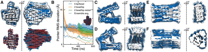
5136 Nucleic Acids Research, 2020, Vol. 48, No. 9
MD has been used to computationally examine these prop- The spline representation mediates conversion from one res-
erties. On the other hand, higher resolution modeling can olution to another; after a CG simulation is performed, new
be prohibitively slow for routine structural characterization, splines are generated that pass through the coordinates of
or for sampling the configurational space of a nanostruc- the beads obtained at the end of the simulation.
ture, particularly when the equilibrium, relaxed conforma- The segment classes can be used directly for algorith-
tion of an object departs strongly from its initial idealized mic design of DNA nanostructures or can be used to write
configuration as obtained from popular CAD tools such as scripts that automatically convert a model from another
cadnano (45), vHelix (16) or DAEDALUS (17). Although software package into an mrdna model. For example, a
reorienting components of the design prior to simulation class was developed using the new cadnano2.5 Python API
can facilitate proper structural relaxation (46), it may be to read in a cadnano json design file, allowing direct ac-
nontrivial to find an appropriate transformation for many cess to the cadnano data structures for conversion to the
structures, especially those containing programmed bends segment-based model. Similar classes have been developed
and twists. for converting vHelix mesh models and atomic Protein Data
Downloaded from https://academic.oup.com/nar/article/48/9/5135/5814051 by guest on 01 October 2020
Here we present a computational framework that com- Bank (PDB) files into mrdna models.
bines low- and high-resolution models of DNA objects to
enable fast and accurate predictions of the objects’ structure Conversion of spline model to bead model
and dynamics suitable for iterative design applications, Fig-
Beads representing interaction sites within double- and
ure 1. Starting from an idealized design of a DNA object,
single-stranded segments are iteratively placed through the
our structure prediction protocol performs rapid relaxation
structure and assigned positions within the segment, see
of the design using a low-resolution model. The outcome
Supplementary Figure S1. First, beads are placed at the
of the relaxation simulation is further refined through sev-
middle of every intrahelical (or intrastrand) connection.
eral simulations performed at increasing resolution to pro-
Beads are next placed on both sides of each crossover. When
duce an accurate, fully atomistic representation of the ob-
two beads are intrahelically adjacent and placed within the
ject’s in situ structure. Our computational framework can
user-provided resolution of the model, they are merged into
also characterize the conformational dynamics of the object
one bead. Next, beads are placed at the ends of each seg-
at multiple time scales and characterize the behavior of the
ment if none already exists. Beads are placed between each
objects in the presence of external potentials and/or under
pair of existing beads so that the linear density of beads
non-equilibrium conditions.
is lower than a user-specified threshold. Finally, the intra-
helical distance between adjacent beads is calculated from
MATERIALS AND METHODS their positions and used to assign a nucleotide count to each
bead. The beads are hierarchically clustered according to
All CG simulations of DNA origami objects were per-
the nucleotide count, ensuring a tractable number of bead
formed using an in-house developed GPU-accelerated CG
types is provided to the simulation engine. If the model is
simulation package, Atomic Resolution Brownian Dynam-
generated with a local representation of twist, each dsDNA
ics (ARBD) (47). ARBD supports Brownian and Langevin
bead receives an orientation bead shifted by the vector R
dynamics simulations of systems composed of isotropic
· (1.5 Å, 0, 0), where the orientation matrix R is obtained
point-like particles interacting through tabulated bonded
from the spline fit through quaternions. If no such spline is
and non-bonded interactions. ARBD simulations are cur-
available, the orientation is taken to be a rotation of = n ×
rently configured through system-specific files that the
34.48◦ (for a helical rise of 10.44) about the local tangential
mrdna framework writes automatically.
axis, where n is contour length at the bead given in units of
base pairs.
Overview of the mrdna framework
Variable-resolution bead model of DNA nanostructures
The mrdna framework is shipped as a Python package that
describes DNA nanostructures using several levels of ab- Once the beads have been placed throughout a DNA nanos-
straction. At the base level are classes that represent collec- tructure, bonded and non-bonded potentials are specified
tions of beads or atoms interacting through user-specified to describe the bead–bead interactions. A tabulated non-
bonded and non-bonded potentials. These classes provide bonded potential was developed by iteratively tuning inter-
a file-based interface to our simulation engine, ARBD. The actions until the osmotic pressure in an array of 256 two-
second level of abstraction provides classes that describe turn DNA helices––each made effectively infinite through
contiguous double-stranded and single-stranded regions or periodic boundary conditions––matched the experimental
‘segments’ of DNA that can be joined together through in- measurements of Rau and Parsegian for DNA in a 25 mM
trahelical, terminal and crossover connections. The spatial MgCl2 electrolyte (48), see Supplementary Figure S2. If the
configuration of each DNA segment is described by a spline user supplies a custom Debye length, a correction is applied
function defined in cartesian coordinates. For dsDNA, an to the tabulated non-bonded potential corresponding to the
additional four-dimensional spline fit through quaternions difference of Yukawa potentials for the user-supplied and
optionally provides a parametric representation of the lo- 11.1-Å Debye lengths, the latter representing the ion condi-
cal orientation throughout the duplex. The use of splines tion of the Rau and Parsegian data. The non-bonded inter-
allows the classes to generate bead-based models with user- action between any pair of beads is scaled by the nucleotide
specified resolution. Alternatively the segment classes can count of each bead involved. DNA array simulations ver-
generate an atomistic or oxDNA model of the structure. ified that the pressure was insensitive to the resolution of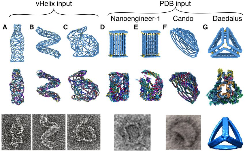
Nucleic Acids Research, 2020, Vol. 48, No. 9 5137
Downloaded from https://academic.oup.com/nar/article/48/9/5135/5814051 by guest on 01 October 2020
Figure 1. DNA nanostructure design workflow augmented by the multi-resolution modeling framework, mrdna. The top row depicts a traditional design
cycle involving several steps that result in a latency of several weeks before feedback is obtained. A DNA origami bundle with a programmed 180◦ bend (56)
is shown as an example. The bottom row illustrates how mrdna can be used to obtain the equilibrium in situ configuration of the DNA nanostructure.
The snapshots within the arrow illustrate several stages of a CG simulation trajectory; the structure immediately to the right of the arrow shows the final
all-atom configuration. The resulting all-atom structures are directly suitable for subsequent all-atom MD simulations, the right most image shows an
example of a solvated all-atom system. TEM and gel images are reproduced from Ref. 56 with permission.
DNA under 1, 3, 5 and 7 bp/bead conditions, with and placed between consecutive intrahelical beads and each of
without a local representation of the twist. The same poten- the orientation beads. A harmonic potential is placed on the
tial is used for non-bonded interactions involving ssDNA. dihedral angle formed by each orientation bead, its parent
For dsDNA, non-bonded exclusions are placed between all intrahelical bead, the adjacent intrahelical bead and the cor-
pairs of beads intrahelically within 20 bp of one another. responding orientation bead with a separation-dependent
Additionally, exclusions are placed between all beads within rest angle (10.44 helical rise) and a spring constant ktw (s)
3 bp of a crossover bead. For ssDNA, exclusions are placed designed to produce a 90-nm twist-persistence length for ds-
between pairs of beads in the same strand that are within DNA to match the measured twist persistence length under
five nucleotides (nts). tension (50), see Supplementary Figures S3 and S4.
The bonded interactions within the model are realized by At last, a harmonic bond is placed between intrahelical
harmonic potentials with spring constants and rest lengths beads on either side of a junction spanning two helices (such
determined from known polymer properties of DNA, Sup- as a DNA origami crossover connection). The spring con-
plementary Figure S3. A harmonic bond connects each stant (4 kcal mol−1 Å−2 ) and the rest length (18.5 Å) of the
consecutive pair of beads in a helix or strand with a bond were derived from the distribution of crossover dis-
separation-dependent rest length (3.4 Å/bp or 6.4 Å/nt) tances observed in atomistic simulations (33). When neither
and a spring constant derived from the elastic moduli of side of the junction occurs at the end of a DNA helix, the
dsDNA and ssDNA (1000 and 800 pN, respectively) (49). junction is assumed to represent a DNA origami crossover,
A harmonic angle potential is placed on the angle formed and a heuristic dihedral angle potential is placed on an in-
by the bonds between consecutive pairs of beads within a trahelical bead on one side of the first junction bead, the first
helix or strand with a 0◦ rest angle and spring constant kp junction bead, the second junction bead and the intrahelical
derived from the persistence length of DNA. Specific val- bead on the same side of the junction (kspring = 0.25 kp ; de-
ues of kp were obtained by numerically solving the following fault rest angle of zero) to keep the adjacent helices roughly
equation: parallel. We chose this rest dihedral angle value, which de-
π viates from the ∼55◦ value observed experimentally for free
sin θ dθ cos θ e−β 2 kp θ
1 2
−s/L p Holliday junctions in solution (51), because the junctions
e = cos θ = 0 π , (1)
−β 12 kp θ 2 in a DNA origami nanostructure have equilibrium angles
0 sin θ dθ e
near to zero (33,52). However, the user may specify their
where s is a half of the contour length between the first and own rest angle so that some off-lattice DNA nanostructures
the third bead, Lp is the persistence length (taken to be 50 may be modeled that have crossovers occuring in the middle
and 1.2 nm for dsDNA and ssDNA), is the angle between of a helix, such as for gridiron nanostructures (53). If twist
the beads, kp is the spring constant used in the simulation is locally represented, an additional harmonic potential is
and β = 1/kB T, where kB T is the thermodynamic tempera- added to the dihedral angle formed on each helix by the
ture. junction bead on the opposite helix, the junction bead, the
The mrdna models can be generated with or without a lo- bead adjacent to the junction bead and the junction bead’s
cal representation of the orientation of each base pair. When orientation bead (kspring = 0.25 kp ; rest angle = ±120, de-
the twist is represented locally, an orientation bead repre- pending on the strand) to ensure that the connecting strand
senting the major groove is placed alongside each dsDNA on each side of the junction faces the helix on the other side
backbone bead, attached to its parent bead by a harmonic of the junction, Supplementary Figure S5A. In the event
potential (1.5 Å rest length; kspring = 30 kcal mol −1 Å−2 ). A that twist is not locally represented, the torsion within each
harmonic angle potential (90◦ rest angle; kspring = 0.5 kp ) is helix is described by a dihedral angle potential applied be-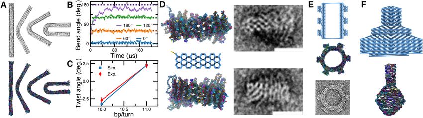
5138 Nucleic Acids Research, 2020, Vol. 48, No. 9
Downloaded from https://academic.oup.com/nar/article/48/9/5135/5814051 by guest on 01 October 2020
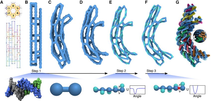
Figure 2. Multiresolution structural relaxation of a curved DNA origami object. (A) Cadnano (45) design of a curved six-helix bundle. A regular pattern
of insertions (blue loops) in the two helices on the left and deletions (red crosses) in the two helices on the right induce the curvature (56). (B–D) Low-
resolution CG simulation of the bundle. In just 40 nanoseconds, the bundle adopts a curved conformation. The bundle is shown using a bead-and-stick
representation, where each bead represents 5 bp, on average. (E and F) High-resolution CG simulations of the bundle. A spline-based mapping procedure
yields a high-resolution (2 beads per base pair) model by interpolation. The high-resolution model includes a local representation of each base pair’s
orientation (teal beads). The twist dihedral angle potential between adjacent orientation beads is smoothly truncated to allow the linking number to relax
during a brief, 10 ns simulation (E). A subsequent 10 ns simulation performed without the truncation of the twist potential allows complete relaxation of
the bundle (F). (G) Resulting atomic model of the curved six-helix bundle. Canonical base pairs are placed throughout the structure using the spline-based
mapping procedure. The images below each of the three CG simulation steps illustrate schematically how the model represents one turn of DNA.
tween the beads forming consecutive junctions in the helix. procedure begins with the construction of a low-resolution,
The rest angle is calculated as described above for the lo- 5 base pair (bp)/bead CG model, Figure 2B. By default,
cal representation of twist, except if the junctions occur on and throughout this paper, the low-resolution model is gen-
different strands, 120◦ will be added to (or removed from) erated without a local representation of twist, though this
the rest angle if the 5 -to-3 direction at each junction site behavior is easily adjusted (see ‘Materials and Methods’ sec-
points away from (or toward) the other junction, Supple- tion). The non-bonded interactions between the beads are
mentary Figure S5B. The spring constant is calculated from described by empirical potentials that have been calibrated
three springs in series, the first and last taken to be one eight to reproduce the experimentally measured osmotic pressure
kp for a single nucleotide span to represent some intrinsic, of a DNA array in 25 mM MgCl2 electrolyte (48), Supple-
resolution-independent flexibility of the crossovers, and the mentary Figure S2.
central spring being calculated as described for the twist di- Our model can accommodate other ionic strength con-
hedral potential when a local twist is applied. ditions at the level of the Debye–Hückel approximation,
At the time of writing, all potentials are independent accounting for ionic strength conditions through a Debye
of the DNA sequence. Additionally, twist–bend and twist– length parameter. The beads within each double- or single-
stretch coupling have been neglected. stranded region are connected by harmonic bond and an-
gle potentials with separation-dependent rest-lengths and
spring constants derived to match the experimentally mea-
RESULTS sured elastic moduli and persistence lengths, Supplemen-
Multi-resolution model of self-assembled DNA nanostruc- tary Figures S3 and S4. At last, bond, angle and dihedral
tures potentials were defined for the beads at each junction, see
‘Materials and Methods’ section for further details. The re-
The mrdna framework performs an automatic multi-stage sulting model is relaxed from its initial, idealized geometry
simulation of a DNA nanostructure starting from its ide- using the simulation engine, ARBD (47). Figure 2B–D illus-
alized initial configuration, which can come from a variety trate the relaxation simulation of the curved six-helix bun-
of sources. Here, we illustrate the basic workflow of mrdna dle.
using, as an example, a cadnano (45) design of a curved six- The structure obtained at the end of the above relax-
helix bundle. ation process is further refined through additional simula-
Starting from a configuration file generated by the de- tions performed using models of increasing resolution. To
sign software, Figure 2A, the standard mrdna relaxation facilitate the change to a higher (2 beads/bp) resolution, a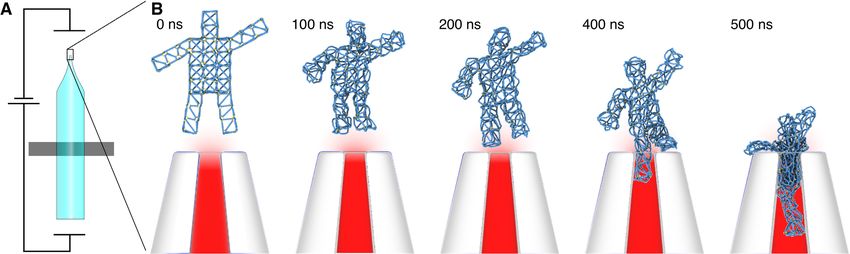
Nucleic Acids Research, 2020, Vol. 48, No. 9 5139

Figure 3. Structure prediction with mrdna. (A) Comparison of the structural models obtained through an mrdna simulation and cryo-EM reconstruction

                                                                                                                                                              Downloaded from https://academic.oup.com/nar/article/48/9/5135/5814051 by guest on 01 October 2020
(EMD-2210) of the ‘pointer’ structure (52). The top row depicts the simulated and experimental structures as isosurfaces of the CG bead (blue, 1 bp/Å3
isovalue) and electron (white, 0.08 isovalue) densities, respectively. The bottom row compares the simulated (blue) and experimental (red) all-atom models.
In the top row, simulated structural fluctuations are visualized using a semi-transparent surface showing the average density of CG beads during the final
600 ns of a 5-bp/bead simulation. Note that some helices along the periphery of the structure were not resolved in the cryo-EM model (52). (B) RMSD of
the center of mass of each base pair in the simulated pointer structure from those of the cryo-EM-derived atomic structure (52) at 5 bp/bead (blue) and
2 beads/bp (orange and green) resolutions. For each resolution, an ensemble of sixteen simulations was performed, the RMSD for each trajectory was
computed every 2 ns, and the ensemble of RMSD values was averaged at each moment in time to provide the solid line. The shaded regions around each
solid line shows the corresponding standard deviation of the RMSD. The dashed lines depict the RMSD between the ensemble’s average structure and the
cryo-EM-derived structure. For all RMSD calculations, the splines traced through the instantaneous configurations were used to determine the position
of each base pair. The configurations of the 5-bp/bead ensemble after 400-ns were used to initialize an ensemble of 2-beads/bp simulations (green). (C–F)
Comparison of the mrdna (blue, 1 bp/Å3 isovalue) and cryo-EM (white, 0.02 isovalue) models of the objects designed and characterized by the Dietz
group (28): v-brick structures without (C; EMD-3828) and with (D; EMD-3828) twist correction, the connector block (E; EMD-3827) and the triangular
vertex (F; EMD-3826).

spline function is first defined to trace the center line of each               NVIDIA Quadro RTX 5000, see Supplementary Anima-
double- or single-stranded DNA region in the structure                          tion 1. The resulting conformations were compared with
obtained at the end of the low-resolution simulation, Fig-                      the high-resolution 3D reconstruction obtained using cryo-
ure 2D. The spline function is used to generate the coor-                       EM (52).
dinates of the finer resolution model by interpolation, Fig-                       To enable comparison of the structures, the simulated
ure 2E. Two beads are used to represent each base pair of ds-                   configurations were first aligned to the initial, idealized con-
DNA: one bead representing the base pair’s center of mass                       figuration by minimizing the root mean squared deviations
and one representing the base pair’s orientation––the loca-                     (RMSD) of the beads’ coordinates. The bead coordinates
tion of the major groove. The initial placement of the ori-                     from the final 600-ns fragment of the simulation were then
entation beads is equilibrated in a brief simulation using                      averaged, producing a simulation-derived structure, shown
a harmonic dihedral potential that is smoothly truncated                        in blue in Figure 3A. For visual comparison, the result-
at 1 kcal mol−1 , which allows the linking number of DNA                        ing time-averaged 5-bp/bead structure was docked into the
to change during this equilibration simulation. Following                       cryo-EM density (white) using the brute-force voltool fit al-
that, the model is simulated using a full (non-truncated)                       gorithm of VMD (54), revealing a good overall match for
harmonic twist potential, producing a high-resolution equi-                     the global configuration, Figure 3A (top row). To quantita-
librated structure, Figure 2F. Center line and orientation                      tively monitor the relaxation of the structure in the mrdna
spline fits through the average coordinates obtained at the                     simulation, we used, as a reference, the all-atom model (52)
end of the 2 beads/bp equilibration simulation are used                         produced by flexible fitting of the idealized conformation
to construct an all-atom structure, Figure 2G. The Meth-                        into the cryo-EM density (55). In addition, we performed
ods section provides a complete description of the map-                         an ensemble of 16 simulations as described above, provid-
ping and model parameterization procedures. It is also pos-                     ing a statistical description of the relaxation process. The
sible to use mrdna to generate coarser models that in-                          center of each base pair in the pointer was estimated from
clude the local representation of the twist, although do-                       each CG trajectory by fitting splines through the bead co-
ing so may impact the computational performance of the                          ordinates (see ‘Materials and Methods’ section).
model.                                                                             The RMSD of the simulated base pair centers from
                                                                                the experimentally derived structure was seen to decrease
Structure prediction                                                            rapidly at first and more steadily as the simulation pro-
                                                                                gressed, see blue curve in Figure 3B. Because the simula-
Using the mrdna framework, one can obtain an equilib-                           tion was performed at 310 K, the ensemble of structures
rium, fully atomistic structure of a complex DNA origami                        provided a significant spread of values for the instanta-
object that almost perfectly matches a cryo-electron mi-                        neous RMSD of each simulation due to equilibrium fluc-
croscopy (cryo-EM) reconstruction in 30 min or less. To                         tuations of the underlying structures, which are averaged
demonstrate the accuracy of the model, the ‘pointer’ DNA                        out in the cryo-EM reconstruction. To reduce the effect of
origami object (52) was simulated for 800 ns (4 million                         the fluctuations, we aligned and averaged the base pair co-
steps) at 5-bp/bead resolution, requiring ∼5 min on an                          ordinates of the sixteen structures at each 2-ns interval to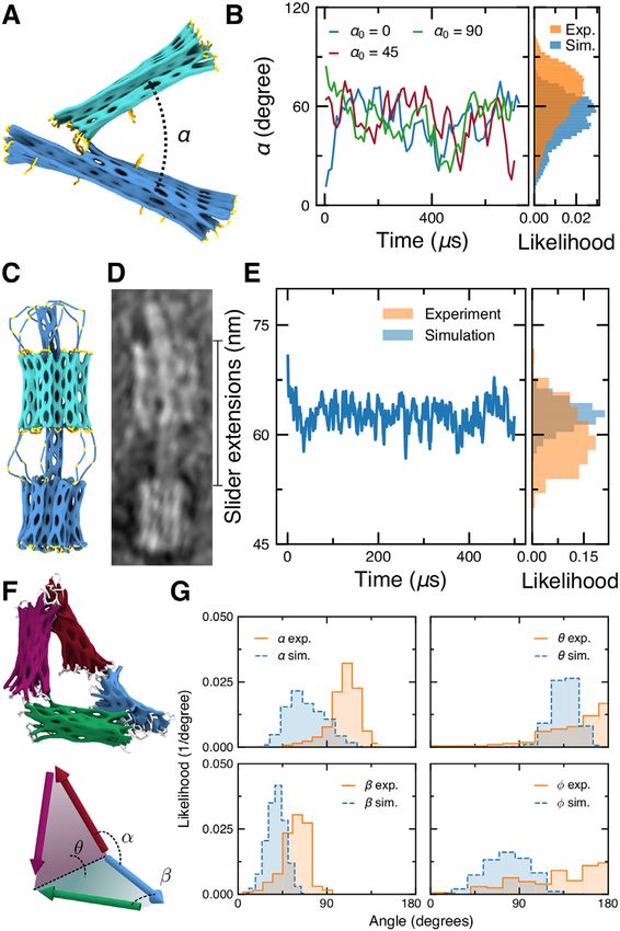
5140 Nucleic Acids Research, 2020, Vol. 48, No. 9
Downloaded from https://academic.oup.com/nar/article/48/9/5135/5814051 by guest on 01 October 2020
Figure 4. Structural relaxation of curved and twisted DNA origami objects. (A) Structures with programmed bends as characterized by TEM in Ref. 56
(top row) and reconstructed at atomic resolution using mrdna (bottom row). From left-to-right, the structures were designed to have a bend of 0◦ , 60◦ ,
120◦ and 180◦ . (B) Bend angle timeseries during 5-bp/bead mrdna relaxation of the structures shown in panel A starting from an initial configuration of a
straight rod. Dashed lines depict the target angles. (C) Dependence of the twist angle on the helical rise imposed by crossovers for honeycomb brick origami
structures (56) in 5-bp/bead mrdna simulations and as measured using TEM (56). (D) All-atom models of brick origami structures (left, top and bottom)
resulting from mrdna simulations starting from an idealized configuration (left, center). The right column shows the corresponding TEM images. (E) Gear
nanostructure (56) before (top; 5 bp/bead) and after (center; atomistic) an mrdna simulation. The gear is depicted using a bead-and-stick representation.
The bottom panel depicts the corresponding TEM image. (F) Flask nanostructure (57) before (top; 5 bp/bead) and after (center; atomistic) an mrdna
simulation. The top panel shows a bead-and-stick representation including long bonds that span the structure. All TEM images were reproduced from
Ref. 56 with permission.
calculate the time-dependent RMSD between the experi- twist within each prism and, where applicable, the skew
mental and average simulated structures, which dropped be- between connected prisms––matched the cryo-EM density
low 1 nm, dashed blue line in Figure 3B. For comparison, very closely, Supplementary Animations 2–6.
we performed the same set of simulations, but using the 2- Many DNA origami objects are designed to bend or twist
beads/bp model. Due to the greater number of beads used via a pattern of base pair insertions and deletions (56), Fig-
to represent the structure and the higher density of beads, ure 4A. Atomistic (or ENRG-MD) simulations of such ob-
the high-resolution simulations required substantially more jects that start from the idealized configuration can become
computation, that is, nearly 4 hours to obtain a single 800- trapped in a local energy minimum, preventing complete
ns simulation (20 million steps) on the same hardware, see relaxation of the object. The mrdna framework was used
orange lines in Figure 3B. The relaxation of the structure oc- to relax an ensemble of curved and twisted structures, Fig-
cured more slowly, but the average structure was even closer ure 4 B–C. A 5-bp/bead simulation was performed for each
to the experimentally derived structure with the structure- bent or twisted structure lasting 200 and 4 s, respectively,
averaged RMSD approaching 8 Å by the end of the sim- followed by 160 ns of 2-beads/bp simulations. All of the
ulation. Mapping the final configuration of the ensemble- objects relaxed to configurations that closely resembled the
averaged structure into an atomistic model provided an all- experimental transmission electron microscopy (TEM) im-
atom structure that matched the cryo-EM derived all-atom ages, providing qualitative validation of the model. The sim-
model very closely, bottom row of Figure 3A. Thus, the ulations suggest that the bent nanostructures exhibit in-
mrdna simulation resulted in a model with comparable de- creasing left-handed twist with increasing curvature with
viation from the experimentally derived structure (8.3 Å) average out-of-plane angles of 8, 1 and −26◦ for structures
as the more detailed models, ENRG-MD (32) (9.1 Å) and with 60, 120 and 180◦ bends, respectively, where a positive
oxDNA (39,40) (8.4 Å (44)) but at a fraction of the compu- out-of-plane angle corresponds to a right-handed twist in
tational cost. It is worth noting that all of the above models the curved region of the structure. Such out-of-plane twists
yielded exceptional agreement with experiment, considering may have been difficult to observe experimentally because
the reported 1.15-nm resolution of the reconstruction. the nanostructures were deposited on a surface prior to
Further validation of the mrdna structure prediction imaging. In addition, we note that the structure designed
protocol was obtained by simulating a set of four hon- to have a 180◦ bend has a slightly lower bend angle in Fig-
eycomb lattice structures that were recently designed and ure 4B due to the structure’s relatively large out-of-plane
characterized through 3D cryo-EM reconstructions by the angle.
Dietz group (28). The structures included ‘v-brick’ objects, The idealized conformation of many DNA nanostruc-
without and with twist correction, a rectangular prism ‘con- tures can sometimes deviate so dramatically from the ob-
nector’ and a triangular prism ‘vertex’. The protocol de- ject’s equilibrated configuration that CG methods, includ-
scribed above for the pointer structure was applied to each ing mrdna, are unable to relax the structure. For exam-
object, resulting in average models that could be docked ple, in its idealized geometry, the flask (57) object from the
very nicely in the corresponding cryo-EM densities, Fig- Yan group is a highly symmetric, flat DNA object that is
ure 3C–F. The ends of the bricks were seen to spread slightly quickly crushed by intrahelical connections that span the
more in mrdna simulation than in the cryo-EM reconstruc- object, see Supplementary Animation 7. To make it possi-
tions. However, the overall simulated shape––including ble to model such complicated structures, the mrdna frame-
Nucleic Acids Research, 2020, Vol. 48, No. 9 5141
work has been implemented as a Python package, which al-
lows one to script modifications to a model. For example,
many designs are intended to self-assemble into larger as-
semblies. The scripting interface was used to combine two
models of the half-gear design (56), applying a 180◦ rotation
to one of the models and adding intrahelical connections
between the two half-gears before simulating for 2 s with
5 bp/bead and 8 ns with 2 beads/bp, Figure 4E. A second
Python script was used to apply a 90◦ rotation to half of
each helix in a DNA flask before running 2-s, 5-bp/bead
and 8-ns, 2-beads/bp simulations, Figure 4F and Suppple-
mentary Animation 8. In both cases, scripting was neces-
sary to prevent the rapid collapse of bonds that span the
Downloaded from https://academic.oup.com/nar/article/48/9/5135/5814051 by guest on 01 October 2020
initially straight structure, which would result in a crushed,
tangled model of the structure. The resulting relaxed struc-
ture of the gear was seen to closely resemble TEM images
of the object, Figure 4D.
Conformational dynamics
DNA origami nanostructures are sometimes designed to
adopt multiple conformations. The mrdna framework can
be used to predict the average structure and the conforma-
tional fluctuations of such objects. For example, the Di-
etz and Castro groups have designed nanoscale calipers us-
ing DNA origami that allow measurement of intermolec-
ular forces between proteins attached to each arm of the
caliper (58,59). TEM can be used to determine the dis-
tribution of angles of the caliper with and without at-
tached proteins, allowing inference of intermolecular forces.
To demonstrate the utility of the mrdna framework for
sampling conformational space, three 700-s simulations
(3.5 billion steps each) of the caliper design from the Di- Figure 5. Mrdna simulation of dynamic DNA origami nanostructures. (A)
etz group were performed using a 5-bp/bead model, Fig- Molecular graphics representation of a DNA origami caliper designed by
ure 5A and Supplementary Animation 9. The angle be- the Dietz group for measuring inter-nucleosome forces (58). (B) The angle
tween the two arms was extracted at each frame of the sim- ␣ between the two arms of the caliper in three 5-bp/bead mrdna simula-
ulation trajectory, providing the distribution of the angles tions, starting with the initial angle of 0, 45 or 90◦ . The caliper angle was
computed as the angle between the axes of the arms of the caliper as deter-
between the two arms, Figure 5B. Substantial overlap be- mined by single-value decomposition (58). Histograms show the distribu-
tween the mrdna-generated and experimentally measured tion of the simulated angles alongside the distribution extracted from the
distributions was observed, though the mrdna distribution TEM images (58). (C and D) Molecular graphics representation (C) and
is skewed toward more acute angles. TEM image (D) of a DNA slider nanostructure designed and characterized
by the Castro group (60), reproduced with permission from Ref. 60. (E)
The slider origami nanostructure designed by the Castro The distance between the bearing and the base of the nanostructure dur-
group (60) was simulated next for 500 s (5 bp/bead; 2.5 ing a 5-bp/bead mrdna simulation. Histograms show the distribution of
billion steps), Figure 5C and D and Supplementary Ani- the simulated distances alongside the distribution extracted from the TEM
mation 10. The slider consists of a large base affixed to a images (60). (F and G) Angle distributions of a Bennett linkage nanostruc-
six-helix bundle shaft that threads through a movable bear- ture designed by the Castro group (62) in 5-bp/bead mrdna simulations.
The distributions observed in simulations are plotted alongside the cor-
ing, which is tethered on opposite ends to the base and to responding distributions extracted from single-particle reconstructions of
the tip of the shaft by a total of twelve flexible linkers. While the linkages (63).
the bearing was seen to dwell ∼5 nm further from the base
structure in mrdna simulations with a more narrow dis-
tribution than observed experimentally, the position of the Animation 11. Using the 5-bp/bead model, we were able to
bearing is overall in qualitative agreement, Figure 5E. In- sample the full range of motion of the Bennett linkage, re-
terestingly, a similar discrepancy was observed for a simi- vealing stochastic transitions between planar and compact
lar slider design when it was simulated for a very long time configurations. Distributions for the large and small angles
using the oxDNA model (61). The simulations using the (␣ and , respectively) between adjacent arms and for the
mrdna framework could be performed quickly and lasted large and small dihedral angles ( and , respectively) be-
for about 2 days. tween the planes formed by adjacent arms were calculated
At last, we performed simulations (four 700-s simula- following the overall approach described by the Ren group
tions, 3.5 billion steps each) of the DNA origami Bennett (63). Briefly, at each frame of the CG simulation trajectory,
linkage designed by the Castro group (62) and imaged using the axis of each arm was determined using single-value de-
electron tomography (63), Figure 5F and Supplementary composition. The angles between the arm axes directly pro-
5142 Nucleic Acids Research, 2020, Vol. 48, No. 9
Downloaded from https://academic.oup.com/nar/article/48/9/5135/5814051 by guest on 01 October 2020
Figure 6. Mrdna simulations of DNA nanostructures designed using programs other than cadnano. (A–C) Structural relaxation of vHelix nanostructures
starting from the vHelix maya files obtained from the Högberg group (16). From top-to-bottom, the panels show the configurations of nanostructures prior
to the low-resolution CG simulation, the resulting atomistic model and an experimentally obtained TEM image, reproduced with permission from Ref. 16.
(D–G) Structural relaxation of nanostructures using all-atom PDB models as input for mrdna. The all-atom PDBs of a box (23) (D and E), hemisphere (57)
(F) and tetrahedron (17) (G) were obtained from Nanoengineer-1 (65), CanDo (30,57) (original design from cadnano) and DAEDALUS (17). From
top-to-bottom, the panels show the configurations of nanostructures prior to the low-resolution CG simulation, the resulting atomistic model and an
experimentally obtained TEM image or reconstruction. The box TEM image is reproduced with permission from Ref. 23. Copyright 2012 American
Chemical Society. The hemisphere TEM image is reproduced from Ref. 57 with permission from AAAS. The tetrahedron reconstruction is reproduced
from Ref. 17 with permission from AAAS.
vided ␣ and . Cross products between the axes of adjacent system. We used that code to simulate a variety of wireframe
arms provided normal vectors for the plane formed by the DNA nanostructures (16) starting directly from their vHe-
two arms, allowing the dihedral angles and to be com- lix Maya designs. The resulting CG MD trajectories charac-
puted from the angle between the normal vectors. The re- terized the amount of structural fluctuations in the designs
sulting distributions are in approximate agreement with dis- and produced representative ensembles of equilibrated all-
tributions experimentally obtained by the Ren group, Fig- atom structures, Figure 6A–C. The results of our mrdna
ure 5G. The discrepancies between experiment and simu- simulations of the vHelix objects showed that the overall
lation could be due to incomplete parameterization of the geometry of each structure was preserved despite relaxation
ssDNA part of the design, a lack of twist–bend and twist– of the DNA.
stretch coupling in the model, the use of an isotropic bend- The mrdna framework also includes an algorithm to au-
ing modulus, or possibly due to a lack of defects such as tomatically identify all backbone connections, base pairs
fraying or partial melting of the Holliday junctions. Nev- and dinucleotide stacks in an all-atom representation of
ertheless, our simulation method can be used to quickly a DNA nanostructure formatted as a PDB file. This
study the structural fluctuations of dynamic DNA nanos- PDB reader module can import the output of many tools
tructures, providing rapid feedback about the mechanics of that export PDBs, including CanDo (30), Nucleic Acid
a design in place of challenging experimental characteriza- Builder (64), oxDNA (39,40) (via the convert to atomic.py
tion. script distributed with oxDNA), Nanoengineer-1 (65) (via
our nanoengineer2pdb web service http://bionano.physics.
illinois.edu/nanoengineer2pdb), DAEDALUS (17) and Tia-
Support for lattice-free DNA nanostructures mat (66), to automatically generate a corresponding mrdna
The mrdna framework can be used to simulate self- model. We demonstrate this capability here by relaxing sev-
assembled DNA nanostructures produced by lattice-free eral DNA nanostructures using all-atom PDBs from var-
approaches. The same set of commands that are used to ious sources as inputs, Figure 6D–G. The mrdna simu-
construct the spline/bead-based models of cadnano designs lations demonstrated substantial relaxation of the designs
can introduce new types of connections between DNA re- away from their idealized initial geometries, with the corre-
gions, extending the application of the mrdna framework sponding final atomistic models having an average RMSD
to other kinds of DNA nanostructures. For example, the of 24 Å compared to the initial structures. The ability of the
mrdna package already includes a code that constructs a mrdna framework to read PDB files immediately allows in-
multi-resolution model from lists of base pairs, dinucleotide terfacing with a wide array of design and simulation tools,
stacks and backbone connections for all the nucleotides in a but not all PDB structures are interpreted perfectly by the
Nucleic Acids Research, 2020, Vol. 48, No. 9 5143

                                                                                                                                                                Downloaded from https://academic.oup.com/nar/article/48/9/5135/5814051 by guest on 01 October 2020
Figure 7. Coupling continuum models with mrdna simulations. (A) Initial configuration of a simulation system consisting of a vHelix-designed nanostruc-
ture (16) near a glass nanopipette under a 300 mV applied bias. The electric potential distribution in and around the pipette was obtained using Comsol as
previously reported (20). The electric potential and a steric grid potential representing the presence of the nanopipette are incorporated in the mrdna model
as external potentials. (B) Mrdna simulation of the DNA nanostructure capture by a nanopipette. From left to right, snapshots depict the configuration
of the system during a 500-ns 5-bp/bead CG simulation.

framework because base pairs or stacks may be assigned                              We demonstrate this capability here by simulating the
incorrectly. Hence, users are cautioned to carefully exam-                       capture of a DNA nanostructure in a nanopipette, Fig-
ine their resulting models to ensure proper conversion and,                      ure 7 and Supplementary Animation 12. The spatial distri-
where possible, use an original design file as the input source                  bution of the electrostatic potential near the pipette under
for mrdna.                                                                       a 300-mV applied bias was obtained using the Comsol con-
   The atomic configuration resulting from the mrdna sim-                        tinuum model, as described previously (20). The potential
ulations could be used to inform design modifications or to                      was applied to each bead in the system with a weight pro-
perform further refinement of the structure using ENRG-                          portional to the number of nucleotides represented by the
MD (32) implicit solvent simulations or explicit solvent                         bead. In our mrdna simulation, we observe one leg of the
all-atom simulations. Moreover, the mrdna framework can                          DNA nanostructure to be captured initially, followed by a
generate all files needed to perform additional relaxation                       collapse of the wireframe mesh, which allowed the object
using the oxDNA model. When invoked, mrdna will write                            to pass fully through the aperture of the pipette. While the
sequence-specific oxDNA2 (67) topology and configuration                         above simulation is just an illustrative example, we have pre-
files and an input file that prescribes 1000 steps of minimiza-                  viously used similar multiscale/multiphysics simulations to
tion to relax the long bonds that are likely to be present in                    obtain a quantitatively correct description of the voltage-
the model, and it will call oxDNA using these files. Then,                       dependent deformation of a DNA nanostructure subject to
mrdna writes a second input file for additional relaxation                       applied electric field of various magnitudes (20). We expect
using an MD integrator, again calling oxDNA to run the                           the combination of particle-based simulations with contin-
simulation.                                                                      uum modeling to bring much anticipated advances in the
                                                                                 field of plasmonic DNA nanostructures, DNA nanostruc-
Integration with continuum models                                                tures that respond to fluid flow and, eventually, modeling
                                                                                 DNA nanostructures in the crowded environment of a bio-
DNA nanostructures are usually designed to operate in the                        logical cell.
environment of a biological system or in the context of a
nanotechnological device. In either case, it is useful to be
                                                                                 DISCUSSION
able to couple a model of a DNA nanostructure to other
models because a DNA object’s conformation and function                          We have presented an automated workflow for the predic-
can both be influenced by its environment.                                       tion of the in situ structure and conformational dynam-
   The mrdna framework uses ARBD (47), a GPU-                                    ics of self-assembled DNA objects. Uniquely, our multi-
accelerated biomolecular simulation engine, which supports                       resolution approach enables fast relaxation of an initial
mixed models of point particles and rigid body objects as                        structural model through low-resolution CG simulations,
well as 3D grid potentials. Hence, configuration files written                   progressive refinement of the structural models through
by the mrdna framework can be manually edited to include                         finer-resolution CG simulations and, ultimately, provides a
interactions with additional entities (e.g. cellular proteins                    fully atomistic representation of the object’s in situ struc-
or small molecules) that are not described by the package.                       ture. In comparison to existing computational models of
The scripting interface for the mrdna framework already                          self-assembled DNA nanostructures, our multi-resolution
exposes commands for applying grid-based potentials to                           approach is computationally much more efficient, providing
DNA nanostructures, allowing one to exert effective forces                       relaxed, fully atomistic models of typical 3D DNA origami
extracted from continuum models to, for example, simulate                        nanostructures within 30 minutes or less. The quantitative
DNA nanostructures under the influence of spatially vary-                        accuracy of the resulting structural model allows the mrdna
ing electric or plasmonic optical fields (68).                                   framework to be used routinely for DNA nanostructure de-
5144 Nucleic Acids Research, 2020, Vol. 48, No. 9

sign and prototyping, replacing labor- and time-consuming         Han for sharing a cadnano design of the DNA flask. C.M.
cryo-EM reconstruction.                                           thanks Carlos Castro for sharing cadnano designs of the
   By taking into account non-trivial environmental condi-        slider and Bennet linkage structures. C.M. thanks Hen-
tions, such as variable ionic strength, applied electric field,   drik Dietz and Klaus Wagenbauer for sharing cadnano de-
non-homogeneous temperature and optical intensity, the            signs of the v-brick, connector block, triangular vertex and
framework enables computational study of, not only the            caliper structures. C.M. thanks Will Kaufhold for his help
structural, but also the functional aspects of the nanos-         debugging the vHelix importer and Chao-Min Huang for
tructure designs. The speed of the model also makes it            adding MagicDNA support for mrdna.
well-suited for the study of structural fluctuations, although
slight differences between simulated and experimentally-
obtained angle and distance distributions were noted. We          FUNDING
speculate that these are caused by small deficiencies in the
model, such as the simple polymer description of ssDNA,           National Science Foundation [OAC-1740212, DMR-
                                                                  1827346]; National Institutes of Health [P41-GM104601].

                                                                                                                                             Downloaded from https://academic.oup.com/nar/article/48/9/5135/5814051 by guest on 01 October 2020
the treatment of ssDNA–dsDNA junctions and the lack
of twist–stretch and twist–bend coupling. The availability        Funding for open access charge: National Science Founda-
of an ensemble of experiments probing the configurational         tion [DMR-1827346].
space explored by such nanostructures will prove valuable         Conflict of interest statement. None declared.
in future refinements of the model.
   The mrdna framework is distributed as an open source
Python package (https://gitlab.engr.illinois.edu/tbgl/tools/      REFERENCES
mrdna), enabling future customization of the algorithms            1. Seeman,N.C. (1982) Nucleic acid junctions and lattices. J. Theor.
by the end users. A command-line interface allows simula-             Biol., 99, 237–247.
                                                                   2. Yan,H., Park,S.H., Finkelstein,G., Reif,J.H. and LaBean,T.H. (2003)
tions to be performed and customized with a few keystrokes.           DNA-templated self-assembly of protein arrays and highly
The package provides additional capabilities to the users             conductive nanowires. Science, 301, 1882–1884.
through simple Python scripting, such as the application of        3. Lin,C., Liu,Y., Rinker,S. and Yan,H. (2006) DNA tile based
geometric transformations to parts of a DNA nanostruc-                self-assembly: building complex nanoarchitectures. Chemphyschem, 7,
                                                                      1641–1647.
ture, interfacing with continuum models through 3D grid-           4. Rothemund,P.W.K. (2006) Folding DNA to create nanoscale shapes
specified potentials, and generating or modulating DNA                and patterns. Nature, 440, 297–302.
nanostructures algorithmically. A step-by-step guide to us-        5. Douglas,S.M., Dietz,H., Liedl,T., Högberg,B., Graf,F. and
ing mrdna is available (https://gitlab.engr.illinois.edu/tbgl/        Shih,W.M. (2009) Self-assembly of DNA into nanoscale
tutorials/multi-resolution-dna-nanostructures).                       three-dimensional shapes. Nature, 459, 414–418.
                                                                   6. Seeman,N.C. (2010) Nanomaterials based on DNA. Annu. Rev.
   Immediate future developments of the mrdna frame-                  Biochem., 79, 65–87.
work will focus on improving the description of ssDNA–             7. Pinheiro,A.V., Han,D., Shih,W.M. and Yan,H. (2011) Challenges and
dsDNA junctions, dsDNA nicks and end-stacking interac-                opportunities for structural DNA nanotechnology. Nat. Nanotech., 6,
tions. The physical realism of the model will be further en-          763–772.
                                                                   8. Seeman,N.C. and Sleiman,H.F. (2017) DNA nanotechnology. Nat.
hanced by supplementing the mrdna framework with CG                   Rev. Mater., 3, 17068–17090.
models that explicitly account for the sequence-dependence         9. Chen,J.H. and Seeman,N.C. (1991) Synthesis from DNA of a
of DNA elasticity and the possibility of DNA hybridiza-               molecule with the connectivity of a cube. Nature, 350, 631–633.
tion and dehybridization reactions. Complementing devel-          10. Fu,T.J. and Seeman,N.C. (1993) DNA double-crossover molecules.
opment of ARBD, the framework will be extended to incor-              Biochemistry, 32, 3211–3220.
                                                                  11. Winfree,E., Liu,F., Wenzler,L.A. and Seeman,N.C. (1998) Design
porate grid- and particle-based models of proteins and inor-          and self-assembly of two-dimensional DNA crystals. Nature, 394,
ganic nanostructures. At last, our multi-resolution simula-           539–544.
tion framework may be integrated with other DNA nanos-            12. Mao,C., Sun,W. and Seeman,N.C. (1999) Designed two-dimensional
tructure design and visualization tools to provide near real-         DNA holliday junction arrays visualized by atomic force microscopy.
                                                                      J. Am. Chem. Soc., 121, 5437–5443.
time feedback to the designer.                                    13. Shih,W.M., Quispe,J.D. and Joyce,G.F. (2004) A 1.7-kilobase
                                                                      single-stranded DNA that folds into a nanoscale octahedron. Nature,
                                                                      427, 618–621.
SUPPLEMENTARY DATA                                                14. Seeman,N.C. (2005) Structural DNA nanotechnology: an overview.
                                                                      Mol. Microbiol., 303, 143–166.
Supplementary Data are available at NAR Online.                   15. Ke,Y., Ong,L.L., Shih,W.M. and Yin,P. (2012) Three-dimensional
                                                                      structures self-assembled from DNA bricks. Science, 338, 1177–1183.
                                                                  16. Benson,E., Mohammed,A., Gardell,J., Masich,S., Czeizler,E.,
ACKNOWLEDGEMENTS                                                      Orponen,P. and Högberg,B. (2015) DNA rendering of polyhedral
                                                                      meshes at the nanoscale. Nature, 523, 441–444.
The authors gladly acknowledge supercomputer time pro-
                                                                  17. Veneziano,R., Ratanalert,S., Zhang,K., Zhang,F., Yan,H., Chiu,W.
vided through XSEDE Allocation Grant MCA05S028, the                   and Bathe,M. (2016) Designer nanoscale DNA assemblies
Blue Waters supercomputer system (UIUC) and the Fron-                 programmed from the top down. Science, 352, 1534–1534.
tera supercomputer (TACC). The authors thank Shawn                18. Maye,M.M., Kumara,M.T., Nykypanchuk,D., Sherman,W.B. and
Douglas, Hendrik Dietz, Björn Högberg, Tim Liedl and                Gang,O. (2010) Switching binary states of nanoparticle superlattices
                                                                      and dimer clusters by DNA strands. Nat. Nanotech., 5, 116–120.
many other practitioners of the structural DNA nanotech-          19. Modi,S., Swetha,M., Goswami,D., Gupta,G.D., Mayor,S. and
nology field for their valuable feedback on the pre-released          Krishnan,Y. (2009) A DNA nanomachine that maps spatial and
version of the mrdna framework. A.A. thanks Dongran                   temporal pH changes inside living cells. Nat. Nanotech., 4, 325–330.
Nucleic Acids Research, 2020, Vol. 48, No. 9 5145
20. Hemmig,E., Fitzgerald,C., Maffeo,C., Hecker,L., Ochmann,S., 42. Snodin,B., Romano,F., Rovigatti,L., Ouldridge,T., Louis,A. and
Aksimentiev,A., Tinnefeld,P. and Keyser,U. (2018) Optical voltage Doye,J. (2016) Direct simulation of the self-assembly of a small DNA
sensing using DNA origami. Nano Lett., 18, 1962–1971. origami. ACS Nano, 10, 1724–1737.
21. Funke,J.J., Ketterer,P., Lieleg,C., Schunter,S., Korber,P. and Dietz,H. 43. Reshetnikov,R.V., Stolyarova,A.V., Zalevsky,A.O., Panteleev,D.Y.,
(2016) Uncovering the forces between nucleosomes using DNA Pavlova,G.V., Klinov,D.V., Golovin,A.V. and Protopopova,A.D.
origami. Sci. Adv., 2, e1600974. (2017) A coarse-grained model for DNA origami. Nucleic Acids Res.,
22. Andersen,E.S., Dong,M., Nielsen,M.M., Jahn,K., Subramani,R., 46, 1102–1112.
Mamdouh,W., Golas,M.M., Sander,B., Stark,H., Oliveira,C. 44. Snodin,B.E.K., Schreck,J.S., Romano,F., Louis,A.A. and Doye,J.P.K.
et al.2009) Self-assembly of a nanoscale DNA box with a controllable (2019) Coarse-grained modelling of the structural properties of DNA
lid. Nature, 459, 73–76. origami. Nucleic Acids Res., 47, 1585–1597.
23. Zadegan,R.M., Jepsen,M.D.E., Thomsen,K.E., Okholm,A.H., 45. Douglas,S.M., Marblestone,A.H., Teerapittayanon,S., Vazquez,A.,
Schaffert,D.H., Andersen,E.S., Birkedal,V. and Kjems,J. (2012) Church,G.M. and Shih,W.M. (2009) Rapid prototyping of 3D
Construction of a 4 zeptoliters switchable 3D DNA box origami. DNA-origami shapes with caDNAno. Nucleic Acids Res., 37,
ACS Nano, 6, 10050–10053. 5001–5006.
24. Kang,S.H., Hwang,W.S., Lin,Z., Kwon,S.H. and Hong,S.W. (2015) A 46. Huang,C.-M., Kucinic,A., Le,J.V., Castro,C.E. and Su,H.-J. (2019)
robust highly aligned DNA nanowire array-enabled lithography for Uncertainty quantification of a DNA origami mechanism using a
Downloaded from https://academic.oup.com/nar/article/48/9/5135/5814051 by guest on 01 October 2020
graphene nanoribbon transistors. Nano Lett., 15, 7913–7920. coarse-grained model and kinematic variance analysis. Nanoscale, 11,
25. Du,K., Park,M., Ding,J., Hu,H. and Zhang,Z. (2017) Sub-10nm 1647–1660.
patterning with DNA nanostructures: a short perspective. Nanotech., 47. Comer,J. and Aksimentiev,A. (2012) Predicting the DNA sequence
28, 442501–442509. dependence of nanopore ion current using atomic-resolution
26. Gopinath,A., Miyazono,E., Faraon,A. and Rothemund,P.W. (2016) Brownian dynamics. J. Phys. Chem. C, 116, 3376–3393.
Engineering and mapping nanocavity emission via precision 48. Rau,D.C., Lee,B. and Parsegian,V.A. (1984) Measurement of the
placement of DNA origami. Nature, 535, 401–405. repulsive force between polyelectrolyte molecules in ionic solution:
27. Kühler,P., Roller,E.M., Schreiber,R., Liedl,T., Lohmüller,T. and Hydration forces between parallel DNA double helices. Proc. Natl.
Feldmann,J. (2014) Plasmonic DNA-origami nanoantennas for Acad. Sci. U.S.A., 81, 2621–2625.
surface-enhanced raman spectroscopy. Nano Lett., 14, 2914–2919. 49. Cocco,S., Marko,J.F. and Monasson,R. (2002) Theoretical models for
28. Wagenbauer,K.F., Sigl,C. and Dietz,H. (2017) Gigadalton-scale single-molecule DNA and RNA experiments: from elasticity to
shape-programmable DNA assemblies. Nature, 552, 78–83. unzipping. Comptes Rendus Physique, 3, 569–584
29. Ong,L.L., Hanikel,N., Yaghi,O.K., Grun,C., Strauss,M.T., Bron,P., 50. Mosconi,F., Allemand,J.F., Bensimon,D. and Croquette,V. (2009)
Lai-Kee-Him,J., Schueder,F., Wang,B., Wang,P. et al. (2017) Measurement of the torque on a single stretched and twisted DNA
Programmable self-assembly of three-dimensional nanostructures using magnetic tweezers. Phys. Rev. Lett., 102, 078301–078304.
from 10,000 unique components. Nature, 552, 72–77. 51. Lilley,D.M. and Clegg,R.M. (1993) The structure of the four-way
30. Kim,D.-N., Kilchherr,F., Dietz,H. and Bathe,M. (2011) Quantitative junction in DNA. Annu. Rev. Biophys. Biomol. Struct., 22, 299–328.
prediction of 3D solution shape and flexibility of nucleic acid 52. Bai,X.-C.C., Martin,T.G., Scheres,S.H.W. and Dietz,H. (2012)
nanostructures. Nucleic Acids Res., 40, 2862–2868. Cryo-EM structure of a 3D DNA-origami object. Proc. Natl. Acad.
31. Yoo,J. and Aksimentiev,A. (2013) In situ structure and dynamics of Sci. U.S.A., 109, 20012–20017.
DNA origami determined through molecular dynamics simulations. 53. Han,D., Pal,S., Yang,Y., Jiang,S., Nangreave,J., Liu,Y. and Yan,H.
Proc. Natl. Acad. Sci. U.S.A., 110, 20099–20104. (2013) DNA gridiron nanostructures based on four-arm junctions.
32. Maffeo,C., Yoo,J. and Aksimentiev,A. (2016) De novo prediction of Science, 339, 1412–1415.
DNA origami structures through atomistic molecular dynamics 54. Humphrey,W., Dalke,A. and Schulten,K. (1996) VMD: Visual
simulation. Nucleic Acids Res., 44, 3013–3019. molecular dynamics. J. Mol. Graph., 14, 33–38.
33. Slone,S., Li,C.-Y., Yoo,J. and Aksimentiev,A. (2016) Molecular 55. Trabuco,L.G., Villa,E., Mitra,K., Frank,J. and Schulten,K. (2008)
mechanics of DNA bricks: in situ structure, mechanical properties Flexible fitting of atomic structures into electron microscopy maps
and ionic conductivity. New J. Phys., 18, 055012–055022. using molecular dynamics. Structure, 16, 673–683.
34. Li,C.-Y., Hemmig,E.A., Kong,J., Yoo,J., Hernández-Ainsa,S., 56. Dietz,H., Douglas,S.M. and Shih,W.M. (2009) Folding DNA into
Keyser,U.F. and Aksimentiev,A. (2015) Ionic conductivity, structural twisted and curved nanoscale shapes. Science, 325, 725–730.
deformation, and programmable anisotropy of dna origami in electric 57. Han,D., Pal,S., Nangreave,J., Deng,Z., Liu,Y. and Yan,H. (2011)
field. ACS Nano, 9, 1420–1433. DNA origami with complex curvatures in three-dimensional space.
35. Göpfrich,K., Li,C.-Y., Mames,I., Bhamidimarri,S.P., Ricci,M., Science, 332, 342–346.
Yoo,J., Mames,A., Ohmann,A., Winterhalter,M., Stulz,E. et al.2016) 58. Funke,J.J., Ketterer,P., Lieleg,C., Korber,P. and Dietz,H. (2016)
Ion channels made from a single membrane-spanning DNA duplex. Exploring nucleosome unwrapping using DNA origami. Nano Lett.,
Nano Lett., 16, 4665–4669. 16, 7891–7898.
36. Göpfrich,K., Li,C.-Y., Ricci,M., Bhamidimarri,S.P., Yoo,J., 59. Le,J.V., Luo,Y., Darcy,M.A., Lucas,C.R., Goodwin,M.F.,
Gyenes,B., Ohmann,A., Winterhalter,M., Aksimentiev,A. and Poirier,M.G. and Castro,C.E. (2016) Probing nucleosome stability
Keyser,U.F. (2016) Large-conductance transmembrane porin made with a DNA origami nanocaliper. ACS Nano, 10, 7073–7084.
from DNA origami. ACS Nano, 10, 8207–8214. 60. Marras,A., Zhou,L., Kolliopoulos,V., Su,H. and Castro,C. (2016)
37. Ohmann,A., Li,C.-Y., Maffeo,C., Al-Nahas,K., Baumann,K.N.B., Directing folding pathways for multi-component DNA origami
Keyser,U.F. and Aksimentiev,A. (2018) A synthetic enzyme built nanostructures with complex topology. New J. Phys., 18,
from DNA flips 107 lipids per second in biological membranes. Nat. 055005–055013.
Commun.9, 2426–2434. 61. Sharma,R., Schreck,J.S., Romano,F., Louis,A.A. and Doye,J.P.K.
38. Hinckley,D.M., Freeman,G.S., Whitmer,J.K. and de Pablo,J.J. (2013) (2017) Characterizing the motion of jointed DNA nanostructures
An experimentally-informed coarse-grained 3-site-per-nucleotide using a coarse-grained model. ACS Nano, 11, 12426–12435.
model of DNA: Structure, thermodynamics, and dynamics of 62. Marras,A.E., Zhou,L., Su,H.-J. and Castro,C.E. (2015)
hybridization. J. Chem. Phys., 139, 144903–144917. Programmable motion of DNA origami mechanisms. Proc. Natl.
39. Ouldridge,T.E., Louis,A.A. and Doye,J.P.K. (2011) Structural, Acad. Sci. U.S.A., 112, 713–718.
mechanical, and thermodynamic properties of a coarse-grained DNA 63. Lei,D., Marras,A.E., Liu,J., Huang,C.-M., Zhou,L., Castro,C.E.,
model. J. Chem. Phys., 134, 085101–085122. Su,H.-J. and Ren,G. (2018) Three-dimensional structural dynamics of
40. Šulc,P., Romano,F., Ouldridge,T.E., Rovigatti,L., Doye,J.P.K. and DNA origami Bennett linkages using individual-particle electron
Louis,A.A. (2012) Sequence-dependent thermodynamics of a tomography. Nat. Commun., 9, 592–600.
coarse-grained DNA model. J. Chem. Phys., 137, 135101–135114. 64. MacKerell,A.D. Jr, Bashford,D., Bellott,M., Dunbrack,R.L. Jr.,
41. Uusitalo,J.J., Ingólfsson,H.I., Akhshi,P., Tieleman,D.P. and Evanseck,J.D., Field,M.J., Fischer,S., Gao,J., Guo,H., Ha,S. et al.
Marrink,S.J. (2015) Martini coarse-grained force field: extension to (1998) All-atom empirical potential for molecular modeling and
DNA. J. Chem. Theory Comput., 11, 3932–3945. dynamics studies of proteins. J. Phys. Chem. B, 102, 3586–3616.
65. Nanorex, Inc. (2006) Nanoengineer-1, v.a8.You can also read