Photophysics of DFHBI bound to RNA aptamer Baby Spinach
←
→
Page content transcription
If your browser does not render page correctly, please read the page content below
www.nature.com/scientificreports
OPEN Photophysics of DFHBI bound
to RNA aptamer Baby Spinach
Nguyen Thuan Dao1,2,5*, Reinhard Haselsberger1,3,5, Mai Thu Khuc1, Anh Tuân Phan1,
Alexander A. Voityuk4 & Maria‑Elisabeth Michel‑Beyerle1,3*
The discovery of the GFP-type dye DFHBI that becomes fluorescent upon binding to an RNA aptamer,
termed Spinach, led to the development of a variety of fluorogenic RNA systems that enable genetic
encoding of living cells. In view of increasing interest in small RNA aptamers and the scarcity of
their photophysical characterisation, this paper is a model study on Baby Spinach, a truncated
Spinach aptamer with half its sequence. Fluorescence and fluorescence excitation spectra of DFHBI
complexes of Spinach and Baby Spinach are known to be similar. Surprisingly, a significant divergence
between absorption and fluorescence excitation spectra of the DFHBI/RNA complex was observed
on conditions of saturation at large excess of RNA over DFHBI. Since absorption spectra were not
reported for any Spinach-type aptamer, this effect is new. Quantitative modelling of the absorption
spectrum based on competing dark and fluorescent binding sites could explain it. However, following
reasoning of fluorescence lifetimes of bound DFHBI, femtosecond-fluorescence lifetime profiles would
be more supportive of the notion that the abnormal absorption spectrum is largely caused by trans-
isomers formed within the cis-bound DFHBI/RNA complex. Independent of the origin, the unexpected
discrepancy between absorption and fluorescence excitation spectra allows for easily accessed
screening and insight into the efficiency of a fluorogenic dye/RNA system.
One of the most challenging problems in cell biology is the control of RNA activity by employing genetically
encoded fluorescent labels. To this goal the non-toxic, membrane-permeable reporter 3,5-DiFluoro-4-Hydroxy-
Benzylidene Imadazolinone (DFHBI) mimicking the fluorophore of the Green Fluorescent Protein (GFP) has
been engineered that is non-fluorescent in solution but emits green fluorescence when bound to the core G-quad-
ruplex of the RNA aptamer S pinach1,2. This discovery paved the road for engineering a large variety of fluorogenic
RNA aptamers as powerful tools for imaging genetically encoded domains in living c ells3–5. Apart from poor
photo- and low thermal stability, the applicability of the initial Spinach aptamers to living cells5 was expected to be
limited also by its propensity to misfold. In the meantime, many limitations are removed either by miniaturizing
the Spinach aptamer, or optimized superfolding aptamers, or by variation of the small reporter fluorophore6,7.
In search for small aptamers that are easier to integrate into arbitrary RNA sequences, the crystal structure
of Spinach allowed for a rational reduction by removing non-essential regions of the sequence, thereby min-
iaturizing Spinach to Baby S pinach1,5,8,9 and, the even smaller B roccoli10–12 as well as still shorter variants such
as Corn13,14. Surprisingly, when genetically inserted into ribosomal RNAs, both of the small aptamers, Baby
Spinach and Broccoli, have shown superior fluorescence efficiency as compared to the identical constructs under
in-vitro conditions8 and as compared to several full-sequence Spinach aptamers. Thus, the early c onjecture1 that
the compact Baby Spinach, only half as large as the mother Spinach, may reduce live-cell artefacts when fused
to ribosomal R NA8, gained confidence. Baby Spinach is also resistant to nuclease cleavage and degradation.
Moreover, the smaller and more compact Baby Spinach results in higher mechanical stability as compared to a full
Spinach sequence15. The structure of Baby Spinach in comparison with full sequence Spinach is shown in Fig. S1.
In view of the increasing interest in small RNA a ptamers9 and the scarcity regarding their photophysical
characterization, this paper is a model study on Baby Spinach. Although there is no high-resolution X-ray
structure available, identical NMR spectra of Spinach (SP) and Baby Spinach (bSP) in the imino region suggest
that the structure of the binding pocket is retained upon miniaturization4. Another feature, though under some
debate8, is the fluorescence yield from bSP aptamers. More recently, the high fluorescence yield reported for the
fluorophore-tagged Baby Spinach approaching 95% of the fluorescence intensity of Spinach is under d ebate8.
1
School of Physical and Mathematical Sciences, Nanyang Technological University, Singapore 637371,
Singapore. 2Institute of Materials Science, Vietnam Academy of Science and Technology, Hanoi 100000,
Vietnam. 3TUMCREATE, Singapore 138602, Singapore. 4Institució Catalana de Recerca I Estudis Avancats,
08010 Girona, Spain. 5These authors contributed equally: Nguyen Thuan Dao and Reinhard Haselsberger *email:
thuandn@ims.vast.ac.vn; mariaelisabeth@ntu.edu.sg
Scientific Reports | (2021) 11:7356 | https://doi.org/10.1038/s41598-021-85091-y 1
Vol.:(0123456789)www.nature.com/scientificreports/
A B
Fluorescene Inetensity (a. u.)
[DFHBI]:[RNA] [DFHBI]:[RNA]
C D
[DFHBI]:[RNA]
Normalized Absorbance
Normalized Excitation
Excitation
Figure 1. Absorption (A) and fluorescence (B) spectra (λexc 460 nm) of free DFHBI (2 µM) in the absence
of bSP (black) and its mixture with increasing bSP concentrations, all measured in a flow-cell at low intensity.
(C) Fluorescence excitation spectra (λprobe 501 nm) of free DFHBI and in the presence of increasing bSP
concentrations. (D) Normalized calculated absorption spectra for increasing bSP concentrations as derived from
Kd = 4.4 µM in comparison with the fluorescence excitation spectrum (red, dotted).
In this paper, we report on DFHBI/Baby Spinach studied by steady-state absorption and fluorescence spec-
troscopy complemented by femtosecond time-resolved fluorescence measurements. While quantitative modelling
of the absorption spectrum is consistent with competitive binding to a fluorescent and dark RNA aptamer site
in the case of bSP, femtosecond lifetime profiles suggest cis–trans isomerisation of the DFHBI/bSP c omplex16,17
within the binding pocket as main contribution to the large amplitude of the absorption spectrum on condition
of RNA excess over DFHBI and saturation of fluorescence.
Results
Absorption and fluorescence spectra of the DFHBI/bSP system. Absorption and Fluorescence. As
shown in Fig. 1A, the absorption spectrum of DFHBI anion is strongly affected by increasing amounts of bSP.
At saturating bSP concentration the spectrum is broadened, and its peak is red shifted from 416 to 427 nm,
while its molar extinction coefficient decreases by ≈ 30% (data in Fig. S2). Such strong hypochromism known to
accompany intercalation of dyes between base stacks of DNA is not only consistent with the X-ray structure5,18
of the parent SP, but supports also a similar stacking pattern for bSP.
The fluorescence spectrum of the complex DFHBI/bSP (Fig. 1B) is independent of the bSP concentration
(Fig. S3). Its peak position is well matched (Fig. S4) with the one observed for DFHBI bound to full-sequence
SP1,5.
The corresponding fluorescence excitation spectra peaking at 460 nm (Fig. 1C) are blue shifted (9 nm) as
compared to the DFHBI/SP complex2. In difference to the parent DFHBI/SP complex, excitation and fluores-
cence spectra of the bSP complex (Fig. S4) are significantly broadened indicating a collection of conformational
substates in the complex that seem to depend on preparative details.
Determination of the dissociation constant Kd. The binding affinity of DFHBI (D) to bSP (RNA) can be cal-
culated by applying Eq. (1) to the formation of the fluorescing complex [D•RNA]FL assuming non-cooperative
binding (n = 1):
[D • RNA]FL / [RNA]0 = [D • RNA]FL / (Kd + [D]0 − [D • RNA]FL ) (1)
The dissociation constant K d = 4.4 µM has been obtained by fitting fluorescence titration data with Eq. (1). This
d values1,16–18
high Kd value is indicative of a relatively low binding efficiency. It is beyond the range of literature K
Scientific Reports | (2021) 11:7356 | https://doi.org/10.1038/s41598-021-85091-y 2
Vol:.(1234567890)www.nature.com/scientificreports/
for the full Spinach sequence, although these are scattered over a wide range between 0.3 μM16,18 and 1.3 μM17.
The scatter depends on details of the preparative protocol and the measuring method used (see SI). Note that
these measurements in the literature employed only fluorescence data, and their analysis is based on 1:1 com-
plexation of DFHBI and Spinach RNA.
With the value K d = 4.4 µM obtained from the fluorescence titration, the equilibrium concentrations of the
free dye [D] and fluorescent complex [D•RNA]FL were calculated for different ratios of [D] and [RNA]. On the
assumption that a 1:1 fluorescent complex is formed, the calculated absorption spectrum of the fluorescent
complex in each mixture (Fig. S5) was derived by subtracting the absorption of the free dye from the observed
absorption of the mixture according to
1
Abs[D• RNA]FL ( ) = {AbsMIXTURE ( ) − AbsD ( )} (2)
[D • RNA]FL
where AbsD ( ) and AbsMIXTURE ( ) are the respective absorption spectra of free DFHBI and its mixture with
the aptamer. Figure 1D compares the normalized calculated absorption spectra of the fluorescing complex with
the normalized excitation spectrum that is independent of the RNA concentration. Most strikingly, in Fig. 1D
there is a large discrepancy between the apparent absorption and excitation spectra of the fluorescent complex
independent of the concentration ratio. Thus, the simplest and commonly used binding model cannot be applied
to derive the absorption spectrum of the fluorescent complex, and an extended model for binding should be
considered. We also noted that even for the Spinach-DFHBI complex, there are large discrepancies in the exci-
tation spectra reported by different groups in the literature (Fig. S6). Although none of these groups reported
the absorption spectrum of the complex, the large discrepancy in excitation spectra reported by these groups
would clearly modify the discrepancy between absorption and excitation spectra of the Spinach-DFHBI complex.
Modelling of absorption spectra. In the simplest model, DFHBI forms a fluorescent complex with bSP
RNA that can be described by the equilibrium constant Kd
[D].[RNA]
Kd = (3)
[D • RNA]FL
Since the fluorescence intensity IFL is proportional to the concentration of the complex: IFL = f [D•RNA]FL,
Eq. (3) can also be written as
IFL IFL IFL
K = [D]0 − [RNA]0 − (4)
f f f
where [D]0 and [RNA]0 are the initial concentrations of these components and Kd = K. As shown in Fig. 1D,
this simple model leads to a significant discrepancy of the absorption and excitation spectra of the fluorescent
complex.
What comes to mind first, is that the relatively small bSP RNA may be not perfectly folded. If one assumes
that a part of RNA exists in unfolded or misfolded conformations Z which do not form the fluorescent complex,
the equilibrium RNA Z, defined by a dissociation constant K z, should be considered. In this case Eq. (4) reads
IFL IFL IFL
K(1 + Kz ) = [D]0 − [RNA]0 − (4a)
f f f
Denoting K*(1 + Kz) by Kd in Eq. (4a), we obtain Eq. (3) implying that K found by fitting of experimental
fluorescence data depends on the value of Kz. More important, however, the factor f remains unchanged. Thus, the
ratio [D•RNA]FL/[D] does not depend on Kz. In other words, accounting for the formation of the inactive form
Z of RNA aptamer structural diversity will not help to resolve the discrepancy of the absorption and excitation
spectra of the fluorescent complex.
Multiple‑site binding model. Now we consider that in addition to the fluorescent complex also a multitude of
non-fluorescent complexes [D•RNA]DARK = [X] is formed. For the sake of simplicity, the multitude of independ-
ent binding sites is subsumed by the notion dark complex X. The stability of the dark complex is defined by the
dissociation constant
[D][RNA]
KX = (5)
[X]
For convenience, we will use a parameter p = 1 + Kd/Kx. Note that p = 1 corresponds to the standard model
with a single binding site.
In this extended model, p > 1 and Eq. (4) transforms to Eq. (6)
IFL IFL IFL
K = [D]0 − p [RNA]0 − p (6)
f f f
Replacing f by f′/p and K by K′/p in Eq. (4) we obtain
Scientific Reports | (2021) 11:7356 | https://doi.org/10.1038/s41598-021-85091-y 3
Vol.:(0123456789)www.nature.com/scientificreports/
A Kd = 7.9 M B
Excitation spectrum
Normalized Excitation
A
Kd = 7.9
p = 1.8
Figure 2. (A) Comparison of experimental and calculated fluorescence intensity. The continuous line
represents the best fit of the experimental data to the multiple-site model. (B) The normalized calculated
absorption spectrum of the DFHBI/bSP complex derived with K d = 7.9 µM in comparison to the observed
fluorescence excitation spectrum (Fig. 1D).
IFL ′ ′ IFL ′ IFL ′
K = [D]0 − [RNA]0 − (7)
f f f
Equation (7) has the same form as Eq. (4). It means that the dissociation constant and the factor f within the
extended model can be directly expressed by the corresponding quantities derived within the standard model,
f′ = f * p and K′ = K* p. The concentration of the fluorescent complex is by the factor p smaller than that derived
within the simple model, [D•RNA]FL = IFL/f′ = IFL/(f * p). The factor p cannot be derived from the fluorescence
data. However, this parameter can be determined using the absorption spectra shown in Fig. 1A
A416
0 − Ai
416 ǫ460
p= 460
. 416 (8)
Ai ǫ
where A4160 is the absorbance of the pure dye at 416 nm, Ai and Ai are absorbance of the mixture i (at different
416 460
416 460
concentration ratios) at 416 nm and 460 nm, ε and ε are the corresponding molecular extinction coefficients
at 416 nm and 460 nm. From the absorption spectra of the mixtures given in Fig. 2A we obtain p = 1.8 ± 0.1. With
this value for p and K = 4.4 μM, we obtain a larger dissociation constant K d for the fluorescent complex, Kd = K
* p = 7.9 ± 0.2 μM than the one extracted from the fluorescence titration. As compared to Kd of the fluorescing
complex, the dissociation constant Kx for the dark complex, Kx = Kd/(p − 1) = 9.8 ± 0.2 μM is only slightly larger.
Figure 2A shows good agreement between experimental data and calculations using the multiple-site binding
model. Also using Kd = 7.9 µM to derive the absorption spectrum of the fluorescing complex largely removes
the discrepancy between absorption and fluorescence excitation spectra in Fig. 1D. Both Fig. 2A,B validate
our multiple-site binding model. To our knowledge, it is the first time a discrepancy between the absorption
and excitation spectra of the DFHBI/RNA system has been recognized. However, we cannot exclude that this
discrepancy is a special feature of the system DFHBI/bSP and less relevant for Spinach-analogue aptamers with
higher binding affinity.
The parameter p (p > 1) determines the ratio of the fluorescent to the dark complexes [D•RNA] FL/
[D•RNA]DARK = Kd/Kx = (p − 1). Therefore, the difference of the free energies of these species can be expressed as
(9)
� = �G((D • RNA)FL ) − �G((D • RNA)DARK ) = RTln p − 1
while (D•RNA)FL is thermodynamically more stable than X (Δ < 0) when p < 2. By contrast, in systems where
p > 2, the formation of the dark states becomes dominating (Δ < 0). For bSP, p = 1.8 yields Δ = -0.17 kcal/mol.
Because the ratio [D•RNA]DARK/[D•RNA]FL is extremely sensitive to Δ, small changes in the structure or in
energy parameters of either RNA or the dye can seriously affect the fluorescent properties of the system.
It is interesting to add that for SP the deletion of both the knob region—a non-complimentary stem in
SP—and the U50 nucleotide destroyed the fl uorescence19. The effect has been attributed to folding problems.
This observation is in contrast to the strong fluorescence reported for bSP where also the knob region and the
nucleotide U50 are deleted10. A direct comparison between the two variants is not possible since the SP mutant10
still contains the S3 stem which has been removed in bSP. This comparison demonstrates that a small change in
the sequence—even in the region outside of the binding pocket—may lead to a minor change in the structure
that can largely affect the fluorescence.
Quantifying fluorescent complexes and the dark states. The absorption spectrum of the mixture
2 µM DFHBI/20 µM RNA comprises two components: the absorption of the fluorescing complex saturated in
the presence of 20 µM RNA and the combined absorption of non-fluorescing species consisting of the free dye
and the dark complex. The separation of the two components is achieved as follows: Since the absorption spec-
Scientific Reports | (2021) 11:7356 | https://doi.org/10.1038/s41598-021-85091-y 4
Vol:.(1234567890)www.nature.com/scientificreports/
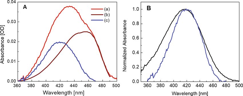
Figure 3. Absorption spectra of the system DFHBI/bSP at saturating conditions 2 µM DFHBI/20 µM bSP.
(A) (a) Measured absorption spectrum of the mixture Fig. 1A (red), (b) calculated absorption spectrum of
the fluorescing complex (D•RNA)FL (brown), and (c) calculated absorption spectrum of free dye and dark
complexes (D•RNA)DARK (blue). (B) Absorption spectrum of the free dye (black) and absorption spectrum of
the mixture of free dye and non-fluorescent complex from (A) (blue). Note that the absorption spectrum of the
mixture is narrowed and slightly red shifted from 416 to 419 nm.
trum of the fluorescent complex (D•RNA)FL should be identical to the excitation spectrum of its fluorescence
at the low concentration of 2 μM used, the combined absorption spectra of the free dye and the dark complex
can be constructed for the various DFHBI/bSP ratios as given in Fig. 3A for saturating conditions at a tenfold
excess of bSP over the fluorescence reporter DFHBI. (We note in passing that at the low dye concentration used,
absorbance measured in OD based on the 10log scale and the excitation spectrum are similar although the latter
based on the number of absorbed photons is directly dependent on concentration.)
The combined absorption spectrum of the free dye and the dark complex (Fig. 3B) is slightly red-shifted peak-
ing at 419 nm and narrower than the absorption spectra of free DFHBI in Fig. 1A. At saturating conditions (2 µM
DFHBI/20 µM bSP) and with the dissociation constants for the fluorescing and the dark complex, Kd = 7.9 μM
and Kx = 9.8 μM, respectively, the mixture contains 1.3 μM fluorescing complex, 0.55 μM dark complex, and
0.15 μM free dye. The error bar of these values is about ± 8%.
In the case of bSP, the large dissociation constant of the fluorescing complex indicates poor binding, i. e. a
binding efficiency of 65 ± 8% that is similar to the 55 ± 7% binding efficiency reported for DFHBI/bSP when fold-
ing was achieved upon heating the RNA aptamer and slow c ooling5. In the present case, folding was performed
at room temperature. It is added in passing that the binding efficiency in Ref.5 was estimated using a previously
described assay7 that is based on the standard 1-site model. However, at the saturating conditions treated, the
1-site standard model and the multiple-site model are equivalent.
Fluorescence decay kinetics of DFHBI free in solution and in bSP complex. The excited state of
DFHBI in complex with bSP decays almost monoexponentially within 4 ± 0.1 ns (Fig. 4A). This lifetime which
has been reported also for the parent DFHBI/SP complex16 is indicative of a similar complex in the case of the
smaller aptamer complex with bSP.
In Fig. 4B we compare the fluorescence decay patterns of the free DFHBI and of the DFHBI/RNA mixture
under saturating conditions. Both measurements were performed in fluorescence up-conversion with a time
resolution better than 100 fs in a flow-cell. The fluorescence decay of the free DFHBI could be fitted using a
single exponential with a time constant of 1.2 ± 0.02 ps. This time constant is similar the one obtained with
higher (20 fs) resolution that showed a biexponential decay giving a mean decay time of 0.97 p s20. In analogy to
the ~ 1 ps lifetime of the excited GFP chromophore, the ultrashort lifetime of the excited DFHBI is attributed
to photoisomerization leading to internal c onversion21,22.
In contrast to free DFHBI, the fluorescence decay of the DFHBI/bSP mixture fitted to a tri-exponential shows
also slower component. First, there is an obvious offset that is constant in the time window (< 1 ns) accessible
in femtosecond up-conversion experiments. The offset is caused by the 4 ns lifetime of the fluorescing complex
as resolved in the TCSPC measurement (Fig. 4A). In addition to the ~ 1 ps component reflecting the free dye, a
second short component of about 10 ps can be fitted, however only with an amplitude of maximal 10% that is
too small to account for 28%, dark DFHBI/bSP complexes, as determined in the model of this paper. It should
be added that by the nature of fitting and the closeness of the ps time constants, the uncertainties are large. As a
consequence, the lifetimes of the free and the dark components cannot be safely discriminated.
Scientific Reports | (2021) 11:7356 | https://doi.org/10.1038/s41598-021-85091-y 5
Vol.:(0123456789)www.nature.com/scientificreports/
4
10 A B
1.0
Fluorescence Intensity (norm.)
0.8
Fluorescence (counts)
3
10
0.6
2
10
0.4
10
1 0.2
0.0
0
10
0 2 4 6 8 10 0 1 2 3 4 5 6 200 400
Time delay (ns) Time delay (ps)
Figure 4. Fluorescence decay probed at 500 nm of DFHBI (2 μM) in absence (black) and presence (red) of
bSP (20 μM) under flow cell conditions. (A) Time Correlated Single PhotonCounting (TCSPC) measurements
of the free dye (black) in solution excited at 416 nm and ofthe complex excited at 470 nm (red), excitation
power 10 μW.The fluorescence decay of thefree dye follows the instrument function IRF and is resolved in
Fig. 4B. (B) Fluorescence up-conversionmeasurements of the free DFHBI (black) and the DFHBI/bSP mixture
(red),excitation at 400 nm (25 mW).
Discussion
How could an ultrashort fluorescence decay time of a bound DFHBI be explained? Following the successful
modelling of the steady state absorption spectra (Fig. 3) there may be one or more non-specific binding modes.
These may either involve grooves or loops of the bSP structure allowing for hydrogen-bonding and electrostatic
interactions in the dark bound states.
Apart from the unknown identity of the dark complexes, also the nature of fluorescence quenching is of
interest. In principle, mechanisms that come to mind are photoinduced electron transfers preferentially involv-
ing guanine, and internal conversion enabled by almost full rotational freedom of DFHBI in the unspecified
dark binding sites. A priori, oxidative electron transfer seems to be improbable considering the anionic state of
DFHBI and the large coupling needed to satisfy the fast 10 ps rate in a non-stacked structure. Alternatively, there
are a few arguments supportive of short lifetimes of a weakly bound complex. (i) It could be that H-bonding
of the dark complex accompanied by some bending of the exocyclic bridge in the DFHBI fluorophore already
approaches the conical intersection and it needs only minute rotational motion to cross over to the electronic
ground state. (ii) If we translate features of the GFP chromophore to the fluorinated analogue DFHBI where the
same methyne-group bridges the phenol and the heterocycle, the underlying photoisomerization may also in
DFHBI be a volume conserving process23,24 that is unlikely to involve a large scale structural rearrangement and
that may therefore be fast. (iii) Arguing on the level of absorption spectra, quantum chemical calculations show
(SI) that H-bonding of DFHBI to the neighbouring guanine G31 has little effect on the absorption spectrum.
H-bonding, however, may well be the predominant binding mode of a multitude of the complexes. This specu-
lation could gain support by the similarity of the combined absorption spectra of the free DFHBI and the dark
complex (Fig. 3B) showing that the absorption maxima are by-and-large overlapping although the contribution
of the dark complex is predominant.
However, the very same spectral argument holds also for the assumed overlapping of cis- and trans-conformer
of DFHBI. Although all these considerations can support non-specific H-bonding of DFHBI associated with fast
fluorescence decay, so far no argument is compelling enough to devalue the observation that the fluorescence
amplitude of the apparent dark complex is far too small to explain the large contribution (28%) of the apparent
dark state to the absorption spectrum.
In this situation we resort to a molecular specificity of DFHBI. As revealed by X-ray structural analysis5,18 of
the DFHBI/Spinach complex, DFHBI is sandwiched between the upper G tetrad of a G-quadruplex stretch and
the triple bases (UAU, see Fig. S7). The pocket binds the planar cis-isomer of DFHBI by π-π stacking to both, the
quadruplex platform and the UAU cap and allows, in addition, for hydrogen bonding of DFHBI to the co-planar
guanine (G31). Most importantly, binding of the DFHBI anion in the RNA pocket has been shown not to prohibit
photo-isomerisation and escape of the non-planar trans-conformer into the s olution16,17. Although this loss is
reversible, it is the origin of the photochemical instability that is particularly harmful for imaging experiments.
In the experiments of this paper, the accumulation of non-fluorescent trans-isomer of DFHBI bound to
Baby Spinach is at least partially suppressed by performing all steady state measurements at lowest possible light
intensity and in a flow-cell. Experiments on kinetics of the DFHBI/bSP system in order to quantitatively assess
contributions of the trans-conformer to the absorption spectrum together with kinetic modelling are in progress.
Scientific Reports | (2021) 11:7356 | https://doi.org/10.1038/s41598-021-85091-y 6
Vol:.(1234567890)www.nature.com/scientificreports/
In parallel, the suggestion that the accumulation of trans-conformers with short lifetimes is at least partially
responsible for the discrepancy of absorption and excitation spectra should certainly be tested on its general
validity in experiments spanning the role of preparative details and truncated vs full sequence of Spinach,
sequence as well as on other miniaturized aptamers as Broccoli10–12 and C orn13,14. Another attractive line of
experiments this paper might stimulate is along the recent developments of modified DFHBI fluorophores that
carry bulky s ubstituents25 for slowing down internal rotation and thus cis–trans isomerisation. Finally, address-
ing again the present issue of absorption vs. fluorescence excitation spectra. Recently a synthetic dye termed
HPC has been r eported26 that maintains the core structure of the GFP chromophore. HPC is non-fluorescent
in solution but shows enhanced fluorescence by more than a factor 3000 when expressed in E. coli cells and also
in vitro upon binding to the miniaturized RNA aptamer Pepper530. Pepper530 in complex with HBC showed
high stability, i.e. a dissociation constant Kd = 3.5 nM that is not yet understood in structural and photophysical
terms. However, most relevant in the context of the present paper is that Pepper530 folds independently of the
potassium concentration suggesting that it does not contain a G-quadruplex and, in contrast to DFHBI/bSP,
absorption and fluorescence excitation spectra are perfectly overlapping as demonstrated in Fig. S8.
In conclusion, the entire field of encoding RNA aptamers for live cell imaging will profit from new approaches
and improved insights into mechanistic details of reporter/RNA systems as impressively demonstrated in a recent
study of viral RNA d ynamics11,12. A more general approach to treat these experimental results and remove some
of the uncertainties has been developed and will be published elsewhere.
Materials and methods
The fluorophore DFHBI and Baby Spinach RNA. DFHBI (Lucerna Technologies, USA) was dissolved
in DMSO to get 40 mM stock solution and stored at − 20 °C. From that, each stock of diluted solution (200 µM)
is made in HEPES buffer (10 mM pH 7.5) containing DEPC treated water, 50 mM KCl and 5 mM M gCl2.
Baby spinach RNA. RNA (sequence: 5′- GGU GAA GGA CGG GUC CAG UAG UUC GCU ACU GUU GAG
UAG AGU GUG AGC UCC -3′) was purchased from Dharmacon Inc in protected form. It was then de-pro-
tected following Dharmacon’s protocol using de-protection buffer, purified by HPLC, and lyophilized to dryness.
The dry pellet was stored at − 80 °C and before use, suspended in HEPES buffer (10 mM pH 7.5) containing
DEPC treated water, 50 mM KCl and 5 mM MgCl2. The 2-ml Eppendorf tube containing RNA in buffer was put
in a water bath (2 L), then heated to 65 °C for 3 min and left cooling overnight to room temperature to let the
RNA slowly fold to the correct conformation. The tube was then stored at -30 °C fridge and used as RNA stock
after thawing to room temperature. The integrity of the RNA was confirmed by gel electrophoresis experiment
as shown in Fig. S9.
The mixture of the DFHBI and Baby Spinach RNA. DFHBI at 200 µM stock was diluted in HEPES buffer
(10 mM pH 7.5) containing DEPC treated water, 50 mM KCl and 5 mM M gCl2 to get 2 µM DFHBI solution,
which contains less than 0.1% DMSO. RNA aliquots were added from the RNA stock to the 2 µM DFHBI solu-
tion to achieve different ratios of RNA to DFHBI concentrations in the mixture. The mixture was vortexed for
1 min and left in the dark for 2 h for completion of the binding process. Spectroscopic experiments were carried
out right after that. The formation and the integrity of the G-quadruplex binding pocket before and after forming
the complex was confirmed by CD spectra as shown in Fig. S10.
Suppression of bleaching by using a flow‑cell. All our measurements were performed under flow-cell conditions
to minimize photo-bleaching unless otherwise stated. A total of 20 ml of the DFHBI/RNA mixture (at different
ratios) was prepared in HEPES buffer (10 mM pH 7.5) containing DEPC treated water, 50 mM KCl and 5 mM
MgCl2. The solution was circulated between a 20-ml bottle and a flow-cell quartz cuvette by a Gilson Minipulse 3
peristaltic pump with a rotation speed of 48 rpm. Fig. S11 clearly shows photostability of bound DFHBI by using
the flow-cell setup for recording fluorescence upon excitation at 460 nm with an irradiation power of 650 µW at
an averaged photon flux density of 1014 photons/mm2s.
RNA folding efficiency. To determine the folding efficiency of the RNA, we employed a method which was
reported in Folding assay section in Online Methods in Ref.6. That method compared fluorescence (excited
at 469 nm) of the mixture under 2 extreme conditions: one in which the RNA is in excess relative to the
DFHBI (0.1 µM DFHBI + 10 µM RNA), and one in which the DFHBI is in excess relative to the RNA (10 µM
DFHBI + 0.1 µM RNA). For each condition, the signal from DFHBI without RNA was subtracted from each
signal. The signal from the first condition (limiting RNA) was divided by the signal from the second condition
(limiting dye) to determine the fraction folded. From Fig. S12, the fraction of properly folded RNA is estimated
to be 91%. Identical CD spectra of the Baby Spinach before and after adding DFHBI (Fig. S10) confirmed the
formation and the integrity of the G-quadruplex binding pocket before and after forming the complex.
Absorption and fluorescence measurements. Quantum‑chemical calculations. Absorption
measurements. Absorption measurements were performed on a Varian CARY-100 spectrophotometer at room
temperature using a 10 × 2 mm quartz cuvette with 10-mm path length. At the low light intensity (7.1 × 10¹⁰ pho-
tons/mm2 s) used in our absorption measurements, no photo-bleaching has been observed.
Scientific Reports | (2021) 11:7356 | https://doi.org/10.1038/s41598-021-85091-y 7
Vol.:(0123456789)www.nature.com/scientificreports/
Fluorescence and fluorescence excitation spectra. Fluorescence and fluorescence excitation spectra were
recorded on a Jobin-Yvon-Spex Fluorolog3-11 fluorometer using a 10 × 1 mm quartz cuvette in front-face con-
figuration at high RNA concentration or a 10 × 2 mm quartz cuvette with 10-mm path length in right angle con-
figuration for low RNA concentration. All fluorescence spectra are scanned with an integration time of 0.5 s, an
excitation and emission slit width of 2 nm, and step size of 1 nm. In all cases, for the fluorescence spectra three
continuous scans were averaged.
Time‑correlated single photon counting (TCSPC). The time-resolved fluorescence decay with time con-
stants > 15 ps was measured using a-time-correlated single photon counting (TCSPC) set-up from PicoQuant as
described elsewhere27. Briefly, the output of a Titan:Sapphire Laser (780–1000 nm, 80 MHz, 100 fs) was frequency
doubled (SHG) to obtain a 416 (469)-nm excitation and focused onto the sample (average power ~ 10 µW). A
portion of the excitation light was used as the start signal for the measurement cycle controlled by a histogram
accumulating real-time processor (PicoHarp 300). A time-resolving spectrometer FluoTime 200 with wave-
length resolution of 1 nm or better was used to collect the fluorescence signal. This signal was then recorded by
a Multi-Channel-Plate Photomultiplier Tube (MCP-PMT) with an overall IRF (Instrument Response Function)
FWHM of 40 ps. The samples were held in a quartz flow cuvette (1 mm).
Fluorescence up‑conversion. Femtosecond time-resolved fluorescence was measured using our fluorescence
up-conversion spectrometer (FOG100, CDP). The samples were excited with the second harmonic of a tita-
nium-sapphire laser (Chameleon, Coherent Inc.) at 400 nm (100 fs, 80 MHz). The fundamental beam of the
laser at 800 nm travelled through an optical delay line. The fluorescence of the samples was collected and focused
onto a 1 mm BBO crystal together with the delayed fundamental beam. The sum frequency (upconverted fluo-
rescence) beam was focused into the entrance of a double-monochromator. The samples were held in a 1 mm
quartz flow cuvette.
To ensure photostability all steady-state and time-resolved fluorescence measurements were performed in
a flow-cell (Fig. S11).
Quantum chemical calculations. For all calculation the program Gaussian 09 (rev. E01)28 was used. Vertical
excitation energies were calculated using TDA formalism29 with the long-range corrected functional CAM-
B3LYP30. The standard 6-31G* basis set was used. The equilibrium and non-equilibrium solvation energy in
a medium with dielectric constant ε was estimated using a COSMO-like polarizable continuum model in the
monopole approximation31.
Received: 20 September 2020; Accepted: 19 February 2021
References
1. Paige, J. S., Wu, K. Y. & Jaffrey, S. R. RNA mimics of green fluorescent protein. Science 333, 642–646 (2011).
2. Paige, J. S., Nguyen-Duc, T., Song, W. & Jaffrey, S. R. Fluorescence imaging of cellular metabolites with RNA. Science 335, 1194–1194
(2012).
3. Strack, R. L. & Jaffrey, S. R. Live-cell imaging of mammalian RNAs with Spinach2. Method. Enzymol. 550, 129–146 (2015).
4. You, M., Littke, J. L. & Jaffrey, S. R. Imaging metabolite dynamics in living cells using a Spinach-based riboswitch. Proc. Natl. Acad.
Sci. 112, E2756–E2765 (2015).
5. Warner, K. D. et al. Structural basis for activity of highly efficient RNA mimics of green fluorescent protein. Nat. Struct. Mol. Biol.
21, 658–663 (2014).
6. Strack, R. L., Disney, M. D. & Jaffrey, S. R. A superfolding Spinach2 reveals the dynamic nature of trinucleotide repeat-containing
RNA. Nat. Methods 10, 1219–1224 (2013).
7. Song, W., Strack, R. L., Svensen, N. & Jaffrey, S. R. Plug-and-play fluorophores extend the spectral properties of Spinach. J. Am.
Chem. Soc. 136, 1198–1201 (2014).
8. Okuda, M., Fourmy, D. & Yoshizawa, S. Use of Baby Spinach and Broccoli for imaging of structured cellular RNAs. Nucleic Acids
Res. 45, 1404–1415 (2017).
9. Soni, R., Sharma, D., Krishna, A. M., Sathiri, J. & Sharma, A. A highly efficient Baby Spinach-based minimal modified sensor
(BSMS) for nucleic acid analysis. Org. Biomol. Chem. 17, 7222–7227 (2019).
10. Filonov, G. S., Moon, J. D., Svensen, N. & Jaffrey, S. R. Broccoli: Rapid selection of an RNA mimic of green fluorescent protein by
fluorescence-based selection and directed evolution. J. Am. Chem. Soc. 136, 16299–16308 (2014).
11. Nilaratanakul, V., Hauer, D. A. & Griffin, D. E. Visualization of cell-type dependent effects of anti-E2 antibody and interferon-
gamma treatments on localization and expression of Broccoli aptamer-tagged alphavirus RNAs. Sci. Rep. 10, 5259 (2020).
12. Nilaratanakul, V., Hauer, D. A. & Griffin, D. E. Development of encoded Broccoli RNA aptamers for live cell imaging of alphavirus
genomic and subgenomic RNAs. Sci. Rep. 10, 5233 (2020).
13. Warner, K. D., Sjekloca, L., Song, W., Filonov, G. S., Jaffrey, S. R. & Ferré-D’Amaré, A. R. A homodimer interface without base pairs
in an RNA mimic of red fluorescent protein. Nat. Chem. Biol. 13, 1195–1201 (2017).
14. Song, W., Filonov, G. S., Kim, H., Hirsch, M., Li, X., Moon, J. D. & Jaffrey, S. R. Imaging RNA polymerase III transcription using
a photostable RNA-fluorophore complex. Nat. Chem. Biol. 13, 1187–1194 (2017).
15. Mitra, J. & Ha, T. Nanomechanics and co-transcriptional folding of Spinach and Mango. Nat. Commun. 10, 4318 (2019).
16. Han, K. Y., Leslie, B. J., Fei, J. Y., Zhang, J. C. & Ha, T. Understanding the photophysics of the Spinach-DFHBI RNA aptamer-
fluorogen complex to improve live-cell RNA imaging. J. Am. Chem. Soc. 135, 19033–19038 (2013).
17. Wang, P. C. et al. Photochemical properties of Spinach and its use in selective imaging. Chem. Sci. 4, 2865–2873 (2013).
18. Huang, H. et al. A G-quadruplex–containing RNA activates fluorescence in a GFP-like fluorophore. Nat. Chem. Biol. 10, 686–691
(2014).
19. Ketterer, S., Fuchs, D., Weber, W. & Meier, M. Systematic reconstruction of binding and stability landscapes of the fluorogenic
aptamer spinach. Nucleic Acids Res. 43, 9564–9572 (2015).
Scientific Reports | (2021) 11:7356 | https://doi.org/10.1038/s41598-021-85091-y 8
Vol:.(1234567890)www.nature.com/scientificreports/
20. Laptenok, S. P. et al. Photoacid behaviour in a fluorinated green fluorescent protein chromophore: Ultrafast formation of anion
and zwitterion states. Chem. Sci. 7, 5747–5752 (2016).
21. Kummer, A. D. et al. Viscosity-dependent fluorescence decay of the GFP chromophore in solution due to fast internal conversion.
J. Phys. Chem. B 106, 7554–7559 (2002).
22. Litvinenko, K. L., Webber, N. M. & Meech, S. R. Internal conversion in the chromophore of the green fluorescent protein: Tem-
perature dependence and isoviscosity analysis. J. Phys. Chem. A 107, 2616–2623 (2003).
23. Conyard, J., Heisler, I. A., Chan, Y., Bulman Page, P. C., Meech, S. R. & Blancafort, L. A new twist in the photophysics of the GFP
chromophore: A volume-conserving molecular torsion couple. Chem. Sci. 9, 1803–1812 (2018).
24. Carrascosa, E. et al. Reversible photoisomerization of the isolated green fluorescent protein chromophore. J. Phys. Chem. Lett. 9,
2647–2651 (2018).
25. Li, X., Kim, H., Litke, J. L., Wu, J. & Jaffrey, S. R. Fluorophore-promoted RNA folding and photostability enables imaging of single
broccoli-tagged mRNAs in live mammalian cells. Angew. Chem. Int. Ed. 59, 4511–4518 (2020).
26. Chen, X. et al. Visualizing RNA dynamics in live cells with bright and stable fluorescent RNAs. Nat. Biotechnol. 37, 1287–1293
(2019).
27. Dao, N. T., Haselsberger, R., Michel-Beyerle, M.-E. & Phan, A. T. Following G-quadruplex formation by its intrinsic fluorescence.
FEBS Lett. 585, 3969–3977 (2011).
28. Frisch, M., Trucks, G., Schlegel, H., Scuseria, G., Robb, M., Cheeseman, J., Scalmani, G., Barone, V., Mennucci, B. & Petersson, G.
Gaussian 09 Revision D. 01, 2013. (Gaussian Inc., 2013).
29. Hirata, S. & Head-Gordon, M. Time-dependent density functional theory within the Tamm-Dancoff approximation. Chem. Phys.
Lett. 314, 291–299 (1999).
30. Yanai, T., Tew, D. P. & Handy, N. C. A new hybrid exchange-correlation functional using the Coulomb-attenuating method (CAM-
B3LYP). Chem. Phys. Lett. 393, 51–57 (2004).
31. Klamt, A. Calculation of UV/Vis spectra in solution. J. Phys. Chem. 100, 3349–3353 (1996).
Acknowledgements
This research was supported by the Vietnam National Foundation for Science and Technology Development
(NAFOSTED) under grant number 106.02-2018.11 (to Nguyen Thuan Dao), Nanyang Technological University
grants to M.-E. Michel-Beyerle and to Anh Tuan Phan. Alexander Voityuk is grateful for financial support from
the MINECO, Spanish Ministerio de Economía y Competitividad. This work was supported in part by the Sin-
gapore National Research Foundation under its Campus for Research Excellence and Technological Enterprise
(CREATE) programme.
Author contributions
M.-E. M.-B., A.T.P., and N.T.D. designed experiments. N.T.D., R.H., and M.T.K. conducted steady-state and time-
resolved spectroscopy, as well as data processing. A.A.V. analysed kinetics and performed quantum chemical
calculations. M.-E. M.-B., R.H., and N.T.D. wrote the paper.
Competing interests
The authors declare no competing interests.
Additional information
Supplementary Information The online version contains supplementary material available at https://doi.org/
10.1038/s41598-021-85091-y.
Correspondence and requests for materials should be addressed to N.T.D. or M.-E.M.-B.
Reprints and permissions information is available at www.nature.com/reprints.
Publisher’s note Springer Nature remains neutral with regard to jurisdictional claims in published maps and
institutional affiliations.
Open Access This article is licensed under a Creative Commons Attribution 4.0 International
License, which permits use, sharing, adaptation, distribution and reproduction in any medium or
format, as long as you give appropriate credit to the original author(s) and the source, provide a link to the
Creative Commons licence, and indicate if changes were made. The images or other third party material in this
article are included in the article’s Creative Commons licence, unless indicated otherwise in a credit line to the
material. If material is not included in the article’s Creative Commons licence and your intended use is not
permitted by statutory regulation or exceeds the permitted use, you will need to obtain permission directly from
the copyright holder. To view a copy of this licence, visit http://creativecommons.org/licenses/by/4.0/.
© The Author(s) 2021
Scientific Reports | (2021) 11:7356 | https://doi.org/10.1038/s41598-021-85091-y 9
Vol.:(0123456789)You can also read