Prognostic value of microRNA-4521 in non-small cell lung cancer and its regulatory effect on tumor progression
←
→
Page content transcription
If your browser does not render page correctly, please read the page content below
Open Medicine 2021; 16: 1150–1159
Research Article
Butong Sun#, Dan Cong#, Kang Chen, Yuansong Bai*, Jun Li*
Prognostic value of microRNA-4521 in non-small
cell lung cancer and its regulatory effect on tumor
progression
https://doi.org/10.1515/med-2021-0312 which was significantly related to poor prognosis. In
received October 28, 2020; accepted May 23, 2021 addition, the low expression of miR-4521 significantly pro-
Abstract moted cell proliferation, migration, and invasion with
Background ‒ Non-small cell lung cancer (NSCLC) is a highly expressed related protein levels. FOXM1 might be
malignant tumor with the highest mortality rate in our a direct target of miR-4521.
country. It has been found in many studies that microRNA- Conclusion ‒ The results of this study showed that the
4521 (miR-4521) is abnormally expressed and plays a role in low expression of miR-4521 indicated the poor prognosis
clear cell renal cell carcinoma and other cancers. of NSCLC and promoted cell proliferation, migration, and
Objective ‒ The purpose of this study was to explore the invasion by targeting FOXM1.
relationship between miR-4521 expression and clinical Keywords: NSCLC, miR-4521, biomarker, proliferation,
prognosis, as well as its influence on cell biological migration, invasion
behavior.
Methods ‒ The expression differences of miR-4521 in
NSCLC tissues and cells were examined by qRT-PCR tech-
nology. Kaplan–Meier survival analysis and Cox regres- 1 Introduction
sion analysis were used to analyze the clinical information
and survival status of patients to explore the relationship. Lung cancer is the fastest-increasing incidence rate and
Using the vitro cell MTT assay, Transwell assay, and wes- the major malignant tumor among death-related cancers
tern-blot analysis, the effects of miR-4521 on cell prolifera- worldwide [1]. Non-small cell lung cancer (NSCLC) accounts
tion, migration, and invasion were analyzed. for about 80% of lung cancer, of which lung adenocarci-
Results ‒ The expression of miR-4521 in NSCLC tissues noma (LUAD) and lung squamous cell carcinoma (LUSC)
and cells was significantly downregulated. miR-4521 can are the most common subtypes [2,3]. It is difficult to detect
be used as an independent prognostic factor. The survival NSCLC in the early stage and most patients are already in
rate of the miR-4521 low expression group was lower, the advanced stages when they are discovered, with poor
clinical prognosis and a low 5 year survival rate [4,5]. The
diagnosis and treatment of NSCLC have made progress in
the past two decades, with surgical resection and adjuvant
treatments as the mainstay [6,7]. The guidelines for the
# The first two authors contributed equally. treatment of NSCLC that stands today were established
with more biomarker-driven and effective-matched targeted
therapies [8]. Tissue- and blood-based biomarker testing
* Corresponding author: Yuansong Bai, Department of Hematology
is vital for determining the optimal treatment for NSCLC
and Oncology, China-Japan Union Hospital of Jilin University,
No. 126 of Xiantai Street, Changchun, Jilin, 130031, China, patients. However, due to its high recurrence rate and meta-
e-mail: baiys@jlu.edu.cn, tel: +86-43184641026 stasis rate, the survival rate after treatment is still very low
* Corresponding author: Jun Li, Department of Hematology and [9]. At present, in the postoperative monitoring of NSCLC,
Oncology, China-Japan Union Hospital of Jilin University, No. 126 of TNM (tumor-node-metastasis) comprehensive staging and
Xiantai Street, Changchun, Jilin, 130031, China,
common tumor markers are generally used as prognostic
e-mail: ljun01@jlu.edu.cn, tel: + 86-13756471376
Butong Sun, Dan Cong, Kang Chen: Department of Hematology and
factors, but their accuracy and specificity are poor [8]. There-
Oncology, China-Japan Union Hospital of Jilin University, fore, there is an urgent need to find new targets and markers
Changchun, Jilin, 130031, China for the treatment and prognosis of NSCLC.
Open Access. © 2021 Butong Sun et al., published by De Gruyter. This work is licensed under the Creative Commons Attribution 4.0
International License.Role of miR-4521 in NSCLC 1151
MicroRNAs (miRNAs) are small noncoding RNAs with Table 1: Correlation between miR-4521 expression levels and clinical
a length of about 17–22 nucleotides [10]. miRNAs regulate features in NSCLC patients
the gene expression of target proteins through transcrip-
tion and can also be combined with other miRNAs to Parameters Case No. miR-4521 P
(n = 131) expression
regulate target genes [11,12]. Studies have shown that
miRNAs are involved in basic cell activities such as cell High Low
proliferation, differentiation, and apoptosis [13,14]. Recent (n = 61) (n = 70)
studies have found that miRNAs are closely related to the Age 0.622
development of cancers and can be used as cancer onco-1152 Butong Sun et al.
used as a blank control. All cells were quantified by qRT-PCR culturing for 24 h at 37°C, the cells in the upper chamber
technology after transfecting. were fixed in methanol for 30 min and then stained with
1% crystal violet for 15 min. Then, the number of cells
were counted using a microscope.
2.4 RNA isolation and quantitative
real-time PCR
2.7 Western blot analysis
We used TRIzol reagent (TaKaRa, Otsu, Shiga, Japan) to
extract RNA from tissues and cell lines, and then used A549 cell lysates were prepared with RIPA buffer (Santa
NanoDrop to determine the concentration of the extract Cruz Biotechnology) containing 1% phenylmethylsulfonyl
to quantify RNA. Next, we used miRNA first-strand cDNA fluoride on ice and then centrifuged at 12,000 rpm at 4°C.
synthesis kit (Applied Biosystems, Foster City, CA) to The supernatant was collected and the protein concentra-
reverse transcribe miRNA. According to the manufac- tions were measured using the bicinchoninic acid method
turer’s instructions, we performed qRT-PCR on the 7900 (Thermo Scientific). Lysates with equal amounts of protein
real-time PCR system (Applied Biosystems, Foster City, CA) were used for electrophoresis using 10% SDS-PAGE for
and used SYBR Green (GenePharma, Shanghai, China) to protein separation. Then, the protein was transferred onto
detect the miR-4521 expression in the tissues and cell lines. the poly (vinylidene fluoride) membrane (Millipore, Bedford,
U6 was selected as the endogenous control and the relative USA) in Tris/glycine/SDS buffer and blocked in 5% milk for
value of its expression level was calculated by the 2−ΔΔCt 1 h. The membranes were incubated with the following pri-
method. mary antibodies: anti-cyclin D1 antibody (ab16663; Abcam,
USA), anti-Cdk4 antibody (ab108357; Abcam, USA), anti-
MMP2 antibody (ab92536, Abcam, USA), anti-MMP9 anti-
body (ab76003, Abcam, USA), and anti-β-actin (ab8227,
2.5 Cell proliferation Abcam, USA) overnight. The blots were washed with TBST
and were exposed to HRP-labeled goat anti-rabbit IgG sec-
In this study, an 3-(4,5-dimethylthiazol-2-yl)-2,5-diphe- ondary antibodies (ab7090, Abcam, USA) for 1 h. Protein
nyltetrazolium bromide) tetrazolium (MTT) assay was bands were visualized by electrochemiluminescence (ECL)
used to verify the effect of miR-4521 on cell proliferation. and scanned by the software ChemoiDox MP system (Bio-
The transfected cells (1 × 10⁴ cells/well) were cultured in Rad, USA).
a 96-well plate and an MTT reagent (Sigma-Aldrich,
St. Louis, MO, USA) was added to each well at 0, 24,
48, and 72 h. After incubating for 4 h, the cell suspension
was removed and dimethyl sulfoxide (Sigma-Aldrich, St. 2.8 Bioinformatic analysis and dual-
Louis, MO, USA) was added. Finally, the absorbance was luciferase reporter assay
measured at 490 nm using a microplate reader.
Bioinformatic analysis using TargetScan 7.2 database
showed that FOXM1 possesses binding sites of miR-4521
with a high context score percentile of 98. FOXM1 with
2.6 Cell migration and invasion assay miR-4521-binding sites was inserted into the pGL3 plasmid
to construct the FOXM1 wild-type (WT) plasmid. Besides,
The cell migration and invasion assays were verified with the mutant miR-4521-binding sequence was synthesized
the Transwell chamber (24-well plate, 8 μm pore size; to construct the FOXM1 mutant (MUT) plasmid. A549
Corning Costar, Lowell, MA, USA), and the invasion assay cells (with the lowest miR-4521 expression levels) were
required Matrigel (BD Biosciences, Franklin Lakes, NJ, incubated in 48-well plates for 24 h. Then, cells were trans-
USA) pre-coated on the bottom of the upper chamber. fected with FOXM1-WT or FOXM1-MUT plasmid in combi-
We cultured the transfected cells in a serum-free medium nation with miR-4521 mimic, inhibitor, or controls using
for a certain period and then added the cell suspension Lipofectamine 2000 (Invitrogen; Thermo Fisher Scientific,
(5 × 10⁴ cells/well) to the upper chamber of the Transwell. Inc.). After transfection for 48 h, a Dual-Luciferase Assay
At the same time, the serum-containing medium as a kit (Promega) was used to analyze the relative luciferase
chemokine in the lower chamber was also added. After activities.Role of miR-4521 in NSCLC 1153
2.9 Statistical analysis (P < 0.001, Figure 1b). We selected two groups of cells A549
and H460 with lower relative expression from the four cell
SPSS22.0 software (SPSS Corporation, Chicago, Illinois, lines for the next experiments.
USA) and GraphPad 7.0 (GraphPad, La Jolla, California,
USA) were used for statistical analysis. The significant
differences between different groups were analyzed by
Student’s t-test or one-way ANOVA. Kaplan–Meier ana- 3.2 Expression of miR-4521 was correlated
lysis and Cox regression analysis were used to determine with clinicopathological features of
the relationship between miR-4521 expression level and NSCLC patients
clinical characteristics of patients. All determinations
were repeated more than three times, and the results We divided NSCLC patients into high-expression group
are expressed as mean ± SD. P < 0.05 means significant. (n = 61) and low-expression group (n = 70). The grouping
was based on the average expression of miR-4521 in the
tissues of NSCLC patients. Table 1 shows the correlation
between the downregulation of miR-4521 and clinico-
3 Results pathological indicators. It can be concluded from the
table that the low expression of miR-4521 is significantly
correlated with lymph node metastasis (P = 0.016) and
3.1 miR-4521 expression in NSCLC tissues TNM staging (P = 0.011), but is not correlated with other
and cell lines parameters including age, gender, tumor size, and histo-
logical type (all P > 0.05).
We first examined the expression of miR-4521 in 131 pairs
of NSCLC tissues and adjacent noncancerous tissues by
qRT-PCR. It can be seen from the results that the expres-
sion of miR-4521 in NSCLC tissues was significantly lower 3.3 miR-4521 was correlated with poor
than that in adjacent noncancerous tissues (P < 0.001, prognosis in NSCLC patients
Figure 1a). Next, the expression difference of miR-4521 in
NSCLC cell lines and lung epithelial cell lines was deter- The relationship between miR-4521 expression and prog-
mined. Compared with the lung epithelial cells BEAS-2B, nosis was examined by survival curve and Cox regression
the expression of miR-4521 in NSCLC cell lines (H460, model. It can be concluded from the Kaplan–Meier sur-
HCC821, A549, and H1975) was significantly downregulated vival curve in Figure 2 that the 5 year survival rate of the
Figure 1: The relative expression of miR-4521 in NSCLC tissues and cells. (a) Compared with adjacent noncancerous lung tissues, the
expression of miR-4521 in NSCLC tissues was significantly downregulated (***P < 0.001). (b) Compared with the lung epithelial cell
BEAS-2B, the expression of miR-4521 in the NSCLC cell lines (H460, HCC821, A549, H1975) was significantly downregulated (*P < 0.05,
***P < 0.001).1154 Butong Sun et al.
Table 2: Multivariate Cox analysis of clinical parameters in relation
to overall survival
Characteristics Multivariate analysis
HR 95% CI P
miR-4521 0.544 0.329–0.898 0.017
Age 1.542 0.967–2.459 0.069
Gender 0.820 0.518–1.298 0.397
Tumor size (cm) 1.525 0.950–2.446 0.081
Histological type 0.737 0.449–1.211 0.229
Lymph node metastasis 0.613 0.379–0.991 0.046
TNM staging 0.575 0.355–0.932 0.025
HR: hazard ratio; CI: confidence interval.
(P < 0.01) (Figure 3a). The transfected cells were analyzed
by MTT assay to analyze the effect of miR-4521 on cell
proliferation. It can be concluded that miR-4521 mimics
significantly inhibited cell proliferation, while inhibi-
Figure 2: Survival curves of miR-4521 high expression group and low
tors significantly promoted cell proliferation (P < 0.01;
expression group. The 5 year survival rate of the miR-4521 high Figure 3b). Next, we studied the effect of miR-4521 on the
expression group was significantly higher than that of the low migration and invasion of A549 and H460 cells by the
expression group (log-rank test P = 0.004). Transwell assay. It can be seen from the figure that miR-
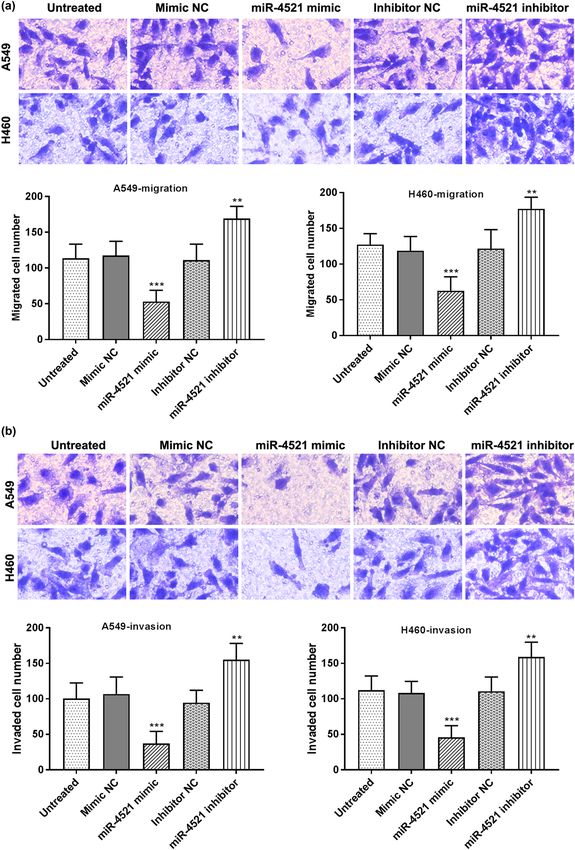
4521 mimics inhibited cell migration and invasion cap-
abilities (P < 0.001), while miR-4521 inhibitors significantly
high expression group was significantly higher than that
enhanced cell migration and invasion capabilities (P < 0.01)
of the low expression group, indicating that the low
(Figure 4a and b).
expression of miR-4521 may indicate the poor prognosis
of NSCLC patients (log-rank test P = 0.004). The multi-
variate Cox risk regression model was used to analyze the
relationship between the expression of miR-4521 in tissues 3.5 Effect of the miR-4521 expression on the
and the survival status of the corresponding patients. It corresponding cell proliferation and
can be concluded that the expression of miR-4521 (HR =
metastasis-related proteins
0.544, 95% CI = 0.329–0.898, P = 0.017), lymph node
metastasis (HR = 0.613, 95% CI = 0.379–0.991, P = 0.046),
Cyclin D1, Cdk4, MMP2, and MMP9 are cell proliferation,
and TNM staging (HR = 0.575, 95% CI = 0.355–0.932, P =
migration, and invasion-related proteins. Thus, we further
0.017) can be used as independent factors for the prognosis
measured the expression of these proteins in A549 cells
of NSCLC (Table 2).
transfected with mimic, inhibitor, or NCs using Western
blot assay. miR-4521 overexpression led to the downregu-
lation of cyclin D1, Cdk4, MMP2, and MMP9 (Figure 5).
3.4 Low expression of miR-4521 promoted
proliferation, invasion, and migration of
NSCLC cells
3.6 FOXM1 is directly regulated by miR-4521
In vitro cell assays were conducted to explore the effects
of miR-4521 on the proliferation, migration, and invasion To uncover the potential mechanism of the decreased
of NSCLC cells by transfecting miR-4521 mimics or inhi- expression of miR-4521 in NSCLC cells, the potential tar-
bitors into A549 and H460 cells. The detection of trans- gets of miR-4521 were identified using Targetscan prediction
fected cells by qRT-PCR revealed that miR-4521 inhibitors tools. Among these targets that were predicted, Forkhead
significantly downregulated the miR-4521 expression in Box M1 (FOXM1) was further studied. The binding sites
A549 and H460 cells (P < 0.001), while the miR-4521 between FOXM1 3′-UTR and miR-4521 are shown in
mimics significantly increased its expression in cells Figure 6a. Subsequently, a dual-luciferase reporter assayRole of miR-4521 in NSCLC 1155
Figure 3: The effect of the miR-4521 expression on cell proliferation. (a) miR-4521 mimic promoted the expression of miR-4521, while miR-
4521 inhibitor suppressed its expression (**P < 0.01, ***P < 0.001). (b) miR-4521 mimics can suppress A549 and H460 cell proliferation,
while miR-4521 inhibitors can promote cell proliferation (**P < 0.01).
was performed to confirm the hypothesis. As shown in finding new biomarkers are the main research directions
Figure 6b, the luciferase activity of cells co-transfected in the current field [24,25]. In recent studies, miRNAs
with FOXM1 WT and miR-4521 mimics was significantly have been found to be used as oncogenes or tumor sup-
lower than that of the control group, while co-transfected pressors and expressed differentially in various cancers
with FOXM1 WT and inhibitors was higher than the control [26,27]. Many studies explored the relevance of miRNAs
group (P < 0.05). However, the luciferase activities of cells to cancer prognosis and their effects on cell activities
co-transfected with FOXM1 MUT and mimics had no sig- [28,29]. For example, Ma et al. have confirmed that the
nificant changes. Thus, we believed that FOXM1 is a down- low expression of miR-100 was significantly associated with
stream target of miR-4521 in NSCLC cells. poor prognosis [30]. Feng et al. have proved that miR-16-1-3p
may suppress the proliferation, migration, and invasion of
NSCLC cells [31]. It was found that miR-4521 was abnor-
mally expressed in cancers such as chronic lymphocytic
4 Discussion leukemia (CLL) and had a certain impact on the migration,
invasion, and apoptosis of ccRCC cells [21,32]. So, our study
NSCLC is the major pathological type of lung cancer, with explored the abnormal expression and role of miR-4521 in
high morbidity and mortality, which is easy to relapse the occurrence and development of NSCLC.
and difficult to be monitored [23]. Therefore, in-depth miR-4521 may be used as a tumor suppressor gene
study of the development mechanism of NSCLC and for NSCLC. Our study has found that compared with the1156 Butong Sun et al. Figure 4: The effect of miR-4521 on cell migration and invasion. (a) miR-4521 mimics inhibited the migration of A549 and H460 cells, while miR-4521 inhibitors promoted cell migration (magnification ×200; **P < 0.01, ***P < 0.001). (b) miR-4521 mimics inhibited the invasion ability of cells, while miR-4521 enhanced the invasion ability of cells (magnification ×200; **P < 0.01, ***P < 0.001). control group, the expression of miR-4521 in NSCLC tis- analyzing the relationship between the expression of sues and cell lines was significantly downregulated. By miR-4521 and the clinical characteristics of patients, it
Role of miR-4521 in NSCLC 1157 Figure 5: Cell proliferation-, migration-, and invasion-related protein levels were measured by western blot analysis. Enhanced miR-4521 expression inhibited the cyclin D1, Cdk4, MMP2, and MMP9 levels (*P < 0.05). can be concluded that the low expression of miR-4521 is miR-4521 may be a prognostic biomarker of NSCLC. significantly related to TNM staging and lymph node metas- We used the Kaplan–Meier survival curve and Cox regres- tasis. These results are consistent with the existing research sion model to explore the relationship between miR-4521 results. Pekarsky et al. have proved that miR-4521 was expression and patient prognosis. It can be concluded downregulated in tumor samples from patients with CLL from the survival curve that the survival rate of patients [32]. Feng Xue et al. have demonstrated that miR-4521 defi- with low expression is lower and low expression of miR- ciency promoted the progression of ccRCC in patients [21]. 4521 indicates a poor prognosis. At the same time, the Cox Figure 6: FOXM1 may be a direct target gene of miR-4521. (a) The binding sites between FOXM1 3′-UTR and miR-4521. (b) Dual-luciferase reporter assay was performed to confirm FOXM1 as a direct target of miR-4521 (*P < 0.05).
1158 Butong Sun et al.
regression model can prove miR-4521 is an independent Funding information: This work was supported by the
prognostic factor for NSCLC. Liao et al. have found that Science and Technology Development Program of Jilin
miR-4521 showed a good prognostic ability and can be province [No. 20200403106SF].
used as an independent prognostic factor for the survival
of pancreatic ductal adenocarcinoma [33]. These results Author contributions: Butong Sun, Dan Cong, Yuansong
are consistent with the results of our research and we Bai, and Jun Li carried out the concepts, design, and defi-
can conclude that miR-4521 may be a marker of cancer nition of intellectual content; Butong Sun, Dan Cong, and
prognosis. Kang Chen provided assistance for data acquisition, data
The low expression of miR-4521 promoted the prolif- analysis, and statistical analysis. All authors performed
eration, migration, and invasion of NSCLC cells. We stu- the experiment. Butong Sun and Dan Cong drafted the
died the effects of miR-4521 mimics or inhibitors on the manuscript. Yuansong Bai and Jun Li revised the manu-
biological behavior of NSCLC cells using MTT and Transwell script critically for important intellectual content. All
assays. These results showed that low expression of miR- authors have read and approved the content of the
4521 can enhance cell proliferation, migration, and invasion manuscript.
ability, while high expression of miR-4521 can reduce its
ability. Furthermore, enhanced expression of miR-4521 Conflict of interest: The author reports no conflicts of
decreased the proliferation-, metastasis-related protein levels, interest in this work.
including cyclin D1, Cdk4, MMP2, and MMP9. These data
suggested that miR-4521 influences A549 cell malignancy Data availability statements: The datasets generated during
via cyclin D1, Cdk4, MMP2, and MMP9. A previous study and/or analyzed during the current study are available from
also demonstrated that the overexpression of miR-4521 can the corresponding author on reasonable request.
reduce the in vitro migration and invasion ability of ccRCC
cells 786-O and ACHN through the TIMP-1/MMP2/MMP9
pathway, and promote their apoptosis through the MDM2/
p53/Bcl2/Bax pathway to reduce its proliferation [21].
References
Senfter et al. have confirmed that miR-4521 transfection
[1] Jonna S, Subramaniam DS. Molecular diagnostics and targeted
reduced the proliferation and invasion of several medullo- therapies in non-small cell lung cancer (NSCLC): an update.
blastoma cell lines by activating caspase 3/7, and induced Discov Med. 2019;27(148):167–70.
programmed cell death [34]. Furthermore, we explored [2] Bai Y, Xiong L, Zhu M, Yang Z, Zhao J, Tang H. Co-expression
the potential downstream target of miR-4521 and FOXM1 network analysis identified KIF2C in association with pro-
gression and prognosis in lung adenocarcinoma. Cancer
might be a direct target of miR-4521. Mammalian transcrip-
Biomark. 2019;24(3):371–82. doi: 10.3233/cbm-181512.
tion factor FOXM1, one of the members of the Forkhead
[3] Tsim S, O’Dowd CA, Milroy R, Davidson S. Staging of non-small
family proteins, plays crucial roles in tumorigenesis in cell lung cancer (NSCLC): a review. Respir Med.
human cancers [35]. FOXM1 was reported to be upregu- 2010;104(12):1767–74. doi: 10.1016/j.rmed.2010.08.005.
lated in lung cancer (including NSCLC) and was associated [4] Liao Y, Cheng S, Xiang J, Luo C. lncRNA CCHE1 increased pro-
with poor prognosis of patients, as well as regulating cell liferation, metastasis and invasion of non-small lung cancer
cells and predicted poor survival in non-small lung cancer
proliferation, apoptosis, and metastasis of cancer cells
patients. Eur Rev Med Pharmacol Sci. 2018;22(6):1686–92.
[36,37]. Based on these previous studies and present results, doi: 10.26355/eurrev_201803_14581.
it is supposed that the abnormal expression of miR-4521 [5] Soria JC, Massard C, Le Chevalier T. Should progression-free
plays a certain role in the activities of NSCLC cells by tar- survival be the primary measure of efficacy for advanced
geting FOXM1. However, the detailed mechanism of miR- NSCLC therapy? Ann Oncol. 2010;21(12):2324–32.
doi: 10.1093/annonc/mdq204.
4521 in the development of NSCLC remains to be explored.
[6] Zhou W, Liu T, Saren G, Liao L, Fang W, Zhao H. Comprehensive
This study indicated that the expression of miR-4521
analysis of differentially expressed long non-coding RNAs in
is downregulated in NSCLC tissues and cell lines, and non-small cell lung cancer. Oncol Lett. 2019;18(2):1145–56.
its low expression is significantly related to the poor doi: 10.3892/ol.2019.10414.
prognosis of patients. The low expression of miR-4521 [7] Naylor EC, Desani JK, Chung PK. Targeted therapy and immuno-
promoted cell proliferation, migration, and invasion by therapy for lung cancer. Surg Oncol Clin N Am. 2016;25(3):601–9.
doi: 10.1016/j.soc.2016.02.011.
targeting FOXM1. In short, miR-4521 may serve as a new
[8] Pennell NA, Arcila ME, Gandara DR, West H. Biomarker testing
prognostic biomarker and the miR-4521/FOXM1 axis for patients with advanced non-small cell lung cancer: real-
may be a therapeutic target for the treatment of NSCLC world issues and tough choices. Am Soc Clin Oncol Educ Book.
patients. 2019;39:531–42. doi: 10.1200/EDBK_237863.2020.test.Role of miR-4521 in NSCLC 1159
[9] Herbst RS, Morgensztern D, Boshoff C. The biology and [23] Chen Z, Fillmore CM, Hammerman PS, Kim CF, Wong KK. Non-
management of non-small cell lung cancer. Nature. small-cell lung cancers: a heterogeneous set of diseases.
2018;553(7689):446–54. doi: 10.1038/nature25183. Nat Rev Cancer. 2014;14(8):535–46. doi: 10.1038/nrc3775.
[10] Lu TX, Rothenberg ME. MicroRNA. J Allergy Clin Immunol. [24] Wang Y, Zhang Q, Guo B, Feng J, Zhao D. miR-1231 Is down-
2018;141(4):1202–7. doi: 10.1016/j.jaci.2017.08.034. regulated in prostate cancer with prognostic and functional
[11] Bak RO, Mikkelsen JG. miRNA sponges: soaking up miRNAs for implications. Oncol Res Treat. 2020;43(3):78–86.
regulation of gene expression. Wiley Interdiscip Rev RNA. doi: 10.1159/000504606.
2014;5(3):317–33. doi: 10.1002/wrna.1213. [25] Yang T, Li H, Chen T, Ren H, Shi P, Chen M. LncRNA MALAT1
[12] Krol J, Loedige I, Filipowicz W. The widespread regulation of depressed chemo-sensitivity of NSCLC cells through directly
microRNA biogenesis, function and decay. Nat Rev Genet. functioning on miR-197-3p/p120 catenin axis. Mol Cell.
2010;11(9):597–610. doi: 10.1038/nrg2843. 2019;42(3):270–83. doi: 10.14348/molcells.2019.2364.
[13] Gong L, Ren M, Lv Z, Yang Y, Wang Z. miR-92b-3p promotes [26] Cai H, Lin H, Cao W, Sun J, Huang Y, Fang Y. Downregulation of
colorectal carcinoma cell proliferation, invasion, and migra- miR-519a predicts poor prognosis and contributes to tumor
tion by inhibiting FBXW7 in vitro and in vivo. DNA Cell Biol. progression in gastric cancer. Oncol Res Treat.
2018;37(5):501–11. doi: 10.1089/dna.2017.4080. 2020;43(1–2):19–26. doi: 10.1159/000504054.
[14] Kay M, Soltani BM, Aghdaei FH, Ansari H, Baharvand H. Hsa- [27] Bai H, Wu S. miR-451: a novel biomarker and potential thera-
miR-335 regulates cardiac mesoderm and progenitor cell dif- peutic target for cancer. Onco Targets Ther. 2019;12:11069–82.
ferentiation. Stem Cell Res Ther. 2019;10(1):191. doi: 10.1186/ doi: 10.2147/ott.s230963.
s13287-019-1249-2. [28] Guo Y, Tao M, Jiang M. MicroRNA-454-3p inhibits cervical
[15] Yang ZQ, Wu CA, Cheng YX. Prognostic value of microRNA-133a cancer cell invasion and migration by targeting c-Met. Exp Ther
expression and its clinicopathologic significance in non-small Med. 2018;15(3):2301–6. doi: 10.3892/etm.2018.5714.
cell lung cancer: a comprehensive study based on meta-ana- [29] Cheng Y, Liu W. MicroRNA-503 serves an oncogenic role in
lysis and the TCGA database. Oncol Res Treat. retinoblastoma progression by directly targeting PTPN12. Exp
2018;41(12):762–8. doi: 10.1159/000492343. Ther Med. 2019;18(3):2285–92. doi: 10.3892/etm.2019.7795.
[16] Feng X, Zhu M, Liao B, Tian T, Li M, Wang Z, et al. Upregulation [30] Ma X, Zhou J, Mo H, Ying Y. Association of miR-100 expression
of miR-552 predicts unfavorable prognosis of gastric cancer with clinicopathological features and prognosis of patients
and promotes the proliferation, migration, and invasion of with lung cancer. Oncol Lett. 2019;18(2):1318–22.
gastric cancer cells. Oncol Res Treat. 2020;43(3):103–11. doi: 10.3892/ol.2019.10393.
doi: 10.1159/000505377. [31] Feng QQ, Dong ZQ, Zhou Y, Zhang H, Long C. miR-16-1-3p
[17] Mei LL, Qiu YT, Zhang B, Shi ZZ. MicroRNAs in esophageal targets TWIST1 to inhibit cell proliferation and invasion in
squamous cell carcinoma: potential biomarkers and thera- NSCLC. Bratisl Lek Listy. 2018;119(1):60–5. doi: 10.4149/
peutic targets. Cancer Biomark. 2017;19(1):1–9. doi: 10.3233/ bll_2018_012.
cbm-160240. [32] Pekarsky Y, Balatti V, Palamarchuk A, Rizzotto L, Veneziano D,
[18] Gu GM, Zhan YY, Abuduwaili K, Wang XL, Li XQ, Zhu HG, et al. Nigita G, et al. Dysregulation of a family of short noncoding
MiR-940 inhibits the progression of NSCLC by targeting RNAs, tsRNAs, in human cancer. Proc Natl Acad Sci USA.
FAM83F. Eur Rev Med Pharmacol Sci. 2018;22(18):5964–71. 2016;113(18):5071–6. doi: 10.1073/pnas.1604266113.
doi: 10.26355/eurrev_201809_15927. [33] Liao X, Wang X, Huang K, Yang C, Yu T, Han C, et al. Genome-
[19] Liu R, Zhang YS, Zhang S, Cheng ZM, Yu JL, Zhou S, et al. MiR- scale analysis to identify prognostic microRNA biomarkers in
126-3p suppresses the growth, migration and invasion of patients with early stage pancreatic ductal adenocarcinoma
NSCLC via targeting CCR1. Eur Rev Med Pharmacol Sci. after pancreaticoduodenectomy. Cancer Manag Res.
2019;23(2):679–89. doi: 10.26355/eurrev_201901_16881. 2018;10:2537–51. doi: 10.2147/cmar.s168351.
[20] Yan C, Zhang W, Shi X, Zheng J, Jin X, Huo J. MiR-760 sup- [34] Senfter D, Samadaei M, Mader RM, Gojo J, Peyrl A, Krupitza G,
presses non-small cell lung cancer proliferation and meta- et al. High impact of miRNA-4521 on FOXM1 expression in
stasis by targeting ROS1. Env Sci Pollut Res Int. medulloblastoma. Cell Death Dis. 2019;10(10):696.
2018;25(19):18385–91. doi: 10.1007/s11356-017-1138-0. doi: 10.1038/s41419-019-1926-1.
[21] Feng X, Yan N, Sun W, Zheng S, Jiang S, Wang J, et al. miR- [35] Gartel AL. FOXM1 in Cancer: Interactions and Vulnerabilities.
4521-FAM129A axial regulation on ccRCC progression through Cancer Res. 2017;77(12):3135–9. doi: 10.1158/0008-5472.can-
TIMP-1/MMP2/MMP9 and MDM2/p53/Bcl2/Bax pathways. 16-3566.
Cell Death Discov. 2019;5:89. doi: 10.1038/s41420-019- [36] Zhang J, Zhang J, Cui X, Yang Y, Li M, Qu J, et al. FoxM1: a novel
0167-5. tumor biomarker of lung cancer. Int J Clin Exp Med.
[22] Zhang Y, Sui J, Shen X, Li C, Yao W, Hong W, et al. Differential 2015;8(3):3136–40.
expression profiles of microRNAs as potential biomarkers for [37] Xiu G, Sui X, Wang Y, Zhang Z. FOXM1 regulates radiosensitivity of
the early diagnosis of lung cancer. Oncol Rep. lung cancer cell partly by upregulating KIF20A. Eur J Pharmacol.
2017;37(6):3543–53. doi: 10.3892/or.2017.5612. 2018;833:79–85. doi: 10.1016/j.ejphar.2018.04.021.You can also read