Regulation of ARL2 in colorectal cancer cell proliferation and tumorigenicity, and its negative association with AXL
←
→
Page content transcription
If your browser does not render page correctly, please read the page content below
ONCOLOGY LETTERS 21: 196, 2021

               Regulation of ARL2 in colorectal cancer cell proliferation
               and tumorigenicity, and its negative association with AXL
                XUNLEI PANG1,2*, YANHONG WANG2*, BEI MIAO2, SUJUAN FEI2 and WEICHANG CHEN1

           1
          Department of Gastroenterology, The First Affiliated Hospital of Soochow University, Suzhou, Jiangsu 215008;
2
    Department of Gastroenterology, The Affiliated Hospital of Xuzhou Medical University, Xuzhou, Jiangsu 221004, P.R. China

                                      Received June 2 2020; Accepted December 4, 2020

                                                   DOI: 10.3892/ol.2021.12457

Abstract. Colorectal cancer (CRC) is the third most common         Introduction
malignant disease in adults. ADP ribosylation factor‑like
GTPase 2 (ARL2) is crucial for controlling the dynamics            As the third most common malignant disease in the world,
of microtubules and mitochondrial functions. However, the          colorectal cancer (CRC) exhibits a 2.27% cumulative risk of
biological function of ARL2 in CRC remains unclear. The            onset between 0‑74 years of age (1). In China, CRC is also one
present study was performed to identify the expression level       of the 5 most common types of cancer, and poses a significant
and functional role of ARL2 in CRC. A total of 19 CRC and 3        disease burden in the aging population (2). The 5‑year rela‑
normal healthy colorectal tissues were collected. Furthermore,     tive survival rate for Chinese patients with CRC has increased
ARL2 expression was analyzed in healthy colorectal and CRC         from 47.2 to 56.9% in the past 10 years. However, the possible
tissues by immunohistochemistry (IHC). ARL2 overexpres‑            of mortality rate of CRC has remained at 50% (3). The overall
sion and knockdown was achieved using lentiviral vectors and       prognosis of CRC was improved from the use of adjuvant
plasmid transfection in HCT8 and HCT116 cells. The protein         chemotherapy and the advance of resection. However, even in
and mRNA expression levels of ARL2 and AXL were analyzed           the early stages of CRC, recurrence following surgery remains
using western blot and reverse transcription‑quantitative PCR      the primary, and ultimate, cause of death (4). The development
in ARL2 knockdown and ARL2 overexpressing HCT8 and                 of a potential therapeutic target, that can aid the detection
HCT116 cells. Cell Counting Kit‑8, colony formation, wound         and treatment of CRC, is important for the prevention of this
healing, and Matrigel assays were used to investigate the          disease.
biological functions of ARL2. Taken together, ARL2 protein             Multiple mitochondrial functions, including motility,
expression level was upregulated in CRC tissues. Furthermore,      mitochondrial morphology, asymmetric division and main‑
ARL2 overexpression decreased proliferation and weakened           tenance of ATP levels (5,6) are necessary for the function
the colony‑formation abilities of the CRC cells, as well as        of phosphodiesterase‑ δ. The ADP ribosylation factor‑like
their migratory and invasive abilities. ARL2 interference          GTPase2 (ARL2) assists in the release of Ras (6). Multiple
enhanced proliferation and colony‑formation rates of the CRC       cell signaling pathways and various cellular functions
cells, as well as their migratory and invasive abilities. ARL2     are regulated by Ras, including proliferation, differentiation,
regulated CRC proliferation and tumorigenicity and was nega‑       vesicle transport, nuclear assembly, and the regulation of the
tively associated with AXL. The results of the present study       cytoskeleton are included (7). Several studies have shown
suggested that the proliferation, migration and tumorigenicity     the contribution of a Ras mutation, such as KRAS‑4B, in the
of the CRC cells could be inhibited by ARL2 overexpression.        development of human tumors (8‑10). It has been shown that
The latter may be used as a predicted and potential therapeutic    ~21% of all human cancers, and ~30, 45, and 90% of lung,
target for CRC.                                                    colon and pancreatic cancers, respectively, originate due
                                                                   to activating mutations in the Ras family of enzymes (8).
                                                                   It has also been found that the regulation of ARL2 protein
                                                                   expression could alter the duration and degree of numerous
                                                                   types of cancer, and the cell migratory and invasive abilities
Correspondence to: Dr Weichang Chen, Department of                 of cancer cells (11,12). ARL2 has been shown to regulate
Gastroenterology, The First Affiliated Hospital of Soochow         p53 localization, resulting in a chemoresistant phenotype
University, 188 Shizi Street, Suzhou, Jiangsu 215008, P.R. China
                                                                   in breast cancer cells (13). As a prognostic marker, ARL2
E‑mail: weichangchen2020@163.com
                                                                   mRNA expression was significantly elevated in hepatocel‑
*
    Contributed equally                                            lular tumors (14). In addition, ARL2 acts as an oncogene in
                                                                   cervical cancer (15). However, the functional role of ARL2
Key words: ARL2, colon cancer, AXL, tumorgenicity                  in CRC is not clear.
                                                                       AXL is a member of the TAM group of receptors, an inhib‑
                                                                   itor of the cytokine receptor‑mediated macrophage/monocyte
                                                                   activation and promoter of apoptotic cell removal. In addition,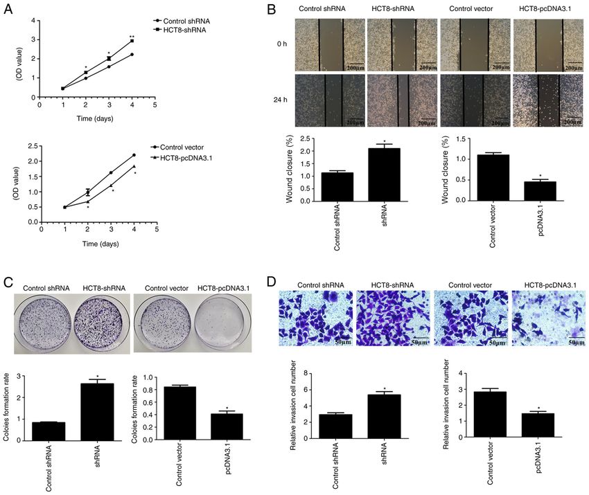
2 PANG et al: ARL2 REGULATES COLORECTAL CANCER CELL PROLIFERATION AND TUMORIGENICITY
AXL is an oncotarget in human CRC (16). Silencing of AXL resistant rabbit IgG; 1:1,000; cat. no. abs957; Absin (Shanghai)
gene expression suppresses proliferation, migration and Biotechnology Co., Ltd.] at room temperature for 1 h. The
survival in CRC cells (16). In CRC, AXL is overexpressed and slices were stained with 3',3'‑diaminobenzidine (DAB) at
its elevated protein expression promotes migration, invasion room temperature for about 7 min [Liquid A and liquid B;
and epithelial‑mesenchymal transition of CRC cell lines (17). 1000:50; cat. no. abs957; Absin (Shanghai) Biotechnology Co.,
The expression and function of ARL2 in CRC was Ltd.]. Finally, the slides were stained with 0.02% hematoxylin
investigated in the current study. It was found that ARL2 over‑ (cat. no. BL702A; Biosharp Life Sciences) and observed under
expression or knockdown regulated the proliferation, colony a light microscope (IX73; Olympus Corporation). The images
formation, migration and invasion abilities of CRC cells and it were analyzed using Photoshop (Adobe Systems, Inc.).
had a negative association with AXL. A total of two pathologists from The Affiliated Hospital
of Xuzhou Medical University (Xuzhou, China) examined
Materials and methods the pathological sections separately under blinded experi‑
mental conditions and all the differences were evaluated by
Patients and samples. The patient samples were collected discussion. High power microscopic fields (two), that were
from The Affiliated Hospital of Xuzhou Medical University representative of the samples were selected for each specimen
(Jiangsu, China) from January to June in 2019, including and 200 tumor cells were counted in each field. The German
19 colon samples (6 well‑differentiated, 6 moderately IHC Score (GIS) (11) was used for scoring. The intensity of
differentiated and 7 poorly differentiated cases). All patients ARL2 immunostaining was scored as follows: 0, negative;
with CRC underwent surgical resection and histological 1, weak; 2, moderate; 3, strong, whereas the percentage
diagnosis was verified by 2 gastroenterologists (Pathology of immunoreactive cells was categorized as 1 (0‑25%), 2
Department of The Affiliated Hospital of Xuzhou Medical (26‑50%), 3 (51‑75%), and 4 (76‑100%). The IHC expression
University) (Xuzhou, China). Patients with CRC who had values of ARL2 and the associated clinical information of
undergone preoperative radiotherapy and chemotherapy each sample are shown in Table II.
were excluded. A total of 19 colon cancer tissues and 3
healthy colorectal tissues (from partial resection in patients Cell lines and culture conditions. The HCT8, HCT116 and
with colorectal trauma. As a regional scientific research and 293T cell lines were acquired from the Cancer Institute,
medical institution, some patients suitable for being assigned Xuzhou Medical University (Jiangsu, China), which were orig‑
to normal controls were informed and signed informed inally purchased from the Shanghai Institute of Biochemistry
consent before or after surgery were used for immuno‑ and Cell Biology, Chinese Academy of Science (Shanghai,
histochemistry (IHC) analysis. Surgical resection was China). The HCT8, HCT116 and 293T cell lines were
performed in all patients with colon cancer, and the histo‑ authenticated using short tandem repeat DNA profiling, and
logical diagnosis was confirmed according to 2016 World subsequently cultured in DMEM containing 10% fetal bovine
Health Organization (WHO) guidelines (18). The present serum (cat. no. SH30022.01B; HyClone; Cytiva), 100 µg/ml
study was approved by the Medical Ethics Committee of streptomycin and 100 U/ml penicillin, and incubated at 37˚C
the Hospital of The Affiliated Hospital of Xuzhou Medical in a humidified incubator with 5% CO2.
University (Jiangsu, China), and written informed consent
was provided from each patient for his/her participation in RNA isolation and reverse transcription‑quantitative PCR
the study (approval no. XYFY2019‑KL157‑01). The clinical (RT‑qPCR). Total RNA was extracted from the HCT8 and
characteristics of the 19 patients with colon cancer are HCT116 cells using TRIzol® (Invitrogen; Thermo Fisher
listed in Table I. Scientific, Inc.), in line with the manufacturer's protocol. The
total RNA was reverse transcribed into cDNA for PCR amplifi‑
IHC assay. IHC analysis of ARL2 was performed on 19 cation. RT‑qPCR was performed using TransStart® Top Green
colon cancer and 3 healthy colorectal tissues. The samples qPCR SuperMix Assays (cat. no. AQ131; Beijing Transgen
were fixed in 10% neutral buffered formalin for ~3‑24 h at Biotech Co., Ltd.) and in a thermal cycler (LightCycler® 480Ⅱ;
room temperature, dehydrated in gradient alcohol solution Roche Diagnostics). Human mRNA sequences, encoding
(30, 50, 70, 80, 90, 95, 100, 100% alcohol each for 30 min) and ARL2 and AXL genes, were retrieved from the nucleotide
paraffin‑embedded. Paraffin‑embedded tissues were cut into database of National Center for Biotechnology Information
~3‑µm thick continuous sections. Routine dewaxing, antigen (NCBI, https://www.ncbi.nlm.nih.gov/). The primers were
retrieval (microwave thermal retrieval) and quenching of designed using Primer Premier v5.0 (Premier Biosoft) and
endogenous peroxidase activity with 3% H2O2 at room temper‑ Basic Local Alignment Search Tool was used in NCBI to
ature was performed on the slices. After blocking with 5% determine the specificity of the primers. The primers were
goat serum [cat. no. abs933; Absin (Shanghai) Biotechnology synthesized by Generay Biotech Co., Ltd. The following
Co., Ltd.] at room temperature for ~1 h, primary antibody thermocycling conditions were used: Initial denaturation at
(anti‑ARL2; 1:100; cat. no. 188322; Cell Signaling Technology, 95˚C for 30 sec, followed by 40 cycles of a two‑step cycling
Inc.) was added to the sections and incubated overnight at 4˚C. program (95˚C for 5 sec and 60˚C for 30 sec). The mRNA
The substrate [primary reactant amplifier; cat. no. abs957; expression was normalized to the expression of β‑actin and
Absin (Shanghai) Biotechnology Co., Ltd.] for the secondary calculated using the 2‑ΔΔCq method (19). The specific primers
antibody was added and incubated at room temperature for for ARL2, AXL and β‑actin were as follows: ARL2 forward,
30 min with the samples. Then the sections were subse‑ 5'‑GGGAGGACATCGACACCA‑3' and reverse, 5'‑AGGACC
quently treated with a secondary antibody [HRP‑labeled goat GCAG GGACTTC‑3'; AXL forward, 5'‑GTTTGGAGCTGT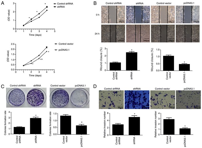
ONCOLOGY LETTERS 21: 196, 2021 3
Table I. Clinical characteristics of the 19 patients with CCGCAAAG‑3' and reverse, 5'‑CTGGAAG GTG GACAG
colorectal cancer. CGAGG‑3'.
Characteristics Number of patients (%) Immunofluorescence. ARL2 pcDNA3.1 HCT8 and HCT116
cells, ARL2 shRNA HCT8 and HCT116 cells, and the
Age, years corresponding control group cells were fixed with 4% para‑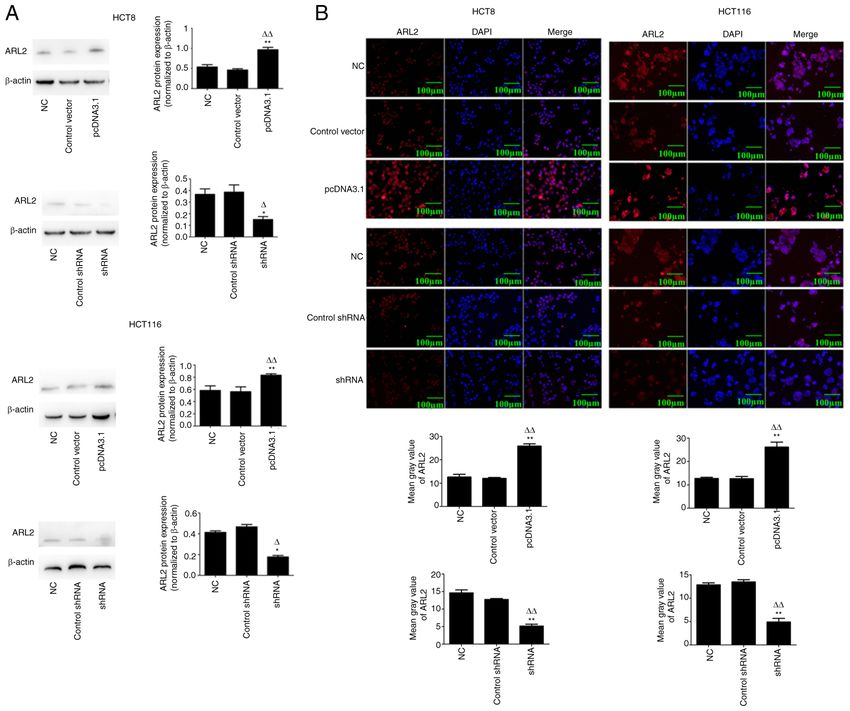
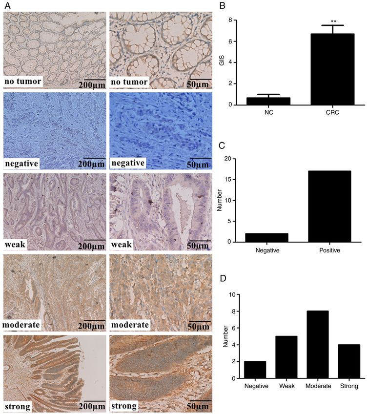
4                  PANG et al: ARL2 REGULATES COLORECTAL CANCER CELL PROLIFERATION AND TUMORIGENICITY

Figure 1. ARL2 protein expression levels are increased in patients with CRC tumor tissues. (A) Representative immunohistochemistry images of the ARL2
protein expression in normal healthy (n=3) and colon cancer samples CRC (n=19). Magnification of the images is x10 and 40 in the left and right panels respec‑
tively. (B) Analysis of GIS between NC and CRC groups. (C) Distribution of ARL2 negative and positive expression levels in CRC tissues. (D) Distribution
of ARL2 expression level using GIS in CRC tissues. **P
ONCOLOGY LETTERS 21: 196, 2021                                                                  5

Figure 2. Generation of pcDNA3.1 and shRNA in the HCT8 and HCT116 cell lines. (A) Representative western blot images and analysis of ARL2 protein
expression level in the HCT8 and HCT116 cells transfected with pcDNA3.1 or shRNA or their respective controls (n=3; ANOVA). *P
6                 PANG et al: ARL2 REGULATES COLORECTAL CANCER CELL PROLIFERATION AND TUMORIGENICITY

Figure 3. ARL2 expression and HCT8 proliferation, migration and invasion. (A) Cell Counting Kit‑8 assays were used to examine HCT8 cell proliferation
in control and ARL2 overexpressing and knockdown HCT8 cells. (B) Wound healing assays were used to examine cell migration in control and ARL2 over‑
expressing and knockdown HCT8 cells. Scale bar, 200 µm. (C) Colony formation assays were used to examine HCT8 cell proliferation in control and ARL2
overexpressing and knockdown HCT8 cells. (D) Matrigel assay was used to examine cell invasion in control, ARL2 overexpressing and knockdown HCT8
cells. Scale bar, 50 µm. The data are presented as the mean ± SD from three independent experiments performed in triplicate. *P
ONCOLOGY LETTERS 21: 196, 2021                                                                  7

Figure 4. ARL2 expression and HCT116 proliferation, migration and invasion. (A) Cell Counting Kit‑8 assays were used to examine HCT116 cell proliferation
in control, ARL2 overexpressing and knockdown HCT8 cells. (B) Wound healing assays were used to examine cell migration in control, ARL2 overexpressing
and knockdown HCT116 cells. Scale bar, 200 µm. (C) Colony formation assays were used to examine HCT116 cell proliferation in control, ARL2 overex‑
pressing and knockdown HCT116 cells. (D) Matrigel assay was used to examine cell invasion in control, ARL2 overexpressing and knockdown HCT116 cells.
Scale bar, 50 µm. The data are presented as the mean ± SD from three independent experiments performed in triplicate. *P
8                 PANG et al: ARL2 REGULATES COLORECTAL CANCER CELL PROLIFERATION AND TUMORIGENICITY

Figure 5. ARL2 and AXL protein and mRNA expression levels. (A) Western blot analysis was performed in ARL2 overexpression and knockdown HCT8 and
HCT116 cells for the detection of AXL and ARL2 protein expression level. (B) Reverse transcriptions‑quantitative PCR was performed to analyze ARL2 and
AXL mRNA expression levels in HCT8 and HCT116 cells, following transfection with ARL2 overexpression and knockdown vectors. The data are presented
as the mean ± SD from three independent experiments performed in triplicate. *P
ONCOLOGY LETTERS 21: 196, 2021                                                       9

    As a member of the TAM group of receptors, AXL acts            Authors' contributions
as an inhibitor of cytokine receptors mediates cell activa‑
tion and promotes apoptotic cell removal (27,28). AXL gene         SF designed the study. XP, YW, BM and WC performed the
silencing and drug inhibition suppressed proliferation, migra‑     experiments and analyzed the data. WC, YW and XP wrote
tion and survival in CRC cells (16,17). In the present study,      the manuscript. SF and BM revised the manuscript. All
the results indicated that ARL2 was inversely associated           authors read and approved the final version of the manuscript
with AXL expression in CRC cells. Furthermore, previous            and agree to be accountable all aspects of the study to ensure
studies have indicated that AXL functions as a tumor               the accuracy or integrity of all parts of the work.
promoter in cancer. For example, AXL induced the activa‑
tion of downstream AKT, and inhibited the proliferation and        Ethics approval and consent to participate
migration of pancreatic cancer cells in vitro (29). In addition,
GAS6 promoted gastric cancer invasiveness by activating            This study was approved by the Institutional Ethics Committee
AXL (30). Previous studies have demonstrated the role of           of the Affiliated Hospital of Xuzhou Medical University
AXL in regulating cell growth, migration and tumorigenesis         (approval no. XYFY2019‑KL157‑01) and adhered to the prin‑
of CRC cells (29,30). AXL acted as a tumor promoter (31,32),       ciples of the Declaration of Helsinki. Each patient provided
which may partially be suppressed by overexpressed ARL2            written informed consent.
expression in CRC cells. The present study provided the first
preliminary evidence of AXL was negatively associated with         Patient consent for publication
ARL2. To elucidate the mechanism by which ARL2 and
AXL expression in CRC cells, western blot and RT‑qPCR              Not applicable.
analyses were used to detect the protein and mRNA expres‑
sion levels of ARL2 and AXL in HCT8 and HCT116 cells,              Competing interests
following overexpression or knockdown of ARL2. At both
the protein and mRNA expression, increased ARL2 led to             The authors declare that they have no competing interests.
decreased AXL and decreased ARL2 led to increased AXL.
The results revealed that the increase in the levels of ARL2       References
was accompanied by decrease in AXL expression level and
may have reduced CRC cell progression. Therefore, ARL2              1. Mattiuzzi C, Sanchis‑Gomar F and Lippi G: Concise update on
may be negatively correlated with the expression level of              colorectal cancer epidemiology. Ann Transl Med 7: 609‑609,
                                                                       2019.
AXL at the gene level.                                              2. Gu X, Zheng R, Xia C, Zeng H, Zhang S, Zou X, Yang Z, Li H
    In conclusion, the present study described the increased           and Chen W: Interactions between life expectancy and the inci‑
expression levels of ARL2 in clinical CRC samples. ARL2                dence and mortality rates of cancer in China: A population‑based
                                                                       cluster analysis. Cancer Commun (Lond) 38: 44, 2018.
could play a crucial role in CRC. In addition, significant          3. Zeng H, Chen W, Zheng R, Zhang S, Ji JS, Zou X, Xia C, Sun K,
evidence was provided demonstrating that increased ARL2                Yang Z, Li H, et al: Changing cancer survival in China during
expression in CRC cells inhibited proliferation, colony                2003‑15: A pooled analysis of 17 population‑based cancer regis‑
                                                                       tries. Lancet Glob Health 6: e555‑e567, 2018.
formation, migration and invasion. Furthermore, the results         4. Dai W, Li Y, Mo S, Feng Y, Zhang L, Xu Y, Li Q and Cai G:
confirmed that ARL2 was negatively associated with AXL                 A robust gene signature for the prediction of early relapse in
in CRC cells and maybe a protective factor in CRC. Taken               stage I‑III colon cancer. Mol Oncol 12: 463‑475, 2018.
                                                                    5. Chen K, Koe CT, Xing ZB, Tian X, Rossi F, Wang C, Tang Q,
together, the results suggested that ARL2 could play an                Zong W, Hong WJ, Taneja R, et al: Arl2‑ and Msps‑dependent
important inhibitory role in the proliferation, migration and          microtubule growth governs asymmetric division. J Cell
tumorigenicity of CRC cells and ARL2 was negatively associ‑            Biol 212: 661‑676, 2016.
                                                                    6. Ozdemir ES, Jang H, Gursoy A, Keskin O and Nussinov R:
ated with AXL expression. Therefore, ARL2 may be a new                 Arl2‑mediated allosteric release of farnesylated KRas4B from
predictive and therapeutic target for CRC.                             shuttling factor PDEδ. J Phys Chem B 122: 7503‑7513, 2018.
                                                                    7. Stettin D, Waldmann A, Wolters M, Trunz B, Schauder P and
                                                                       Hahn A: Infection with Helicobacter pylori‑outcome of a
Acknowledgements                                                       cross‑sectional investigation. Dtsch Med Wochenschr 132:
                                                                       2677‑2682, 2007 (In German).
The authors would like to thank Dr Yingying Cui and                 8. Zhang F and Cheong JK: The renewed battle against RAS‑mutant
                                                                       cancers. Cell Mol Life Sci 73: 1845‑1858, 2016.
Dr Kai Cao (Department of Pathology, The Affiliated Hospital        9. Cox AD, Fesik SW, Kimmelman AC, Luo J and Der CJ:
of Xuzhou Medical University) for discussions pertaining to            Drugging the undruggable RAS: Mission possible? Nat Rev
                                                                       Drug Discov 13: 828‑851, 2014.
the present study.                                                 10. Stephen AG, Esposito D, Bagni RK and McCormick F: Dragging
                                                                       ras back in the ring. Cancer Cell 25: 272‑281, 2014.
Funding                                                            11. Wang Y, Guan G, Cheng W, Jiang Y, Shan F, Wu A, Cheng P
                                                                       and Guo Z: ARL2 overexpression inhibits glioma proliferation
                                                                       and tumorigenicity via down‑regulating AXL. BMC Cancer 18:
No funding was received.                                               599, 2018.
                                                                   12. Li HJ, Sun XM, Li ZK, Yin QW, Pang H, Pan JJ, Li X and
                                                                       Chen W: LncRNA UCA1 promotes mitochondrial function of
Availability of data and materials                                     bladder cancer via the MiR‑195/ARL2 signaling pathway. Cell
                                                                       Physiol Biochem 43: 2548‑2561, 2017.
The datasets used and/or analyzed in the current study are         13. Béghin A, Matera E, Brunet‑Manquat S and Dumontet C:
                                                                       Expression of Arl2 is associated with p53 localization and
available from the corresponding author upon reasonable                chemosensitivity in a breast cancer cell line. Cell Cycle 7:
request.                                                               3074‑3082, 2008.
10 PANG et al: ARL2 REGULATES COLORECTAL CANCER CELL PROLIFERATION AND TUMORIGENICITY
14. Hass HG, Vogel U, Scheurlen M and Jobst J: Gene‑expression 26. Long LM, He BF, Huang GQ, Guo YH, Liu YS and Huo JR:
analysis identifies specific patterns of dysregulated molecular microRNA‑214 functions as a tumor suppressor in human colon
pathways and genetic subgroups of human hepatocellular carci‑ cancer via the suppression of ADP‑ribosylation factor‑like
noma. Anticancer Res 36: 5087‑5096, 2016. protein 2. Oncol Lett 9: 645‑650, 2015.
15. Peng R, Men J, Ma R, Wang Q, Wang Y, Sun Y and Ren J: 27. Zhou C, Cunningham L, Marcus AI, Li Y and Kahn RA: Arl2
miR‑214 down‑regulates ARL2 and suppresses growth and inva‑ and Arl3 regulate different microtubule‑dependent processes.
sion of cervical cancer cells. Biochem Biophys Res Commun 484: Mol Biol Cell 17: 2476‑2487, 2006.
623‑630, 2017. 28. Newman LE, Schiavon CR, Zhou C and Kahn RA: The
16. Martinelli E, Martini G, Cardone C, Troiani T, Liguori G, abundance of the ARL2 GTPase and its GAP, ELMOD2, at mito‑
Vitagliano D, Napolitano S, Morgillo F, Rinaldi B, chondria are modulated by the fusogenic activity of mitofusins
Melillo RM, et al: AXL is an oncotarget in human colorectal and stressors. PLoS One 12: e0175164, 2017.
cancer. Oncotarget 6: 23281‑23296, 2015. 29. Song X, Akasaka H, Wang H, Abbasgholizadeh R, Shin JH,
17. Uribe DJ, Mandell EK, Watson A, Martinez JD, Leighton JA, Zang F, Chen J, Logsdon CD, Maitra A, Bean AJ and Wang H:
Ghosh S and Rothlin CV: The receptor tyrosine kinase AXL Hematopoietic progenitor kinase 1 down‑regulates the onco‑
promotes migration and invasion in colorectal cancer. PLoS genic receptor tyrosine kinase AXL in pancreatic cancer. J Biol
One 12: e0179979, 2017. Chem 295: 2348‑2358, 2020.
18. Bläker H: Grading of tumors in the tubular digestive tract: 30. Bae CA, Ham IH, Oh HJ, Lee D, Woo J, Son SY, Yoon JH,
Esophagus, stomach, colon and rectum. Pathologe 37: 293‑298, Lorens JB, Brekken RA, Kim TM, et al: Inhibiting the
2016 (In German). GAS6/AXL axis suppresses tumor progression by blocking the
19. Livak KJ and Schmittgen TD: Analysis of relative gene expres‑ interaction between cancer‑associated fibroblasts and cancer
sion data using real‑time quantitative PCR and the 2(‑Delta Delta cells in gastric carcinoma. Gastric Cancer 23: 824‑836, 2020.
C(T)) method. Methods 25: 402‑408, 2001. 31. Vajkoczy P, Knyazev P, Kunkel A, Capelle HH, Behrndt S,
20. Hou P, Li L, Chen F, Chen Y, Liu H, Li J, Bai J and Zheng J: von Tengg‑Kobligk H, Kiessling F, Eichelsbacher U, Essig M,
PTBP3‑mediated regulation of ZEB1 mRNA stability promotes Read TA, et al: Dominant‑negative inhibition of the Axl receptor
epithelial‑mesenchymal transition in breast cancer. Cancer tyrosine kinase suppresses brain tumor cell growth and invasion
Res 78: 387‑398, 2018. and prolongs survival. Proc Natl Acad Sci USA 103: 5799‑5804,
21. Qiu C, Bu X and Jiang Z: Protocadherin‑10 acts as a tumor 2006.
suppressor gene, and is frequently downregulated by promoter 32. Cheng P, Phillips E, Kim SH, Taylor D, Hielscher T, Puccio L,
methylation in pancreatic cancer cells. Oncol Rep 36: 383‑389, 2016. Hjelmeland AB, Lichter P, Nakano I and Goidts V: Kinome‑wide
22. Tohidi F, Sadat SM, Bolhassani A and Yaghobi R: Construction shRNA screen identifies the receptor tyrosine kinase AXL as
and production of HIV‑VLP harboring MPER‑V3 for potential a key regulator for mesenchymal glioblastoma stem‑like cells.
vaccine study. Curr HIV Res 15: 434‑439, 2017. Stem Cell Reports 4: 899‑913, 2015.
23. Seetharaman S and Etienne‑Manneville S: Microtubules at focal
adhesions‑a double‑edged sword. J Cell Sci 132: jcs232843, 2019. This work is licensed under a Creative Commons
24. van Vuuren RJ, Botes M, Jurgens T, Joubert AM and Attribution-NonCommercial-NoDerivatives 4.0
van den Bout I: Novel sulphamoylated 2‑methoxy estradiol International (CC BY-NC-ND 4.0) License.
derivatives inhibit breast cancer migration by disrupting micro‑
tubule turnover and organization. Cancer Cell Int 19: 1, 2019.
25. Galmarini CM, Martin M, Bouchet BP, Guillen‑Navarro MJ,
Martínez‑Diez M, Martinez‑Leal JF, Akhmanova A and Aviles P:
Plocabulin, a novel tubulin‑binding agent, inhibits angiogenesis
by modulation of microtubule dynamics in endothelial cells.
BMC Cancer 18: 164, 2018.You can also read