Review-Recent Progress in Portable Fluorescence Sensors - IOPscience
←
→
Page content transcription
If your browser does not render page correctly, please read the page content below
Journal of the Electrochemical
Society

OPEN ACCESS

Review—Recent Progress in Portable Fluorescence Sensors
To cite this article: Young-Ho Shin et al 2021 J. Electrochem. Soc. 168 017502

View the article online for updates and enhancements.

                               This content was downloaded from IP address 46.4.80.155 on 08/01/2021 at 04:05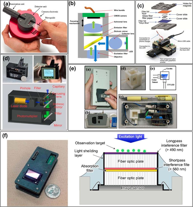
Journal of The Electrochemical Society, 2021 168 017502

                               Review—Recent Progress in Portable Fluorescence Sensors
                               Young-Ho Shin,1 M. Teresa Gutierrez-Wing,2 and Jin-Woo Choi1,3,z
                               1
                                 School of Electrical Engineering and Computer Science, Louisiana State University, Baton Rouge, Louisiana 70803, United
                               States of America
                               2
                                 Department of Renewable Natural Resources, Louisiana State University, Baton Rouge, Louisiana 70803, United States of
                               America
                               3
                                 Center for Advanced Microstructures and Devices, Louisiana State University, Baton Rouge, Louisiana 70803, United
                               States of America

                 Portable fluorescence sensors have been developed for biochemical detection, water quality monitoring, biomedical sensing, and
                 many other applications. With help of advancement in modern electronics, conventional fluorescence-based instrumentations are
                 now integrated into portable sensing devices for remote and resource-limited settings. In this work, fluorescence sensing
                 technology is introduced and different applications of portable fluorescence sensors and their characteristics are reviewed. Current
                 issues, technological challenges, and future direction of the portable fluorescence sensor development are discussed. The goal is to
                 provide a comprehensive survey on the recent advancements in optics, semiconductors, smartphones, and many other
                 manufacturing technologies that increased the portability, miniaturization, and sensitivity of portable fluorescence sensor devices.
                 © 2021 The Author(s). Published on behalf of The Electrochemical Society by IOP Publishing Limited. This is an open access
                 article distributed under the terms of the Creative Commons Attribution 4.0 License (CC BY, http://creativecommons.org/licenses/
                 by/4.0/), which permits unrestricted reuse of the work in any medium, provided the original work is properly cited. [DOI: 10.1149/
                 1945-7111/abd494]

                 Manuscript submitted September 27, 2020; revised manuscript received November 24, 2020. Published January 7, 2021. This
                 paper is part of the JES Focus Issue on IMCS 2020.

    Accurate detection and quantification of biological and chemical                                             Fluorescent Sensing
substances are becoming important in many applications, such as
                                                                                         During the last few decades, significant advances in fluorescence-
environmental monitoring, clinical diagnostics, DNA sequencing,
                                                                                     based instruments have brought a great impact on chemical and
and even biological warfare agent detection.1–6 A desirable sensor
                                                                                     biological sensing. The sensitivity of a modern fluorometer is as low
should be not only highly sensitive and selective but also capable of
                                                                                     as a single photon level, and a fluorescence microscope can distinguish
concurrently detecting and distinguishing multiple target analytes in
                                                                                     two different particles spaced less than 10 nm distance thanks to state-
a simple and rapid way. Optical sensing technology is one of the
                                                                                     of-the-art super-resolution technology.20–22 Fluorescence-based instru-
most promising methods in this aspect due to the many advantages
                                                                                     ments can be categorized into several types based on which
over other sensing methods. The main advantages of optical sensing
                                                                                     fluorescence parameters are being measured. The most basic function
include immunity to electromagnetic interference, durability under
                                                                                     of a fluorometer is to simply measure fluorescence intensity at fixed
severe pressures and temperatures, and most importantly, high
                                                                                     wavelength values of excitation and emission, which is often called a
sensitivity and selectivity because the measurement is performed
                                                                                     steady-state measurement (wavelength-based).23 Fluorescence lifetime
utilizing unique excitation and emission wavelengths specific to the
                                                                                     is determined by the decay of emission intensity, which is a unique
target analytes.7–9
                                                                                     property of different fluorophores.24 Anisotropy is the utilization of
    With the benefits that optical sensing technologies provide, different
                                                                                     polarized excitation light for characterizing the rotational motions of
optical detection techniques, such as absorbance,10 diffraction,11
                                                                                     fluorophores via the detection of fluorescence emission that has the
reflection,12 scattering,13 chemiluminescence,14 and refractive index15
                                                                                     same polarity as the excitation wavelength.25
have been utilized and reported in developing portable biochemical
                                                                                         This review addresses mostly the steady-state fluorescent sensing
sensors. Amongst various optical detection techniques, fluorescent
                                                                                     since the measurements are relatively simple and do not require
sensing is considered highly useful in practical applications for its high
                                                                                     sophisticated systems for the operation; thus, these types of
sensitivity, specificity, and accuracy compared to other optical sensing
                                                                                     measurements are more appropriate for portable systems including
techniques.16–18
                                                                                     spectrometers and fluorometers.
    There are increasing demands for fluorescent portable sensors in
a variety of applications due to the benefits that fluorescent sensing
                                                                                         Spectrometer.—A spectrometer contains five essential compo-
technology offers. Although technologies in benchtop fluorescent
                                                                                     nents as illustrated in Fig. 1a: (1) a light source for broad excitation
instruments are well-established and advanced, challenges still exist
                                                                                     spectrum; (2) an excitation monochromator for wavelength selec-
in applying them to a fully portable fluorescence sensing system
                                                                                     tion; (3) a sample container (usually cuvette and holder jig); (4) an
because most of the conventional fluorescence sensing instruments
                                                                                     emission monochromator for fluorescence wavelength selection; and
(especially spectrometers) require complicated optical components
                                                                                     (5) a photodetector. Light absorbance can be measured as an option
and systems, which makes them expensive, large, and high-power
                                                                                     (spectrophotometry function). Amongst all five main components,
consuming.19
                                                                                     the quality of the measured fluorescence (resolution and intensity) is
    In this review, an overview of recent developments of portable
                                                                                     usually determined by the monochromator components. A mono-
fluorescent sensors is introduced. Then, different applications of the
                                                                                     chromator consists of a collimator, focusing mirror, grating, slits,
sensors and their characteristics are addressed and discussed.
                                                                                     optomechanical components, and peripheral systems for controlling
Finally, current limitations, technological challenges, and future
                                                                                     optics as illustrated in Fig. 1b. Monochromators and detectors are
potential of the portable fluorescence sensor development are
                                                                                     mainly responsible for making it difficult to miniaturize a spectro-
discussed. The goal of this review is to provide a survey on the
                                                                                     meter system unless its performance is being significantly compro-
portable fluorescence sensors, with the main focus on recent research
                                                                                     mised in terms of resolution and sensitivity.26,27 Simple handheld
activities over the past decade (2010–2020).
                                                                                     spectrometers have been reported to inspect the ripeness of fruits and
                                                                                     measure the hemoglobin in blood.28,29 Smartphones are being used
                                                                                     to capture the light rays via embedded cameras or to process and
                                                                                     display captured spectra data through its processor (Figs. 2a and 2b).
  z
      E-mail: choijw@lsu.edu                                                         Wilkes et al. developed a spectrometer having 1 nm resolution with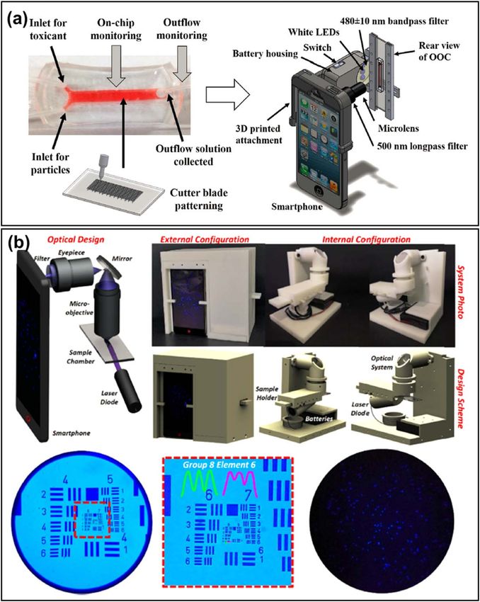
Journal of The Electrochemical Society, 2021 168 017502
Figure 1. Basic spectrometer components: (a) block diagram of spectrometer components and (b) illustration of a basic monochromator for excitation and
emission wavelength selection.
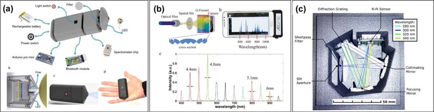
Figure 2. Simple handheld spectrometers: (a) fruit ripeness monitoring spectrometer28; (b) a smartphone-based diffusive reflectance spectrometer for
hemoglobin measurement,29 and (c) a low-cost 3D printed 1 nm resolution spectrometer.30 Licensed under a CC BY license for (a), (b), and (c).
conventional optics and incorporating the Czerny-Turner monochro-
mator configuration.30 However, it requires relatively pricey optics
and makes it difficult to achieve a compact form factor (Fig. 2c).
Regardless of the application of the device, all five fundamental
components are critical in the spectrometer and none of them can be
removed to be fully functional. However, some of the components
that serve the same functions can be replaced for different applica-
tions and purposes. Different components have unique advantages
and disadvantages, which requires careful selection of alternative
components for the proper applications. A portable system may
require alternative components to achieve compact size and less
power-consuming features.
Filter-based fluorometer.—A filter-based fluorometer is the oldest
configuration of all fluorescence-based instruments. Components of
this type of instrument are similar to that of a spectrometer, but some
components including excitation and emission wavelength selectors
are replaced with optical filters as illustrated in Fig. 3. These excitation
and emission optical filters are either single or multiple layers of Figure 3. Illustration of the filter-based fluorometer. Compared to the
color filters or dichroic mirrors; therefore, it is only possible to select conventional spectrometer, filter-based fluorometers are simpler, cheaper,
fixed excitation and emission wavelengths. Nevertheless, multiple compact, and more application-specific.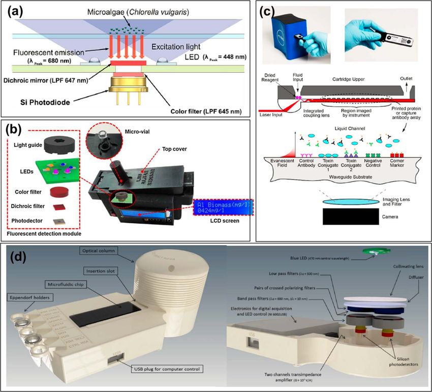
Journal of The Electrochemical Society, 2021 168 017502

wavelengths can be selected with a filter-wheel configuration that             (300 ∼ 450 W), the power consumption is still high for portable
contains a limited number of optical filters to obtain various wave-          systems being powered by a battery. In addition, it requires an
lengths of interest.31                                                       electronic circuitry for a high voltage discharging system, therefore,
   Generally, the configuration for a filter-based fluorometer is               it becomes more challenging to incorporate into a compact system.
simpler, cheaper, and smaller compared to a conventional spectro-                A laser is very useful when wavelength-specific light is required
meter due to the absence of conventional monochromators used for             (⩽1 nm). The light emitted from the laser is monochromatic,
excitation and emission wavelength selection. Therefore, for por-            coherent, directional, and highly powerful. Cowles et al. reported a
table fluorescence sensing applications, the filter-based fluorometer           laser-based fluorometer for in situ measurements of different
configuration is frequently utilized to achieve a simple and compact          phytoplankton groups by stimulating their photopigments (phycoer-
system. However, the optical filters transmit only a single wave-             ythrin and chlorophyll).35 An argon laser (514 nm) coupled with a
length at a time; therefore, selectivity is limited to a set number of       long optical fiber (200 m) was selected for stimulation and detection.
filters for both excitation and emission wavelengths. Filter-based            Chen et al. developed a portable in situ fluorometer to measure the
fluorometers are usually appropriate for applications in which                fluorescence signals of various dissolved organic matters in the
periodic quantitative analysis for a single analyte is needed. In            sea.36 By utilizing a narrow band laser with a center wavelength of
addition, optical filters are often utilized to minimize the autofluor-        405 nm, fluorescence emission from the organic matter was success-
escence noise. Autofluorescence is fluorescence emission from the              fully discriminated from the phytoplankton fluorescence. Although
materials other than the target analytes. There are several transparent      the laser is highly advantageous for fluorescent sensing systems, it
materials that are widely used to load the samples, such as glass,           requires a complicated, expensive, and large system, thus is not
polydimethylsiloxane (PDMS), Polymethyl methacrylate (PMMA),                 suitable for portable applications.
cyclic olefin copolymer (COC), and polycarbonate (PC). The                        An LED is a semiconductor light that has many advantages over
comparison of autofluorescence of different materials was reported            conventional light sources, especially for portable applications, due
by Piruska et al.32 In this study, the highest autofluorescence               to its compact size, high-power efficiency, low-cost, narrow emis-
emission was observed under blue excitation (403 nm). Relative               sion wavelength, and long lifetime. In addition, commercially
autofluorescence of PDMS under 403 nm was about 4 times higher                available LEDs are diverse in wavelength selection (ranging from
than that of borosilicate glass. PMMA exhibited 6 times higher,              ultraviolet to near-infrared); therefore, they are more appropriate as
COC exhibited 20 times higher, and PC showed 41 times higher.                excitation light sources for a portable system than other sources. A
Although thermoplastic materials exhibit relatively higher autofluor-         laser diode, which is also widely used in portable systems, is a class
escence, they are durable against temperature and pressure, thus             of laser fabricated with semiconductor materials. The working
have been used in biological sensing applications with optical filters        principle of laser diode is very similar to that of LED, but it
to minimize the autofluorescence noise.                                       incorporates optical components that a laser has, thus it offers a more
                                                                             coherent and directional emission ray than LEDs. Velpula et al.
    Various light sources and photodetectors for portable fluores-            reported the first axial AlInN ultraviolet core–shell nanowire LEDs
cent sensing systems.—A proper selection of excitation light is              in the ultraviolet wavelength range.37 It exhibited high internal
essential to improve the sensitivity, selectivity, and many other            quantum efficiency of ∼52% for emission at 295 nm. This can help
parameters of the fluorescent sensing system. There are various               to replace the bulky and high-power-consuming conventional ultra-
excitation light sources applied for different applications and each         violet lamps for portable applications. In addition, micro-LEDs
has its advantages and disadvantages. The spectral linewidth of              (μLEDs) and mini-LEDs have been actively studied for next-
excitation light should be as narrow as possible to minimize the             generation displays for their low power consumption and extremely
interference with emission fluorescence. Characteristics of different         small size.38 They can be highly useful for portable applications such
light sources are described below: lamp, light-emitting diode (LED),         as wearable sensors and internet-of-things (IoT). LED or laser diode
and light amplification by stimulated emission of radiation (laser).          is a practical excitation light source for portable fluorescence sensing
    A lamp (low-pressure mercury and xenon arc lamp) is normally             applications.
used for a laboratory-based benchtop spectrometer with monochro-                 Photodetectors used in portable fluorescence sensing application
mators and provides a broad spectral range of light (from deep               should be compact, inexpensive, and low-power consuming, yet
ultraviolet to far infrared). This type of light source is large in size     sensitive. Depending on the requirements of a specific application,
and usually requires high power. For this reason, a small flashing            an appropriate photodetector should be selected. Wavelength range,
xenon lamp combined with an optical filter is an option for portable          light power, electrical bandwidth, gain, and response time (rise time)
fluorometer applications.33 A xenon lamp with excitation and                  are among those requirements to consider in choosing the type of
emission filter wheels was reported for a simpler design compared             photodetectors. For example, photodiode (PD) or CCD is appro-
to the monochromator-based spectrometer.34 This study utilized a             priate for applications expecting plenty of light. In order to detect
charge-coupled device (CCD) as a fluorescence detector in order to            weak light signals, PMT would be the best option except for portable
obtain spatial information similar to information obtained from              applications requiring small size, low cost, and low-power con-
fluorescence microscopy. Replacing conventional monochromators                sumption. The following table compares four different photodetec-
and a photomultiplier tube (PMT) with filter wheels and CCD,                  tors commonly used in portable fluorescence sensing applications:
respectively, provides rapid scanning of fluorescence capabilities            PD, CCD, avalanche photodiode (APD), and silicon photomultiplier
with spatial information. Even though the power requirement is               (SiPM). Table I summarizes the characteristics of different types of
relatively smaller (5 ∼ 60 W) than the conventional xenon lamps              photodetectors for fluorescent detection.

   Table I. Characteristics of different types of photodetectors.

   Parameters                           PMT                         PD                     CCD                       APD                     SiPM

   QE (%) at peak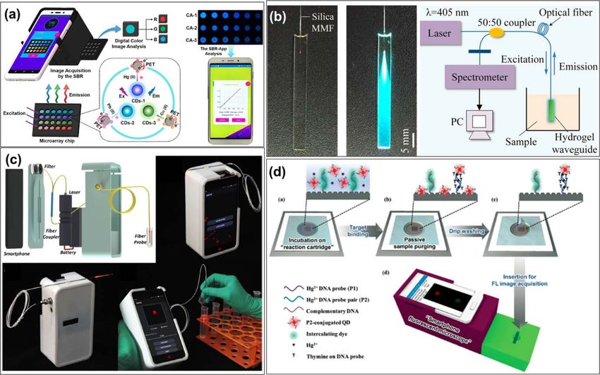
Journal of The Electrochemical Society, 2021 168 017502

          Fluorescent Sensing in Biochemical Detection                        Fang et al. developed a handheld laser-induced fluorescence
                                                                          (LIF) detector for various applications such as capillary electro-
    Portable biochemical sensors are widely used in various areas
                                                                          phoresis, flow cytometry, and scanning detection.50 For an excitation
such as environmental monitoring,39 disease diagnosis,40 drug
                                                                          source, a laser diode with 450 nm peak was selected. The sample
discovery,41 and food quality monitoring.42 Fluorescence-based
                                                                          solution was loaded in the center of the capillary for testing. For an
portable biochemical sensors are recognized as being highly
                                                                          emission part, a 525 nm peak bandpass filter was coupled with a
advantageous over other methods for its high sensitivity, high
                                                                          1.0 mm diameter aperture. A miniaturized PMT was chosen for the
selectivity, rapid response, and simple operation.18 Unlike colori-
                                                                          sensitive detection of fluorescence emission. For demonstration,
metric or absorbance-based sensing techniques, the fluorescence
                                                                          sodium fluorescein was used, and the detection limit was 0.42 nM
signal is directly measured without comparing with a reference
                                                                          (Fig. 4d).
beam. Therefore, fluorescence-based detection is often a more
                                                                              Chang et al. developed a handheld electronic tongue for
attractive option for field applications where only a small amount
                                                                          determining the taste level of astringency and umami in different tea
of biochemical samples are available. The development of new
                                                                          infusions.51 Fluorescence quenching reaction of 3-aminophthalate
fluorophores increased the possibilities for the sensitive detection of
                                                                          (reaction with tannin) and o-phthalaldehyde (OPA) (reaction with
numerous biochemical molecules. For example, near-infrared to far-
                                                                          amino acids) were utilized to detect astringency and umami,
red fluorescent probes have widely been used in bioimaging because
                                                                          respectively. A single excitation ultraviolet LED with 340 nm peak
of minimum photobleaching effect, deep penetration depth, and
                                                                          was selected to stimulate both target analytes. The emission
minimum auto-fluorescence by biomolecules.43,44 In addition, Wang
                                                                          wavelength of fluorescence was 425 nm peak and it was measured
et al. developed rhodamine-based fluorogenic probes in various
                                                                          with an RGB photosensor. Electronic circuits and optical compo-
colors and having increased cell permeability.45 Halabi et al.
                                                                          nents are closely packed in a plastic housing with a dimension of
reported a new type of fluorophore possessing photoswitching
                                                                          120 × 60 × 65 mm3. A standard disposable cuvette was used to hold
capability and fluxionality, which enables to conduct long time-
                                                                          the sample. The detection limits of theanine and tannic acid were
lapse and super-resolution microscopy experiments.46
                                                                          0.2 μg ml−1 and 1 μg ml−1, respectively (Fig. 4e).
    Kozma et al. developed a handheld fluorescent microarray reader
                                                                              Sasagawa et al. demonstrated a portable lensless fluorescence
for point-of-care diagnostics.47 A laser diode with 635 nm was used
                                                                          imaging device using multiple layers of interference filters.52 A
as an excitation source. The ray of light was guided and focused by
                                                                          CMOS image sensor with a dimension of 67 mm2 was selected for a
optical components including a pinhole array, waveguide, microlens
                                                                          large imaging area. A series of optical components were layered
array, and interference filter. Optical components were placed on top
                                                                          right on top of the image sensor to shift the focal plane and block the
of CCD image sensor to selectively measure the fluorescence signal.
                                                                          excitation light. First, a fiber optic plate (FOP) with a thickness of
A thin waveguide, which can be easily loaded into the device, was
                                                                          2.54 mm was placed right on top of the image sensor. Two optical
used to deliver the sample solution. For demonstration, a series of
                                                                          filters, an absorption filter (longpass above 500 nm) and a shortpass
different concentrations of fluorescence-labeled antibody solutions
                                                                          interference filter (490 nm) was placed right on top of the first FOP
    Ghosh et al. reported a miniaturized fluorescence-based micro-
                                                                          to block the excitation ray and autofluorescence. A sample can be
scope for cellular imaging purposes.48 The device includes blue
                                                                          directly loaded on top of the FOP surface. For demonstration,
LEDs (470 nm peak) as an excitation light source soldered on a
                                                                          fluorescence images of the sliced brain of a mouse (stained with a
6 mm × 6 mm printed circuit board (PCB) and assembled with
                                                                          green fluorescent protein (GFP), emission 515 nm) were obtained
optical components including a drum lens and excitation filter. A
                                                                          (Fig. 4f).
dichroic mirror was used for directing the excitation light towards
the sample while selectively allowing the fluorescence signal. An
                                                                                              Water Quality Monitoring
additional bandpass filter with 535 nm peak was placed before a
complementary metal-oxide-semiconductor (CMOS) image sensor                  Water quality monitoring is essential to prevent harm to human
to increase the signal-to-noise ratio. The size of the CMOS sensor        health and the aquatic ecosystem. Nowadays, natural events and
was 5.8 mm × 5.8 mm having a pixel size of 5.6 μm × 5.6 μm.               human activities can lead to water pollution, resulting in poor water
Electronic components were controlled with an interface PCB board         quality. By detecting and analyzing the parameters in the water, it is
containing field programmable gate arrays (FPGA) and an external           possible to identify the impacts and risks to human health and the
PC. For the demonstration, cellular imaging of the active brain of        ecosystem. This can help to plan and implement appropriate
mice and its activity was traced by a cell-permeant fluorescent Ca2+       management measures for water quality control. The following
indicator (Fig. 4b).                                                      subsections review the studies of fluorescence-based portable
    Katzmeier et al. developed a pocket-sized fluorescence detector        sensors for detecting the important parameters in the water.
for point-of-care testing using a paper-based cartridge for a sample
delivery.49 The device has two parts: a detection unit and an assay           Phytoplankton detection.—Phytoplankton monitoring is one of
cartridge. The detection unit includes an excitation light source and a   the important tasks for supporting human health and environmental
photodetector. The assay cartridge includes optical filters and a          issues, especially for water quality control. Detecting and analyzing
paper strip, and a 3D-printed frame that incorporates all the             different groups of phytoplankton in water provides important informa-
components. An LED with 466 nm peak wavelength was used as                tion on aquatic ecological states and nutrient compositions.53 In
an excitation light source (70 mW m−2) and a thin blue filter              addition, early detection of certain species of algae that cause harmful
(440 nm peak) was placed on top of the LED to have narrower               algal blooms (HABs) is essential to protect the water ecosystem and
excitation wavelength. A paper test strip was sandwiched between          human health.54,55 There are various ways to detect and analyze
two slide glass covers to hold the samples and placed over the            phytoplankton species, namely microscopy,56 flow cytometry,57 high-
excitation filter for direct stimulation of the fluorophore samples. An     performance liquid chromatography (HPLC),58 spectrophotometry,59–61
orange filter (longpass 700 nm) was placed on top of the top cover         spectrometry,62–64 and dry weight.65,66 However, these methods are
slide glass to block the stray light while allowing the fluorescence       difficult to be applied in the portable sensing system.
emission from the sample to pass. A cadmium sulfide (CdS) light                Different phytoplankton groups exhibit unique fluorescence
dependent resistor (LDR) was selected to measure the fluorescence          properties due to different photopigment constituents.67–70 A review
emission from the paper strip. For a demonstration, Cas13a-based          on the photopigment constituent of different phytoplankton groups
fluorescence assay was used to detect target RNAs. The reported            revealed that chlorophyll a can be found in every algal species due to
limit of detection was 3.7 nM (Fig. 4c).                                  its essential role in oxygenic photosynthetic reactions.71 Therefore,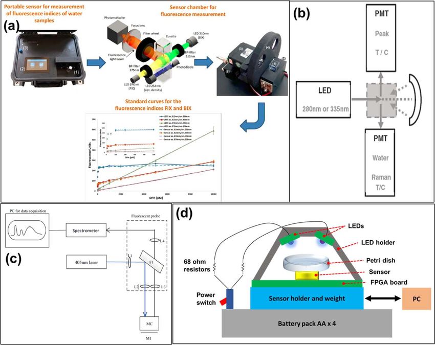
Journal of The Electrochemical Society, 2021 168 017502

even though chlorophyll a fluorescence provides important informa-                   pigments.35 Although all the pigments in green algae are responsible
tion about the spatial mapping of overall phytoplankton abundance,                  for absorbing light and contributing to the photosynthetic reaction,
it only offers information about microalgal species in the water and                chlorophyll a is responsible for majority of fluorescence due to the
does not distinguish one group from another. Different photopig-                    relatively longer lifetime of electrons. For this reason, chlorophyll a
ments within various phytoplankton species are responsible for                      is referred to as a primary photosynthetic pigment, while all the
absorbing photon energy with broader wavelengths for effective                      others are referred to as accessory photosynthetic pigments.70,72
energy collection. Chlorophyll a is called a primary photosynthetic                 Accessory pigments, such as carotene and chlorophyll b, show very
pigment, and all of the others are accessory photosynthetic                         weak or no fluorescence emissions since electrons are transferred to

Figure 4. Portable fluorescence-based biochemical detectors: (a) a handheld fluorescent microarray reader for point-of-care diagnostics47; (b) a miniaturized
fluorescence-based microscope for cellular imaging purposes (after48); (c) a pocket-sized fluorescence detector for point-of-care testing using a paper-based
cartridge for a sample delivery49; (d) a handheld LIF detector for various applications such as capillary electrophoresis, flow cytometry, and scanning detection50;
(e) a handheld electronic tongue for determining the taste level of astringency and umami in different tea infusions,51 and (f) a lensless fluorescence imaging
device using multiple layers of interference filters.52 Copyright 2013 with permission from Elsevier for (a); licensed under a CC BY license for (c); copyright
2016 with permission from Elsevier for (d); copyright 2010 with permission from Elsevier for (e), and licensed under a CC BY license for (f).
Journal of The Electrochemical Society, 2021 168 017502

Figure 5. Portable fluorescence algae sensors: (a) a 3D-printed portable microalgal sensor with a disposable PDMS chip73; (b) a handheld fluorescence platform
for multiple phytoplankton detection (green algae and cyanobacteria)74; (c) a portable toxin meter for detecting microcystin and cylindrospermopsin,75 and (d) a
portable xurography-based microfluidic biosensor for green algae detection via total chlorophyll a fluorescence measurement.76 Copyright 2015 with permission
from Elsevier for (a); copyright 2018 with permission from Elsevier for (b); copyright 2018 with permission from American Chemical Society for (c), and
copyright 2018 with permission from Elsevier for (d).

chlorophyll a immediately upon generation after absorbing photon                   and differentiate different mixture of green algae and cyanobacteria
energy. In this regard, careful selection of excitation light sources,             species using a multivariate algorithm. The limit of detection for green
optics, and detectors is necessary for multiple algal species detection            algae and cyanobacteria were 1 mg l−1 and 4 mg l−1, respectively.
capabilities. This section focuses on portable sensors that measure                (Figs. 5a and 5b).
different phytoplankton species or toxins produced by some species                     Bickman et al. developed a portable toxin meter that can detect
of cyanobacteria.                                                                  marine biotoxins (microcystin and cylindrospermopsin) released
    Shin et al. developed handheld phytoplankton sensors using                     from cyanobacteria using multiplexed fluorescence immunoassay
different excitation wavelengths (385 nm, 448 nm, and 590 nm) of                   technology.75 This approach is more accurate and direct when
LEDs to selectively stimulate different species of phytoplankton.73,74             detecting the toxins released from HAB compared to the conven-
A disposable PDMS chip and micro-vial were used for the sample                     tional indirect detection methods. The main reader device includes a
holding and delivery purposes. Excitation light sources were placed                laser diode (639 nm peak) that is coupled with a plastic light
underneath the sample and the detector was installed at the opposite               waveguide to evenly distribute and project the excitation lights on
side from where the excitation light rays are being projected to                   the sample holding substrate. A simple disposable cartridge that is
minimize the stray noise. Series of optical filters (dichroic and color             pre-coated with antibodies was used to detect the toxins in the
filters) were used to block the remaining stray noise. The emission                 collected sample. The presence of the toxins inhibited the binding of
fluorescent lights from the green algae and cyanobacteria were 680 nm               biotoxin-conjugate spots on the surface of the cartridge where
and 645 nm, respectively. It was successfully demonstrated to detect               fluorescence signal is reduced. The entire surface of a microarray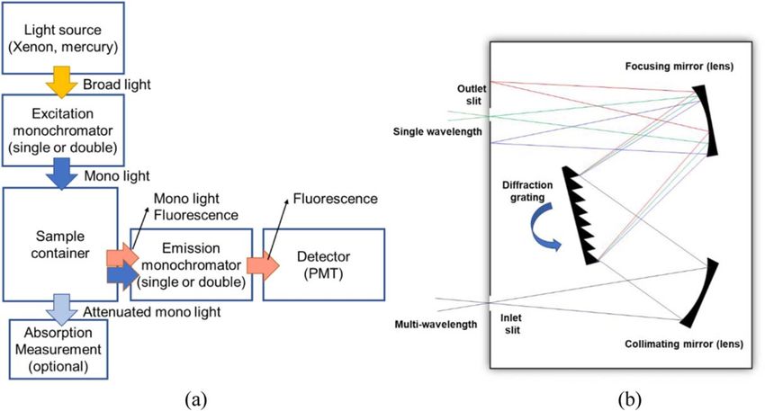
Journal of The Electrochemical Society, 2021 168 017502

was analyzed with a camera to detect the fluorescence emission. The             Brandl et al. developed a portable fluorometer for detecting DOM
limit of detection for microcystin and cylindrospermopsin were             and three different green algae by using different excitation LEDs.91
0.4 μg l−1 and 0.7 μg l−1, respectively (Fig. 5c).                         The fluorometer has three different wavelengths of excitation LEDs
    Gosset et al. developed a portable xurography-based microfluidic        (254, 310, and 370 nm) with two bandpass filters (370 and 310 nm). An
biosensor for green algae detection via total chlorophyll a fluorescence    adjustable filter wheel with four bandpass filters with 380, 430, 450,
measurement.76 The sensor utilizes an LED with 470 nm peak                 and 500 nm wavelengths was utilized to selectively measure the target
wavelength as an excitation source. Optical components such as             fluorescence emission from different analytes. A PMT was used to
collimating lens and diffuser were placed underneath the LED to            detect the fluorescence signals. The DOM concentration was estimated
evenly distribute the light. A disposable microfluidic chip was             by calculating the ratio of two fluorescence emissions from 380 nm and
fabricated with two slide glasses and a double-sided pressure adhesive     430 nm bandpass filters under 310 nm excitation. Consequently, the
film with 100 μm thickness to deliver the microalgal sample to the          ratio of two fluorescence emissions from 450 nm and 500 nm bandpass
device. The chip has two chambers to read a control and a target of        filter under 370 nm excitation, was calculated to determine if the
interest. Different concentrations of herbicide solutions were mixed       measured DOM is from microbial nature or terrestrially derived. A
with the algal samples to analyze the relationship between the             standard cuvette was selected to hold the sample solution. (Fig. 6a).
fluorescence intensity changes to the photosynthesis disturbance rate           Bridgeman et al. reported an LED-based portable fluorescence
of the samples. The fabricated portable fluorescence sensor was             sensor to detect total organic carbon (TOC) and microbial activity of
successfully demonstrated using three different microalgae species         water.92 Two UV excitation LEDs with 280 nm and 335 nm
with the herbicide solution. The detection limit of the microalgae         wavelengths were used to measure microbial activity and organic
measurement was 1 μg l−1. (Fig. 5d).                                       carbon, respectively. An additional UV LED with 310 nm was
    Although the use of those fluorometers was successfully demon-          used to measure the water Raman signal, which is caused by the
strated for phytoplankton detection, limitations for those sensors still   inelastic scattering of the excitation light. This allows to minimize
exist. It is challenging to distinguish multiple species of phyto-         the measurement error when the analyte concentration is low and
plankton, simultaneously, because the fluorescence signals from             the fluorescence is weak. A standard quartz cuvette was used to
those species often overlap and interfere with each other. In addition,    hold the sample solution. Two PMTs were used to measure the
dissolved organic matter in the water becomes another source of the        fluorescence signal and water Raman signal, simultaneously. The
noise.                                                                     dimension of the device was 425 (l) × 300 (w) × 225 (d) mm, with a
                                                                           weight of 3.5 kg. The results showed high correlations (r2) between
    Dissolved organic matter detection.—The increase of human              the new portable system and a conventional benchtop instrument
pollution and the effects of climate change significantly affected the      (ranging from 0.83–1.00). (Fig. 6b).
quality of the water.77–79 The pollutants in this natural water are            Laser-based excitation sources have several benefits over LED-
closely related to the dissolved organic matter (DOM) concentration        based excitation sources. Laser can generate highly monochromatic
and its composition, therefore it is important to understand the           (equal or less than 1 nm), coherent, and collimated excitation light,
characteristics of DOM in the water.80,81 DOM is defined as any             therefore, they show minimal interference with the fluorescent
organic matter dissolved in the water that can pass through the water      emission spectra and require less optical components for beam
filter with a pore size of 0.2 μm. For DOM detection, ultraviolet-          shaping. However, it is difficult to apply conventional laser systems,
visible fluorescence spectroscopy method was widely used to fully           such as gas lasers and solid-state lasers to portable systems because
scan the absorption and the emission spectra.82,83 Since not all           of their large size, high cost, and power consumption. On the
DOMs are light interactive, the light-absorbing portion, of which is       contrary, LEDs with optical filters are desired for portable applica-
defined as colored dissolved organic matter (CDOM), is measured             tions. A LIF technology was utilized to a portable fluorometer
by the fluorescence-based detection. Due to the advantages that             system by Chen et al. to detect chlorophyll a, CDOM, and total
fluorescence-based detection method offers, various studies have            suspended matter (mainly slit and microorganisms).93 A laser with
been conducted to concentrate and develop an on-site detection of          405 nm wavelength was coupled with a fiber-optic probe to guide the
CDOM fluorescence to aid the water quality monitoring. The main             light excitation and emission lights. As an excitation source, laser
advantage of a portable fluorometer system is that in situ measure-         showed several benefits over other light sources such as high pulse
ment of CDOM can report ecological conditions of the water in a            frequency modulation capacity and narrower spectral profile. Those
timely manner, thus more accurate environmental monitoring is              benefits can improve the signal-to-noise ratio of measured fluores-
possible. Natural water typically has CDOM that is responsible for         cence signals. A quartz cuvette was selected to load the sample
strongly absorbing light in the light range from 250 nm to 450 nm          solution. A dichroic beam splitter and longpass filter were deployed
and fluorescing at 400–450 nm.84,85 However, the utilization of blue        to selectively scan the emission fluorescence spectra from the
light excitation for CDOM detection is challenging due to the              sample. Fluorescence emission from the sample was measured
presence of chlorophyll pigments in natural bodies of the water, thus      with a hyperspectral micro spectrometer. The fluorescence emissions
accurate assessment of the emission signal is essential.86 Lewis et al.    in the peak near 685, 508, and 470 nm correspond to chlorophyll a,
studied an algorithm to accurately estimate the CDOM in the arctic         CDOM, and water Raman scattering, respectively. The measurement
ocean to compensate for the overly estimated CDOM when                     of water Raman signal was required to compensate the errors due to
chlorophyll a present in the water.87 For remote sensing, satellite-       the spectral overlapping. The limit of detection for CDOM, total
based measurements were widely used to quickly assess the large            suspended matter, and chlorophyll a were estimated to be
water areas such as the ocean.88,89 In addition, hyperspectral remote      0.75 μg l−1, 1 mg l−1, and 0.2 μg l−1, respectively (Fig. 6c).
sensing was reported to achieve higher resolution than the satellite-          Blockstein et al. reported a portable fluorometer to measure
based sensing while covering a relatively larger area in the last          chlorophyll pigments and CDOM concentration.94 Light emitting
decade and successfully differentiated multiple many important             diodes with 405 nm and 465 nm were utilized to selectively
water quality parameters such as CDOM, chlorophyll a, diatoms,             stimulate the CDOM and chlorophyll, respectively. Two custom
and turbidity.90 However, those remote sensing techniques face             fabricated thin optical filters were directly attached on a single
challenges to report ecological state of deeper water body or              sensor array to selectively measure two different fluorescence
continuous measurement of one single spot of an interest. This             signals. The thin glass substrates were applied to absorb and
section focuses on reviewing the properties and results of water           attenuate the excitation lights. For demonstration, standard fluor-
quality measurements by utilizing the portable fluorescence sensors         escein dye was selected to simulate chlorophyll a. The sensor was
for detecting CDOM excited by the light wavelength in the range of         tested while it is completely submerged under the water. The limit of
250–500 nm.                                                                detection for the fluorescein was 0.7 nM. (Fig. 6d).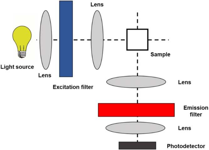
Journal of The Electrochemical Society, 2021 168 017502

Figure 6. Portable DOM fluorescence sensors: (a) a portable fluorescence sensor for DOM measurement using fluorescence index (FIX) and biological index
(BIX)91; (b) a portable LED fluorescence instrumentation for the rapid measurement of TOC92; (c) a light-weight laser-based fluorometer for monitoring
chlorophyll a and CDOM,93 and (d) a miniature fluorometer is designed to measure chlorophyll and CDOM concentration in the aquatic environment (after94).
Licensed under a CC BY license for (a) and (b), and copyright 2015 with permission from Elsevier for (c).

   Dissolved organic matter measurement in natural water is                   reagents and indicator dyes. They only react with desired target metal
challenging since it is a collection of different constituents in water.      ions and absorb a specific wavelength of the color. For reactive agent
The ratio of these elements in the water may highly vary depending            material, colloidal gold nanoparticles (AuNPs) were widely selected to
on the region, time, and the weather, thus accurate assessment is             detect the target metal ions by examining the color shifting of the
required. CDOM showed a broad range of excitation spectrum,                   solution, in which the particles aggregate with the target ions.103,104
usually between 250 nm to 500 nm. The emission spectral range was             Morais et al. developed a portable lead detector to demonstrate a low-
normally between 350 nm to 500 nm.                                            cost colorimetric-based sensor using economical components such as
                                                                              LEDs and microcontroller module.105 Chen et al. reported a mercury
    Heavy metal ion detection.—Heavy metal pollutions in water have           detector by utilizing the paper-based sensor combined with AuNPs to
become a strong threat to marine animals and humans since they can            demonstrate a cheap/disposable sensing platform for resource-limited
bioaccumulate in living organisms either directly or through                  settings.106 Wei et al.,107 Nguyen et al.,108 and Xiao et al.109
consumption.95 Among many metal ions, copper, lead, mercury,                  demonstrated portable smartphone-based heavy metal detectors to
chromium, and cadmium are known to be highly toxic for humans.                show the benefits of incorporating the mobile phones with custom-
For example, copper can cause liver damage, lead is known to damage           designed sensing platforms. However, colorimetric-based sensing
the brain, and low doses of mercury exposure can cause severe damage          techniques face challenges when used at the natural water bodies
to the nervous system for all animals including humans. Therefore, it is      where numerous elements exist as interference factors, therefore
critical to rapidly detect those metal ions from the aqueous system.          fluorescent-based detection method is desirable for a field-deployable
    Various methods have been developed for heavy metal detection             application. The major interfering elements in natural water bodies
such as electrochemical,96 spectroscopic,97 and optical detection.98–101      include CDOMs and suspended particles. CDOMs strongly absorb the
Although both spectroscopic and electrochemical detection methods             spectral range in 250 nm to 500 nm, which may interfere with AuNP-
have been widely used, optical detection methods showed great                 based colorimetric heavy metal sensors. Suspended particles are any
potential for portable sensing platforms.102 One of the widely used           particles that are bigger than 2 μm, such as clay, silt, sand, gravel,
optical detection methods is colorimetric sensing that utilizes selective     bacteria, and algae. It has been reported that suspended particles can
Journal of The Electrochemical Society, 2021 168 017502
negatively affect the detection performance of colorimetric-based Liu et al. reported smartphone-based fluorescence sensor for
sensors by increasing the optical density and scattering of light, Hg2+ detection.112 CdSe/ZnS quantum dot modified optical fiber
however, the detection performance of fluorescence sensors was not probe was coupled with a laser excitation light (405 nm) and an
affected by suspended particles.73 This section focuses on portable optical filter (bandpass with 620 ± 15 nm). The camera on the
fluorescence sensors for detecting heavy metal ions in water. smartphone was placed behind the bandpass filter to measure the
Xiao et al. developed a paper-based microarray using carbon intensity of the fluorescence to quantify the Hg2+ concentration in
nanodots (CDs) to detect the heavy metal ions (Hg2+, Pb2+, and the solution. The detection range was between 1 nM to 1000 nM and
Cu2+).110 Three different CDs were prepared and drop casted on the the limit of detection was 1 nM (Fig. 7c).
pretreated filter paper for selective detection of different heavy metal Lee et al. also introduced a smartphone imaging-based fluorescence
ions. A 3D-printed apparatus was assembled with a smartphone to read microscope for monitoring Hg2+ ions utilizing a biosensor cartridge.113
out the fluorescence signals from the microarray using an image sensor The cartridge was pretreated with an Hg2+ DNA probe to capture the
on the phone. The apparatus includes an excitation LED (365 nm), a Hg2+ ions. The intercalated fluorescence dye that emits green (520 nm)
plastic diffuser, and an optical lens to evenly illuminate the light on the fluorescence was quenched after binding with Hg2+ ions while the
surface of the microarray. The emission spectra for Hg2+, Pb2+, and quantum dot that emits red (655 nm) light became brighter after the
Cu2+ were estimated to be 445, 450, and 475 nm, respectively. The reaction with the Hg2+ ions. The captured image using a camera
image of the microarray was processed with the smartphone and the image sensor was analyzed in red and green channels to quantify the
results were displayed on the screen using the custom-built application. intensities and the results. The limit of detection was 1 pM (Fig. 7d).
The limit of detection for Hg2+, Pb2+, and Cu2+ were 5.8 nM, Heavy metal detection in an aqueous environment is challenging
0.12 μM, and 0.076 μM, respectively. The sensor offers a great since it includes many elements and particles that can potentially
resolution to measure the World Health Organization (WHO) guideline interfere with the target metal ions and it may lower the detection
values for the heavy metal ions in drinking water (Hg2+ (0.006 mg l−1), accuracy.114 There are several strategies to improve the detection
Pb2+ (0.01 mg l−1), and Cu2+(2 mg l−1)) (Fig. 7a). performance of heavy metals in water. Cellulose filter papers can be
Guo et al. reported a carbon dot doped hydrogel waveguide for used to pretreat the water before the test to get rid of the debris from the
detecting Hg2+ in the water.111 Although the reported work is not a solution. In order to minimize the interference from the dissolved matter
stand-alone system, it showed great potential to be a key element of in water, accurate characterization of fluorescence emission spectra of
the device and easily incorporated into a portable device. PEG the sample solution (noise mainly from CDOM and chlorophylls).
diacrylate (PEGDA) was selected as the material for the waveguide
and the fluorescent carbon dots with 7.8 nm diameters were selected
Smartphone-Based Fluorescence Microscope
for the material. The waveguide exhibited a peak absorption at 352 nm
and fluorescence emission at 475 nm. The detection range was Fluorescence microscopy is a very effective method in
between 0 to 5 μM and the limit of detection was 4 nM (Fig. 7b). bioscience, however, its use in portable applications is difficult
Figure 7. Portable fluorescence-based heavy metal detectors: (a) a paper-based microarray using CDs to detect the heavy metal ions with a smartphone-based
fluorescence reader110; (b) a carbon dot doped hydrogel waveguide for detecting Hg2+ in the water111; (c) smartphone-based fluorescence sensor for Hg2+
detection using CdSe/ZnS quantum dot modified optical fiber probe,112 and (d) a smartphone imaging-based fluorescence microscope for monitoring Hg2+ ions
utilizing a biosensor cartridge.113 Copyright 2020 with permission from American Chemical Society for (a); licensed under a CC BY license for (b); copyright
2019 with permission from Elsevier for (c), and copyright 2019 with permission from Royal Society of Chemistry for (d).Journal of The Electrochemical Society, 2021 168 017502
due to the complexity and the size of its instrument. Recently, diode and ball lens were moved vertically by a miniature dovetail
smartphones have been widely used for remote sensing applications stage. An aliquot volume of sample droplet (20 μl) was placed on a
owing to their multiple built-in sensors and communication modules. glass coverslip (22 mm × 50 mm) for sample delivery. A longpass
In addition, smartphones were able to achieve great improvements in emission filter (>514 nm) was placed before the smartphone camera
image sensor and computing power. This allowed to demonstrate a with an external lens to block the excitation light. A smartphone
compact fluorescence microscope for analytical imaging applications in LED (white) was utilized to locate the reference dot and preliminary
field settings. Wei et al. demonstrated a smartphone-based fluorescence focusing of the sample. For testing, bacteria with various concentra-
microscope to detect single bacteria.115 In addition, advanced image tions were prepared with fluorescence labels. The sensor was able to
processing algorithms such as machine learning and deep learning selectively detect the target strand even when mixed with different
techniques were applied to improve sensing accuracy.116,117 In this bacterial strain and infant formula. The fluorescence emission
section, smartphone-based fluorescence microscopes used in various wavelength was 525 nm. The limit of detection was 104 CFUs
applications were reviewed. ml−1 (Fig. 8e).
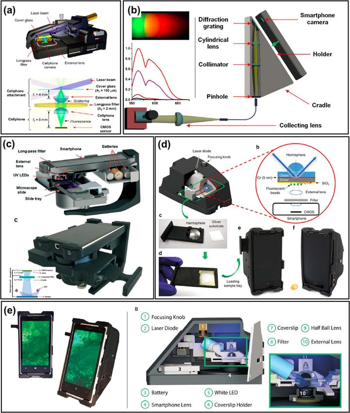
Wei et al. reported a smartphone-based portable fluorescence Cho et al. reported a smartphone-based fluorescence microscope
microscope for imaging of nanoparticles and viruses.115 For an for monitoring of organ-on-a-chip.121 White LED with a bandpass
excitation source, a laser diode with 450 nm peak was used with an filter (480 ± 10 nm) was selected as an excitation source. A longpass
optical lens (focal distance = 4 mm). Two longpass optical filters filter (>500 nm) with an objective lens was placed in front of the
(> 500 nm) were placed above the image sensor to block the stray image sensor to block the noise from the stray light. Organ-on-a-chip
lights from the excitation source. An aliquote volume of sample was fabricated using PDMS, which is a transparent silicon-based
solution was loaded with a standard cover glass slide, which was organic polymer. The microfluidic channel was etched on the glass
held by a 3D-printed tray. The light ray was projected with a high substrate and bonded with PDMS chip. Sample solutions were
incident angle (75°) towards the sample to minimize the excitation rays loaded through the inlet and outlet holes. For demonstration,
from reaching to the image sensor. For demonstration, green fluor- cancerous cells were selected as target analytes. These cells were
escent polystyrene (PS) particles (ex/em: 505/515 nm) and human functionalized on the glass substrate and fluorescently labeled (ex:
cytomegaloviruses were tested. The microscope had 0.6 mm × 0.6 mm 480 nm/em: 510 nm). The solution including the fluorescent particles
field of view. (Fig. 8a). was introduced into the organ-on-a-chip (OOC) with 500 μl h−1 flow
Yu et al. demonstrated smartphone-based fluorescence spectro- rate during the testing. The limit of detection was 10 pg ml−1
scopy for demonstrating microRNA sequencing.14 Two different (Fig. 9a).
lasers with a peak wavelength of 532 nm and 653 nm were selected Shan et al. developed a portable fluorescence-based mercury
as excitation sources. A series of optical components including detector using a smartphone microscope.122 A compact laser diode
pinhole, optical fiber, diffraction grading (1200 lines mm−1), and with a wavelength of 405 nm peak was selected for an excitation
three different optical lenses were used to scan broadband fluores- source. Optical components including micro-objective and eyepiece
cence emission spectra. The sample solution was loaded via standard were arranged for focusing and collimation. A bandpass filter (469 ±
cuvette. For testing, molecular beacon Förster resonance energy 35 nm) was placed before the phone camera to obtain noiseless
transfer (FRET) assay was selected to observe the changes in the fluorescence images. A standard slide glass with a 3D printed tray
quenching efficiency of a miRNA sequence. The limit of detection was used to deliver the sample droplet. The size of a sensor device
was 10 pM. Although the laser excitation sources and the sample was 170 mm (length) × 113 mm (width) × 168 mm (height). For
holder were not completely incorporated into the cradle, the authors detection, fluorescent microspheres (ex: 405 nm, em: 450 nm) were
claimed that they can be easily integrated for a standalone applica- utilized for selected detection of mercury. The limit of detection was
tion. (Fig. 8b). 1 nM (Fig. 9b).
Snow et al. developed a smartphone-based fluorescence micro- A variety of types of smartphone-based fluorescence micro-
scope for imaging and detecting pathogenic spores in honeybees.118 scopes were reviewed. Smartphones and optical components
For excitation light source, four ultraviolet (specific wavelength is were easily incorporated into custom-designed jigs to demonstrate
unclear) LEDs were selected. A longpass filter (>460 nm) and a compact stand-alone systems for remote sensing applications.
simple optical lens were placed before the smartphone camera lens Smartphones have shown great potential to be used in a re-
to minimize the noise from the excitation light. For sample delivery, source-limited setting and point-of-care testing. It has been
a standard microscopic slide glass was selected and it was easily reported that even in-built ambient light sensors on the smart-
inserted with a 3D printed slide-holder. For sample preparation, phones were utilized for biochemical sensing applications.123–125
midgut tissues of honeybees were stained with fluorescent brightener Table II summarizes the characteristics of different types of
to detect the spores. A smartphone application was developed to fluorescence-based portable sensing devices that are reviewed.
transmit the captured image to the main server for image processing. Typical detection speeds for fluorescence-based sensors were
The detection limit was 0.5 × 10⁶ spores per bee (Fig. 8c). almost instantaneous, however incubation and pretreatment times
Wei et al. reported a smartphone-based fluoresce microscope increased the overall test duration.
combined with surface-enhanced fluorescence created with thin
metal-film.119 A compact laser diode with 465 nm peak wavelength
Discussion and Challenges
was utilized as an excitation source and the light was guided through
a glass hemisphere at an incident angle of 58°. The beam from the Fluorescence-based portable sensing systems have been consid-
laser was then filtered by a polarizer to deliver p-polarized rays onto erably studied and developed in the past decade. The main benefits
the silver-coated glass coverslip (22 × 22 mm). A bandpass filter of fluorescence-based portable sensors are compact size, low power
(525 ± 25 nm) and a collimating lens were placed before the phone consumption, low cost, and fast speed. Although fluorescence-based
camera to block the excitation background noise. For demonstration, detection method is highly useful for portable applications, there is
DNA origami nanobeads (average diameter of 23 nm) and quantum still room to improve.
dots (ex: 468 nm/em: 508 nm) were selected. The limit of detection First, a fluorescence labeling process is required to detect the
for the DNA origami nanobeads was reported as 80-fluorophore analytes that are not inherently fluorescent. This may add laborious
DNA origami nanobeads (Fig. 8d). steps for sample preparation to bind the fluorescent tags to the
Müller et al. developed a smartphone-based fluorescence micro- biomolecules such as proteins. Furthermore, the sample solution
scope for pathogenic bacteria identification.120 The laser diode with can contain unwanted matters, which interfere with the target
488 nm wavelength was selected for an excitation source and a half analyte signals. Several studies have demonstrated an integration
ball lens was chosen to focus the light on the sample. Both the laser of microfluidic channels with sample separation/filtration, mixing,Journal of The Electrochemical Society, 2021 168 017502 Figure 8. Smartphone-based fluorescence microscopes: (a) smartphone-based imaging device for nanoparticles and viruses115; (b) smartphone spectro- photometry for detecting fluorescence biological assays14; (c) a smartphone-based fluorescence microscope for imaging and detecting pathogenic spores in honeybees118; (d) a smartphone-based fluoresce microscope combined with surface-enhanced fluorescence created with thin metal-film,119 and (e) a smartphone- based fluorescence microscope for pathogenic bacteria identification.120 Copyright 2013 with permission from American Chemical Society for (a); copyright 2014 with permission from American Chemical Society for (b); copyright 2019 with permission from Royal Society of Chemistry for (c), and Licensed under a CC BY license for (d) and (e). target labeling, and washing to minimize the preparation steps.126,127 based fluorescent paper strip to monitor the glutathione level in In addition, biochemical reagents can be prefabricated in cheap & human serum.128 A mixture of quantum dot and carbon dot solution disposable materials such as paper. Chu et al. reported a nanoprobe- was deposited on a piece of filter paper to fabricate a paper strip
Journal of The Electrochemical Society, 2021 168 017502

Figure 9. Smartphone-based fluorescence microscopes (cont.): (a) reported a smartphone-based fluorescence microscope for monitoring of OOC121 and (b) a
portable fluorescence-based mercury detector using smartphone microscope.122 Copyright 2016, with permission from Elsevier for (a) and copyright 2019, with
permission from Elsevier for (b).

sensor. With those approaches, laborious steps can be significantly             low-cost, and low-power, hence a great option for portable applica-
reduced and increase the practicality of the system.                           tions. Jian et al. demonstrated a portable fluorescence-based imaging
    Secondly, many demonstrated systems still require external                 system for intraoperative display of biliary structure and prevention
instruments to aid the device operation. For example, a laptop or              of iatrogenic injuries during cholecystectomy.129 A single-board
PC was frequently required to control the device or process the data.          computer (Raspberry Pi, Raspberry Pi Foundation, UK) was able to
Single-board computers can be selected as a viable option to handle            handle multiple tasks, such as displaying the LCD screen, control-
complicated tasks such as image processing, data analysis, and                 ling the image sensor, and processing the data. As reviewed in the
controlling peripheral devices, simultaneously. They are compact,              previous section, smartphones can be an alternative option as well,
You can also read