Role of emerging vitamin K dependent proteins: Growth arrest specific protein 6, Gla rich protein and periostin (Review) - Spandidos ...
←
→
Page content transcription
If your browser does not render page correctly, please read the page content below
INTERNATIONAL JOURNAL OF MOlecular medicine 47: 2, 2021

      Role of emerging vitamin K‑dependent proteins: Growth
  arrest‑specific protein 6, Gla‑rich protein and periostin (Review)
                              HUIYU XIAO1, JIEPENG CHEN2, LILI DUAN2 and SHUZHUANG LI1

                         1
                             Department of Physiology, Dalian Medical University, Dalian, Liaoning 116044;
                                2
                                  Sungen Bioscience Co., Ltd., Shantou, Guangdong 515071, P.R. China

                                         Received April 13, 2020; Accepted October 21, 2020

                                                     DOI: 10.3892/ijmm.2020.4835

Abstract. Vitamin K‑dependent proteins (VKDPs) are a group            1. Introduction
of proteins that need vitamin K to conduct carboxylation. Thus
far, scholars have identified a total of 17 VKDPs in the human        Scholars have explored vitamin K since it was discovered by
body. In this review, we summarize three important emerging           Henrik Dam in 1935 (1). The vitamin K family belongs to
VKDPs: Growth arrest‑specific protein 6 (Gas 6), Gla‑rich             the group of naphthoquinone compounds, and their common
protein (GRP) and periostin in terms of their functions in            structure is composed of a 2‑methyl‑1,4‑naphthoquinone ring
physiological and pathological conditions. As examples,               and a hydrophobic polyisoprenoid side chain. Depending
carboxylated Gas 6 and GRP effectively protect blood vessels          on the side chain length and saturation (2), vitamin K can
from calcification, Gas 6 protects from acute kidney injury and       be divided into vitamin K1 (phylloquinone, PK), vitamin K2
is involved in chronic kidney disease, GRP contributes to bone        (menaquinones, MKs), and vitamin K3 (3). Vitamin K2 is often
homeostasis and delays the progression of osteoarthritis, and         represented by MK‑n, where n is represented for isoprene units
periostin is involved in all phases of fracture healing and assists   counts, and MK comprises 15 types. The main food sources
myocardial regeneration in the early stages of myocardial             of PK are green vegetables, especially spinach, broccoli and
infarction. However, periostin participates in the progression        kale. MK‑4, the dominant form of MK, is found in fish, milk,
of cardiac fibrosis, idiopathic pulmonary fibrosis and airway         liver and vegetables. Other MKs are mainly synthesized by
remodeling of asthma. In addition, we discuss the relationship        microorganisms and are also found in Japanese natto (MK‑7),
between vitamin K, VKDPs and cancer, and particularly the             cheese (MK‑8, MK‑9) and other food (4). Bacteria in the large
carboxylation state of VKDPs in cancer.                               intestines of humans are the main synthesizer of MKs, such
                                                                      as MK‑7, MK‑8, MK‑10 and MK‑11, with the exception of
                                                                      MK‑4. MK‑4 is formed by PK or vitamin K3 through in vivo
Contents                                                              tissue‑specific transformation, which is also the mechanism
                                                                      underlying the biological activities of PK and vitamin K3 (3).
1. Introduction                                                       The form and content of MK in different regional food supplies
2. Uptake, distribution and vitamin K cycle                           differs due to the food species limit. Natto is irreplaceable in
3. VKDPs                                                              the traditional Japanese diet, and cheese is a staple of dairy
4. Gas 6                                                              product supply in Europe. It is thus inevitable for MK uptake
5. GRP                                                                across regions to be imbalanced.
6. Periostin                                                              Osteoporosis is a systemic skeletal disorder with a globally
7.	Discussion                                                         high incidence that is exacerbated by the problem of an aging
                                                                      population. Osteoporosis has three types, which are termed
                                                                      primary osteoporosis, secondary osteoporosis and idiopathic
                                                                      osteoporosis (5). The most common type in older women is
                                                                      postmenopausal osteoporosis, which is a form of primary
                                                                      osteoporosis. Subsequent fractures, particularly hip frac‑
                                                                      tures, seriously affect the survival prospects and life quality
Correspondence to: Professor Shuzhuang Li, Department of              of the elderly. Based on predictions of the Asian Federation
Physiology, Dalian Medical University, 9 West Section, Lvshun         of Osteoporosis Societies, the total number of hip fractures
South Road, Dalian, Liaoning 116044, P.R. China                       are due to reach 2.56 million by 2050 in the studied Asian
E‑mail: shuzhuangli@126.com                                           countries (6). Calcium supplements are the most well‑known
                                                                      non‑prescription therapy for strengthening bone mineral
Key words: vitamin K‑dependent proteins, bone, heart, kidney,         density and preventing osteoporosis. However, the calcium
calcification, cancer, idiopathic pulmonary fibrosis, asthma          paradox, a consequence of damaged calcium metabolism, is
                                                                      identified as the loss of calcium in the bones parallel with the
                                                                      formation of calcification in the arteries in the elderly (7), and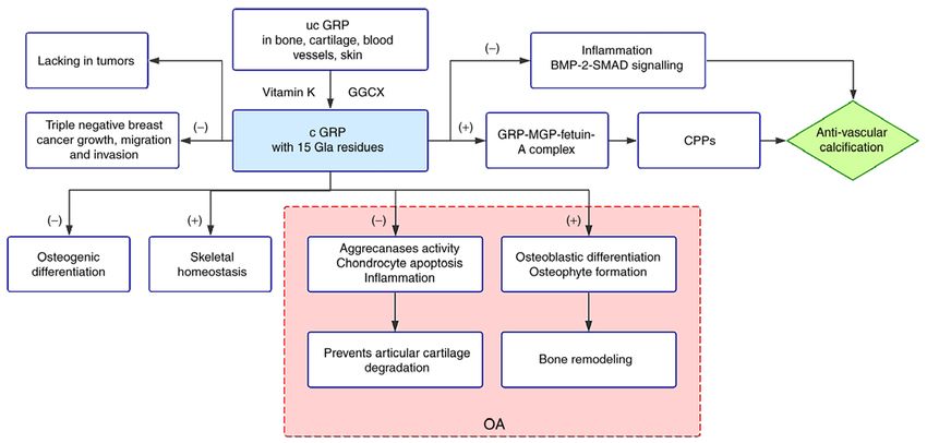
2                                XIAO et al: ROLE OF EMERGING VITAMIN K-DEPENDENT PROTEINS

exists as a potential risk of calcium supplements. Evidence             Vitamin K is metabolised in the human body through
has accumulated that vitamin K can be of benefit in avoiding        the vitamin K cycle (20). The three forms of vitamin K in
the calcium paradox. In addition, VKDPs, such as osteocalcin        this cycle are quinone (K), vitamin K hydroquinone (KH 2)
(OC), indicate a beneficial effect on bone strength loss.           and vitamin K epoxides (KO). K is initially reduced to KH2,
    Cardiovascular diseases (CVDs), such as acute myocardial        which is oxidised into KO under the effect of epoxidase
infarction, atherosclerosis and heart failure, are the main cause   (GGCX). KO is then reduced to KH2, and vitamin K epoxide
of human deaths worldwide. These diseases, not only pose a          reductase (VKOR) participates in the process. After repeti‑
great threat to patients' health, but also disturb their families   tion of the above steps, the vitamin K cycle is formed. It is
and even society. An epidemiological study of 709 multiethnic       worth mentioning that Warfarin exerts anticoagulant effects
adults, with follow‑up at an average of 11.0 years, showed          to inhibit VKOR activity and induce the cellular produc‑
VKDP activity is associated with the incidence of ischemic          tion of a large number of nonreactive substances into the
cardiovascular events (8). The relationship between vascular        coagulation system (21). The protein containing glutamate
calcification and disease has become a research focus due to        (Glu, ‑CH2CH2COOH) residues in the body is also catalysed
the increasing rates of morbidity and mortality of CVDs. An         into γ ‑carboxyglutamate [Gla, ‑CH 2 CH(COOH)2] under
epidemiological study of 116,309 individuals, with follow‑up        the action of the key enzyme gamma‑glutamyl carboxylase
at an average of 28 years, indicated an aortic arch calcifica‑      (GGCX) and co‑actors KH2, carbon dioxide and oxygen (22).
tion exhibited a positive correlation with an increased risk of     The protein containing Glu residues is known as VKDP. The
coronary heart disease (9). Moreover, another epidemiological       Glu residues in VKDPs that can be transformed are usually
study showed coronary artery calcium was independently              located in an amino acid region known as the Gla domain.
associated with cardiac events (10). The matrix Gla protein         It is worth mentioning that the Gla domain formed after
(MGP), a kind of VKDP, synthesized by vascular smooth               carboxylation of VKDP is the key to its biological func‑
muscle cells (VSMCs) is widely expressed in soft tissues, such      tion. For instance, Gla domain at the N‑terminal provides
as cartilages and blood vessels (11), especially in calcified       a special bond for the interaction of vitamin K‑dependent
tissues. It has been suggested that MGP regulates vascular          blood coagulation proteins with cell membranes containing
calcification and various important pathological processes. In      phosphatidylserine, and this binding is requisite for blood
fact, many emerging proteins related to vitamin K are involved      coagulation (23).
in the fight against vascular calcification and are described in
more detail below.                                                  3. VKDPs
    Kidney disease poses a great threat to health. According
to the course duration of the disease, the disease can be           At present, scholars have identified 17 types of VKDPs in
classified as acute kidney disease or chronic kidney disease        humans. Seven of them are dependent on vitamin K1 to play
(CKD). Various causes have been aligned closely with CKD.           their roles in the liver (coagulation factor II, VII, IX, X and
To be specific, diabetes and hypertension are the two main          anticoagulant proteins C, S, Z). Six of them were modi‑
contributing factors of CKD in developed countries. However,        fied by vitamin K after transcription and were involved in
glomerular diseases still occupy an important position in           various physiological and pathological processes in extra‑
developing countries. Sub‑clinical vitamin K deficiency exists      hepatic tissues. They are OC, MGP, Gas6, GRP, periostin
in most CKD patients, with the characteristic of low circu‑         and periostin‑like‑factor. The remaining four proteins need
lating vitamin K level and high inactive VKDP level (12‑14).        further study (proline‑rich Gla protein 1, proline‑rich Gla
The factors that contribute to this situation include low           protein 2, transmembrane Gla protein 3 and transmembrane
vitamin K intake and reduction in the carboxylation process         Gla protein 4) (Table I).
of VKDPs (15). In addition, cardiovascular complications are            OC was the first VKDP to be identified that is synthesized
the main reason for the mortality of CKD patients (16). The         and secreted by bones. Originally, researchers found osteo‑
protective effect of some VKDPs, such as MGP, on both the           calcin has the ability to attract calcium ions. Vitamin K lowers
kidney and cardiovascular system, has been widely explored.         serum undercarboxylated OC (ucOC) concentrations and
    Numerous studies are available on OC and MGP. The aim           increases carboxylated OC (cOC). Furthermore, cOC can bind
of the current review is to focus on three emerging VKDPs           with hydroxyapatite crystals, the material of the bone matrix,
that are increasingly being studied: Growth arrest‑specific         while simultaneously promoting bone mineral density (41,73).
protein 6 (Gas6), Gla‑rich protein (GRP), and periostin and         Moreover, it has been suggested during the past decade that
their roles in various physiological and pathological processes.    OC shows functions of regulating systemic glucose and energy
                                                                    metabolism (42).
2. Uptake, distribution and vitamin K cycle                             MGP plays a beneficial role in vascular calcification and
                                                                    various pathological processes. MGP regulates vascular
Both vitamin K1 and vitamin K 2 are absorbed by the small           calcification by eliminating the calcification effect of
intestine and are transferred to liver in the form of chylomi‑      bone morphogenetic protein (BMP)‑2 and BMP‑4 (46,47).
crons. After absorption into the blood by liver, vitamin K1         Additionally, the MGP‑fetal‑A complex inhibits ectopic
completes the carboxylation of coagulation factors in the liver     mineralization by binding to alkaline calcium phosphate
and be eliminated via circulation rapidly (17). By contrast,        crystals (47). Based on these mechanisms, MGP is related to
vitamin K2, especially long chain derivatives, are reapportioned    the prevention of cardiovascular and chronic kidney disease.
throughout the body due to the long half‑life in circulation and    In a previous review, we presented a new viewpoint, namely,
play vital roles in the extra‑hepatic tissues (18,19).              that osteophyma may be caused by the accumulation of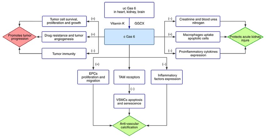
Table I. The 17 types of VKDPs in humans.

Designation                              Main distribution           Gla domain                                Function                                      Related pathological process

Coagulation factor II (prothrombin)   Liver                   10 Gla residues        Pro‑coagulant (24)                                          Thrombosis (24)
Coagulation factor VII (proconvertin) Liver                   10 Gla residues        Pro‑coagulant (24)                                          Thrombosis (24)
Coagulation factorIX (antihemophilic Liver                    10 Gla residues        Pro‑coagulant (24)                                          Thrombosis, ameliorating
factor B)				                                                                                                                                    hemophilia B (24,25)
Coagulation factorX                   Liver                   11 Gla residues        Pro‑coagulant (26)                                          Thrombosis (26)
Anticoagulant protein C               Liver                   9 Gla residues         Anticoagulant, anti‑inflammatory and anti‑apoptotic,        Preventing thrombosis and stroke, resisting severe
			                                                                                  cell protectant (27‑29)                                     sepsis (29‑31)
Anticoagulant protein S               Liver                   11 Gla residues        Anticoagulant, anti‑inflammatory, immunoregulation,         Preventing thrombosis (34), ameliorating
			                                                                                  regulator of apoptotic cell clearance, promoter of          diabetes (35), promoting tumor metastasis (33)
			                                                                                  vasculogenesis and angiogenesis (32,33)
Anticoagulant protein Z               Liver                   13 Gla residues        Anticoagulant                                               Preventing thrombosis, fetal loss and
				                                                                                                                                             antiphospholipid syndrome (36,37)
Proline‑rich Gla protein 1            Spinal cord             The Gla domain exposed Signal transduction (38)                                    Not clear
		extracellularly
Proline‑rich Gla protein 2            Thyroid                 The Gla domain exposed Signal transduction (38,39)                                 Not clear
		extracellularly
Transmembrane Gla protein 3           Heart, brain,           13 Gla residues        Protein turnover, cell‑cycle progression, and signal        Warfarin embryopathy (40)
                                      kidney		                                       transduction (40)
Transmembrane Gla protein 4           Kidney, pancreas,       9 Gla residues         Protein turnover, cell‑cycle progression, and signal        Warfarin embryopathy (40)
                                      placenta		                                     transduction (40)
OC                                    Bone                    3 Gla residues         Regulator of bone homeostasis, bone mineral density,        Preventing osteoporosis, osteoarthritis (44)
			                                                                                  systemic glucose and energy metabolism (41,42,43)
MGP                                   Lung, heart, kidney     5 Gla residues         Inhibitor of soft tissue mineralization (45‑47)             Osteophyma, cardiovascular disease (43)
Gas 6                                 Brain, heart, lung,     11 Gla residues        Anti‑vascular calcification, regulator of cell              Preventing vascular calcification, acute kidney
                                      kidney		                                       proliferation, migration, apoptosis and senescence,         injury, assisting tumor progression (49,50,53,54)
			                                                                                  and anti‑inflammatory (48‑52)
                                                                                                                                                                                                      INTERNATIONAL JOURNAL OF MOlecular medicine 47: 2, 2021

GRP                                   Bone, cartilage         16 Gla residues        Inhibitor of osteogenic differentiation, regulator of       A dual role in osteoarthritis, preventing vascular
			                                                                                  skeletal homeostasis, anti‑vascular calcification, and      calcification and triple‑negative breast
			                                                                                  anti‑inflammatory (55‑59)                                   cancer (56,59-62)
Periostin                             Periosteum, periodontal 4 Gla residues         Regulator of periosteum activation and cardiac fibrosis,    Fracture healing, cardiac fibrosis, idiopathic
                                      ligament		                                     promoter of cell proliferation, differentiation, adhesion   pulmonary fibrosis, asthma (63,68-71))
			                                                                                  and angiogenesis (63-68)
Periostin‑like‑factor                 Heart, bone, vascular   4 Gla residues         Promoter of osteoblast proliferation and                    Fracture healing, heart failure (64,72)
                                      smooth muscle cells		                          differentiation (64)
                                                                                                                                                                                                      3
4 XIAO et al: ROLE OF EMERGING VITAMIN K-DEPENDENT PROTEINS
uncarboxylated MGP, in which vitamin K is required by the of potential cardiovascular risks and can prognosticate
carboxylation process (43). cardiovascular events (52).
In recent years, a number of emerging VKDPs, such as Accumulating evidence has indicated that Gas 6 is
Gas6, GRP, and periostin, have been considered to participate significantly secreted by VSMCs in human atherosclerotic
in multifarious physiological and pathological processes. plaques, but there is no secretion in healthy blood vessels.
The anti‑inflammatory cytokine transforming growth factor β
4. Gas 6 (TGF‑β) induces the secretion of Gas 6 in VSMCs, and then,
stimulated by Gas 6, the VSMCs suppress the expression of
Brief introduction to Gas 6. Gas 6, weighing 75 kDa, is a rela‑ inflammatory factors, such as tumor necrosis factor (TNF) α
tively large member of the VKDP family. The concentration and intracellular adhesion molecule (ICAM)‑1 (85). Thus,
of plasma Gas 6 ranges from approximately 2.5 to 18.8 µg/l in Gas 6 acts as a protective factor in human atherosclerosis. Of
healthy adults (74). Gas 6 is highly homologous with Protein S note, Gas6 levels inversely related to complexity and stability
and carries an N‑terminal Gla domain after vitamin K in patients with carotid atherosclerotic plaques (85,86). In
carboxylation. Gas 6 is widely expressed in brain, heart, lung, particular, it should be noted that Gas6‑deficient mice show
kidney and other tissues, with the exception of the liver (43). more stable atherosclerotic lesions than normal mice, and
In 1995, Gas 6 was reported as the endogenous ligand for inhibition of Gas 6 is considered to be beneficial to plaque
the TAM family for the first time (75). TAM is the acronym stabilization (87). The contradictory feature between humans
for three receptors: Tyro3, Axl and Mer. Among these, Axl and mice is associated with obvious species physiological
has the highest affinity to Gas 6 (76). It has been reported differences (88).
that the laminin‑like globular domain of Gas 6 at C‑terminus Of note, overexpression of Gas 6 has a detrimental effect
appears to be the binding site of TAM receptors (75). However, on some pathological processes. The renin‑angiotensin‑aldo‑
after warfarin inhibits vitamin K‑dependent carboxylation, sterone system is closely connected with cardiovascular and
inactivated Gas6, not only completely inhibits the autophos‑ renal inflammation and fibrosis. It has been emphasized that
phorylation of the Axl receptor, but also fails to bind to the Gas 6 deficiency prevents the damage of aldosterone on target
Axl receptor in vitro (77,78). Therefore, vitamin K‑dependent organs (89). In addition, cardiomyocyte‑specific Gas 6 over‑
carboxylation is the key to the interaction between Gas 6 expression hastens the deterioration of pathological cardiac
and TAM receptor. In light of numerous previous studies, hypertrophy, mainly due to the activation of mitogen‑activated
the binding of Gas 6 and its receptors activated downstream protein kinase (MAPK) kinase 1/2‑ERK 1/2 signaling (90).
signaling, such as of phosphatidylinositol 3‑kinase (PI3K),
extracellular signal regulated kinase (ERK) and nuclear factor Gas 6 and the kidney. The contribution of Gas 6 to acute
kappa‑light‑chain‑enhancer of activated B cells (NF‑κ B) kidney injury is closely related to its biological functions,
pathways, to adjust the processes of apoptosis, survival, such as anti‑inflammation and immunoregulation (91,92).
proliferation, migration and adhesion (48,79‑82). The kidney, despite being a rich blood supplying organ, is
susceptible to hypoxic injury due to the complex balance of
Gas 6 and the cardiovascular system. There is an inseparable renal blood flow, glomerular filtration rate, oxygen consump‑
relationship between Gas 6 and the cardiovascular system. tion and arteriovenous oxygen shunting (93). Previous findings
The binding of Gas 6 and Axl limits the apoptosis of VSMCs suggested that Gas 6 protected against renal ischemia‑reper‑
by activating Akt and PI3K (80). It is worth mentioning that fusion injury in a mouse model (94). To be specific, with
vitamin K 2 can inhibit VSMC calcification and apoptosis the assistance of Gas 6 treatment, creatinine and blood urea
by restoring Gas6 expression and activating downstream nitrogen decreased by 29 and 27%, respectively. Cell apoptosis
signaling by Axl, Akt and Bcl2 (49,50). Endothelial progenitor was significantly decreased, attributable to Gas 6 enhancing
cells (EPCs) are involved in the saving response to ischemic macrophages to uptake apoptotic cells (95). Furthermore, the
tissue through forming new blood vessels or proliferation of expression of pro‑inflammatory cytokines, such as interleukin
pre‑existing vasculature. Autologous EPC transplantation (IL)‑1β and TNF‑α, was markedly reduced by another Gas 6
therapy has been indicated as safe and practical in chronic function, dampening the inflammatory responses (11,91,94).
myocardial ischemia (51). It has been identified that Gas6 Similarly, concentration of Gas 6 rose in sepsis‑induced
has the ability to stimulate EPC proliferation and migration acute kidney injury mice, and improved the survival rate by
in vitro by activating the Akt signaling pathway (48). The reducing serum urea nitrogen, creatinine and renal tissue
finding provides a basis for the further therapy of vascular apoptosis (53). In addition, several reports demonstrated that
re‑endothelialization. Vascular aging, a risk factor of CVDs, Gas6 levels were significantly increased in CKD patients and
is characterized by vascular stiffness, vascular remodeling chronic hemodialysis patients (96,97). Opinions regarding
and endothelial dysfunction (83). Aging vessels provide a the potential mechanisms vary. Researchers tend to associate
good environment for CVDs. Gas6/Axl can delay cell cycle the elevation with endothelial function (Gas6 is expressed by
arrest, which is a key cause in the development of VSMC endothelial cells) and inflammation because pro‑inflammatory
senescence and promotes their transition from the G1 to the cytokines are abundant in the blood of these patients (96,98).
S phase. The PI3K/Akt/Forkhead box O (FoxO) signaling It is reported that endothelial cells in CKD are subjected to
pathway is considered the major target of Gas6/Axl signaling specific stress overtime which leads to accelerated cardio‑
in VSMC senescence, with FoxO being the key factor (84). vascular disease and high mortality (99). Disruption and
Furthermore, clinical investigation has demonstrated that inflammation of glomerular capillaries influence the evolution
Gas 6 plasma levels at admission reflect the existence of CKD, and, consequently, elevated Gas 6 levels (100). ItINTERNATIONAL JOURNAL OF MOlecular medicine 47: 2, 2021 5
is worth noting that Gas 6 is upregulated in many forms of 5. GRP
inflammatory nephropathy, for example, lupus nephritis and
IgA nephropathy (101,102). Brief introduction to GRP. As its name suggests, GRP, which
Diabetic nephropathy is a common complication of diabetes was first identified in sturgeon cartilage, has abundant Gla
that can further develop into end‑stage renal disease. There residues (15 Gla residues in human) (114,115). With unusually
are opposing conclusions on the tendency of plasma Gas 6 in high capacity to bind calcium through Gla resdues, GRP accu‑
diabetes and diabetic nephropathy. Nagai et al first reported that mulates in bone, cartilage and ectopic calcification, such as
the expression of both Gas6 and Axl was distinctly increased blood vessels and skin (112). During physiological conditions,
in diabetic rats and proved Gas 6 can induce mesangial cell GRP participates in the stabilization of cartilage matrix, chon‑
hypertrophy, which further leads to glomerular hypertrophy drogenesis and inhibition of osteogenesis (116‑118). Recently,
in the early stage of diabetic nephropathy (103). Furthermore, GRP has attracted attention due to its crucial performance in
a reliable mechanism was proposed in which high glucose combating ectopic calcification.
stimulates mesangial cells, followed by activating Gas6/Axl
and the Akt/mTOR pathway, which results in mesangial and GRP and bones. The growth of long bones is inseparable from
glomerular hypertrophy (104). By contrast, Hung et al indi‑ the process of endochondral ossification. First, chondrocytes
cated that plasma Gas6 levels in impaired glucose tolerance participate through a combination of proliferation, extracel‑
patients and type 2 diabetes were significantly decreased (105). lular matrix secretion and hypertrophy. Then, hydroxyapatite
A study based on individuals with different degrees of albu‑ crystals are deposited in the extracellular matrix surrounding
minuria offers some insight into this controversy, and showed late hypertrophic chondrocytes, known as mineralization.
the blood level of Gas 6 decreased with the deterioration of Next, chondrocyte death, matrix degradation and contents
proteinuria (106). Silaghi et al formulated a hypothesis that invasion occur. Finally, the growth plates close and the
the interaction between molecular charge and weight may bones mature (119). Surmann‑Schmitt et al reported GRP in
participate in glomerular filtration of Gas 6 (100). More the upper zone of the growth plate, termed unique cartilage
specifically, Gas 6 and albumin (approximately 66 kDa) have matrix‑associated protein, which exhibits a negative correlation
a similar molecular weight and a net negative charge repelled with osteogenic differentiation (116). Both GRP knockdown
the glomerular membrane. Complex interactions eventually zebrafish and warfarin‑exposed zebrafish show irreversible
lead Gas 6 to filter through the glomerular membrane and be growth retardation and altered skeletal development; therefore
excreted from the body (100). Therefore, the concentration of Gla residues are necessary for the function of GRP (117). It
plasma Gas 6 changes in different stages of diabetes. is worth mentioning that a similar feature is found in human
warfarin embryopathy, which results in pregnant women from
Gas 6 and cancer. The contribution of Gas6 to cancer has warfarin therapy (120,121). Surprisingly, GRP is not essential
been reported for a large number of cancer types. For example, for mouse skeletal development (55). However, the fact that
Gas 6 is upregulated in breast cancer, melanoma and ovarian GRP is still expressed in adult mouse cartilage indicates
cancer (107‑109). Tumor cells lack the competence to produce that GRP may contribute to skeletal homeostasis and other
Gas 6, but can educate infiltrating macrophages to promote calcification‑associated pathological processes after infancy.
the production of Gas6 by producing IL‑10 and macro‑ Osteoarthritis (OA), a painful joint disease, is character‑
phage colony‑stimulating factor (M‑CSF) (110). Previous ized by articular cartilage degradation, bone remodeling,
findings have shown the pro‑tumor effects of Gas6/TAM tissue inflammation and abnormal extracellular matrix miner‑
signaling. In the case of Gas 6 overexpression, the survival alization. In fact, GRP plays a dual role in OA. GRP prevents
of myeloma cells was significantly increased in vitro and, articular cartilage degradation in two practical ways. On the
conversely, the deficiency of Gas 6 led to rapid cell death of one hand, GRP blocks the aggrecanase activity of A disin‑
myeloma (111). In addition, the autocrine Gas 6 assists the tegrin and metalloproteinase with thrombospondin motifs
resistance of myeloma cells to bortezomib (111). Recently, (ADAMTS)‑4 and ADAMTS‑5 by physical interaction (56,57).
the pro‑tumor effects of Gas 6 was also reported in lung Aggrecanolysis is considered the main process of cartilage
cancer cells (54). In addition, blocking Gas 6/Mer signaling degradation, thus GRP protects cartilage by increasing its
with Mer receptor inhibitors significantly limits the prolif‑ resistance to aggrecan cleavage in OA. By contrast, enhanced
eration and growth of lung cancer cells (54). Interestingly, chondrocyte apoptosis accelerates the cartilage damage in OA.
a high level expression of Axl and its ligand Gas 6 were It is reported that chondrocyte cell death is markedly increased
recognized in non‑small cell lung cancer patients, who in GRP‑deficient mice; thus, GRP protects articular cartilage by
acquired resistance with epidermal growth factor receptor reducing chondrocyte apoptosis (56). However, GRP has also
tyrosine kinase inhibitors (112). It has been reported that been implicated in bone remodeling, which is mediated with
Gas 6 negatively regulates the proliferation and interferon‑γ the altered function and metabolism of osteoblasts and osteo‑
production of natural killer cells to inhibit tumor immunity clasts in OA (122). Previous findings have shown osteoblasts
through binding with Casitas B cell lymphoma‑b/TAM contribute to phenotypic changes and osteoclasts are associ‑
receptors (113). In addition, Gas 6 prolongs VSMC survival ated with cartilage destruction in OA (56,123). Additionally,
in the tumor microenvironment, which is requisite to tumor GRP, as a downstream gene of runt‑related transcription
angiogenesis (79). Several investigations have indicated the factor 2 and Osterix, stimulates osteoblast differentiation in
roles of Gas 6 in predicting the prognostic risk of cancer. OA (60). Similar results have been found in mice, in which
Gas 6 protein as an independent predictor always indicates a osteoblasts and osteoclasts decreased during experimental
poor prognosis (107,109) (Fig. 1). OA in GRP‑deficient mice, while there was no fluctuation in6 XIAO et al: ROLE OF EMERGING VITAMIN K-DEPENDENT PROTEINS
Figure 1. Functional mechanisms of Gas6. The ‘+’ refers to promotion and ‘‑’ refers to inhibition. Green represents Gas 6 physiological effects and red repre‑
sents its pathological effects. Gas 6 is widely expressed in heart, kidney, brain and other tissues. Abundant vitamin K ensures sufficient carboxylated Gas 6 in
the body. Gas 6 resists vascular calcification through three mechanisms: i) Gas 6 promotes proliferation and migration of endothelial progenitor cells (EPCs);
ii) Gas 6 inhibits apoptosis and senescence of vascular smooth muscle cells (VSMCs) by binding Tyro3, Axl and Mer (TAM) receptors; iii) Gas 6 decreases
expression of inflammatory factors, including TNF‑α and ICAM‑1. Similarly, Gas 6 protects from acute kidney injury: i) Gas 6 significantly reduces creatinine
and blood urea nitrogen; ii) Gas 6 enhances macrophages to uptake apoptotic cells; iii) Gas 6 reduces the expression of pro‑inflammatory cytokines, such
as IL‑1β. However, Gas 6 assists tumor progression: i) Gas 6 is necessary for survival, proliferation and growth of tumor cells; ii) Gas 6 contributes to drug
resistance and tumor angiogenesis; iii) Gas 6 negatively regulates tumor immunity.
normal mice (56). The obvious conflict regarding the effect regulatory proteins, such as BMP‑2, BMP‑4, osteopontin,
of GRP on osteoblastic differentiation may be explained by MGP and OC, are expressed in calcifying vessels (130). In
post‑translational modification of GRP (56,60,116). Moreover, response to the high level of extracellular calcium and the lack
certain data have indicated GRP promotes osteophyte forma‑ of calcification inhibitors, VSMCs release extracellular vesicles
tion in OA and the effect occurs via bone remodeling rather (EVs) into circulation with good mineralization capability and
than cartilage maturation (57,60). In addition, inflammation form a nucleation site for hydroxyapatite (131,132). Fetuin‑A,
presents before the joint structure changes in OA joints (124). a 48 kDa protein, is synthesized in the liver and secreted into
It has been demonstrated that GRP has a similar inhibitory circulation as a powerful calcification inhibitor. Interestingly,
effect on calcification and inflammation processes (58). fetuin‑A is too large to enter the collagen fibril where the
Furthermore, synovial fluid GRP levels in OA patients exhibit mineral grows (133). It is reported that the mineral grows only
a positive correlation with radiographic findings and symp‑ inside the fibril when fetuin‑A exists, whereas it grows beyond
tomatic severity of OA (125). However, it is noteworthy that the fibril without fetuin‑A (134). Therefore, fetuin‑A is the key
studies have shown that vitamin K deficiency is a potential risk factor in determining the location of mineral growth. Moreover,
factor for knee OA (126). Due to the lack of effective treatment inflammatory activity takes part in early calcification. Many
and prevention methods currently, fully carboxylated GRP by studies have indicated a synergistic interaction between
vitamin K supplementation is a convenient and inexpensive macrophage and VSMC calcification. Activated macrophages
candidate for the treatment of OA. produce a large number of proteases to enhance the degrada‑
tion of elastin and collagen (124,135). Macrophages markedly
GRP and vascular calcification. Vascular calcification is a increased BMP‑2 expression in VSMCs and also released
pathological process characterized by the deposition of calcium EVs with calcification capacity (136,137). In addition, many
phosphate crystals in vessel walls (127,128). According to other factors influence the process of vascular calcification,
the location of calcification, it can be classified into intimal for instance, VSMC apoptosis, oxidative stress and endothelial
calcification (related to plaque burden and luminal narrowing) dysfunction (138,139).
and medial calcification (associated with vessel stiffness and GRP, a VKDP, has been identified as a powerful inhibitor
vascular compliance decline) (128). VSMCs are a contractile of vascular calcification. GRP, MGP and fetuin‑A form a large
phenotype in the physiological state that can regulate vascular complex that is loaded in noncalcifying EVs but distinctly
tension. However, they lose expression of contractility‑related lowered in high calcium‑loaded vesicles, thus recommending
genes when vascular injury exists and are further transformed GRP as an important mineralization inhibitor (59,61).
into osteoblast‑like cells (129). In addition, bone matrix Furthermore, calciprotein particle (CPP), a fetuin‑mineralINTERNATIONAL JOURNAL OF MOlecular medicine 47: 2, 2021 7
complex, principally contains mineral, fetuin‑A, MGP and breast cancer. However, recent research may be useful in
GRP, and contributes greatly to the stabilization of minerals. resolving this issue. GRP inhibits the growth, migration and
Research has demonstrated that CKD patients possess CPPs invasion of triple‑negative breast cancer tissues in vitro and
with a lower content of fetuin‑A and GRP compared with in vivo (62). Moreover, according to survival analysis in the
healthy individuals (138). Fetuin‑A is predominant in healthy open database, the relapse‑free survival rate of patients with
CPPs and retards the deterioration toward calcifying CPPs triple‑negative breast cancer was significantly correlated with
through collaboration with GRP (140,141). Moreover, GRP high GRP expression (62).
shows the ability to counteract inflammation and is found in
macrophage‑derived EVs (142). In vitro studies found calcifi‑ 6. Periostin
cation in both GRP‑deficient and normal VSMCs in response
to osteogenic medium after 6 days, yet GRP‑deficient VSMCs Brief introduction to periostin. Periostin, initially known as
calcified about twice as much as normal VSMCs 9 days osteoblast‑specific factor 2, was first cloned from a cDNA
later (143). Of note, there is an apparent increase in the expres‑ library of the mouse osteoblastic cell line MC3T3‑E1 in
sion of BMP‑2 and its downstream marker (small mother Japan (152). Over a decade later, the Gla‑containing protein,
against decapentaplegic, SMAD) and, finally, after comparing periostin, was determined to require vitamin K‑dependent
GRP with two different carboxylation states, the direct carboxylation and became the 13th member of the VKDP
interaction between the carboxylated GRP and BMP‑2 was family (153). Characterized by fasciclin domains, periostin
confirmed (143). Therefore, GRP disturbs the BMP‑2‑SMAD is particularly expressed in connective tissues submitted to
signaling in calcifying VSMCs, playing a central role in VSMC constant mechanical stresses (153). For example, periosteum,
calcification (Fig. 2). the periodontal ligament, heart valves and skin. Periostin
has also been implicated in fibrosis, inflammation, tumor
GRP and cancer. Microcalcification, a small deposit of calcium metastasis and the fracture healing process (67,154-156).
with a diameter less than 1 mm in mammographic images,
is vital for the diagnosis and prognosis of breast cancer (61). Periostin and bone. Fractures are one of the most common
Ductal carcinoma in situ can be as high as 20‑25% in women traumatic injuries to humans. Most fractures can be repaired
with asymptomatic breast cancer (144). Furthermore, 70% of to their pre‑injury state through a process similar to embry‑
ductal carcinoma in situ can be diagnosed only by microcal‑ onic skeletal development. According to the characteristics
cification in mammography (145). Recent findings have shown of fracture healing, the process is divided into four partially
that linear branching microcalcifications in mammography overlapping phases: The inflammation phase, the soft callus
indicate the aggressive of tumor tissue (146). A differential phase, the hard callus phase and the remodeling phase (157).
accumulation pattern of carboxylated GRP (cGRP) and under‑ The inflammation phase is marked by acute inflammation,
carboxylated GRP (ucGRP) by vitamin K has been recently hematoma formation and skeletal stem cell recruitment.
emphasized in human breast cancer (147). In healthy mammary During the soft callus phase, cartilaginous callus and nascent
gland tissues, cGRP was predominant, while ucGRP was found blood vessels form. During the hard callus phase, the most
to be either co‑localized or undetectable. By contrast, ucGRP active phase of osteogenesis, the cartilage is reabsorbed and
was widely detected in tumor cytoplasm, while cGRP was only bone is deposited by osteoblasts (158). Angiogenesis also
intermittently found in certain tumor cells. There are many continues during this phase. During the last phase, primary
explanations for the large quantity of ucGRP in tumors. It has bone is eventually replaced by lamellar bone, which supports
been observed that the decreased level of vitamin K in tumor normal skeletal functions, and vascular remodeling is finally
areas is in contrast to non‑tumorous areas (148). Patients with completed (158,159). There is a vital association between
tumor complications, such as venous thromboembolism, have periosteum and fracture repair. In a mouse model in which
received long‑term therapy with vitamin K antagonists and the graft femoral bone was segmentally transplanted, the peri‑
potential detrimental effects to GRP should be noted (149). In osteum showed positive osteogenic and angiogenic activity,
addition, prolonged subclinical vitamin K deficiency has been leading to superior healing and repair of live isografts (160).
identified in cancer patients. Furthermore, vitamin K preferen‑ However, absence of the periosteum led to poor cartilaginous
tially supports the coagulation factor synthetic process in the callus formation, and even fracture non‑union (160,161). The
liver, and only after the vitamin K supply has met the liver's periosteum is anatomically comprised of an outer fibrous
need is the excess vitamin K transported to extra‑hepatic layer and an inner cambium layer. The fibrous layer contains
tissues (177,150). Thus, ucGRP is widespread in tumor tissues. fibroblasts, collagen, and elastin fibers, along with a nerve and
Furthermore, the formation mechanism of microcalcification microvascular network (162). The cambium layer is directly
in breast tumor tissue is similar to physiological bone miner‑ closed to the bone surface and contains high‑quality mesen‑
alization and pathological vascular mineralization (151). Both chymal progenitor cells, osteoblasts, fibroblasts, microvessels
cGRP and ucGRP showed an advanced affinity to calcium and sympathetic nerves (162,163). In human bones, periostin
mineral deposits in breast cancer tissue. Thus, with the capa‑ is highly expressed in the cambium layer, where it is highly
bility of resisting ectopic calcification, GRP is considered a active during bone remodeling (164). In a mouse model of
novel effective antagonist against cancer. It is worth noting that fracture, rapid periostin gene expression occurred during the
triple‑negative breast cancer is a subtype with low expression of inchoate phase of fracture healing (155). In the first 1‑2 weeks
estrogen receptor, progesterone receptor and human epidermal after fracture, human serum periostin is decreased initially,
growth factor receptor 2 receptor (62). Therefore, there is a prior to a progressive elevation that peaks at 8 weeks, and is
lack of effective targeted therapy drugs for triple‑negative present for about 26 weeks (165).8 XIAO et al: ROLE OF EMERGING VITAMIN K-DEPENDENT PROTEINS
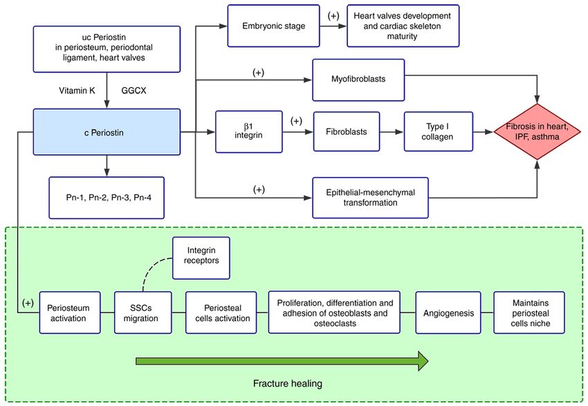
Figure 2. Functional mechanisms of GRP. The ‘+’ refers to promotion and ‘‑’ refers to inhibition. Green represents GRP physiological effects and red represents
its pathological effects. GRP is widely expressed in bone, cartilage, blood vessels and other tissues. GRP develops its role after γ‑carboxylation, which is
regulated by the GGCX enzyme and vitamin K. Gla residues are necessary for GRP to perform its physiological functions: Reducing osteogenic differentiation
and maintaining skeletal homeostasis. GRP plays a dual role in OA. On the one hand, GRP prevents articular cartilage degradation by blocking aggrecanase
activity (ADAMTS‑4 and ‑5) and inhibiting chondrocyte apoptosis and inflammation. By contrast, GRP contributes to bone remodeling in OA via promotion
of osteoblastic differentiation and osteophyte formation. Additionally, GRP also resists vascular calcification: i) GRP, matrix Gla protein (MGP) and fetuin‑A
complex combines the with mineral to form calciprotein particles (CPPs), which contribute greatly to the stabilization of minerals; ii) carboxylated GRP
disturbs inflammation and BMP‑2‑SMAD signaling in calcifying VSMCs. Abundant ucGRP assembles in tumor cells, while cGRP is rare. Moreover, GRP
inhibits the growth, migration and invasion of triple‑negative breast cancer.
Periostin participates in almost all phases of fracture contrast, when periostin‑deficient grafts were transplanted
healing. In the early inflammation phase, as a result of the into wild‑type hosts, the contribution of periosteal cells to
inflammatory response or paracrine effects of the periosteum, repairing of the second fracture injury disappeared, leading
periostin is present at a low level in serum (165). Transplantation to defective callus formation and fibrosis, and furthermore this
of the periosteum of periostin‑deficient mice to the fracture site was not due to deficient proliferation (63). Therefore, periostin
of wild‑type mice induced negative fracture repair, indicating plays a crucial role in maintaining periosteal cell niche and
that periostin regulates periosteum activation (66). Skeletal supporting bone remodeling.
stem cells (SSCs), with local osteogenic potential, are recruited Long‑term anticoagulant therapy with vitamin K antago‑
in the early stage of bone regeneration and periosteum and is nists, such as warfarin, reduces bone density and increases the
considered one of its major sources (166). As an extracellular risk of osteoporosis (169,170). Previous findings have shown
matrix protein, periostin promotes the migration of SSCs that warfarin significantly inhibits osteoblastic differentia‑
by binding integrin receptors on the cell surface (162,167). tion (171). Warfarin interferes in the carboxylation of periostin
Notably, periosteal cells, another form of convened cells by antagonizing the function of vitamin K, and the decrease
that shares a common embryonic origin with SSCs, have of carboxylated periostin is one of the main causes of bone
been revealed to have greater regenerative potential than density reduction (172). By contrast, vitamin K 2 promotes
SSCs (63). Moreover, periosteal cell functions are impaired in mineralization of osteoblasts (173). In recent years, periostin
mice lacking periostin, suggesting that periostin contributes has been recommended as a potential predictive marker
to periosteal cell activation (63). During the callus phases, of bone events. Osteoporotic fracture is a major cause of
induced by BMP‑2, periostin is upregulated in soft callus and disability in the elderly, while the ability of current predicting
osteoblasts (168). Accumulating evidence has indicated that methods is limited. In a cohort of 607 postmenopausal women
abundant periostin facilitates the proliferation, differentiation from France that were followed up for 7 years, a positive
and adhesion of osteoblasts in bone formation (64,65). In correlation between serum periostin and fracture risk was
addition, periostin may interfere with osteoclasts in a similar observed (174). Furthermore, the association was indepen‑
way (65). It is reported that periostin markedly increases arte‑ dent of bone mineral density and prior fractures, indicating
rioles in a calvarial defects model, proving periostin promotes that periostin is an independent predictive marker of fracture
angiogenesis (66). Periostin has a crucial mission in the last risk. This hypothesis was confirmed in another case control
phase of fracture healing, that is, to recover the periosteum study of Korean postmenopausal women (175). Interestingly,
niche of periosteal cells. In a periosteum transplantation high plasma periostin levels prefer non‑vertebral fractures to
model, periosteal cells may still be re‑activated to contribute vertebral fractures, such as limb fractures (175). These clinical
to cartilage within the callus after three injury cycles (63). By outcomes seem contrary to the popular view of periostin. TheINTERNATIONAL JOURNAL OF MOlecular medicine 47: 2, 2021 9
specific mechanisms for these conclusions need further study kinase 3β/cyclin D1 signaling pathway (180). Therefore, peri‑
as they may be related to the carboxylation state of periostin ostin is pivotal for myocardial regeneration at the early stage
or to the distribution of periostin in the body. Specifically, of myocardial infarction, and is involved in fibrillogenesis and
periostin in bone are induced to circulation. Notably, it has scar generation in the later chronic stage.
been demonstrated that lower serum periostin concentrations Previous findings have shown that periostin is abundantly
were related to prevalence of knee OA in women (176). This expressed in patients with atherosclerosis (181‑183). In the
provides a new idea for the application of periostin in bone ‘Pathobiological Determinants of Atherosclerosis in Youth’
event prediction. study, the variant encoding periostin gene was connected with
atherosclerotic lesion traits (181). Matrix metalloproteinases,
Periostin and heart. During embryogenesis, periostin enzymes implicated in atherosclerosis and vascular remod‑
supports normal valve leaflet morphogenesis and cardiac eling, which were induced by periostin, led to valve thickening
skeleton maturity (177). Periostin is implicated in CVDs, in mice fed high‑fat diets (182). Additionally, periostin stimu‑
such as myocardial infarction, atherosclerosis and cardiac lates angiogenesis both in vitro and ex vivo (179). In response
fibrosis‑related diseases (67). Cardiac fibrosis is a prominent to injury, periostin was markedly upregulated in neointimal
feature of cardiac remodeling that can further lead to heart SMCs and adventitial myofibroblasts, and promoted cell
failure and impaired cardiac function. Fibroblasts, the most migration (183). By contrast, the plaques of periostin‑deleted
abundant cell population in the heart except cardiomyocytes, mice, not only had a smaller necrotic core and fibrous cap, but
rapidly differentiate into myofibroblasts in the cardiac fibrosis also possessed more cholesterol clefts (184). The deficiency
process (67). Abundant differentiated myofibroblasts found in of periostin also reduced the infiltration of macrophages into
hearts suffering failure also support the transformation (179). the plaque (184). Thus, periostin plays a considerable role in
Emerging evidence suggests that the myofibroblast phenotype atherosclerosis, and targeted periostin treatment may delay
still has latent reversibility in end‑stage heart failure (67,178). progression of diseases associated with atherosclerosis (Fig. 3).
Of note, periostin, as the most specific product, is expressed
in essentially all myofibroblasts (67,68). Certain data have Periostin and the respiratory system. In the last decade, the
indicated targeted ablated periostin‑expressing myofibroblasts role of periostin in airway development and diseases has been
led to a diminished fibrotic area and improved the ejection widely emphasized. For instance, periostin was reduced in
fraction in hearts in AngII‑induced fibrosis mice (68). In addi‑ tracheal aspirate fluid of bronchopulmonary dysplasia during
tion, not only was cardiac fibrosis reduced, but treatment also the window period (185). Then, TGF‑β upregulated the expres‑
did not affect scar stability in myocardial infarction mice (68). sion of periostin in the interstitial fibrosis region (185,186).
Moreover, periostin antibody treatment visibly restricted cell Thus, periostin is recognized as a potential biomarker that
viability of myofibroblasts in vitro (69). Therefore, periostin is predicts the risk of bronchopulmonary dysplasia and the need
a novel central factor contributing to the function of myofibro‑ for preventative therapies in preterm infants. Periostin has
blasts during cardiac fibrosis. Research has demonstrated that been involved in many respiratory disorders, such as idiopathic
ginsenoside Rb1, the bioactive component of ginseng, reduced pulmonary fibrosis (IPF), asthma, chronic rhinosinusitis,
the expression levels of periostin and protected rats against idiopathic eosinophilic pneumonia and allergic bronchopul‑
myocardial fibrosis (179). monary aspergillosis (156,187‑190). The most notable of these
According to whether exons 17 and 21 exist or not, periostin are IPF and asthma.
can be divided into four isoforms, i.e., Pn‑1 to Pn‑4. In detail, IPF, a common pulmonary fibrotic conditions, is a chronic
Pn‑1 is a full‑length form, Pn‑2 is short of exon 17, Pn‑3 is short progressive parenchymal lung disease of unclear cause that
of exon 21, and Pn‑4 is short of exons 17 and 21 (69). Using an is limited to the lungs (191,192). Patients are predominantly
antibody that specifically inhibits exon 17, the dispute regarding older individuals and typically have progressive worsening
the functions of different periostin isoforms has been settled. lung function, leading to a grave prognosis (193). It has
It is not surprising that the expression of periostin increased been indicated that periostin was elevated in IPF patients'
in the border zone on day 5 after myocardial infarction (69). circulation (190). Furthermore, more periostin was found in
However, total infarction and fibrosis size were notably the lungs of IPF patients and concentrated in areas of active
reduced in an adult mouse model by selectively neutralizing an fibrosis (187). Interestingly, the exon 21 of the periostin gene
antibody against exon 17 (69). In addition, cardiac dysfunctions is more likely to be spliced out in IPF lung samples than in
were improved. Moreover, Pn‑2 contributes to angiogenesis in the control (194). Injury factors activate alveolar epithelial
in vitro experiments, while Pn‑1 does not (69). Low expression cells disrupting the homeostatic balance between epithelial
of TGF‑β, a fibrosis‑related gene, was associated with the inhi‑ and mesenchymal cells, thus fibrotic response is driven. As
bition of fibrosis. Thus, Pn‑1 contributes to fibrosis and heart an extracellular matrix protein, periostin and TGF‑β regulate
remodeling after myocardial infarction, and there is potential each other in fibroblasts (195); specifically, TGF‑β increases
to improve the prognosis of myocardial infarction via selec‑ the expression of periostin. In return, periostin significantly
tively inhibited Pn‑1 treatment. Nevertheless, neonatal mice upregulates the production of TGF‑ β in fibroblasts and
were capable of regenerating myocardium after myocardial increases type I collagen production (70,195). However,
infarction. On day 21 after myocardial infarction, the infarcted periostin activates fibroblasts to produce type I collagen via
areas of neonatal mice almost disappeared (180). However, β1 integrin, rather than the TGF‑β signal (195). Similar to
myocardial regeneration was inhibited in periostin‑deficient heart fibrosis, periostin promotes differentiation of fibroblasts
neonatal mice, presenting with a larger infarcted area, which to myofibroblasts. By mediating epithelial‑mesenchymal
was attributed to the inhibition of PI3K/glycogen synthase transformation, periostin induces alveolar epithelial cells10 XIAO et al: ROLE OF EMERGING VITAMIN K-DEPENDENT PROTEINS Figure 3. Functional mechanisms of periostin. The ‘+; refers to promotion. Green represents periostin physiological effects and red represents pathological effects. Periostin is particularly expressed in connective tissues, such as the periodontal ligament, periosteum and heart valves. Vitamin K and GGCX are two vital enzymes in the carboxylation of periostin. According to whether exons 17 and 21 exist or not, periostin can be divided into four isoforms: Pn‑1, Pn‑2, Pn‑3 and Pn‑4. Periostin is involved in all phases of fracture healing. Periostin promotes periosteum activation in the early stage. Subsequently, periostin facilitates the migration of SSCs via binding integrin receptors. Periostin contributes to the activation of periosteal cells, revealing greater regenerative potential than SSCs. Periostin facilitates the proliferation, differentiation and adhesion of osteoblasts and osteoclasts in bone formation. Periostin accelerates angiogenesis and maintains periosteal cell niche in the later period of fracture healing. At the embryonic stage of the heart, periostin supports heart valve development and cardiac skeleton maturity. However, periostin participates in progression of cardiac fibrosis, idiopathic pulmonary fibrosis (IPF) and asthma airway remod‑ eling. Expressed in essentially all myofibroblasts, periostin is a central factor contributing to the function of myofibroblasts. Periostin activates fibroblasts to produce type I collagen via β1 integrin in IPF. Moreover, periostin induces epithelial‑mesenchymal transformation, which leads to alveolar epithelial cells taking on the characteristics of mesenchymal cells and accelerates the aggravation of fibrosis. to take on the characteristics of mesenchymal cells, which Asthma, as a heterogeneous disease, has been defined as leads to the aggravation of fibrosis (70). Emerging evidence several phenotypes according to different clinical features and suggests that periostin silencing drives the fibroblasts into physiological indexes. Nevertheless, type‑2 airway inflamma‑ G1 arrest of the cell cycle and retards the proliferation in tion is one of the main causes of asthma, which is supported by IPF (196). Thus, periostin plays a pivotal role in lung fibro‑ activity of type 2 cytokines, such as IL‑4 and IL‑13. As a result blast proliferation. Currently, early lung transplantation is a of chronic airflow limitation, airway remodeling develops in beneficial therapeutic option for IPF patients, and another two chronic severe asthma. Many studies have shown periostin is available drugs (Pirfenidone and Nintedanib) are able to limit deeply involved in the process of asthma, from airway inflam‑ IPF progress (197). Recently, a compound known as CP4715 mation to remodeling. The periostin gene is highly induced was found to prohibit the interaction between TGF‑ β and in asthmatic airway epithelial cells with a 4.4‑fold increase periostin (197). CP4715, not only lessened bleomycin‑induced compared to healthy controls (199). In a cohort of asthmatics pulmonary fibrosis, but also disturbed TGF‑β signals in fibro‑ from Sweden, a negative correlation between serum periostin blasts from IPF (195). Therefore, CP4715 may become a latent and lung function was observed (71). Type‑2 inflammation drug therapy to provide more therapeutic possibilities for IPF. attracts large numbers of immune cells to release cytokines, It is worth mentioning that vitamin K antagonists are related such as IL‑4, IL‑13 and TGF‑β. These cytokines stimulate to the rising mortality of IPF (198). The carboxylation status the production of periostin from fibroblasts, epithelial cells of periostin in IPF patients deserves further study. Some and endothelial cells, which are known as the main sources scholars have proposed that the use of vitamin K instead of of periostin in asthma (200), and some researchers have vitamin K antagonists may help reduce the progression of hypothesized that eosinophils also secrete periostin (201). IPF, but this idea needs further verification (198). As an integrin ligand, periostin binds to integrin αMβ2 and
INTERNATIONAL JOURNAL OF MOlecular medicine 47: 2, 2021                                         11

α4β1 on eosinophil, guiding recruitment of eosinophils and          inflammation‑stimulated cells and can inhibit the expression
increasing eosinophil adhesion to fibronectin (156,202). In         of inflammatory cytokines (e.g., TNF‑α, IL‑6 and IL‑8) (215).
addition, through its fibrogenic function, periostin participates   Finally, vitamin K 2 is involved in immune regulation.
in the process of subepithelial fibrosis, which is feature of       Specifically, T‑cell proliferation was inhibited with vitamin K2
airway remodeling in asthma (185). Periostin, secreted from         instead of vitamin K1 (216).
airway epithelial cells, activates TGF‑β and upregulates type I         In this review, we highlighted three emerging VKDPs
collagen via autocrine effects (70,203). Similarly, periostin       (Gas 6, GRP and periostin) that need vitamin K to conduct
activates TGF‑β‑mediated fibroblasts to increase the produc‑        carboxylation and then perform various biological func‑
tion of type I collagen (70,203). Clinical studies from Japan       tions in the human body, such as bone homeostasis, heart
have reported that vitamin K2 therapy has an effective rate         development and anti‑vascular calcification. In combina‑
of up to 90.9% in patients with mild asthma (204). The effec‑       tion with previous studies, we believe that a high intake
tive rate was 86.7 and 72.7% in moderate and severe patients,       of vitamin K, especially vitamin K 2, is beneficial for the
respectively (204). In addition, vitamin K2 has a powerful          cardiovascular system and bones. However, some ques‑
ability to inhibit the release of inflammatory cytokines (205).     tions about the relationship between vitamin K and cancer
It has also been shown that vitamin D, also a fat‑soluble           remain unsolved. Many studies have shown vitamin K 2 has
vitamin, can regulate inflammatory chemokines in asthma             anticancer effects. Ishizuka et al reported that vitamin K 2
and significantly inhibit airway smooth muscle cell prolif‑         has a moderately suppressive effect on hepatocellular
eration (205). Therefore, whether vitamin K2 can regulate the       carcinoma recurrence (217). Zhong et al indicated that
release of inflammatory factors in asthma and thus inhibit the      vitamin K2 reduces the hepatocellular carcinoma recurrence
production of large amounts of periostin remains to be further      rate after 1 year (218). Similarly, vitamin K 2 exerts anti‑
studied. Additionally, periostin increases gel elasticity formed    cancer effects in cancer cell lines, such as cholangiocellular
by type 1 collagen, thus mediating the biomechanical capa‑          carcinoma, ovarian cancer and pancreatic cancer (219‑221).
bilities of the airway and leading to airway remodeling (206).      Accumulating evidence has indicated that vitamin K 2 not
Accumulated evidence has indicated that high serum periostin        only inhibits the proliferation and differentiation of tumor
concentrations were implicated in certain characteristics of        cells, but also induces the apoptosis and autophagy of tumor
asthma. It is reported that serum periostin concentrations          cells (222). In addition, however, some VKDPs represented
were not combined with atopic status or treatment status            by Gas6 have been indicated to facilitate the survival and
of asthma, while high level serum periostin was related to          metastasis of cancer cells. Moreover, as mentioned above,
older patients at the onset of asthma, aspirin intolerance or       GRP carboxylation status in breast cancer tissues is signifi‑
nasal disorders (207‑209). As serum biomarkers are more             cantly different from those in normal tissues, but there are
convenient than lung function tests in some special cases of        few studies measuring this in other diseases. Thus, the
asthma, periostin has become one of the practical biomarkers        relationship between measurement of VKDP carboxylation
of asthma. For instance, periostin rises significantly in severe    status and disease progression remains to be further inves‑
asthma and acute asthma exacerbation of children, which is          tigated. Furthermore, periostin is a newly identified VKDP
an important serum biomarker in assessing the severity of           that has been extensively studied in the heart and respiratory
asthma (206). Of note, periostin is a helpful biomarker to          system. However, the role of periostin as a VKDP has been
detect long‑term bronchial obstruction in severe asthmatic          rarely studied. A large number of studies have shown that the
patients, as well as the sensitivity of sputum periostin beyond     Gla domain after vitamin K carboxylation is an important
the serum periostin (210).                                          structure for VKDPs to play a role; thus, this review provides
                                                                    a new idea for the further exploration of periostin. Overall,
7. Discussion                                                       the process of γ‑carboxylation modification has a significant
                                                                    effect on biological functions, although the functional results
In recent years, numerous physiological benefits of vitamin         of γ‑carboxylation for these proteins are not yet clear. These
K 2 have been identified, such as anti‑vascular calcification,      three emerging proteins act in different directions, so their
glycemic control and lipid‑lowering effects (49,211). In general,   specific roles with vitamin K 2 need further study.
the mechanisms by which vitamin K 2 has been found to                   In conclusion, Gas6, GRP and periostin are involved in
exhibit functional pluripotency can be summarized as follows.       a variety of physiological and pathological processes in the
First of all, vitamin K‑dependent proteins (VKDPs) regu‑            body. Vitamin K is essential for their function, and thus may
lated by vitamin K play important roles in various biological       be a potential preventive and therapeutic agent for many
processes. In addition, vitamin K2 is a powerful antioxidant.       diseases. Additionally, VKDPs are expected to be biomarkers
The antioxidant activity of vitamin KH 2 far exceeds that of        for many diseases.
known free radical scavengers such as alpha‑tocopheroland
ubiquinone (212). Vitamin K2, not only increased the number         Acknowledgements
of surviving oxidative stress cells, but can also limit the
amount of reactive oxygen species in cells (213). Moreover,         Not applicable.
vitamin K2 is effective in protecting mitochondrial function.
Previous findings have shown that vitamin K2 can be used to         Funding
substitute for ubiquinone to produce enough ATP to main‑
tain mitochondrial function during electron transfer (214).         This study was funded by the National Nature Science
In addition, vitamin K 2 exerts anti‑inflammatory activity to       Foundation of China (grant no. 30971065), the Science and
You can also read