The Builders of the Junction: Roles of Junctophilin1 and Junctophilin2 in the Assembly of the Sarcoplasmic Reticulum-Plasma Membrane Junctions in ...
←
→
Page content transcription
If your browser does not render page correctly, please read the page content below
biomolecules

Review
The Builders of the Junction: Roles of Junctophilin1 and
Junctophilin2 in the Assembly of the Sarcoplasmic
Reticulum–Plasma Membrane Junctions in Striated Muscle
Stefano Perni

                                          Department of Physiology and Biophysics, Anschutz Medical Campus, University of Colorado,
                                          Aurora, CO 80045, USA; stefano.perni@cuanschutz.edu

                                          Abstract: Contraction of striated muscle is triggered by a massive release of calcium from the
                                          sarcoplasmic reticulum (SR) into the cytoplasm. This intracellular calcium release is initiated by
                                          membrane depolarization, which is sensed by voltage-gated calcium channels CaV 1.1 (in skeletal
                                          muscle) and CaV 1.2 (in cardiac muscle) in the plasma membrane (PM), which in turn activate the
                                          calcium-releasing channel ryanodine receptor (RyR) embedded in the SR membrane. This cross-
                                          communication between channels in the PM and in the SR happens at specialized regions, the SR-PM
                                          junctions, where these two compartments come in close proximity. Junctophilin1 and Junctophilin2
                                          are responsible for the formation and stabilization of SR-PM junctions in striated muscle and actively
                                          participate in the recruitment of the two essential players in intracellular calcium release, CaV and
                                          RyR. This short review focuses on the roles of junctophilins1 and 2 in the formation and organization
                                          of SR-PM junctions in skeletal and cardiac muscle and on the functional consequences of the absence

                                   or malfunction of these proteins in striated muscle in light of recently published data and recent
Citation: Perni, S. The Builders of the   advancements in protein structure prediction.
Junction: Roles of Junctophilin1 and
Junctophilin2 in the Assembly of the      Keywords: striated muscle; ER-PM junctions; junctophilins; excitation-contraction coupling
Sarcoplasmic Reticulum–Plasma
Membrane Junctions in Striated
Muscle. Biomolecules 2022, 12, 109.
https://doi.org/10.3390/                  1. Introduction
biom12010109
                                                Striated muscle evolved in early free-living invertebrates to confer locomotion to
Academic Editors: Rosario                 the individual and allow the search for food and the avoidance of predators or harmful
Francesco Donato and                      environments. The term striated arises from the typical striated pattern of this tissue when
Guglielmo Sorci                           observed in light and electron microscopy and defines both skeletal and cardiac muscle.
Received: 17 December 2021
                                          This striation is the manifestation of the finely organized contractile apparatus in each of the
Accepted: 7 January 2022
                                          functional contractile units of the muscle, the sarcomeres, which are arranged in series in
Published: 10 January 2022                the longitudinal direction of the muscle fiber. By the 1960s, it was suspected that a chemical
                                          activator, later identified as calcium ions (Ca2+ ), was responsible for muscle contraction,
Publisher’s Note: MDPI stays neutral
                                          but it was still puzzling how a soluble activator, with a relatively slow diffusion speed,
with regard to jurisdictional claims in
                                          could be responsible for the fast and uniform contraction of a muscle fiber that can be tens
published maps and institutional affil-
                                          of microns thick. Experiments conducted by Andrew Huxley and colleagues on frog, lizard
iations.
                                          and crab skeletal muscle revealed that the minimal electrical stimulus necessary to achieve
                                          local contractions in the muscle fiber was lower in areas of the fiber that fell along specific
                                          sections of the sarcomere [1,2]. Such sections corresponded to regions where particular
Copyright:     © 2022 by the author.      structures, called triads, were observed in electron microscopy [3] (Figure 1).
Licensee MDPI, Basel, Switzerland.              Ultrastructural studies of the triad revealed a tri-partite organization, hence the name
This article is an open access article    triad, in which two enlarged regions of the sarcoplasmic reticulum (SR), called terminal
distributed under the terms and           cisternae, sandwich another membrane structure, the T-tubule, which is in direct continuity
conditions of the Creative Commons        with the plasma membrane (PM) [7,8]. The continuity of the T-tubule with the plasma
Attribution (CC BY) license (https://     membrane ensures fast transmission of the membrane depolarization into the interior of the
creativecommons.org/licenses/by/          fiber, allowing for a uniform contraction. Therefore, the triad in skeletal muscle represents
4.0/).

Biomolecules 2022, 12, 109. https://doi.org/10.3390/biom12010109                                     https://www.mdpi.com/journal/biomolecules
Biomolecules 2022, 12, 109 2 of 14
Biomolecules 2022, 12, x FOR PEER REVIEW 2 of 15
a highly specialized form of ER-PM junction in which the SR membrane and the T-tubule
are joined together in close proximity.
Figure 1. Organization
Figure 1. Organization of of ER-PM
ER-PM junctions
junctions in skeletal and
in skeletal and cardiac
cardiac muscle.
muscle. Thin section electron
Thin section electron
micrographs
micrographs illustrating the different organization of ER-PM junctions in skeletal and cardiac cardiac
illustrating the different organization of ER-PM junctions in skeletal and muscle.
muscle. The sarcoplasmic reticulum and the T-tubule are pseudo-colored in yellow and pink,
The sarcoplasmic reticulum and the T-tubule are pseudo-colored in yellow and pink, respectively. The
respectively. The gap separating the T-tubule and the SR membrane is pseudo-colored in blue. The
gap separating the T-tubule and the SR membrane is pseudo-colored in blue. The T-tubule is absent
T-tubule is absent in cardiac peripheral couplings since the SR is juxtaposed to the plasma
in cardiac peripheral couplings since the SR is juxtaposed to the plasma membrane at the periphery
membrane at the periphery of the fiber. Images are from Perni et al. [4] (triad), Lavorato et al. [5]
of the fiber. Images are from Perni et al. [4] (triad), Lavorato et al. [5] (dyad) and Perni et al. [6]
(dyad) and Perni et al. [6] (peripheral coupling).
(peripheral coupling).
Ultrastructural studies of the triad revealed a tri-partite organization, hence the name
Mammalian ventricular cardiac muscle shows a similar organization to that of skeletal
triad, in which two enlarged regions of the sarcoplasmic reticulum (SR), called terminal
muscle with T-tubules and SR terminal cisternae. However, the specialized ER-PM junctions
cisternae,
are arranged sandwich
as dyads with another membrane
a T-tubule structure,
contacting a singlethe T-tubule,cisterna
SR terminal which[9]israther
in direct
than
continuity with the plasma membrane (PM) [7,8]. The continuity
triads (Figure 1). Mammalian atrial myocytes and cardiac muscle from non-mammalian of the T-tubule with the
plasma membrane
vertebrates ensures
lack T-tubules, and fast
thetransmission
ER-PM junctions of thearemembrane
in the formdepolarization into the
of peripheral couplings,
interiorthe
where of the
SR is fiber, allowing
in direct for awith
contact uniform contraction.
the plasma Therefore,
membrane at the theperiphery
triad in skeletal
of the
muscle represents
fiber [6,10–13]. a highly specialized form of ER-PM junction in which the SR membrane
and the T-tubule
Triads are joined
in skeletal muscle together in close
and dyads andproximity.
peripheral couplings in cardiac muscle are
Mammalian ventricular cardiac muscle
the sites at which membrane depolarization is translated shows a into similar
Ca2+organization
release from the to SR
thatand,of
skeletal muscle with T-tubules and SR terminal cisternae. However,
eventually, muscle contraction. This process, called excitation–contraction (EC) coupling, the specialized ER-
PM junctions
requires are arranged
the cross-talk between as two
dyads mainwith a T-tubule
players: contacting
the ryanodine a single
receptor SR terminal
(RyR), a highly
cisterna
conductive[9] rather than triads
Ca2+ channel (Figure in
embedded 1).the
Mammalian
SR membrane atrial and
myocytes and cardiac
responsible for themuscle
rapid
from non-mammalian
2+ vertebrates lack T-tubules, and
2+
release of Ca from the SR, and the L-type Ca channel in the plasma membrane the ER-PM junctions are in the form(T-
of
tubule), which senses membrane depolarization and activates the RyR. The way RyR at
peripheral couplings, where the SR is in direct contact with the plasma membrane is
the periphery
activated of the
differs in thefiber [6,10–13].
cardiac and skeletal muscle systems. In the former, the opening
of theTriads
cardiacin skeletal
muscle L-Typemuscle channel
and dyads CaV and
1.2 peripheral rapid Ca2+
generates acouplings in cardiac muscle the
influx through are
the sites from
channel at which membrane depolarization
the extracellular environment, is translated
causing intoincrease
a rapid Ca2+ release 2+ concentration
in Cafrom the SR and,
eventually,
in the narrow muscle
space contraction.
separatingThis process, called
the T-tubule and the excitation–contraction
SR in the dyad, and(EC) coupling,
inducing the
requires
opening of thethe
cross-talk
cardiac RyR between two RyR2.
isoform, main players: the ryanodine
This mechanism receptor
is defined (RyR), a highly
as calcium-induced
conductive
calcium releaseCa2+(CICR)
channel[14]. embedded
In skeletal in muscle,
the SR membrane
the release of and 2+ from the for
Caresponsible SR isthe rapid
directly
release
triggeredof by
Cathefrom
2+ the SR,ofand
activation the the L-type
skeletal Ca channel
muscle 2+ in the plasma
L-type channel, CaV 1.1.membrane
The voltage- (T-
tubule),
induced which senses membrane
conformational change in Ca depolarization and activates
V 1.1 is mechanically the RyR.
transmitted The [15],
to RyR1 waycausing
RyR is
its opening. Thisinmechanism, which is independent of CaIn2+ influx through Ca 1.1, is
activated differs the cardiac and skeletal muscle systems. the former, the opening V of
known
the as voltage-induced
cardiac muscle L-Typecalcium channel release
CaV1.2 (VICR).
generates a rapid Ca2+ influx through the
In addition to the voltage sensor in the membrane 2+ -releasing
channel from the extracellular environment, causing a rapid(Ca V ) and
increase inthe
Ca2+Caconcentration
channel in the SR (RyR), the voltage-gated 2+
Ca and channels
in the narrow space separating the T-tubule the SR βinsubunits
the dyad, areand
alsoinducing
essential thefor
EC coupling [16,17].
opening of the cardiac RyR Ca V 1.1 and Ca 1.2 channels bind to the skeletal
isoform,VRyR2. This mechanism is defined as calcium-induced β1a and cardiac β2a,
calcium release (CICR) [14]. In skeletal muscle, the release of Ca2+ from the SR is directly
triggered by the activation of the skeletal muscle L-type channel, CaV1.1. The voltage-
induced conformational change in CaV1.1 is mechanically transmitted to RyR1 [15],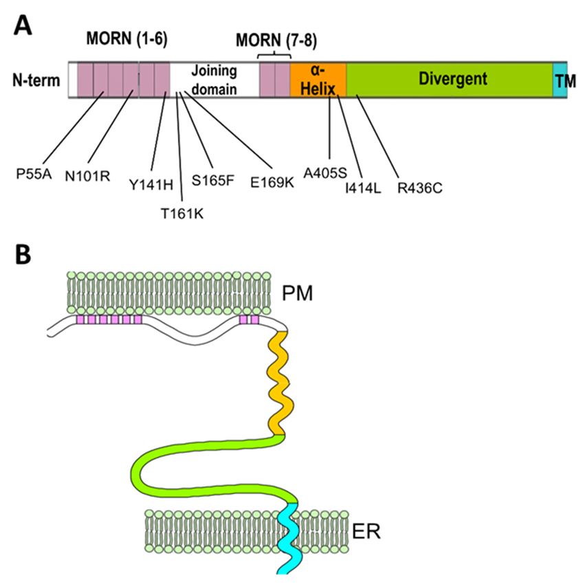
Biomolecules 2022, 12, 109                                                                                            3 of 14

                             respectively, through their alpha interacting domain (AID) located in the intracellular loop
                             connecting transmembrane domains I and II [18]. Beta subunits are crucial for facilitating
                             the trafficking of the channel into the plasma membrane and for modulating the channel
                             activity [16,19]. In skeletal muscle, the adapter protein Stac3 is also required for voltage-
                             induced calcium release [20,21]. The exact role of Stac3 in skeletal muscle EC coupling is
                             yet to be elucidated. Stac3 facilitates, but is not essential for, the membrane trafficking of
                             CaV 1.1 [22,23]; nonetheless, knocking out Stac3 completely abolishes the voltage-induced
                             Ca2+ release [20,21]. The observation that Stac3 binds to the II-III intracellular loop of
                             CaV 1.1 [24], which is critical for the cross-talk between CaV 1.1 and RyR1 [25], suggests that
                             Stac3 might allow or facilitate the mechanical coupling between these two channels.
                                   It appears evident that the association of all the EC coupling essential and accessory
                             proteins and the efficient cross-talk between CaV 1 in the plasma membrane and RyR in the
                             SR require the accurate formation and organization of ER (SR)-PM junctions, making the
                             proteins responsible for the organization of such junctions also essential for EC coupling.
                             This short review focuses on the proteins responsible for the formation of SR-PM junctions
                             in striated muscle, junctophilin1 and junctophilin2. Junctophilins’ physiological role in the
                             structural and functional assembly of triads and dyads and the pathological consequences
                             of their absence or malfunction in skeletal and cardiac muscle will be discussed. Finally,
                             new developments in predicting the structure of junctophilins, and the possible implication
                             for their functions will be touched on.

                             2. The Junctophilin Family
                                  Junctophilins (JPH1, JPH2, JPH3 and JPH4) were discovered by Takeshima and col-
                             laborators in the early 2000s in muscles and neurons [26,27]. All four isoforms of this
                             family contain a C-terminal transmembrane domain that embeds in the ER membrane and
                             eight N-terminal domains called membrane occupation and recognition nexuses (MORN),
                             thought to be responsible for the association with the plasma membrane (Figure 2).
                                  This particular organization allows the junctophilins to link the ER membrane and
                             the plasma membrane to one another. The MORN motifs are known to bind to the inter-
                             nal leaflet of the plasma membrane and specifically to phosphatidylinositol phosphate
                             species [29–31], although the role of the MORN motif as a bona fide membrane-binding
                             domain has been recently called into question after the observation that a number of pro-
                             teins that also contain MORN repeats are either not associated with membranes or employ
                             alternative domains to anchor to the membrane, or utilize their MORN motifs as protein-
                             protein–protein interaction domains rather than membrane association domains [32–34].
                             Therefore, the ability of the MORN motifs in the junctophilins to associate with the mem-
                             brane might arise from the presence of positively charged residues in the less conserved
                             regions or from post-transcriptional modifications. Jiang and colleagues [35] have recently
                             shown that palmitoylation at cysteine residues in MORN 1 and 8 of JPH2, which are largely
                             conserved in other junctophilin isoforms, is involved in the formation of stable ER-PM
                             junctions in CHO cells and is instrumental for the association of JPH2 with lipid rafts.
                                  In addition to the MORN motifs and the transmembrane domain, junctophilins contain
                             a mostly disordered region that connects the sixth and seventh MORN motifs, termed
                             joining domain, a putative α-helical region following the eighth MORN domain and an
                             extended disordered divergent region that separates the α-helical region and the C-terminal
                             transmembrane domain (Figure 2). As suggested by its designation, the divergent domain
                             is poorly conserved among different isoforms, while the rest of the sequence shows high
                             (MORN motif and transmembrane domain), moderate (α-helical domain) or mild (joining
                             domain) conservation among isoforms [27].
                                  Junctophilin expression can be detected in all excitable cells, with different isoforms
                             having tissue-specific expressions. JPH3 and JPH4 were first discovered in the brain, where
                             they are expressed almost ubiquitously [26] and in sensory neurons [36]. Junctophilins
                             3 and 4 were also detected in other non-neuronal tissues such as pancreatic β-cells [37]
                             and T-type lymphocytes [38]. Junctophilin 1 and 2 are mostly expressed in muscle tissues.
Biomolecules 2022, 12, 109 4 of 14
In particular, Junctophilin 1 is expressed primarily in skeletal muscle. Junctophilin 2 is
expressed in skeletal muscle and is the only isoform expressed in cardiac muscle [27].
Biomolecules 2022, 12, x FOR PEER REVIEW 4 of 15
Junctophilin 1 expression has also been found in the peripheral nervous system [39], and
Junctophilin 2 is also highly expressed in smooth muscle [40].
Figure 2. Schematic
Schematic representation
representation ofof junctophilin’s
junctophilin’s domain domain structure
structure and
and its
its arrangement
arrangement in in the
ER-PM
ER-PM junction.
junction. (A)
(A) Linear
Linear map
map showing
showing the the first
first (I(I through
through IV)
IV) and
and second
second (VII
(VII to
to VIII)
VIII) set
set of
MORN
MORN domains
domains (in(in pink)
pink) separated
separated by
by the
the joining
joining domain
domain (white).
(white). The
The α-helical
α-helical domain
domain (orange)
(orange)
follows
follows MORN
MORN VIIIVIII and
and is
is separated
separated from
from thethe transmembrane
transmembrane (TM, (TM, inin cyan)
cyan) by
by the
the long
long divergent
divergent
domain (green). The numbers underneath the map indicate mutations associated with human
domain (green). The numbers underneath the map indicate mutations associated with human
cardiomyopathies identified in JPH2 and their relative positions. (B) Schematic representation of
cardiomyopathies identified in JPH2 and their relative positions. (B) Schematic representation of
junctophilin’s organization in the ER-PM junctions (adapted from Garbino et al. [28]). The MORN
junctophilin’s organization in the ER-PM junctions (adapted from Garbino et al. [28]). The MORN
motifs associated with the internal leaflet of the plasma membrane and the C-terminal
motifs associated with
transmembrane the internal
domain leafletin
embedded of the
theplasma membrane and the
endo/sarcoplasmic C-terminal
reticulum transmembrane
membrane allow
domain embedded
junctophilins in the
to bridge theendo/sarcoplasmic
two membrane systems reticulum membrane
together. allowdomains
Different junctophilins to bridge the
are color-coded as
two membrane
indicated in (A).systems together. Different domains are color-coded as indicated in (A).
While,
This in addition
particular to junctophilins,
organization neuronal
allows the cells express
junctophilins a variety
to link the ER of proteins and
membrane that
are able to form stable ER-PM junctions [41], JPH1 and JPH2 are the
the plasma membrane to one another. The MORN motifs are known to bind to the internal only proteins that
form ER-PM
leaflet of thejunctions in striated muscle.
plasma membrane The forcestothat
and specifically keep these junctions
phosphatidylinositol together
phosphate
are considerably strong since they have to withstand mechanical stress
species [29–31], although the role of the MORN motif as a bona fide membrane-binding provided by the
repetitive contractions and stretching of the muscle fiber, which in
domain has been recently called into question after the observation that a number some cases can reach
of
extreme levels [42,43]. However, junctophilins’ role is not limited to
proteins that also contain MORN repeats are either not associated with membranes or building such strong
structures;
employ they alsodomains
alternative actively to
recruit
anchor andtointeract with the or
the membrane, components that
utilize their populate
MORN these
motifs as
SR-PM junctions and form the functional apparatus responsible for EC coupling.
protein-protein–protein interaction domains rather than membrane association domains
[32–34]. Therefore,
3. Recruitment the ability Proteins
of Junctional of the MORN motifs in the and
by Junctophilins1 junctophilins
2 to associate with
the membrane might arise from the presence of positively charged residues in the less
3.1. Recruitment of Skeletal Muscle Junctional Proteins
conserved regions or from post-transcriptional modifications. Jiang and colleagues [35]
JPH1 and JPH2, and specifically a region in the two junctophilins spanning approxi-
have recently shown that palmitoylation at cysteine residues in MORN 1 and 8 of JPH2,
matively from the second half of the joining domain to the first half of the putative α-helical
which are largely conserved in other junctophilin isoforms, is involved in the formation
domain, co-immunoprecipitate with Ca 1.1 [44]. A 20-residue sequence in the C-terminal
of stable ER-PM junctions in CHO cellsV and is instrumental for the association of JPH2
with lipid rafts.
In addition to the MORN motifs and the transmembrane domain, junctophilins
contain a mostly disordered region that connects the sixth and seventh MORN motifs,
termed joining domain, a putative α-helical region following the eighth MORN domainBiomolecules 2022, 12, 109 5 of 14
domain of CaV 1.1 is directly involved in the interactions with junctophilins and in the
recruitment of CaV 1.1 to triads [45]. Additionally, CaV 1.1 is recruited to junctions formed by
JPH2 when the proteins are expressed in non-muscle cell models together with the CaV 1.1
auxiliary subunit β1a and Stac3 [46]. Overall, this indicates that JPH1 and JPH2 have an
active role in recruiting the voltage-gated Ca2+ channel to triads by binding directly to the
channel; the disruption of this interaction interferes with the assembly of the triad [44,45].
JPH1 co-immunoprecipitates with RyR1, a behavior that has not been observed for
JPH2 [47]. Nonetheless, JPH1 KO mice can still perform EC coupling [48], suggesting
that the presence of RyR1 in junctions does not depend solely on JPH1. Therefore, the
recruitment of RyR1 in JPH2-induced junctions might be due to a weak interaction that is
not detected in biochemical assays or requires the presence of additional proteins, with
CaV 1.1 being a likely candidate. Interestingly, a 28-residue region involved in the direct
interaction with the cytoplasmic domain of RyR1 was identified in the divergent domain of
the neuronal isoform JPH3 [49], but no homologous sequences have been found in either
JPH1 or JPH2, suggesting that different junctophilins might employ different strategies to
recruit RyR1 to junctions.
3.2. Recruitment of Cardiac Muscle Junctional Proteins
The cardiac L-type Ca2+ channel CaV 1.2 co-immunoprecipitates with JPH2 [50,51],
indicating that, as in skeletal muscle, JPH2 likely plays a role in recruiting the voltage-
sensor channel in the dyads and peripheral couplings of cardiac muscle. Notably, the
same C-terminal sequence identified as the CaV 1.1 site of interaction with junctophilins, is
conserved in CaV 1.2 [45], suggesting that this sequence might also be involved in CaV 1.2–
JPH2 interactions.
Differently from what was found with JPH2 and RyR1, co-immunoprecipitation was
observed between JPH2 and RyR2 [50,51], indicating a stronger interaction between the
two proteins. This interaction is disrupted by the E169K substitution located towards the
N-terminal end of the JPH2 joining domain [52] and weakened by the R420Q mutation in
RyR2 [53]. A stronger interaction with RyR2 might be required by JPH2 because it is the
only junctophilin isoform expressed in cardiac muscle and possibly because cardiac muscle
lacks the additional stabilization provided by the mechanical connection between RyR1
and CaV 1.1 that exists in skeletal muscle [15].
4. Functional Studies on Junctophilins 1 and 2
4.1. Junctophilin 1
JPH1 knock-out mice die within 24 h after birth due to suckling defects leading to
undernourishment. The suckling defect is likely due to muscle weakness since the neuronal
suckling reflexes are normal in knock-out mice [48]. Functional studies on isolated hindlimb
muscle showed abnormal twitch tension and a greater dependency on extracellular calcium
in KO mice muscles, suggesting that a significant fraction of RyR1s in the junctional SR
are not directly coupled with the CaV 1.1 channels in the T-tubules and therefore operate
via calcium-induced calcium release. Nonetheless, knock-out (KO) mice are still relatively
mobile and show skeletal muscle-type EC coupling to a certain degree, indicating that JPH2
can support voltage-induced Ca2+ release in the absence of JPH1. From a structural point
of view, although no major disorganization of the fiber is noticed at the light microscopy
level, evident alterations are noticeable at the ultrastructural level [48,54]. In particular, the
skeletal muscle of wt and JPH1 KO mice show a similar development in the embryonic
stages until shortly after birth. At this age, wt muscle experiences a significant increase
in JPH1 expression, which is temporally correlated with the transition from immature
SR-PM junctions, mainly organized in dyads at this stage, into fully formed triads. This
transition is absent in JPH1 KO muscle [48,54], suggesting that JPH2 is important in
forming the dyads, while JPH1 has a crucial role in the conversion from dyads to triads
in the fully mature skeletal muscle. The knocking down of junctophilins using sh-RNA,
leads to the impairment of store-operated Ca2+ entry (SOCE), altered intracellular calciumBiomolecules 2022, 12, 109 6 of 14
release and intracellular calcium stores [55] and to a reduction in RyR1 and CaV 1.1 co-
clustering associated with a decrease in CaV 1.1 membrane expression [44] both in myotubes
and muscle fibers. In both these studies, a shRNA against both JPH1 and JPH2 was
used; hence, it was impossible to distinguish each isoform’s relative contribution to the
resulting phenotype. In 2010 Li and collaborators [56] showed that store-operated Ca2+
entry was severely impaired in JPH1 KO mice myotubes, suggesting that JPH1 substantially
contributes to maintaining an efficient SOCE in skeletal muscle.
4.2. Junctophilin 2
JPH2 knock-out mice die in utero due to cardiac failure. Ultrastructural analyses
on embryonic myotubes of KO mice revealed a substantial reduction in the number and
extension of peripheral couplings [27]. To avoid the complication related to the early
mortality of KO mice, van Oort et al. generated a conditional JPH2 knockdown mice
model to assess the effect of JPH2-reduced expression in the mature heart [57]. Inducing
JPH2 knockdown led to an increased frequency of heart failure events. At the cellular
level, this was explained structurally by T-tubule remodeling and destabilization and
disorganization of the dyads, and functionally by CaV 1.2 and RyR2 uncoupling and the
consequent reduction in the efficiency of calcium-induced calcium release. An increase in
the frequency of calcium sparks was noticed in knocked down isolated cardiomyocytes,
suggesting that JPH2 might also modulate RyR2 by reducing its activity.
A number of point mutations in JPH2 have been discovered in association with
hypertrophic cardiomyopathy and atrial fibrillation. The localization of these mutations
spans from the N-terminal MORN motifs to the divergent domain at the C-terminus
(Figure 2, Table 1), indicating that multiple regions of JPH2 are involved in supporting
cardiac muscle structure and function. A subset of all JPH2 mutations associated with
cardiomyopathies has been functionally characterized in cardiomyocytes or cardiomyocyte-
derived cell lines (Table 1).
Amino acid substitutions N101R and Y141H in the MORN IV and VI, respectively,
and S165F in the joining domain, cause similar phenotypes such as JPH2 mislocalization,
reduction in spontaneous Ca2+ signaling and increased cell size in HL-1 and H9c2 cell
lines [59]. Mutations Y141H and S165F were also tested in skeletal muscle myotubes [60],
where they were found to induce myocytes hypertrophy, reduce EC coupling gain and
increase intracellular Ca2+ concentration. Additionally, Y141H but not S165F pathogenically
increased store-operated calcium entry [60]. Mutation E169K, located in the joining domain,
causes weaker binding between JPH2 and RyR2 and increased spontaneous Ca2+ leakage
from the SR in the form of a spontaneous Ca2+ release and increased Ca2+ sparks in
isolated cardiomyocytes from a pseudo-knock-in mouse model [52]. The A405S mutation
is located in the putative α-helical region of JPH2. The equivalent mutation introduced in
mice (A399S) resulted in cardiomyocytes with an irregular T-tubule pattern but otherwise
relatively normal Ca2+ signaling with only a moderate increase in sarco–endoplasmic
reticulum Ca2+ ATPase (SERCA) activity.
It is still unclear whether most of these mutations specifically disrupt binding sites
crucial for the interaction of JPH2 with other junctional proteins or whether they alter the
structure and stability of the protein with detrimental effects on JPH2 folding and trafficking
and, consequently, T-tubular remodeling and impaired SR-PM junction formation. Recent
work from Gross and collaborators [66] shows reduced co-immunoprecipitation in the
plasma membrane fractions of ventricular myocytes between CaV 1.2 and a JPH2 mutant in
which seven random mutations were introduced in the joining domain. Based on structure
prediction simulations, the authors found that the seven mutations did not compromise the
overall organization of JPH2. This work supports the possible role of the joining domain
in the binding with CaV 1.2. However, the mutated JPH2 showed a significantly reduced
ability to form dyads in ventricular myocytes, suggesting that the lack of interactions with
CaV 1.2 is likely not the only consequence of these mutations for JPH2 function.Biomolecules 2022, 12, 109 7 of 14
Table 1. Identified mutations in JPH2 linked to hypertrophic cardiomyopathy (HCM) and atrial
fibrillation (AF).
Mutation Position Reference Functional Characterization Human Phenotype
P55A MORN II [58] Uncharacterized HCM
JPH2 mislocalization, disrupted Ca2+
N101R MORN IV [59] HCM
signaling, cardiomyocytes hypertrophy.
JPH2 mislocalization, disrupted Ca2+
signaling, cardiomyocytes hypertrophy.
In skeletal muscle: abnormal ER-PM
Y141H MORN VI [59,60] HCM
junctions, increased store-operated Ca2+
entry, decreased EC coupling gain,
myotubes hypertrophy.
T161K Joining domain [61] Uncharacterized HCM
JPH2 mislocalization, disrupted Ca2+
signaling, cardiomyocytes hypertrophy.
S165F Joining domain [59,62] In skeletal muscle: Abnormal ER-PM HCM
junctions, decreased EC coupling gain,
myotubes hypertrophy,
Reduced binding to RyR2, increases
E169K Joining domain [52] spontaneous Ca2+ release and Ca2+ AF
sparks frequency
Irregular T-tubule pattern, mild effect on
A405S α-helical domain [52,63] calcium signaling in the equivalent HCM
mutation (A399S) in mice.
Dilated
I414L α-helical domain [64] Uncharacterized
Cardiomyopathy
R436C Divergent domain [65] Uncharacterized HCM
5. Post-Transcriptional Regulation of JPH1 and JPH2
Conditions of elevated cytosolic Ca2+ concentrations lead to fragmentation of junc-
tophilins 1 and 2 in skeletal and cardiac muscle. Murphy and colleagues [67] determined
that exposure to elevated (≥20 µM) intracellular [Ca2+ ] for 60 min led to the almost com-
plete loss of full-length JPH1 and JPH2 in skeletal muscle fibers. This loss is mirrored by a
loss of contractile force in skinned skeletal muscle fibers after just one minute of exposure
to 40 µM Ca2+ . The same authors also observed fragmentation of JPH1 after raising the
intracellular [Ca2+ ] by supraphysiological stimulation of the muscle fiber. Interestingly, the
proteolysis of JPH1 temporally matched the autolytic activation of calpain-µ (calpain1).
The link between calpain, specifically calpain1, and JPH1 cleavage was recently confirmed
by data from Tammineni and colleagues in patients with malignant hyperthermia sus-
ceptibility (MHS) and muscle cell lines [68]. Patients with MHS carry mutations in the
RyR1 (most often) or other proteins involved in EC coupling that cause leakage of Ca2+
from the SR and chronic increases in cytoplasmic Ca2+ concentration. Tammineni and
colleagues observed increased fragmentation of JPH1 in MHS individuals compared to
healthy subjects. Furthermore, the fragmented JPH1 abandons the triad, and its C-terminal
fragment translocates into nuclei, where it regulates the transcription of genes known to
be altered in MHS. Upon identification of a potential calpain1 cleavage site in JPH1, it
was confirmed that treatment of human muscle lysate with Calpain1 generated the same
fragments of JPH1 observed in MHS individuals, and the addition of a calpain1 inhibitor
prevented fragmentation.
After the identification of several putative calpain binding sites also in JPH2 [51,69],
the implication of calpain in the proteolytic regulation of JPH2 was verified using cal-
pain inhibitors to rescue the loss of JPH2 in an inducible heart failure mouse model and
in mice cardiomyocytes after ischemia/reperfusion [70]. Akin to what was shown byBiomolecules 2022, 12, 109 8 of 14
Tammineni and colleagues in skeletal muscle, Guo and collaborators also observed that
digestion by calpain1 releases several fragments of JPH2 [51]. Among these fragments,
a ~ 75 kDa N-terminal peptide (JPH2-NTP), generated by calpain1 cleavage at residues
R565/T566 in the JPH2 divergent region, migrates into the nucleus, where it binds to TATA
box regions and interacts with the transcription machinery [69]. Somewhat in contrast
with the findings of Guo et al., recent work from Lahiri and colleagues [71] describes the
presence of a C-terminal JPH2 fragment (JPH2-CPT), generated by cleavage of full-length
JPH2 at residues G482/T493, in the heart of human patients with heart failure (HF) and
of mice HF models. Unlike the JPH2-NTP fragment described earlier, JPH2-CTP seems
to be specifically generated by calpain2 in vivo. This specificity might be of importance
since calpain2 is activated by millimolar cytoplasmic Ca2+ concentrations, which are more
likely to be reached in pathological conditions, while calpain1 is activated at more physio-
logical (micromolar) Ca2+ concentrations. Interestingly, JPH2-CTP shares the same nuclear
localization signal sequence as JPH2-NTP and similarly localizes into the nuclei. However,
while JPH2-NTP associates with chromatin and acts as a transcription factor, JPH2-CTP
is confined in nuclear sub-compartments and does not seem to affect transcription. The
nuclear localization of JPH2-NTP and JPH2-CTP leads to opposite functional outcomes.
While JPH2-NTP regulates genes involved in calcium homeostasis to possibly protect the
cells from the consequences of elevated intracellular calcium, JPH2-CTP nuclear localiza-
tion is causative of cardiomyocytes hypertrophy, an early marker of pathogenic cardiac
remodeling. Altogether, these results indicate a fine modulation of junctophilin 1 and 2 as a
way to regulate intracellular Ca2+ homeostasis and possibly reduce EC coupling gain in
conditions of excessive intracellular Ca2+ concentration. The work from Tammineni et al.
and Guo et al. also opens the possibility that cleaved junctophilins might serve as tran-
scription modulators to further contribute to the Ca2+ regulation of striated muscle. At the
same time, the seemingly contradictory results from different authors indicate that further
research is still needed to fully elucidate the different pathways and functional implications
of the post-transcriptional regulation of junctophilins.
To further add to the complexity of junctophilin post-transcriptional regulation, cal-
pains might not be the only proteins that cleave junctophilins; work from Chan and
colleagues [72] showed a protective effect of a metalloproteinase-2 (MMP-2) inhibitor in
murine hearts after acute ischemia-reperfusion injury. This effect was attributed to a reduc-
tion in JPH2 cleavage by MMP-2. Such cleavage was demonstrated in vitro in mouse heart
extracts after incubation with MMP-2 with or without an inhibitor.
6. New Insights from Deep Learning Protein Structure Prediction
Remarkable advancements in protein folding prediction were recently achieved by the
artificial intelligence software Alphafold2 [73]. Alphafold2 is a giant leap forward in the
reliability of protein folding prediction compared to similar existing software [74], and it
has already been used to predict the structure of nearly the entire human proteome. Based
on Aplhafold2 prediction models, junctophilin 1 and 2 show a similar 3D structure, also
shared by the neuronal isoforms JPH3 and JPH4 (UniProt protein ID: Q9HDC5, Q9BR39,
Q8WXH2, Q96JJ6 for human JPH1, JPH2, JPH3 and JPH4, respectively). The structure of
the most ordered domains of junctophilin, specifically the MORN repeats, the α-helical
region and the transmembrane domain, are predicted with high confidence by Alphafold2.
In contrast, the joining and divergent domains are likely disordered, at least in the isolated
protein, and the structure cannot be predicted with reasonable confidence. According to
the prediction model (Figure 3), the MORN domains are arranged in an extended “half-
pipe” configuration, with the α-helical domain lying on the convex side of this half-pipe
(Figure 3B,C) and establishing interactions with charged residues in the MORN domains
(see the zoomed-in region in Figure 3C for an example).Biomolecules 2022, 12, x FOR PEER REVIEW 10 of 15
Biomolecules 2022, 12, 109 9 of 14
Figure Figure
3. Structure of the MORNs
3. Structure and α-helical
of the MORNs domains
and α-helical of human
domains junctophilin1
of human and junctophilin2.
junctophilin1 and junctophilin2.
(A) Schematic representation of junctophilin domain as shown in Figure 2; the
(A) Schematic representation of junctophilin domain as shown in Figure 2; the solid solid red lines indi-indicate
red lines
cate the regions for which the structure is predicted with high fidelity by Alphafold2 and illustrated
the regions for which the structure is predicted with high fidelity by Alphafold2 and illustrated
in (B,C). (B,C) predicted structures of the MORNs-α-helical domains of junctophilin1 (B) and junc-
in (B,C). (B,C) predicted structures of the MORNs-α-helical domains of junctophilin1 (B) and junc-
tophilin2 (C). The α-helical domain (in red) lies on the convex side of the MORN domains half-pipe
tophilin2
structure (C).inThe
(in blue) α-helical
both domainand
junctophilin1 (in red) lies on the convex
junctophilin2. β-sheet side of theforming
hairpins MORN MORN
domainsdo- half-pipe
structure (in blue) in both junctophilin1 and junctophilin2. β-sheet hairpins forming
mains I and VIII (green parentheses) and the position of the N-terminal end (N-t) and C-terminal MORN domains
end (C-t) of the joining domain (in pink), which is absent in this representation, are indicated in (B). (C-t)
I and VIII (green parentheses) and the position of the N-terminal end (N-t) and C-terminal end
of the
The inset joining
in (C) shows domain
some (in pink),
of the whichthat
residues is absent in this
form the representation,
hydrogen arestabilize
bonds that indicatedthe
in associ-
(B). The inset
ation between the MORN
in (C) shows some ofdomains and that
the residues the α-helical
form the domain
hydrogen ofbonds
junctophilin2.
that stabilize the association between
the MORN domains and the α-helical domain of junctophilin2.
The structure obtained using Alphafold2 is substantially different from what was
previously The structure
predicted obtained
using RaptorX using Alphafold2
software is and
by Gross substantially
colleaguesdifferent from
[66]. In the what was
struc-
previously predicted using RaptorX software by Gross and colleagues [66].
ture described by Gross et al., the α-helical domain extends beyond the MORN domains In the struc-
ture
without describedwith
interacting by Gross
themetatal.,
all.the domain extends
α-helicalAlphafold2
However, beyond
software the MORN
is considered to domains
beBiomolecules 2022, 12, 109 10 of 14
without interacting with them at all. However, Alphafold2 software is considered to be
more accurate than most (if not all) of the currently existing structure-predicting software,
especially for proteins for which no homologous structures exist [75,76], and the recipro-
cal arrangement of JPH2 MORN motifs and α-helical domain predicted by Alphafold2
agrees with data from Li and collaborators [32] based on the crystal structure of the protein
MORN4. MORN4 contains a series of MORN motifs arranged in a half-pipe configuration
followed by a brief α-helical region. The helical region stabilizes the MORN domains
by lying over part of the convex side of the half-pipe. The structure solved by Li and
colleagues is in many ways very similar to the sequence predicted by Alphafold2 for JPHs.
Furthermore, in MORN 4, the concave side of the MORN half-pipe structure, containing
most of the conserved residues that define the MORN domain, engages in the binding with
the α-helical region of myosin3a. It is conceivable that the concave side of the junctophilin
MORN motifs could also participate in protein–protein interactions with components of the
EC coupling machinery. The particular arrangement of the α-helical domain with respect
to the MORN motifs predicted by Alphafold2 and suggested by the observations of Li and
colleagues challenges the classic view of the α-helical domain as the spacer that spans most
of the junctional gap (see schematic representation in Figure 2) and points to the divergent
domain as the region that most likely fulfills this role.
7. Closing Remarks
Junctophilin1 and 2 are crucial players in EC coupling and striated muscle physiology.
They form and stabilize the specialized ER-PM junctions, which are the functional platforms
at which EC coupling is executed and actively participate in the recruitment of crucial
components of EC coupling into such junctions. Under particular pathological conditions
leading to elevated cytoplasmic Ca2+ concentrations, both JPH1 and JPH2 can be potentially
utilized by the cell to ameliorate the consequences of such elevated Ca2+ . Specifically,
Ca2+ -induced cleavage of JPH1 or JPH2 has the dual effect of uncoupling CaV and RyR,
reducing calcium release from the SR and producing JPH fragments that traffic to the
nucleus and up-regulate or down-regulate specific genes involved in intracellular Ca2+
homeostasis. Therefore, it is not surprising that knocking out or knocking down JPH1
and JPH2 severely impairs EC coupling in animal models. While mutations in JPH1 are
generally not associated with significant muscle diseases in humans, possibly due to the
compensatory effect of the concurrent expression of JPH2 in skeletal muscle, numerous
point mutations in JPH2 have been identified in patients with cardiomyopathies. Although
the functional effects of some of these mutations have been explored in animal models, it is
still, for the most part, unclear whether JPH2 mutations destroy the ability of the protein to
interact with other partners or whether they cause JPH2 misfolding or instability. If, on
one end, recent progress in protein structure prediction can help to infer the effect of at
least some of JPH2 mutation, some disagreement between different structure-predicting
software still exists. Solving the protein’s actual structure, preferably in conjunction with its
binding partners, would represent a big step forward in investigating and understanding
the nature of junctophilins’ interactions with other junctional proteins and the pathological
consequences of the disruption of such interactions.
Funding: This study was supported by the National Institutes of Health grant AR070298 to Dr
Kurt Beam.
Institutional Review Board Statement: Not applicable.
Informed Consent Statement: Not applicable.
Data Availability Statement: Not available.
Conflicts of Interest: The author declares no conflict of interest.Biomolecules 2022, 12, 109 11 of 14
References
1. Huxley, A.F.; Taylor, R.E. Local activation of striated muscle fibres. J. Physiol. 1958, 144, 426–441. [CrossRef]
2. Huxley, A.F. Local activation of striated muscle from the frog and the crab. J. Physiol. 1957, 135, 17–18. [PubMed]
3. Porter, K.R.; Palade, G.E. Studies on the endoplasmic reticulum. III. Its form and distribution in striated muscle cells. J. Biophys.
Biochem. Cytol. 1957, 3, 269–300. [CrossRef]
4. Perni, S.; Marsden, K.C.; Escobar, M.; Hollingworth, S.; Baylor, S.M.; Franzini-Armstrong, C. Structural and functional properties
of ryanodine receptor type 3 in zebrafish tail muscle. J. Gen. Physiol. 2015, 145, 253. [CrossRef]
5. Lavorato, M.; Huang, T.Q.; Iyer, V.R.; Perni, S.; Meissner, G.; Franzini-Armstrong, C. Dyad content is reduced in cardiac myocytes
of mice with impaired calmodulin regulation of RyR2. J. Muscle Res. Cell Motil. 2015, 36, 205–214. [CrossRef]
6. Perni, S.; Iyer, V.R.; Franzini-Armstrong, C. Ultrastructure of cardiac muscle in reptiles and birds: Optimizing and/or reducing
the probability of transmission between calcium release units. J. Muscle Res. Cell Motil. 2012, 33, 145–152. [CrossRef] [PubMed]
7. Franzini-Armstrong, C. Fine Structure of Sarcoplasmic Reticulum and Tranverse Tubular System in Muscle Fibers. Fed. Proc.
1964, 23, 887–895. [PubMed]
8. Franzini-Armstrong, C. STUDIES OF THE TRIAD: I. Structure of the Junction in Frog Twitch Fibers. J. Cell Biol. 1970, 47, 488–499.
[CrossRef] [PubMed]
9. Franzini-Armstrong, C.; Protasi, F.; Ramesh, V. Shape, size, and distribution of Ca2+ release units and couplons in skeletal and
cardiac muscles. Biophys. J. 1999, 77, 1528–1539. [CrossRef]
10. Bossen, E.H.; Sommer, J.R. Comparative stereology of the lizard and frog myocardium. Tissue Cell 1984, 16, 173–178. [CrossRef]
11. Bossen, E.H.; Sommer, J.R.; Waugh, R.A. Comparative stereology of the mouse and finch left ventricle. Tissue Cell 1978, 10,
773–784. [CrossRef]
12. Grimley, P.M.; Edwards, G.A. The ultrastructure of cardiac desnosomes in the toad and their relationship to the intercalated disc.
J. Biophys. Biochem. Cytol. 1960, 8, 305–318. [CrossRef] [PubMed]
13. Santer, R.M.; Cobb, J.L. The fine structure of the heart of the teleost, Pleuronectes platessa L. Z. Zellforsch. Mikrosk. Anat. 1972, 131,
1–14. [CrossRef] [PubMed]
14. Fabiato, A. Time and calcium dependence of activation and inactivation of calcium-induced release of calcium from the
sarcoplasmic reticulum of a skinned canine cardiac Purkinje cell. J. Gen. Physiol. 1985, 85, 247–289. [CrossRef] [PubMed]
15. Block, B.A.; Imagawa, T.; Campbell, K.P.; Franzini-Armstrong, C. Structural evidence for direct interaction between the molecular
components of the transverse tubule/sarcoplasmic reticulum junction in skeletal muscle. J. Cell Biol. 1988, 107, 2587–2600.
[CrossRef] [PubMed]
16. Schredelseker, J.; Dayal, A.; Schwerte, T.; Franzini-Armstrong, C.; Grabner, M. Proper restoration of excitation-contraction
coupling in the dihydropyridine receptor β1 -null zebrafish relaxed is an exclusive function of the β1a subunit. J. Biol. Chem. 2009,
284, 1242–1251. [CrossRef]
17. Weissgerber, P.; Held, B.; Bloch, W.; Kaestner, L.; Chien, K.R.; Fleischmann, B.K.; Lipp, P.; Flockerzi, V.; Freichel, M. Reduced
cardiac L-type Ca2+ current in Cav β2 −/− embryos impairs cardiac development and contraction with secondary defects in
vascular maturation. Circ. Res. 2006, 99, 749–757. [CrossRef]
18. Pragnell, M.; De Waard, M.; Mori, Y.; Tanabe, T.; Snutch, T.P.; Campbell, K.P. Calcium channel β-subunit binds to a conserved
motif in the I-II cytoplasmic linker of the alpha 1-subunit. Nature 1994, 368, 67–70. [CrossRef]
19. Chien, A.J.; Zhao, X.; Shirokov, R.E.; Puri, T.S.; Chang, C.F.; Sun, D.; Rios, E.; Hosey, M.M. Roles of a membrane-localized β
subunit in the formation and targeting of functional L-type Ca2+ channels. J. Biol. Chem. 1995, 270, 30036–30044. [CrossRef]
20. Nelson, B.R.; Wu, F.; Liu, Y.; Anderson, D.M.; McAnally, J.; Lin, W.; Cannon, S.C.; Bassel-Duby, R.; Olson, E.N. Skeletal muscle-
specific T-tubule protein STAC3 mediates voltage-induced Ca2+ release and contractility. Proc. Natl. Acad. Sci. USA 2013, 110,
11881–11886. [CrossRef] [PubMed]
21. Horstick, E.J.; Linsley, J.W.; Dowling, J.J.; Hauser, M.A.; McDonald, K.K.; Ashley-Koch, A.; Saint-Amant, L.; Satish, A.; Cui,
W.W.; Zhou, W.; et al. Stac3 is a component of the excitation-contraction coupling machinery and mutated in Native American
myopathy. Nat. Commun. 2013, 4, 1952. [CrossRef]
22. Polster, A.; Perni, S.; Bichraoui, H.; Beam, K.G. Stac adaptor proteins regulate trafficking and function of muscle and neuronal
L-type Ca2+ channels. Proc. Natl. Acad. Sci. USA 2015, 112, 602–606. [CrossRef] [PubMed]
23. Polster, A.; Nelson, B.R.; Olson, E.N.; Beam, K.G. Stac3 has a direct role in skeletal muscle-type excitation-contraction coupling
that is disrupted by a myopathy-causing mutation. Proc. Natl. Acad. Sci. USA 2016, 113, 10986–10991. [CrossRef] [PubMed]
24. Polster, A.; Nelson, B.R.; Papadopoulos, S.; Olson, E.N.; Beam, K.G. Stac proteins associate with the critical domain for excitation-
contraction coupling in the II-III loop of CaV1.1. J. Gen. Physiol. 2018, 150, 613–624. [CrossRef]
25. Tanabe, T.; Beam, K.G.; Adams, B.A.; Niidome, T.; Numa, S. Regions of the skeletal muscle dihydropyridine receptor critical for
excitation-contraction coupling. Nature 1990, 346, 567–569. [CrossRef] [PubMed]
26. Nishi, M.; Sakagami, H.; Komazaki, S.; Kondo, H.; Takeshima, H. Coexpression of junctophilin type 3 and type 4 in brain. Brain
Res. Mol. Brain Res. 2003, 118, 102–110. [CrossRef]
27. Takeshima, H.; Komazaki, S.; Nishi, M.; Iino, M.; Kangawa, K. Junctophilins: A novel family of junctional membrane complex
proteins. Mol. Cell 2000, 6, 11–22. [CrossRef]
28. Garbino, A.; van Oort, R.J.; Dixit, S.S.; Landstrom, A.P.; Ackerman, M.J.; Wehrens, X.H. Molecular evolution of the junctophilin
gene family. Physiol. Genom. 2009, 37, 175–186. [CrossRef]Biomolecules 2022, 12, 109 12 of 14
29. Bennett, H.J.; Davenport, J.B.; Collins, R.F.; Trafford, A.W.; Pinali, C.; Kitmitto, A. Human junctophilin-2 undergoes a structural
rearrangement upon binding PtdIns(3,4,5)P3 and the S101R mutation identified in hypertrophic cardiomyopathy obviates this
response. Biochem. J. 2013, 456, 205–217. [CrossRef]
30. Kakizawa, S.; Moriguchi, S.; Ikeda, A.; Iino, M.; Takeshima, H. Functional cross-talk between cell-surface and intracellular
channels mediated by junctophilins essential for neuronal functions. Cerebellum 2008, 7, 385–391. [CrossRef]
31. Rossi, D.; Scarcella, A.M.; Liguori, E.; Lorenzini, S.; Pierantozzi, E.; Kutchukian, C.; Jacquemond, V.; Messa, M.; De Camilli, P.;
Sorrentino, V. Molecular determinants of homo- and heteromeric interactions of Junctophilin-1 at triads in adult skeletal muscle
fibers. Proc. Natl. Acad. Sci. USA 2019, 116, 15716–15724. [CrossRef]
32. Li, J.; Liu, H.; Raval, M.H.; Wan, J.; Yengo, C.M.; Liu, W.; Zhang, M. Structure of the MORN4/Myo3a Tail Complex Reveals
MORN Repeats as Protein Binding Modules. Structure 2019, 27, 1366–1374.e1363. [CrossRef]
33. Sajko, S.; Grishkovskaya, I.; Kostan, J.; Graewert, M.; Setiawan, K.; Trubestein, L.; Niedermuller, K.; Gehin, C.; Sponga, A.;
Puchinger, M.; et al. Structures of three MORN repeat proteins and a re-evaluation of the proposed lipid-binding properties of
MORN repeats. PLoS ONE 2020, 15, e0242677. [CrossRef]
34. Mikami, K.; Saavedra, L.; Hiwatashi, Y.; Uji, T.; Hasebe, M.; Sommarin, M. A dibasic amino acid pair conserved in the activation
loop directs plasma membrane localization and is necessary for activity of plant type I/II phosphatidylinositol phosphate kinase.
Plant Physiol. 2010, 153, 1004–1015. [CrossRef]
35. Jiang, M.; Hu, J.; White, F.K.H.; Williamson, J.; Klymchenko, A.S.; Murthy, A.; Workman, S.W.; Tseng, G.N. S-Palmitoylation of
junctophilin-2 is critical for its role in tethering the sarcoplasmic reticulum to the plasma membrane. J. Biol. Chem. 2019, 294,
13487–13501. [CrossRef] [PubMed]
36. Hogea, A.; Shah, S.; Jones, F.; Carver, C.M.; Hao, H.; Liang, C.; Huang, D.; Du, X.; Gamper, N. Junctophilin-4 facilitates
inflammatory signalling at plasma membrane-endoplasmic reticulum junctions in sensory neurons. J. Physiol. 2021, 599,
2103–2123. [CrossRef] [PubMed]
37. Li, L.; Pan, Z.F.; Huang, X.; Wu, B.W.; Li, T.; Kang, M.X.; Ge, R.S.; Hu, X.Y.; Zhang, Y.H.; Ge, L.J.; et al. Junctophilin 3 expresses in
pancreatic beta cells and is required for glucose-stimulated insulin secretion. Cell Death Dis. 2016, 7, e2275. [CrossRef]
38. Woo, J.S.; Srikanth, S.; Nishi, M.; Ping, P.; Takeshima, H.; Gwack, Y. Junctophilin-4, a component of the endoplasmic reticulum-
plasma membrane junctions, regulates Ca2+ dynamics in T cells. Proc. Natl. Acad. Sci. USA 2016, 113, 2762–2767. [CrossRef]
[PubMed]
39. Pla-Martin, D.; Calpena, E.; Lupo, V.; Marquez, C.; Rivas, E.; Sivera, R.; Sevilla, T.; Palau, F.; Espinos, C. Junctophilin-1 is a
modifier gene of GDAP1-related Charcot-Marie-Tooth disease. Hum. Mol. Genet. 2015, 24, 213–229. [CrossRef]
40. Pritchard, H.A.T.; Griffin, C.S.; Yamasaki, E.; Thakore, P.; Lane, C.; Greenstein, A.S.; Earley, S. Nanoscale coupling of junctophilin-2
and ryanodine receptors regulates vascular smooth muscle cell contractility. Proc. Natl. Acad. Sci. USA 2019, 116, 21874–21881.
[CrossRef] [PubMed]
41. Chen, Y.J.; Quintanilla, C.G.; Liou, J. Recent insights into mammalian ER-PM junctions. Curr. Opin. Cell Biol. 2019, 57, 99–105.
[CrossRef]
42. Close, M.; Perni, S.; Franzini-Armstrong, C.; Cundall, D. Highly extensible skeletal muscle in snakes. J. Exp. Biol. 2014, 217,
2445–2448. [CrossRef]
43. Rome, L.C.; Syme, D.A.; Hollingworth, S.; Lindstedt, S.L.; Baylor, S.M. The whistle and the rattle: The design of sound producing
muscles. Proc. Natl. Acad. Sci. USA 1996, 93, 8095–8100. [CrossRef] [PubMed]
44. Golini, L.; Chouabe, C.; Berthier, C.; Cusimano, V.; Fornaro, M.; Bonvallet, R.; Formoso, L.; Giacomello, E.; Jacquemond, V.;
Sorrentino, V. Junctophilin 1 and 2 proteins interact with the L-type Ca2+ channel dihydropyridine receptors (DHPRs) in skeletal
muscle. J. Biol. Chem. 2011, 286, 43717–43725. [CrossRef] [PubMed]
45. Nakada, T.; Kashihara, T.; Komatsu, M.; Kojima, K.; Takeshita, T.; Yamada, M. Physical interaction of junctophilin and the CaV1.1
C terminus is crucial for skeletal muscle contraction. Proc. Natl. Acad. Sci. USA 2018, 115, 4507–4512. [CrossRef]
46. Perni, S.; Lavorato, M.; Beam, K.G. De novo reconstitution reveals the proteins required for skeletal muscle voltage-induced Ca2+
release. Proc. Natl. Acad. Sci. USA 2017, 114, 13822–13827. [CrossRef] [PubMed]
47. Phimister, A.J.; Lango, J.; Lee, E.H.; Ernst-Russell, M.A.; Takeshima, H.; Ma, J.; Allen, P.D.; Pessah, I.N. Conformation-dependent
stability of junctophilin 1 (JP1) and ryanodine receptor type 1 (RyR1) channel complex is mediated by their hyper-reactive thiols.
J. Biol. Chem. 2007, 282, 8667–8677. [CrossRef]
48. Ito, K.; Komazaki, S.; Sasamoto, K.; Yoshida, M.; Nishi, M.; Kitamura, K.; Takeshima, H. Deficiency of triad junction and
contraction in mutant skeletal muscle lacking junctophilin type 1. J. Cell Biol. 2001, 154, 1059–1067. [CrossRef]
49. Perni, S.; Beam, K. Neuronal junctophilins recruit specific CaV and RyR isoforms to ER-PM junctions and functionally alter
CaV2.1 and CaV2.2. eLife 2021, 10, e64249. [CrossRef]
50. Jiang, M.; Zhang, M.; Howren, M.; Wang, Y.; Tan, A.; Balijepalli, R.C.; Huizar, J.F.; Tseng, G.N. JPH-2 interacts with Cai-handling
proteins and ion channels in dyads: Contribution to premature ventricular contraction-induced cardiomyopathy. Heart Rhythm
2016, 13, 743–752. [CrossRef]
51. Guo, A.; Hall, D.; Zhang, C.; Peng, T.; Miller, J.D.; Kutschke, W.; Grueter, C.E.; Johnson, F.L.; Lin, R.Z.; Song, L.S. Molecular
Determinants of Calpain-dependent Cleavage of Junctophilin-2 Protein in Cardiomyocytes. J. Biol. Chem. 2015, 290, 17946–17955.
[CrossRef]Biomolecules 2022, 12, 109 13 of 14
52. Beavers, D.L.; Wang, W.; Ather, S.; Voigt, N.; Garbino, A.; Dixit, S.S.; Landstrom, A.P.; Li, N.; Wang, Q.; Olivotto, I.; et al. Mutation
E169K in junctophilin-2 causes atrial fibrillation due to impaired RyR2 stabilization. J. Am. Coll. Cardiol. 2013, 62, 2010–2019.
[CrossRef] [PubMed]
53. Yin, L.; Zahradnikova, A., Jr.; Rizzetto, R.; Boncompagni, S.; Rabesahala de Meritens, C.; Zhang, Y.; Joanne, P.; Marques-Sule, E.;
Aguilar-Sanchez, Y.; Fernandez-Tenorio, M.; et al. Impaired Binding to Junctophilin-2 and Nanostructural Alteration in CPVT
Mutation. Circ. Res. 2021, 129, e35–e52. [CrossRef] [PubMed]
54. Komazaki, S.; Ito, K.; Takeshima, H.; Nakamura, H. Deficiency of triad formation in developing skeletal muscle cells lacking
junctophilin type 1. FEBS Lett. 2002, 524, 225–229. [CrossRef]
55. Hirata, Y.; Brotto, M.; Weisleder, N.; Chu, Y.; Lin, P.; Zhao, X.; Thornton, A.; Komazaki, S.; Takeshima, H.; Ma, J.; et al. Uncoupling
store-operated Ca2+ entry and altered Ca2+ release from sarcoplasmic reticulum through silencing of junctophilin genes. Biophys.
J. 2006, 90, 4418–4427. [CrossRef]
56. Li, H.; Ding, X.; Lopez, J.R.; Takeshima, H.; Ma, J.; Allen, P.D.; Eltit, J.M. Impaired Orai1-mediated resting Ca2+ entry reduces the
cytosolic [Ca2+ ] and sarcoplasmic reticulum Ca2+ loading in quiescent junctophilin 1 knock-out myotubes. J. Biol. Chem. 2010,
285, 39171–39179. [CrossRef] [PubMed]
57. van Oort, R.J.; Garbino, A.; Wang, W.; Dixit, S.S.; Landstrom, A.P.; Gaur, N.; De Almeida, A.C.; Skapura, D.G.; Rudy, Y.; Burns,
A.R.; et al. Disrupted junctional membrane complexes and hyperactive ryanodine receptors after acute junctophilin knockdown
in mice. Circulation 2011, 123, 979–988. [CrossRef]
58. De Bruijn, S.; Galloo, X.; De Keulenaer, G.; Prihadi, E.A.; Brands, C.; Helbert, M. A special case of hypertrophic cardiomyopathy
with a differential diagnosis of isolated cardiac amyloidosis or junctophilin type 2 associated cardiomyopathy. Acta Clin. Belg.
2021, 76, 136–143. [CrossRef]
59. Landstrom, A.P.; Weisleder, N.; Batalden, K.B.; Bos, J.M.; Tester, D.J.; Ommen, S.R.; Wehrens, X.H.; Claycomb, W.C.; Ko, J.K.;
Hwang, M.; et al. Mutations in JPH2-encoded junctophilin-2 associated with hypertrophic cardiomyopathy in humans. J. Mol.
Cell. Cardiol. 2007, 42, 1026–1035. [CrossRef]
60. Woo, J.S.; Cho, C.H.; Lee, K.J.; Kim, D.H.; Ma, J.; Lee, E.H. Hypertrophy in skeletal myotubes induced by junctophilin-2 mutant,
Y141H, involves an increase in store-operated Ca2+ entry via Orai1. J. Biol. Chem. 2012, 287, 14336–14348. [CrossRef]
61. Vanninen, S.U.M.; Leivo, K.; Seppala, E.H.; Aalto-Setala, K.; Pitkanen, O.; Suursalmi, P.; Annala, A.P.; Anttila, I.; Alastalo, T.P.;
Myllykangas, S.; et al. Heterozygous junctophilin-2 (JPH2) p.(Thr161Lys) is a monogenic cause for HCM with heart failure. PLoS
ONE 2018, 13, e0203422. [CrossRef]
62. Woo, J.S.; Hwang, J.H.; Ko, J.K.; Weisleder, N.; Kim, D.H.; Ma, J.; Lee, E.H. S165F mutation of junctophilin 2 affects Ca2+ signalling
in skeletal muscle. Biochem. J. 2010, 427, 125–134. [CrossRef] [PubMed]
63. Quick, A.P.; Landstrom, A.P.; Wang, Q.; Beavers, D.L.; Reynolds, J.O.; Barreto-Torres, G.; Tran, V.; Showell, J.; Philippen, L.E.;
Morris, S.A.; et al. Novel junctophilin-2 mutation A405S is associated with basal septal hypertrophy and diastolic dysfunction.
JACC Basic Transl. Sci. 2017, 2, 56–67. [CrossRef] [PubMed]
64. Miura, A.; Kondo, H.; Yamamoto, T.; Okumura, Y.; Nishio, H. Sudden Unexpected Death of Infantile Dilated Cardiomyopathy
with JPH2 and PKD1 Gene Variants. Int. Heart J. 2020, 61, 1079–1083. [CrossRef] [PubMed]
65. Matsushita, Y.; Furukawa, T.; Kasanuki, H.; Nishibatake, M.; Kurihara, Y.; Ikeda, A.; Kamatani, N.; Takeshima, H.; Matsuoka, R.
Mutation of junctophilin type 2 associated with hypertrophic cardiomyopathy. J. Hum. Genet. 2007, 52, 543–548. [CrossRef]
66. Gross, P.; Johnson, J.; Romero, C.M.; Eaton, D.M.; Poulet, C.; Sanchez-Alonso, J.; Lucarelli, C.; Ross, J.; Gibb, A.A.;
Garbincius, J.F.; et al. Interaction of the Joining Region in Junctophilin-2 With the L-Type Ca2+ Channel Is Pivotal for Cardiac
Dyad Assembly and Intracellular Ca2+ Dynamics. Circ. Res. 2021, 128, 92–114. [CrossRef]
67. Murphy, R.M.; Dutka, T.L.; Horvath, D.; Bell, J.R.; Delbridge, L.M.; Lamb, G.D. Ca2+ -dependent proteolysis of junctophilin-1 and
junctophilin-2 in skeletal and cardiac muscle. J. Physiol. 2013, 591, 719–729. [CrossRef]
68. Tammineni, E.R.; Figueroa, L.; Kraeva, N.; Manno, C.; Ibarra, C.A.; Klip, A.; Riazi, S.; Rios, E. Fragmentation and roles of
junctophilin1 in muscle of patients with cytosolic leak of stored calcium. J. Gen. Physiol. 2022, 154, e2021ecc32. [CrossRef]
69. Guo, A.; Wang, Y.; Chen, B.; Wang, Y.; Yuan, J.; Zhang, L.; Hall, D.; Wu, J.; Shi, Y.; Zhu, Q.; et al. E-C coupling structural protein
junctophilin-2 encodes a stress-adaptive transcription regulator. Science 2018, 362, eaan3303. [CrossRef] [PubMed]
70. Wu, C.Y.; Chen, B.; Jiang, Y.P.; Jia, Z.; Martin, D.W.; Liu, S.; Entcheva, E.; Song, L.S.; Lin, R.Z. Calpain-dependent cleavage
of junctophilin-2 and T-tubule remodeling in a mouse model of reversible heart failure. J. Am. Heart Assoc. 2014, 3, e000527.
[CrossRef]
71. Lahiri, S.K.; Quick, A.P.; Samson-Couterie, B.; Hulsurkar, M.; Elzenaar, I.; van Oort, R.J.; Wehrens, X.H.T. Nuclear localization of a
novel calpain-2 mediated junctophilin-2 C-terminal cleavage peptide promotes cardiomyocyte remodeling. Basic Res. Cardiol.
2020, 115, 49. [CrossRef] [PubMed]
72. Chan, B.Y.H.; Roczkowsky, A.; Cho, W.J.; Poirier, M.; Lee, T.Y.T.; Mahmud, Z.; Schulz, R. Junctophilin-2 is a target of matrix
metalloproteinase-2 in myocardial ischemia-reperfusion injury. Basic Res. Cardiol. 2019, 114, 42. [CrossRef] [PubMed]
73. Jumper, J.; Evans, R.; Pritzel, A.; Green, T.; Figurnov, M.; Ronneberger, O.; Tunyasuvunakool, K.; Bates, R.; Zidek, A.; Potapenko,
A.; et al. Highly accurate protein structure prediction with AlphaFold. Nature 2021, 596, 583–589. [CrossRef] [PubMed]
74. Callaway, E. ‘It will change everything’: DeepMind’s AI makes gigantic leap in solving protein structures. Nature 2020, 588,
203–204. [CrossRef] [PubMed]You can also read