THE DEVELOPMENT OF A BIOLOGICAL NOVELTY: A DIFFERENT WAY TO MAKE APPENDAGES AS REVEALED IN THE SNOUT OF THE STAR-NOSED MOLE
←
→
Page content transcription
If your browser does not render page correctly, please read the page content below
The Journal of Experimental Biology 202, 2719–2726 (1999)                                                                                           2719
Printed in Great Britain © The Company of Biologists Limited 1999
JEB2151

 THE DEVELOPMENT OF A BIOLOGICAL NOVELTY: A DIFFERENT WAY TO MAKE
    APPENDAGES AS REVEALED IN THE SNOUT OF THE STAR-NOSED MOLE
                        CONDYLURA CRISTATA
                  KENNETH C. CATANIA1,*, R. GLENN NORTHCUTT2 AND JON H. KAAS1
 1Department of Psychology, Vanderbilt University, 301 Wilson Hall, Nashville, TN 37240, USA and 2Department of
   Neurosciences, 0201, University of California, San Diego, 9500 Gilman Drive, La Jolla, CA 92093-0201, USA
                                                                    *e-mail: Catania@ctrvax.vanderbilt.edu

                                                     Accepted 8 July; published on WWW 30 September 1999

                                                        Summary
  The nose of the star-nosed mole Condylura cristata is a   only at the tip of the snout. As a result of this unique
complex biological novelty consisting of 22 epidermal       ‘unfolding’ formation, the rostral end of each adult
appendages. How did this new set of facial appendages       appendage is derived from caudal embryonic facial tissue,
arise? Recent studies find remarkable conservation of the   while the caudal end of each appendage is derived from
genes expressed during appendage formation across phyla,    rostral facial tissue. This developmental process has
suggesting that the basic mechanisms for appendage          essentially no outgrowth phase and results in the reversal
development are ancient. In the nose of these moles,        of the original embryonic orientation of each appendage.
however, we find a unique pattern of appendage              This differs from the development of other known
morphogenesis, showing that evolution is capable of         appendages, which originate either as outgrowths of the
constructing appendages in different ways. During           body wall or from subdivisions of outgrowths (e.g. tetrapod
development, the nasal appendages of the mole begin as a    digits). Adults of a different mole species (Scapanus
series of waves in the epidermis. A second deep layer of    townsendii) exhibit a star-like pattern that resembles an
epidermis then grows under these superficial epidermal      embryonic stage of the star-nosed mole, suggesting that the
waves to produce 22 separate, elongated epidermal           development of the star recapitulates stages of its evolution.
cylinders embedded in the side of the mole’s face. The
caudal end of each cylinder later erupts from the face and  Key words: ontogeny, phylogeny, evolution, recapitulation,
rotates forward to project rostrally, remaining attached    homology, star-nosed mole, Condylura cristata.

                                   Introduction
   Animal appendages come in a variety of forms, such as the                             1998). This raises basic questions about the process and course
legs, wings and antennae of arthropods, the fins of fish, the                            of evolution. Does the invention of new methods of appendage
limbs and digits of mammals and the defensive spines of sea                              morphogenesis represent a major hurdle to the process of
urchins, to name a few. Despite a wide variety in structure and                          evolution? Or has it simply been most efficient to re-use and
function, all these appendages originate in the same basic way,                          modify existing developmental pathways?
as outgrowths of the body wall (for reviews, see Panganiban                                 One way to address these questions is to examine the
et al., 1997; Shubin et al., 1997) or as subdivisions of                                 development of novel and recently evolved animal appendages
outgrowths in the case of tetrapod digits (Hamburger and                                 to see whether they make use of conserved mechanisms of
Hamilton, 1951; Hinchliffe and Johnson, 1980). Perhaps this                              morphogenesis. The star-nosed mole (Condylura cristata)
is not too surprising, since recent studies reveal a remarkable                          possesses just such a set of appendages in a ring around its
conservation of the regulatory genes and signaling molecules                             snout. The star-nosed mole is a eutherian mammal thought to
that underlie early appendage development (Averof and                                    have diverged from other moles in the last 30 million years
Cohen, 1997; Basler and Struhl, 1994; Ferrari et al., 1995;                              (Moore, 1986; Skoczen, 1993). Its nose is surrounded by 22
Panganiban et al., 1994, 1995, 1997; Popadic et al., 1998;                               fleshy and mobile appendages that are not homologous to
Shubin et al., 1997). The conservation of these regulatory                               body-wall extensions in other species. They are unique not
elements across metazoan phyla suggests that new appendages                              only in their location and appearance, but also in their function.
may have arisen primarily from the redeployment of ancient                               They form a touch organ of unparalleled complexity and acuity
developmental mechanisms (Panganiban et al., 1997; Raff,                                 (Catania, 1995a, 1996). The star functions much like a tactile
1996; but see also Gerhart and Kirschner, 1997a; Williams,                               eye, with a small but high-resolution pair of central appendages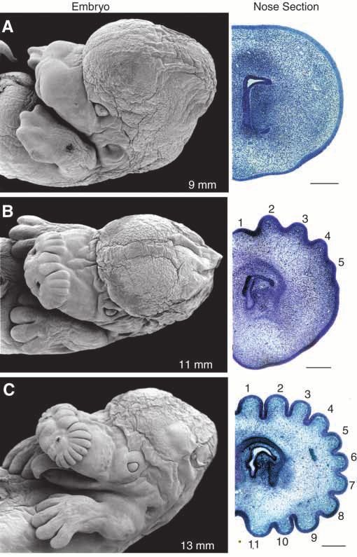
2720 K. C. CATANIA, R. G. NORTHCUTT AND J. H. KAAS
and a number of larger, low-resolution peripheral appendages       electron microscope and serial thin-sectioned for light
(Catania and Kaas, 1995, 1997).                                    microscopy. In addition, innervation patterns were determined
   One might ask whether these nasal structures are comparable     using the lipophilic neuronal tracer 1,1′-dioctadecyl-3,3,3′,3′-
as ‘appendages’ to the body-wall extensions of other               tetramethylindocarbocyanine perchlorate (DiI) (Molecular
metazoans. Though of relatively recent origin, an examination      Probes Inc.) and a scanning confocal microscope. For scanning
of their anatomy reveals them to be elaborate structures that      electron microscopy, specimens were dehydrated through an
integrate a number of tissue systems for their sensory functions   ethanol series to 100 % ethanol, and then transferred into a
(Catania, 1995a, 1996). The outer surface of each appendage        critical-point dryer where the alcohol was replaced with liquid
is an epidermal layer covered with complex tactile sensory         carbon dioxide. After drying, specimens were sputter-coated
organs termed Eimer’s organs. Each appendage is densely            with gold and viewed in a Cambridge 360 Stereoscan scanning
innervated by a separate branch of the infraorbital nerve, which   electron microscope.
runs through its center. The appendages are moved by a series         To examine the histology of the nose, tissue was post-fixed
of tendons that attach to muscles on the side of the face, and     in 1 % osmium tetroxide, dehydrated in a graded ethanol series
each appendage has its own blood supply (Grand et al., 1998).      and transferred into propylene oxide. Tissue was embedded
In addition, the touch centers of the central nervous system of    in Embed 812 epoxy resin (EM Sciences). Semi-thin
the star-nosed mole are subdivided to represent sensory            serial sections (1–2 µm) were cut transversely on an LKB
information from each appendage in a separate module               ultramicrotome and stained with 1 % Toluidine Blue. To
(Catania and Kaas, 1995, 1996). Thus, these appendages are         determine innervation patterns, the tip of the snout was
clearly on a par with other animal appendages in complexity,       removed from selected paraformaldehyde-fixed specimens,
and their evolution has involved the integrated manipulation of    and DiI crystals were applied to exposed nerve fascicles. After
multiple tissue systems, including the dermis, epidermis,          4 weeks of transport at room temperature, the tissue was
tendons, muscles and peripheral nervous and circulatory            examined under the scanning confocal microscope. All
systems.                                                           procedures were approved by the Vanderbilt University
   Were established mechanisms of appendage morphogenesis          Animal Care and Use Committee and are in compliance with
co-opted to produce this recently evolved set of appendages?       NIH guidelines for the care and use of laboratory animals.
We examined the development of the nasal appendages in
embryonic and juvenile moles and found that they form in a
manner unlike any developmental sequence previously                                             Results
described.                                                            In this study, the development of the nasal appendages is
                                                                   examined for a series of five prenatal (Figs 1–3) and three
                                                                   postnatal stages (Fig. 4). The star is formed by 11 symmetrical
                     Materials and methods                         pairs of appendages surrounding the nostrils. The appendages
   To examine the development of the nasal appendages that         are numbered from 1 to 11 on each side of the face, beginning
form the star, we collected a developmental series of embryos      with the dorsal-most appendage. Figs 2 and 3 show DiI-filled
from wild-caught star-nosed moles (Condylura cristata Illiger      and labeled nerve fibers for selected specimens, illustrating
1811). For post-natal stages, captured pregnant moles were         important stages in the development of innervation patterns.
housed in our laboratory where (for the first time) they gave         The earliest stage described is the 9 mm crown–rump length
birth and raised litters of young in captivity. Star-nosed moles   embryo (Fig. 1A). At this stage, there is no sign of the nasal
are seasonal breeders and give birth to a single litter of young   appendages that will eventually form the star. The tip of the
in the spring (Eadie and Hamilton, 1956). We were able to          nose appears smooth and uniform in the scanning electron
collect pregnant females during March and April using              microscope. Transverse sections through the nasal epidermis
Sherman traps under Pennsylvania scientific collecting permit      (nose sections, Fig. 1A) also reveal a smooth thin epidermis
number COL00087. The day of fertilization is unknown               approximately 40 µm in thickness surrounding the underlying
because mating took place in the wild, and all embryos were        dermis and mesenchyme. At this early stage, the epidermis is
therefore staged by crown–rump length. Moles were killed           contacted by many fascicles of nerve fibers that form a dense
with Nembutal (110 mg kg−1), and embryos were fixed in a           subcutaneous network (Fig. 2A). At high magnification (not
mixture of 1 % paraformaldehyde and 2 % glutaraldehyde in          shown), growth cones are visible at the distal ends of the nerve
phosphate buffer (for scanning electron microscopy or serial       fascicles. But there is no indication, however, from either the
sections) or in 4 % paraformaldehyde in phosphate buffer (for      epidermal tissue or the innervation patterns of how or where
neuronal tract tracing). Postnatal development was examined        the appendages of the star will later form.
in newborn moles from females that gave birth and raised              The first signs of the nasal appendages are seen in 11 mm
young in the laboratory. Pregnant females were kept in moist       embryos (Fig. 1B). A series of rostro–caudally oriented
peat moss, given free access to water and fed night-crawlers       swellings appears along the side of the face. Each swelling is
(Lumbricus terrestris) ad libitum. Juvenile moles were killed      the precursor to a single nasal appendage, and each consists of
and fixed as above.                                                simply a ‘buckling’ or slight wave in the epidermis. The
   Tissue from each stage was examined under the scanning          swellings form first along the midline, and later laterally so that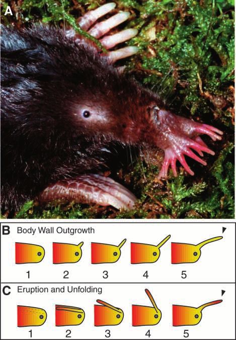
Development of the mole’s star nose 2721
Fig. 1. The embryonic development of the star. (A–E) A series of
successive stages in the prenatal development of the star-nosed mole.
(A) In 9 mm embryos, the nose is undifferentiated and consists of a
smooth thin epidermis. (B) In 11 mm embryos, the primordial
appendages first appear as a series of epidermal swellings. Nose
sections show that these swellings consist of simple ‘waves’ in the thin
epidermis (numbered 1–5). (C) In 13 mm embryos, all 11 primordial
appendages are present as pronounced raised areas of the epidermis.
Although the star appears superficially well formed, the appendages are
still simple waves in the epidermis with no separation from the lumen
of the central snout. (D) In 15 mm embryos, the bottom wall of each
appendage forms as a new deep layer of epidermis (arrow) extends to separate each epidermal wave from the dermis and mesenchyme of the
snout. (E) Just before birth, in 20 mm embryos, there is a proliferation of cells in the epidermis of the snout, and each separate appendage forms
a cylinder embedded in the hypertrophied epidermis (arrowheads). At the same time, the external pattern of appendages on the snout is
obscured. Scale bar, 250 µm.
the precursors to appendages 1–5 and 10 and 11 appear before the side of the face, but this external appearance is an illusion.
the swellings for the lateral appendages. Transverse sections In 13 mm embryos, the appendages are still simple waves or
through the snout reveal the epidermal waves that make up the swellings of the epidermis (see nose section, Fig. 1C) and are
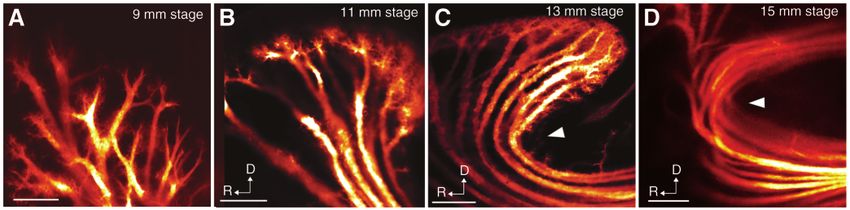
appendages (Fig. 1B). At these early stages, the myelin-poor open to the mesenchyme of the central snout. The epidermis
nerve fascicles are not apparent in light microscopy, but DiI of the waves has become more densely innervated from in-
labeling reveals a very dense innervation of the epidermis. The growing fibers, and the nerve fascicles are well segregated to
crest of each epidermal wave is contacted by numerous nerve individual crests in the epidermis (Figs 2C, 3B). At high
fascicles that project from the center of the snout (Fig. 2B). magnification, there is not yet any sign of the orderly
The simple conformational change of the epidermis into crests arrangements of nerve fibers that characterize the punctate
and troughs seems to be the first foundation for the selective epidermal sensory organs in the adult skin (Catania, 1995a).
elimination or preservation of innervating nerve fibers. Where Instead, the nerve fascicles end in amorphous collections of
the waves have formed, nerve fibers densely contact the crests terminals extending numerous filopodia and often distinct
of the waves and are sparse in the troughs (Fig. 3A1), but in growth cones (arrowhead, Fig. 3B2).
the lateral areas where the waves have not yet formed, there is During these stages (13–15 mm crown–rump), a complex
a uniform distribution of nerve fibers (Fig. 3A2). sequence of tissue growth and movement takes place,
In 13 mm and 15 mm embryos, all 22 appendages of the star separating each of the epidermal waves into a distinct unit with
are clearly apparent (Fig. 1C,D). Here, the star appears a lumen and surrounding walls. A new deep layer of epidermis
remarkably adult-like, as if the appendages were folded against begins to grow beneath the innervated epidermal waves. This
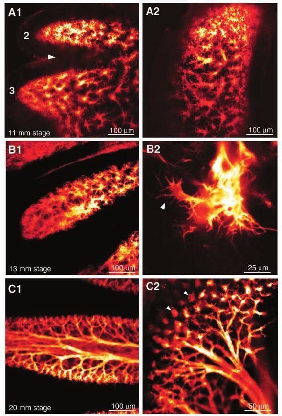
2722 K. C. CATANIA, R. G. NORTHCUTT AND J. H. KAAS

Fig. 2. The innervation of the nose at selected developmental stages, revealed by the lipophilic neuronal tracer DiI. (A) In 9mm embryos, there is
a uniform innervation of the epidermal walls that form the snout, but no pattern that reflects the appendages. (B) In 11 mm embryos, the crests of
a series of epidermal waves (see Fig. 1B) are densely innervated from below. The innervation of a single epidermal crest is viewed from the side,
showing the many nerve fascicles terminating at the skin surface. (C) The bottom epidermal wall of each appendage forms from caudal to rostral.
In 13 mm embryos, just prior to the growth of this lower layer of epidermis (see arrowhead, Fig. 1D), the deep portion of the innervating fascicles
begins to move rostrally (arrowhead). (D) In 15 mm and later embryos, the innervating nerve fascicles must make a complete
‘U                                         -turn’ to go around the deep layer of epidermis and reach their peripheral skin targets (see also Fig. 1E).

Fig. 3. Refinement of innervation patterns during the
course of development. (A1,2) In 11 mm stage embryos,
two different areas of the nose reveal the beginnings of
appendage-specific innervation patterns. In A1, an area
where the epidermal crests and troughs have formed
shows dense innervation of the crest and loss of
innervation from the trough (arrowhead) between the
precursors to appendages 2 and 3. In a more lateral area
(A2), where crests and troughs have not yet formed,
there is a uniform pattern of innervation. (B1,2) At a
later stage, the innervation of the crests is more refined
(B1). Higher magnification (B2) reveals the presence of
numerous filopodia and growth cones (arrowhead). The
adult pattern of punctate neurite distribution has not yet
emerged. (C1,2) In 20 mm embryos, the epidermal
cylinders of each appendage have formed, and the
internal walls are very densely innervated by many nerve
fascicles (C1). A punctate distribution of neurites has
formed in a pattern similar to the adult distribution (C2).
The clusters of nerve terminals reflect the future
distribution of epidermal sensory organs in the adult (see
Catania, 1995a,b and arrowheads in Fig. 6).
Development of the mole’s star nose 2723

Fig. 4. Postnatal development of the star. (A) In newborn moles, the appendages remain embedded in the epidermis of the face. (B) At
approximately 1 week, the appendages emerge from the side of the face as the surrounding epidermis sloughs. (C) Shortly thereafter the
appendages detach and bend forward into the adult pattern. As a result of this ‘backward’ formation, the rostralmost tissue of the adult nose is
derived from the caudalmost tissue of the embryonic nose (see Fig. 5C). Scale bars, 500 µm.

layer forms the bottom wall of each appendage, separating the             With these processes complete, the basic outline of the
connective tissue of the dermis within each appendage from                appendages is in place. Each now consists of a separate
the dermis and mesenchyme that form the center of the snout               cylindrical unit with a densely innervated epidermal wall
(arrow, Fig. 1D). The deep epidermal layer grows from the                 (Fig. 3C1). At this stage (20 mm), the nerve terminals that
posterior snout towards the anterior snout, and the troughs of            contact the skin surface form a series of clusters in a periodic
the waves simultaneously extend slightly downward to meet                 pattern consistent with the distribution of sensory organs seen
and fuse with the new layer of epidermis. At the same time,               later in the adult (arrowheads, Fig. 3C2).
the deep portions of the innervating nerve fibers move ahead                 In newborn moles (Fig. 4A), the appendages remain
of the extending epidermis to maintain contact with their                 embedded in the facial epidermis, giving an external
targets (arrowheads, Fig. 2).                                             appearance that is surprisingly less adult-like than previous
   Towards the end of this process, in 20 mm embryos (just                embryonic stages. But the nasal appendages soon emerge from
prior to birth), there is a proliferation of epidermal cells that
seems to involve the entire rostral snout. As a result, each
primordial appendage forms a separate epidermal cylinder
within the hypertrophied epidermis (arrowheads, Fig. 1E) open
to the deep connective tissue only at the rostral end, near the
nares. The many nerve fascicles innervating an appendage
must pass through this small remaining opening, making a ‘U-
turn’ to reach targets within the appendage (arrow, Fig. 2D).

Fig. 5. An adult mole and two ways that nasal appendages might
develop. (A) A star-nosed mole emerges from its tunnel, showing the
unusual star consisting of 22 fleshy appendages. The appendages are
densely innervated epidermis, have a rich blood supply and are
moved by tendons that connect to facial muscles. (B) A simplified
diagram of appendage formation through body-wall outgrowth.
Some variation of body-wall outgrowth is a basic stage in the
formation of nearly all animal appendages. This mechanism may be
conserved from a Precambrian ancestor of modern metazoans (see
text). (C) The unique developmental sequence for the nasal
appendages of the star. Rather than developing by outgrowth, they
begin as longitudinal cylinders embedded in the side of the face. The
cylinders of epidermis subsequently emerge and rotate forward, so
that the original embryonic orientation of the precursor tissue is
reversed. The colors denote the embryonic origins of the tissues that
make up the appendages in each of the two processes. During the
formation of the nasal appendages, caudal embryonic tissue of the
snout (red) detaches and becomes the most rostral tissue on the adult
nose. This is contrasted with the result of body wall outgrowth
(arrowheads).
2724 K. C. CATANIA, R. G. NORTHCUTT AND J. H. KAAS

Fig. 6. The adult nose of Townsend’s mole (Scapanus townsendi)
showing separate raised subdivisions of epidermis on the side of the
                                                                        Fig. 7. Two examples of common abnormalities of the star. (A) A
face. The star may have evolved from a similar ancestral condition
                                                                        nose with an extra nasal appendage on the animal’s left side. Sudden
through progressive elevation of the raised epidermis, which contains
                                                                        duplications such as this could provide the basis for progressive
numerous sensitive touch organs called Eimer’s organs (small raised
                                                                        elaboration from an ancestor with fewer appendages (see Fig. 6).
domes, arrowheads). The same small receptor organs cover the
                                                                        (B) A nose missing a nasal ray (although there is a partial duplication
appendages of the star-nosed mole (Catania, 1995a,b). The receptors
                                                                        of the tip of ray 6). We estimate that approximately 5–6 % of the
in Scapanus townsendii are elevated relative to the nearby caudal
                                                                        population has an abnormal nose. Clearly, there is relatively
epidermis that does not contain sensory organs, which may allow for
                                                                        widespread variation of the nasal anatomy upon which selection
increased tactile receptivity for the sensitive touch organs. Compare
                                                                        might act, although we have not explored the genetic basis of these
the appearance of the nose of this adult mole with the early
                                                                        variations.
embryonic stage in Fig. 1B.

the side of the face, presumably through programmed cell                tetrapods, a limb bud extends from the body wall to
death, as the once hypertrophied epidermis sloughs from                 successively produce differentiated cells in a proximal-to-
around the appendages (Fig. 4B). Roughly 1 week after birth,            distal sequence of limb elements (Hinchliffe and Johnson,
the appendages break free from the side of the face and rotate          1980). Later, the individual digits are initially sculpted by the
forward to form the adult star (Figs 4C, 5A).                           selective death (Hamburger and Hamilton, 1951) of
   To summarize this complex developmental sequence                     intervening cells in the handplate, and this is followed by
(Fig. 5C), the nasal appendages form and emerge from the side           growth (Martin, 1990). Several stages of tetrapod digit
of the face and then rotate forward to form the adult pattern.          formation are shown alongside the nose formation in the mole
As a result of this ‘backwards’ formation, the rostral tissue of        embryos (Fig. 1A–C) and their development can be contrasted
the appendages is derived from caudal embryonic tissue of the           with nasal appendage development.
snout while the caudal tissue of the appendages is derived from            Tetrapod digits form by separation from a flattened
the rostral embryonic tissue of the snout. There is essentially         outgrowth or handplate and retain their original embryonic
no outgrowth phase in this developmental sequence, and it               orientation. In contrast, the nasal appendages of the star-nosed
results in a reversal of the original embryonic orientation of the      mole unfold from the side of the face. This process requires
tissue that forms the appendages. This developmental sequence           the growth of a new deep layer of epidermis to form both the
is manifestly different from the formation of other animal              bottom wall of the appendages and the remaining side of the
appendages (Fig. 5B).                                                   face from which the appendages later emerge. It also requires
                                                                        the rearrangement of innervating fibers (Fig. 2) and the rich
                                                                        blood supply to the appendages (not shown), and results in the
                         Discussion                                     previously described reversal of the orientation of precursor
   The development and evolution of animal appendages have              tissues (Fig. 5C). What might account for such an unusual, and
been extensively studied in a number of species. In each case,          seemingly inefficient, way of constructing the nasal
animal appendages originate as outgrowths of the body wall.             appendages?
Well-known examples include the embryonic formation of                     A remarkable feature of appendage development is the
arthropod appendages and the tetrapod limb (for an account of           conservation of gene expression during morphogenesis in
postnatal horn and antler development, see Goss, 1983). The             different phyla. Homologous genes provide anterior–posterior
legs, antennae, mouthparts and wings of insects form from               (hedgehog/Sonic hedgehog) and proximal–distal (fringe/
imaginal disks that telescope outwards to produce each                  Radical fringe) patterning information in both insects and
respective structure (Gerhart and Kirschner, 1997b). In                 tetrapods (Shubin et al., 1997). The Distal-less homeotic gene
Development of the mole’s star nose 2725
or its homologue are expressed during appendage formation in unpublished data). This demonstrates that sufficient variability
diverse animal phyla, e.g. during the formation of echinoderm exists upon which selection might act to increase (or decrease)
tube feet, onychophoran lobopodia, tetrapod limbs, insect the number of nasal appendages. It also shows that there seems
appendages and sea urchin spines (Panganiban et al., 1997). to be a surprisingly high degree of variation in this presumably
These studies (Panganiban et al., 1997; Raff, 1996; Shubin et critical structure, which contrasts with a much lower rate of
al., 1997) suggest that the genetic machinery for appendage mutation in the tetrapod limb (Castilla et al., 1996; Zguricus et
formation is phylogenetically ancient and may have helped al., 1998). Darwin (1859) made special note of such examples
signal and regulate the formation of body-wall outgrowths in in support of the Origin of Species, stating “in those cases in
a Precambrian ancestor. which the modification has been comparatively recent and
In the star-nosed mole, however, the development of the extraordinarily great... we ought to find the generative
recently evolved nasal appendages suggests precursors of a variability, as it might be called, still present in a high degree”.
different form, such as sheets of sensory receptors or ‘proto- The argument was that selection has had less time to ‘fix’ the
appendages’ on the side of an ancestor’s snout, which became characteristics of recently evolved complex structures, which
progressively elevated over successive generations. Such a are the recent product of selection for variations.
recapitulatory argument (Dawkins, 1987; Gould, 1977;
Northcutt, 1990) is appealing, but would be more compelling Flexibility or historical constraint?
with fossil evidence or supporting comparative data. Although At one level, these results demonstrate the flexibility of
no fossilized mole noses have been found, nearly all other evolutionary processes. While there seems to be a remarkable
extant moles have sheets of the same complex sensory receptors conservancy in the basic mechanisms of appendage
(Eimer’s organs) making up the epidermis of their snout in a development across metazoan phyla, there is also clearly the
small, uniform epithelium around the unspecialized nares potential for new developmental solutions to this ancient
(Catania, 1995b; Quilliam, 1966; Shibanai, 1988). But in one morphogenetic problem. It has been stated that it is difficult to
North American species (Townsend’s mole Scapanus think up anything very different from some actual product of
townsendi), however, we discovered a set of ‘proto- evolution (Maynard Smith, 1990). Here, we find a product of
appendages’ extending caudally on the snout (Fig. 6). The evolution (the developmental sequence of the star) that we
resemblance of the adult nose of this mole to the early might find hard even to imagine prior to this investigation. This
embryonic stages of the star-nosed mole (Fig. 1B) is striking. is perhaps because the developmental sequence of the nasal
This species might be called the Archaeopteryx of moles, appendages is not only unusual, but might also be considered
representing fairly well what would be expected of an inefficient. To use an old example, it is difficult to envisage an
intermediate form between the least specialized moles and the engineer proposing such a literally ‘backwards’ manner for
enigmatic star-nosed mole. Of course, this extant species is not development of the nasal appendages.
an ancestor of the star-nosed mole and, in fact, studies of mole Thus, at a different level of analysis, the development of the
phylogeny suggest that its nose subdivisions evolved nose seems to reflect the constraints imposed by biological
independently of the appendages of the star-nosed mole history (Gould, 1977, 1979, 1980). Perhaps there are more
(Whidden, 1995; Yates and Moore, 1990). But Scapanus efficient ways to produce the nasal appendages, such as the
townsendii does illustrate the tenability of such a hypothetical outgrowth of the body wall seen for other metazoan
ancestral species. Taken together with the developmental data, appendages, but evolution cannot plan ahead, it can only
it seems reasonable to propose that the star evolved through the ‘tinker’ with the materials at hand (Jacob, 1977). The ontogeny
progressive elevation of strips of sensory receptors on the side of the star provides a classic example of the outcome of such
of the face, and that the stages of these ancestral ontogenies tinkering. While different developmental stages may be altered
have been conserved and built upon to produce the star. The during the course of evolution (for a review, see Gould, 1977),
result is a novel developmental sequence that seems to several early stages of the development of the nose of the mole
recapitulate many stages of ancestral anatomy (Gould, 1977). may be recalcitrant to change. For example, the crests of the
Scapanus townsendi has only eight subdivisions on its face, epidermal waves may produce the neurotrophic factors that
fewer than the 22 modules (11 per side) found on the star-nosed attract extending neurites to their appropriate appendage-
mole. Once the basic developmental mechanism for appendage specific targets. This possibility seems to be strengthened by
production was in place, however, they might simply be the presence of growth cones extending numerous filopodia
duplicated to produce the 22 appendages of the star. Such beneath the crests, up to at least 13 mm stage embryos
meristic changes are a common occurrence in evolution (Raff, (Fig. B2). This ‘crest-and-trough’ stage may have existed in
1996) since duplication of somatic modules provides an the ancestor, which presumably had a snout similar to
efficient means of adding to the body plan without the need to Scapanus townsendi (Fig. 6). If this and other stages of the
reinvent the regulatory elements that produce each module. In development were critical during ancestral ontogenies, as
fact, duplications of nasal appendages are common in wild seems likely, then alteration of the nose may have occurred
populations of star-nosed moles (Fig. 7). We estimate that over primarily through modifications at the terminal end of ancestral
5 % of the population has an abnormal nose, consisting ontogenies, resulting in the apparent recapitulation of nasal
of either extra or fewer nasal appendages (K. C. Catania, appendage evolution during development.2726 K. C. CATANIA, R. G. NORTHCUTT AND J. H. KAAS
This work was supported by NIH grants MH58909 to proboscis and rays of the star-nosed mole, Condylura cristata. J.
K.C.C. and NS 16446 to J.H.K. Mammal. 79, 492–501.
Hamburger, V. and Hamilton, H. L. (1951). A series of normal
stages in the development of the chick embryo. J. Morph. 88, 49–92.
References Hinchliffe, J. R. and Johnson, D. R. (1980). The Development of the
Averof, M and Cohen, S. M. (1997). Evolutionary origin of insect Vertebrate Limb. An Approach through Experiment, Genetics and
wings from ancestral gills. Nature 385, 627–630. Evolution. Oxford: Oxford University Press.
Basler, D. and Struhl, G. (1994). Compartment boundaries and the Jacob, F. (1977). Evolution and tinkering. Science 196, 1161–1166.
control of drosophila limb pattern by hedgehog protein. Nature 368, Martin, P. (1990). Tissue patterning in the developing mouse limb.
208–214. Int. J. Dev. Biol. 34, 323–326.
Castilla, E. E., Lugarinho da Fonseca, R., Dutra, M., Bermejo, E., Maynard Smith, J. (1990). Concluding remarks. In Organizational
Cuevas, L. and Martínez-Frías, M. (1996). Epidemiological Constraints on the Dynamics of Evolution (ed. J. Maynard Smith
analysis of rare polydactylies. Am. J. Med. Genet. 65, 295–303. and G. Vida), pp. 433–437. Manchester: Manchester University
Catania, K. C. (1995a). The structure and innervation of the sensory Press.
organs on the snout of the star-nosed mole. J. Comp. Neurol. 351, Moore, D. W. (1986). Systematic and biogeographic relationships
536–548. among the Talpinae (Insectivore: Talpidae). PhD dissertation,
Catania, K. C. (1995b). A comparison of the Eimer’s organs of three Albuquerque, University of New Mexico, USA.
North American moles: the star-nosed mole (Condylura cristata) Northcutt, R. G. (1990). Ontogeny and phylogeny: A re-evaluation
the hairy-tailed mole (Parascalops breweri) and the eastern mole of conceptual relationships and some applications. Brain Behav.
(Scalopus aquaticus). J. Comp. Neurol. 354, 150–160. Evol. 36, 116–140.
Catania, K. C. (1996). Ultrastructure of the Eimer’s organ of the star- Panganiban, G., Irvine, S. M., Lowe, C., Roehl, H., Corley, L. S.,
nosed mole. J. Comp. Neurol. 36, 343–354. Sherbon, B., Grenier, J. K., Fallon, J. F., Kimble, J., Walker,
Catania, K. C. and Kaas, J. H. (1995). The organization of the M., Wray, G. A., Swalla, B. J., Martindale, M. Q. and Carroll,
somatosensory cortex of the star-nosed mole. J. Comp. Neurol. 351, S. B. (1997). The origin and evolution of animal appendages. Proc.
549–567. Natl. Acad. Sci. USA 94, 5162–5166.
Catania, K. C. and Kaas, J. H. (1996). The unusual nose and brain Panganiban, G., Sebring, A., Nagy, L. and Carroll, S. (1995). The
of the star-nosed mole. BioSci. 46, 578–586. development of crustacean limbs and the evolution of arthropods.
Catania, K. C. and Kaas, J. H. (1997). Somatosensory fovea in the Science 270, 1363–1366.
star-nosed mole: behavioral use of the star in relation to innervation Panganiban, G., Nagy, L. and Carroll, S. B. (1994). The role of the
patterns and cortical representation. J. Comp. Neurol. 387, Distal-less gene in the development and evolution of insect limbs.
215–233. Curr. Biol. 4, 671–675.
Darwin, C. (1859). On the Origin of Species by Natural Selection, or Popadic, A., Panganiban, G., Rusch, D., Shear, W. A. and
the Preservation of Favored Races in the Struggle for Life. London: Kaufman, T. C. (1998). Molecular evidence for the gnathobasic
John Murray. 191pp. derivation of arthropod mandibles and for the appendicular origin
Dawkins, R. (1987). The Blind Watchmaker. New York: W. W. of the labrum and other structures. Dev. Genes Evol. 208, 142–150.
Norton & Co. Inc. pp. 91–92. Quilliam, T. A. (1966). The mole’s sensory apparatus. J. Zool., Lond.
Eadie, W. R. and Hamilton, W. J. (1956). Notes on reproduction in 149, 76–88.
the star-nosed mole J. Mammal. 37, 223–231. Raff, R. A. (1996). The Shape of Life. Chicago: University Chicago
Ferrari, D., Sumoy, L., Gannon, J., Sun, H., Brown, A. M., Press.
Upholt, W. B. and Kosher, R. A. (1995). The expression pattern Shibanai, S. (1988). Ultrastructure of the Eimer’s organs of the
of the Distal-less homeobox-containing gene Dlx-5 in the Japanese shrew mole Urotrichus talpoides (Insectivora,
developing chick limb bud suggests its involvement in apical Mammalia) and their changes following infraorbital axotomy.
ectodermal ridge activity, pattern formation, and cartilage Anat. Anz. Jena 165, 105–129.
differentiation. Mech. Dev. 52, 257–264. Shubin, N., Cliff, T. and Carroll, S. (1997). Fossils, genes, and the
Garstang, W. (1922). The theory or recapitulation: a critical evolution of animal limbs. Nature 388, 639–648.
restatement of the biogenic law. Zool. J. Linn. Soc. Lond. 35, Skoczen, S. (1993). New records of Parascalops, Neurotrichus and
81–101. Condylura (Talpinae, Insectivora) from the Pliocene of Poland.
Gerhart, J. and Kirschner, M. (1997a). Cells, Embryos and Acta Ther. 38, 125–137.
Evolution. Malden, MA: Blackwell Science Inc. pp. 574–575. Whidden, H. P. (1995). The comparative myology of North
Gerhart, J. and Kirschner, M. (1997b). Cells, Embryos and American moles with comments on the phylogeny of the Talpidae.
Evolution. Malden, MA: Blackwell Science Inc. pp. 555–567. PhD dissertation, University of Massachusetts, Amherst, USA.
Goss, R. J. (1983). Deer Antlers: Regeneration, Function and Williams, T. A. (1998). Distalless expression in crustaceans and the
Evolution. New York: Academic Press. patterning of branched limbs. Dev. Genes Evol. 207, 427–434.
Gould, S. J. (1977). Ontogeny and Phylogeny. Cambridge, MA: Yates, T. L. and Moore, D. W. (1990). Speciation and evolution in
Harvard University Press. the family Talpidae (Mammalia: Insectivora). In Evolution of
Gould, S. J. (1979). The spandrels of San Marco and the Panglossian Subterranean Mammals at the Organismal and Molecular Levels.
paradigm: a critique of the adaptationist programme. Proc. R. Soc. New York: Wiley-Liss. pp. 1–22.
Lond. B 205, 581–598. Zguricus, J., Bakker, W. F., Heus, H., Lindhout, D., Heutink, P.
Gould, S. J. (1980). The Panda’s Thumb. New York: W. W. Norton and Hovious, S. E. (1998). Genetics of limb development and
& Company. congenital hand malformations. Plast. Reconstr. Surg. 101,
Grand, T., Gould, E. and Montali, R. (1998). Structure of the 1126–1135.You can also read