The eyes of suckermouth armoured catfish (Loricariidae, subfamily Hypostomus): pupil response, lenticular longitudinal spherical aberration and ...
←
→
Page content transcription
If your browser does not render page correctly, please read the page content below
The Journal of Experimental Biology 205, 3425–3433 (2002)                                                                                     3425
Printed in Great Britain © The Company of Biologists Limited 2002
JEB4345

       The eyes of suckermouth armoured catfish (Loricariidae, subfamily
   Hypostomus): pupil response, lenticular longitudinal spherical aberration and
                               retinal topography
                                       Ron H. Douglas1,*, Shaun P. Collin2,3 and Julie Corrigan1
 1Applied  Vision Research Centre, Department of Optometry & Visual Science, City University, Northampton Square,
       London EC1V 0HB, UK, 2Department of Anatomy & Developmental Science, School of Biomedical Science,
           The University of Queensland, Brisbane 4072, Queensland, Australia and 3Anatomisches Institut,
                        Universität Tübingen, Österbergstrasse 3, Tübingen, 72074, Germany
                                                      *Author for correspondence (e-mail: r.h.douglas@city.ac.uk)

                                                                      Accepted 8 August 2002

                                                        Summary
   The dilated, round pupils of a species of suckermouth     examination of the catfish retina shows the photoreceptors
armoured catfish (Liposarcus pardalis) constrict slowly on   to be exclusively single cones interspersed with elongate
illumination (over 35–40 min) to form crescent-shaped        rods and demonstrates the presence of multiple optic
apertures. Ray tracing of He–Ne laser beams shows            nerve head papillae. Two areas of high ganglion cell
that the lenses of a related species (Pterygoplichthys       density, each side of a vertically oriented falciform
etentaculus), which also has a crescent-shaped pupil, are    process, provide increased spatial resolving power along
well corrected for longitudinal spherical aberration,        the axes examining the substrate in front of and behind
suggesting that the primary purpose of the irregular pupil   the animal.
in armoured catfish is not to correct such aberration. It is
suggested that the iris operculum may serve to camouflage    Key words: catfish, pupil, retina, iris, aberration, Liposarcus
the pupil of these substrate-dwelling species. An            pardalis, Pterygoplichthys etentaculus.

                                   Introduction
   The pupil of the majority of teleost fish, unlike in most other                   will be very different to those of P. notatus, because although
vertebrates, does not change in size in response to variation in                     the fully dilated pupil is, as in most other vertebrates, round,
ambient illumination, and the iris is generally assumed to be                        when constricted it takes on the shape of a ‘crescent moon’
immobile. However, a review of earlier literature shows that                         owing to the presence of a dorsal iris operculum.
some teleosts are, in fact, able to change the size of their pupil                      Iris opercula are common among some elasmobranch fish,
quite considerably, and recently we provided the first                               such as skates and batoid rays (Beer, 1894; Franz, 1905, 1931;
quantitative assessment of such changes in fish, describing                          Young, 1933; Walls, 1963; Kuchnow and Martin, 1970;
pupillary mobility in the marine plainfin midshipman                                 Kuchnow, 1971; Gruber and Cohen, 1978; Nicol, 1978; Collin,
Porichthys notatus that was as rapid and extensive as that in                        1988), and also occur in some bottom-dwelling teleosts
humans (Douglas et al., 1998).                                                       (Bateson, 1890; Beer, 1894; Walls, 1963; Munk, 1970; Collin
   The aim of the present study was to quantify the pupil                            and Pettigrew, 1988a; Nicol, 1989). Despite the relatively
responses of another group of teleosts; the suckermouth                              widespread occurrence of such asymmetric pupils, their
armoured catfish, which belong to the Loricariid family of                           function remains uncertain. One explanation is that they may
Siluriform catfish resident in the freshwaters of Panama and                         reduce the effect of any longitudinal spherical aberration the
South America. Specifically, we investigated members of the                          lens might possess by restricting light to the lens periphery
subfamily Hypostomus, which, along with some other                                   (Murphy and Howland, 1991). Most fish suffer little from
members of the Loricariidae, are popularly referred to by the                        longitudinal spherical aberration owing to a refractive index
generic name ‘Pleco’ or ‘Plecostomus’, but, in fact, comprise                        gradient within the lens (see Sivak, 1990 for a review).
over 100 species in approximately 18 genera (Nelson, 1994).                          However, if crescent-shaped pupils do function primarily to
This group was chosen for study because Walls (1963)                                 reduce such aberration, one might expect the lenses of animals
suggested that they may show extensive pupil mobility,                               with these pupils to be poorly corrected for it. We therefore
although no details of the extent and speed of migration were                        determined the longitudinal spherical aberration of the lens in
provided. Clearly, however, the pupil responses of these catfish                     a species of suckermouth armoured catfish.
3426 R. H. Douglas, S. P. Collin and J. Corrigan
Finally, the retinal ganglion cell topography of these catfish solution containing small amounts of scatter liquid concentrate
was assessed. Although few studies have investigated the (Edmund Scientific, Barrington, USA). The beam of an He–Ne
density of retinal ganglion cells with crescent-shaped laser (emission maximum 632.8 nm) was then passed through
pupils, the crescent-shaped iso-density contours in retinal the lens along the antero-posterior axes. A camera positioned
wholemounts of the sharp-nosed weaver Parapercis cylindrica at the side of the lens was used to ensure that the laser beam
(Teleostei; Collin and Pettigrew, 1988a) and the shovel-nosed passed through its vertical midpoint. This was achieved by
ray Rhinobatos batillum (Elasmobranchii; Collin, 1988) in adjusting the height of the laser beam until it was not deflected
conjunction with a similarly shaped pupil suggest that a by the lens from the horizontal plane. The laser beam was then
relationship may exist. filmed from above while traversing the lens in the horizontal
plane. Individual video frames were analysed using software
to determine the back vertex distance (BVD) for a number of
Materials and methods beam entry positions. A non-linear regression was applied to
Animals the data using the following equation for back vertex distance
All animals were obtained from local aquarium suppliers in (x): x=a+by2+cy4, where a represents back vertex focal length,
London, UK and Tübingen, Germany and identified by the b represents the 3rd order spherical aberration, c represents the
British Museum of Natural History, London. 5th order spherical aberration, and y represents the normalised
beam entry position. This equation represents the longitudinal
Measurement of pupil response ray aberration of a rotationally symmetric optical system on
Three individual Liposarcus pardalis Castelnau 1855 axis.
[standard lengths (SL) 140–150 mm] were held in a 12 h:12 h
L:D cycle for at least 3 months. To examine their pupil Retinal structure and ganglion cell topography
response, they were removed from their home tanks during the The eyes of a single L. pardalis (SL 220 mm) were
dark phase of their L:D cycle using a dim red torch and placed embedded in resin for light microscopy, and two interrupted
in a small aquarium. Following 1 h of acclimation to this tank, series of 0.5 µm sections were cut on an ultramicrotome and
pupillary responses were filmed for 60 min using infrared stained with Toluidine Blue. One eye was serially sectioned in
illumination during continual exposure to one of 13 intensities the dorso-ventral axis and the other along the naso-temporal
of white light. Each fish was examined at the same time each axis.
day to avoid any circadian influences on the pupil response, Both eyes of two individuals of Liposarcus multiradiatus
receiving only one light exposure per day. As these animals Hancock, 1828 (SL 88 mm) and one L. pardalis individual
naturally tend to stay motionless in the light, the only form of (SL 158 mm) were used for topographic analyses of retinal
restraint necessary during filming was a Perspex ‘tent’ placed ganglion cell distribution. After 3 h of dark adaptation and
over them. Stimuli were delivered from directly overhead, via immersion in a lethal dose of MS 222, eyes were enucleated
a shutter-controlled opening and a mirror, from a Kodak with at least 2–3 mm of the optic nerve still attached.
projector located in an adjacent room, which also housed Extraocular muscle tissue was removed, with care taken not to
all recording apparatus. The intensity of illumination was puncture the eyecup. While immersed in oxygenated teleost
controlled by neutral density filters. One eye of each animal Ringer solution, a further 1–2 mm of the optic nerve was
was videotaped using an infrared-sensitive camera (Cohu, San removed, and the remaining nerve stump swabbed dry with
Diego, USA) positioned in a plane parallel to the cornea. Pupil a tissue wick. Crystals of fluorescein-conjugated dextran
area was subsequently determined from individual video (3000 MW, anionic, lysine-fixable; Molecular Probes, Leiden,
frames using NIH-image. To facilitate comparison among The Netherlands) were then applied to the entire diameter of
individuals of different eye size, all measurements were the lesioned optic nerve. After approximately 5 min, to allow
expressed relative to the fully dilated pupil area of each animal uptake of the dextran label, the whole eye was re-immersed in
just prior to experimental light exposure. oxygenated Ringer solution for 24 h at 21°C.
Following incubation, the limbus of each eye was pierced
Determination of lens longitudinal spherical aberration and the cornea and lens were removed. A small dorsal incision
Following immersion in a lethal dose of methane tricaine was made in the retina for orientation. The eyecup was
sulfonate salt (MS 222), both lenses were removed from a immersion fixed in 4% paraformaldehyde in 0.1 mol l–1
single Pterygoplichthys etentaculus Spix and Agassiz 1829 phosphate buffer (pH 7.4) for 40 min before dissection in
(SL 200 mm). P. etentaculus, whose pupil is also crescent 0.1 mol l–1 phosphate buffer. Each retina was removed from its
shaped when constricted, was preferred to L. pardalis for this scleral eyecup and wholemounted (ganglion cell layer
part of the study owing to its larger size. The lenses of P. uppermost) on a subbed slide (double-dipped in 5% gelatin),
etentaculus, which, like those of other suckermouth catfish (L. covered in either Fluoromount (Calbiochem, San Diego, USA)
pardalis, Glyptoperichthys lituratus and Sturisomatichthys sp.; or 0.1 mol l–1 phosphate buffer and coverslipped. Dehydration
R. H. Douglas and S. P. Collin, unpublished data), are antero- was inhibited by sealing the edges of the coverslip with nail
posteriorly flattened, were glued to the tip of a fine pipette, polish. One retina of L. multiradiatus was wholemounted and
placed in a small glass tank and immersed in a teleost Ringer prepared as above but left overnight in a humidified Petri dishThe eyes of catfish 3427
1.0
1.0 Complete darkness
Minimum relative pupil area
0.9 8.4×10⁻² µW cm–2
0.8
Relative pupil area
0.8
0.7 1.37 µW cm–2 0.6
0.6
4.6×101 µW cm–2 0.4
0.5
0.4 2.65×10² µW cm–2
0.2
5.6×10³ µW cm–2 1.00E–02 1.00E+00 1.00E+02 1.00E+04
0.3
0 20 40 60 80 log corneal irradiance
Time (min)
Fig. 2. Minimum pupil area in response to different intensities of
Fig. 1. Pupil response of a Liposarcus pardalis individual during white light. Corneal irradiance is measured in µW cm–2. The
60 min exposure to different intensities of white light. different symbols represent data from three individual Liposarcus
pardalis.
before being stained for Nissl substance with 0.05% Cresyl Once constricted, the pupil remained so for the duration of the
Violet for 3 min. The wholemount was then dehydrated in experiment, showing no signs of re-dilating in continual
a series of alcohols and mounted in DPX to reveal the illumination (Fig. 1).
distribution of glial cells. Pupillary constriction in L. pardalis consists of two
Topographic analysis of the retrogradely labelled ganglion components; a general reduction in the diameter of the pupil
cells was carried out following the protocol of Collin and and the outgrowth of an operculum from the dorsal margin of
Northcutt (1993), where up to 200 regions per retina (50–60% the iris (Fig. 3). Consequently, while the fully dilated pupil of
of the retinal area) were sampled, allowing small fluctuations L. pardalis is more or less round (Fig. 4A), when constricted
in density to be noted. Iso-density contours were constructed it appears as a ‘crescent moon’ with an irideal flap obscuring
by joining areas of similar cell density. Retinal shrinkage was the central pupil (Fig. 4B). In general, especially at higher light
assumed to be minimal because all retinae were examined levels, the decrease in overall pupil diameter occurs more
while hydrated. The total number of labelled ganglion cells rapidly than the increase in opercular area (Fig. 3).
was calculated by multiplying the average cell density between
each iso-density contour by its area. Area measurements were Longitudinal spherical aberration of the lens
calculated by scanning the topography map into a PC The lenses of P. etentaculus are well corrected for
and analysing the area of each contour using NIH-Image. longitudinal spherical aberration, showing only relatively
Retrogradely labelled ganglion cells were viewed and small differences in back vertex distance (BVD) for laser
photographed on a Zeiss Axiophot 135M
fluorescence microscope (Jena, Germany) fitted
with a fluorescein filter block using Kodak Tmax 1.2
400 ASA film.
1.0
Normalized dimension
Results 0.8
Pupil response 0.6
Fig. 1 shows the pupil response of a single L.
pardalis individual to six different intensities of 0.4
illumination. The maximum amount of pupil
constriction as a function of light intensity for all 0.2
three L. pardalis examined is shown in Fig. 2.
The threshold to elicit a measurable pupil 0
0 10 20 30 40 50 60 70
response lies between a corneal irradiance of
2.9×10⁻² µW cm–2 and 8.4×10⁻² µW cm–2 for all Time exposed to light (min)
animals. Overall, the response of the pupil to light Fig. 3. Pupil response of a Liposarcus pardalis individual to 60 min exposure to
was very slow. The time taken for half maximal white light at an intensity of 5.6×10³ µW cm–2. The three curves represent
contraction (t0.5max) was in the order of 1–8 min, normalized lines for area of irideal operculum (squares), pupil area (crosses) and
with full constriction usually taking 35–45 min. horizontal pupil diameter at its widest point (triangles).3428 R. H. Douglas, S. P. Collin and J. Corrigan
A 1 A
0.5
BVD (mm)
0
0.5 1 1.5 2 2.5 3
–0.5
Normalized beam entry
–1
1 B
0.5
BVD (mm)
0
B 0.5 1 1.5 2 2.5 3
–0.5
–1
Fig. 5. Back vertex distance (BVD) as a function of entry position of
an He–Ne laser beam into the (A) right (1.65 mm×1.88 mm diameter)
and (B) left (1.63 mm×1.85 mm diameter) lens of a Pterygoplichthys
etentaculus individual. The triangles represent individual data points,
and the solid lines represent fitted non-linear regression lines [(A)
x=2.50–0.92y2+0.80y4; (B) x=2.44–0.68y2+0.55y4]. The beam-entry
positions have been normalised so that the edges of the lens represent
1 and –1.
which gives rise to an extensive system of vitreal blood vessels.
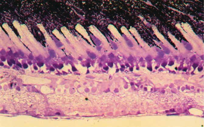
In radial sections examined by light microscopy, the
photoreceptors of the L. pardalis retina are exclusively single
cones interspersed with elongate rods (Fig. 6).
Fig. 4. Infra-red video images of the eye of Liposarcus pardalis
(standard length 14 cm) in (A) the absence of any visible light and Multiple optic nerve head papillae
(B) after 60 min exposure to 5.6×10³ µW cm–2 white light.
Illumination resulted both in a decrease of pupil diameter and the
Histological sections cut parallel to the embryonic fissure
outgrowth of an iris flap, such that overall pupil area following reveal several discrete optic discs (papillae) running along
illumination in this example is 34% of the dark-adapted value. Scale the fissure. Ganglion cell axons backfilled with fluorescein-
bar, 2 mm. conjugated dextran, which are dispersed relatively evenly in
peripheral retina, form discrete bundles or ‘fascicles’ as they
approach the embryonic fissure (Figs 7A–C, 8). The thickness
beams passing through the lens at different points and of each fascicle varies from 12 µm to 80 µm in diameter
displaying a balance between negative 3rd order and positive (Fig. 7D), and up to six fascicles may converge to form a single
5th order aberrations (Fig. 5). The maximum difference in optic papilla (Fig. 7A,B). The optic nerve head, therefore,
BVD for beams passing through the lens at different points comprises a series of optic papillae, each fascicle converging
within the lens is approximately 10% of the total focal length. from either unilateral or bilateral regions of the retina. At the
dorsal end of the optic nerve head, the fascicles converge
Retinal structure from temporal, dorsal and nasal retinal regions. Although
In the isolated eyecup of all the various species of armoured predominantly located in the ganglion cell layer and separated
catfishes examined, an elongated embryonic fissure extends from the optic fascicles, the retinal ganglion cells form loosely
from the central retina to the ventral margin. The choroid distributed columns lying between each optic fascicle (Fig. 8).
protrudes through the fissure to form a falciform process, In Cresyl Violet-labelled retinae, well-defined glial columnsThe eyes of catfish 3429
RPE
Fig. 6. Transverse section of ROS
a Liposarcus pardalis retina.
The arrow indicates the COS
RIS
position of the external
limiting membrane. The
only photoreceptors seen CIS
throughout the retina are
large rods and small single
cones. Abbreviations: RPE,
retinal pigment epithelium;
ROS, rod outer segment; RIS,
rod inner segment; COS,
cone outer segment; CIS,
cone inner segment. Scale
bar, 50 µm.
also emanate from the elongated series of optic nerve heads However, the greatest difference between the pupil response
like the spokes of a wheel. dynamics of P. notatus and L. pardalis is in the speed of
migration. As in humans and many other vertebrates, the t0.5max
Retinal ganglion cell topography in P. notatus occurs in less than one second, while in L.
Retrogradely labelled ganglion cells are distributed non- pardalis the t0.5max is in the order of several minutes, with full
uniformly throughout the retina. Peaks in ganglion cell density, constriction usually taking 35–45 min. The responses recorded
or areae centrales, lie on each side of the vertically oriented here for L. pardalis are again similar in their time course to
falciform process, just dorsal of the horizontal meridian. those observed in nocturnal elasmobranchs (Kuchnow, 1971;
In L. pardalis, densities peak at 18.6×10² cells mm–2 and Douglas et al., 1998).
24.2×10² cells mm–2 in nasal and temporal retinal regions, Function of pupil closure
respectively (Fig. 9). In L. multiradiatus, densities are similar, In most lenses, light rays entering at different eccentricities
with 18.6×10² cells mm–2 and 16.4×10² cells mm–2 in nasal and will be focused at different distances behind the lens,
temporal retinal regions, respectively. Although not analysed inevitably blurring the image on the retina. In most terrestrial
topographically, a small population of displaced ganglion cells animals, the effects of longitudinal spherical aberration within
that lie in the inner nuclear layer were also labelled with a peak the lens are minimised, as the cornea is the major refractive
density of 2.5×10² cells mm–2 in L. multiradiatus. The total surface and the pupil can constrict to limit passage of light to
number of retinal ganglion cells that lie either within the only part of the lens. Lens quality, although important to any
ganglion cell layer or within the inner nuclear layer in L. visual animal, is much more an issue for fish, as the cornea is
pardalis is 33 000. effectively neutralised underwater, resulting in the lens being
the only refractive surface, and in most species the lens
protrudes through an immobile pupil with the whole lens
Discussion consequently being involved in image formation. The degree
Dynamics of the pupil response of longitudinal spherical aberration recorded for fish lenses
The data indicate that armoured catfish are capable of varies between species and with the age of the animal (Sivak,
extensive changes in pupil size that are directly related to the 1990). However, not surprisingly, the lens is generally well
level of illumination, constricting to approximately 30% of corrected for such aberration through the possession of a
their dark-adapted area in response to the highest light levels refractive index gradient (Sroczynski, 1976, 1978, 1979, 1981;
used (Figs 1, 2). While these migrations are extensive, they are Sivak and Kreuzer, 1983; Fernald and Wright, 1983; Kreuzer
not as great as those shown in comparable light levels by P. and Sivak, 1984; Jagger, 1992; Kröger and Campbell, 1996;
notatus, the only other teleost whose pupil response has been Kröger et al., 1994, 2001; Garner et al., 2001). The degree of
characterised in the same detail (Douglas et al., 1998). aberration observed here for the catfish is comparable with that
Furthermore, unlike in P. notatus, where pupil constriction in recorded for other fish with immobile pupils. This suggests that
response to light is immediately followed by a redilation it is unlikely that the main purpose of the mobile crescent-
despite the continued presence of light, in L. pardalis, as in shaped pupil is to decrease the effects of longitudinal spherical
some nocturnal elasmobranchs (Kuchnow, 1971; Douglas et aberration. This is in line with the observation reported for the
al., 1998), the pupil showed no signs of such a redilation. clearnose skate Raja elanteria, which also has a crescent-3430 R. H. Douglas, S. P. Collin and J. Corrigan
Fig. 7. Optic axon fasciculation. Ganglion cell axon fascicles labelled with fluorescein-tagged dextran (A) entering the dorsal region of the
optic nerve head and (B) along the embryonic fissure in the retina of Liposarcus multiradiatus. (C) Low- and (D) high-power micrographs of
retrogradely labelled ganglion cells lying in between axon fascicles. The embryonic fissure lies to the left in (C). Scale bars, 100 µm.
shaped pupil yet displays very little longitudinal spherical 1976; Nicol, 1989; Pavan, 1946; Wagner, 1970; Ali and
aberration (Sivak, 1991; Sivak and Luer, 1991). Wagner, 1975; Verrier, 1927, 1928; Douglas and Wagner,
We have previously suggested that, since the majority of 1984; Nag and Sur, 1992; Sillman et al., 1993), the retina of
teleost fish with extensive pupil mobility are bottom-dwelling L. pardalis is composed of single cones and large rods (Fig. 6).
animals that attempt to blend in with the substrate, the Double cones, which are present in most teleosts, appear to be
constriction of the pupil may aid in camouflaging the animal absent from these species. This is surprising, as catfish often
through obscuring the otherwise very visible pupil (Douglas et inhabit what could be classed as low-light-level environments
al., 1998). The same argument could be applied to L. pardalis and might therefore be expected to have retinae ‘designed for
and other bottom-dwelling suckermouth catfish, as the irideal sensitivity’. Thus, many catfish, for instance, have a retinal
operculum distorts the shape of the eye and blends with the tapetum lucidum, a feature thought to enhance photon capture
rest of the fish’s body markings when viewed by potential (Nicol et al., 1973; Arnott et al., 1974). There is evidence that
predators from above. The animal would, however, be able to double cones are a further mechanism for enhancing
maintain vision through its crescent-shaped pupil in the sensitivity. Ontogenetic development of double cones, for
anterior, posterior and ventral directions, with two areas of instance, is often associated with a change in lifestyle from
increased acuity examining the substrate in front of and behind brightly lit surface waters to inhabiting dimmer, deeper waters
the animal (see below). (e.g. Boehlert, 1978), and psychophysical experiments in birds
suggest that double cones code for luminosity rather than being
Retinal morphology involved in colour vision (Maier and Bowmaker, 1993). Thus,
In agreement with other studies on catfish (Ali and Anctil, one might expect large numbers of double cones in catfishThe eyes of catfish 3431
Fig. 8. Retinal ganglion cell labelling. (A,B) Retrogradely labelled retinal ganglion cells in the relatively high-density regions of the retina in
Liposarcus multiradiatus. Note the heterogeneous cell soma size. Scale bars, 50 µm.
rather than their complete absence. However, there is unknown but a number of theories have been presented to
circumstantial evidence that double cones are involved in explain the existence of multiple nerve heads in catfish. These
movement detection in both birds (Campenhausen and include reducing the size of a large scotoma into several smaller
Kirschfeld, 1998) and fish (Levine and MacNichol, 1982), scotomata (Walls, 1963; Dunn-Meynell and Sharma, 1987),
especially when arranged in a square mosaic (Collin and reducing image degradation, which may be evident as light
Collin, 1999). The propensity of catfish to feed on non-mobile travels through the thick layers of optic fibres near a large optic
prey in generally turbid water may account for the lack of nerve head (Wagner, 1970), and enhancing stimulus perception
double cones. Alternatively, double cones seem to have a role (Walls, 1963). Although breaking up the large scotoma resulting
in mediating polarisation sensitivity (Hawryshyn, 2000), an from a single papilla into small, less-obtrusive scotomata may
ability that catfish might therefore not possess. be advantageous, the fact that the optic papillae in L. pardalis
As in members of most catfish families (Deyl, 1895; Ströer, are not dispersed as in other catfish (e.g. the channel catfish
1939; Herrick, 1941; Wagner, 1970; Arnott et al., 1974; Ali and Ictalurus punctatus; Dunn-Meynell and Sharma, 1987) suggests
Anctil, 1976; Wagner et al., 1976; Frank and Goldberg, 1983, that this novel arrangement is associated with the topographic
Dunn-Meynell and Sharma, 1987; Nag and Sur, 1992), the order of axons as they enter the optic nerve (Dunn-Meynell and
retinal ganglion cell axons of suckermouth catfish form discrete Sharma, 1988).
fascicles within the nerve fibre layer, leading to multiple optic
papillae. The functional significance of this arrangement is Retinal topography
A variety of patterns of peaks in retinal ganglion cell
density have been found in a range of teleosts (Ito and
Murakami, 1984; Collin and Pettigrew, 1988a,b; Collin,
1999) and elasmobranchs (Hueter, 1991; Collin, 1988;
Bozzano and Collin, 2000). The nasal and temporal areae
centrales in L. pardalis and L. multiradiatus would provide
increased sampling, and therefore increased spatial resolving
power, in the rostro-ventral and caudo-ventral axes,
respectively. The arrangement of iso-density contours
broadly follows the shape of the pupil, where the retinal
region underlying the operculum possesses relatively low
densities of ganglion cells, creating a high centro-peripheral
density gradient of more than 25:1 (Fig. 9). Similarly, the
lack of a dorsal pupillary operculum is also reflected in the
topographic distribution of retinal ganglion cells in I.
Fig. 9. Iso-density contour map of the distribution of retrogradely
labelled retinal ganglion cells in the right eye of Liposarcus
pardalis (standard length 158 mm). The elongated optic nerve
head is depicted in black. All densities are ×102 cells mm–2. T,
temporal; V, ventral.3432 R. H. Douglas, S. P. Collin and J. Corrigan
punctatus. Using horseradish peroxidase as a retrograde tracer Ali, M. A. and Wagner, H.-J. (1975). Visual pigments; phylogeny and
from the optic nerve, Dunn-Meynell and Sharma (1987) ecology. In Vision in Fishes: New Approaches in Research (ed. M. A. Ali),
pp. 481-516. New York: Plenum Press.
revealed a naso-temporal elongation of iso-density contours Arnott, H. J., Best, A. C. G. and Nicol, J. A. C. (1974). Studies on the eyes
with a continuous peak of >1.5×10² cells mm–2 lying across the of catfishes with special reference to the tapetum lucidum. Proc. R. Soc.
dorsal hemifield. The decrease in ganglion cell density Lond. B Biol. Sci. 186, 13-36.
Bateson, W. (1890). Contractility of the iris in fishes and cephalopods. J. Mar.
underlying the operculum in L. pardalis is not present, which Biol. Assoc. UK 1, 215-216.
is indicative of an increased dependence on scanning a wider Beer, T. (1894). Die Akkommodation des Fischauges. Pflügers Arch. Ges.
panoramic visual field in I. punctatus. A similar relationship to Physiol. 58, 523-650.
Boehlert, G. W. (1978). Intraspecific evidence for the function of single and
that seen in L. pardalis occurs in the eyes of the sharp-nosed double cones in the teleost retina. Science 202, 309-311.
weaverfish Parapercis cylindrica (Collin and Pettigrew, Bozzano, A. and Collin, S. P. (2000). Retinal ganglion cell topography in
1988a) and the shovel-nosed ray Rhinobatos batillum (Collin, elasmobranchs. Brain Behav. Evol. 55, 191-208.
Campenhausen, M. V. and Kirschfeld, K. (1998). Spectral sensitivity of the
1988), where the shape of a crescent-shaped pupil is reflected accessory optic system of the pigeon. J. Comp. Physiol. A 183, 1-6.
in the distribution of ganglion cells, with two areae divided Collin, S. P. (1988). The retina of the shovel-nosed ray, Rhinobatos batillum
by a region of low density. Interestingly, the pupils of (Rhinobatidae): morphology and quantitative analysis of the ganglion,
amacrine and bipolar cell populations. Exp. Biol. 47, 195-207.
many cetacea (Kröger and Kirschfeld, 1993) and some Collin, S. P. (1999). Behavioural ecology and retinal cell topography. In
elasmobranchs (Douglas et al., 1998) close down to two Adaptive Mechanisms in the Ecology of Vision (ed. S. N. Archer, M. B. A.
roughly horizontally aligned pinholes, and both of these groups Djamgoz, E. R. Loew, J. C. Partridge, S. Vallerga), pp. 509-535. London:
Kluwer Academic Publishers.
also often have two retinal areas of maximal resolution Collin, S. P. and Collin, H. B. (1999). The foveal photoreceptor mosaic in
(cetacea – Dral, 1983; Mass and Supin, 1995; Murayama et al., the pipefish, Corythoichthyes paxtoni (Syngnathidae, Teleostei). Histol.
1995; Murayama and Somiya, 1998: elasmobranchs – Peterson Histopathol. 14, 369-382.
Collin, S. P. and Northcutt, R. G. (1993). The visual system of the Florida
and Rowe, 1980; Bozzano and Collin, 2000; Collin, 1999). garfish, Lepisosteus platyrhincus (Ginglymodi): III. Retinal ganglion cells.
However, it is in many ways surprising to find such Brain Behav. Evol. 42, 295-320.
similarity between ganglion cell distribution and pupil shape, Collin, S. P. and Pettigrew, J. D. (1988a). Retinal topography in reef teleosts.
I. Some species with well-developed areae but poorly-developed streaks.
as the pupil functions as an aperture stop and not a field stop. Brain Behav. Evol. 31, 269-282.
So it is perhaps not unexpected that a similar topographic Collin, S. P. and Pettigrew, J. D. (1988b). Retinal topography in reef teleosts.
II. Some species with prominent horizontal streaks and high density areae.
regionalisation of the retina into two areae is found in a Brain Behav. Evol. 31, 283-295.
number of elasmobranchs (Peterson and Rowe, 1980; Bozzano Deyl, J. (1895). Über den Sehnerven bel siluroiden und acanthopsiden. Anat.
and Collin, 2000; Collin, 1999; L. Litherland and S. P. Collin, Anz. 11, 8-16.
Douglas, R. H. and Wagner, H.-J. (1984). Action spectra of photomechanical
unpublished data) that appear not to have a pupillary cone contraction in the catfish retina. Invest. Ophthalmol. Vis. Sci. 25, 534-
operculum or double pinhole apertures, suggesting that the 538.
need to optimise acuity in the frontal and caudal visual axes Douglas, R. H., Harper, R. D. and Case, J. F. (1998). The pupil response
of a teleost fish, Porichthys notatus: description and comparison to other
may be the driving force behind the development of retinal cell species. Vision Res. 38, 2697-2710.
gradients rather than the presence of an irregular pupil. Dral, A. D. G. (1983). The retinal ganglion cells of Delphinus delphis and
their distribution. Aquat. Mamm. 10, 57-68.
Dunn-Meynell, A. A. and Sharma, S. C. (1987). Visual system of the channel
The authors are grateful to Darrell Siebert (British Museum catfish (Ictalurus punctatus): II. The morphology associated with the
of Natural History), Michael Hardman (University of Illinois) multiple optic papillae and retinal ganglion cell distribution. J. Comp.
and Nigel Merrett (British Museum of Natural History) for Neurol. 257, 166-175.
Dunn-Meynell, A. A. and Sharma, S. C. (1988). Visual system of the channel
identification of specimens and to Christine Wood for the gift catfish (Ictalurus punctatus): III. Fibre order in the optic nerve and optic
of one of the animals. Thanks also to Minal Patel for tract. J. Comp. Neurol. 268, 299-312.
assistance with light microscopy, Paul Dyer for performing Fernald, R. D. and Wright, S. E. (1983). Maintenance of optical quality
during crystalline lens growth. Nature 301, 618-620.
some preliminary pupillometry, Manesh Patel for help Frank, B. L. and Goldberg, S. (1983). Multiple optic fiber patterns in the
in determining lens aberrations and Jochen Wagner for catfish retina. Invest. Ophthalmol. Vis. Sci. 24, 1429-1432.
providing the facilities for the topographic studies. Special Franz, V. (1905). Zur Anatomie, Histologie und functionellen Gestaltung des
Selachierauges. Jenaische Zeitschrift für Naturwissenschaft 40, 697-840.
thanks go to Ronald Kröger for designing the software used to Franz, V. (1931). Die Akkommodation des Selachierauges und seine
analyse lenticular spherical aberration and to Chris Hull for Abblendungsapparate, nebst Befunden an der Retina. Zool. Jahrb. Abt. Allg.
fitting curves to the data. Dan-Eric Nilsson gave helpful Zool. Physiol. Tiere 49, 323-462.
Garner, L. F., Smith, G., Yao, S. and Augusteyn, R. C. (2001). Gradient
comments on an earlier version of the manuscript. Part of this refractive index of the crystalline lens of the black oreo dory (Allocyttus
work was funded by a bursary from the Nuffield Foundation niger): comparison of magnetic resonance imaging (MRI) and laser ray-
to R.H.D. S.P.C. was funded by the Alexander von Humboldt trace methods. Vision Res. 41, 973-979.
Gruber, S. H. and Cohen, J. L. (1978). Visual system of the elasmobranchs:
Stiftung while in Germany and an ARC QE II Research state of the art 1960-1975. In Sensory Biology of Sharks and Rays (ed. E.
Fellowship in Australia. S. Hodgson and R. F. Mathewson), pp. 11-105. Arlington, VA, USA: Office
of Naval Research.
Hawryshyn, C. W. (2000). Ultraviolet polarization vision in fishes: possible
mechanisms for coding e-vector. Phil. Trans. Roy. Soc. Lond. B Biol. Sci.
References 355, 1187-1190.
Ali, M. A. and Anctil, M. (1976). Retinas of Fishes: An Atlas. Berlin: Springer Herrick, C. J. (1941). The eyes and optic paths of the catfish, Ameiurus. J.
Verlag. Comp. Neurol. 75, 255-286.The eyes of catfish 3433 Hueter, R. E. (1991). Adaptations for spatial vision in sharks. J. Exp. Zool. 5 Nicol, J. A. C. (1989). The Eyes of Fishes. Oxford: Oxford University Press. Suppl., 130-141. Nicol, J. A. C., Arnott, H. J. and Best, A. C. G. (1973). Tapeta lucida in Ito, H. and Murakami, T. (1984). Retinal ganglion cells in two teleost bony fishes (Actinopterygii): a survey. Can. J. Zool. 51, 69-81. species, Sebasticus marmoratus and Navodon modestus. J. Comp. Neurol. Pavan, C. (1946). Observations and experiments on the cavefish Pimelodella 229, 80-96. kronei and its relatives. Am. Natural. 80, 343-361. Jagger, W. S. (1992). The optics of the spherical fish lens. Vision Res. 32, Peterson, E. H. and Rowe, M. H. (1980). Different regional specializations 1271-1284. of neurons in the ganglion cell layer and inner plexiform layer of the Kreuzer, R. O. and Sivak, J. G. (1984). Spherical aberration of the fish lens; California horned shark, Heterodontus francisci. Brain Res. 201, interspecies variation and age. J. Comp. Physiol. A 154, 415-422. 195-201. Kröger, R. H. H. and Campbell, M. C. W. (1996). Dispersion and Sillman, A. J., Ronan, S. J. and Loew, E. R. (1993). Scanning electron longitudinal chromatic aberration of the crystalline lens of the African microscopy and microspectrophotometry of the photoreceptors of ictalurid cichlid fish Haplochromis burtoni. J. Opt. Soc. Am. A 13, 2341-2347. catfishes. J. Comp. Physiol. A 173, 801-807. Kröger, R. H. H. and Kirschfeld, K. (1993). Optics of the harbor porpoise Sivak, J. G. (1990). Optic variability of the fish lens. In The Visual System of eye in water.. J. Opt. Soc. Am. A 10, 1481-1489. Fish (ed. R. H. Douglas and M. B. A. Djamgoz), pp. 63-80. London: Kröger, R. H. H., Campbell, M. C. W., Munger, R. and Fernald, R. D. Chapman & Hall. (1994). Refractive index distribution and spherical aberration in the Sivak, J. G. (1991). Elasmobranch visual optics. J. Exp. Zool. 5 Suppl., 13- crystalline lens of the African cichlid fish, Haplochromis burtoni. Vision 21. Res. 34, 1815-1822. Sivak, J. G. and Kreuzer, R. O. (1983). Spherical aberration of the crystalline Kröger, R. H. H., Campbell, M. C. W. and Fernald, R. D. (2001). The lens. Vision Res. 23, 59-70. development of the crystalline lens is sensitive to visual input in the African Sivak, J. G. and Luer, C. A. (1991). Optical development of the ocular lens cichlid fish, Haplochromis burtoni. Vision Res. 41, 549-559. of an elasmobranch, Raja elanteria. Vision Res. 31, 373-382. Kuchnow, K. P. (1971). The elasmobranch pupillary response. Vision Res. Sroczynski, S. (1976). Die chromatische Aberration der Augenlinse der 11, 1395-1406. Regenbogenforelle (Salmo gairdneri RICH.). Zool. Jahrb. Abt. Allg. Zool. Kuchnow, K. P. and Martin, R. (1970). Fine structure of elasmobranch iris Physiol. Tiere 80, 432-450. muscle and associated nervous structures. Exp. Eye Res. 10, 345-351. Sroczynski, S. (1978). Die chromatische Aberration der Augenlinse der Levine, J. S. and MacNichol, E. F., Jr (1982). Colour vision in fishes. Sci. Bachforelle (Salmo trutta fario L.). Zool. Jahrb. Abt. Allg. Zool. Physiol. Am. 246, 108-117. Tiere 82, 113-133. Maier, E. J. and Bowmaker, J. K. (1993). Colour vision in the passeriform Sroczynski, S. (1979). Das optische System des Auges des Flußbarsches bird, Leiothrix lutea: correlation of visual pigment absorbance and oil droplet (Perca fluviatilis L.). Zool. Jahrb. Abt. Allg. Zool. Physiol. Tiere 83, 224- transmission with spectral sensitivity. J. Comp. Physiol. A 172, 295-301. 252. Mass, A. M. and Supin, A. Y. (1995). Ganglion cell topography of the retina in Sroczynski, S. (1981). Optical system of the eye of the Ruff (Acerina cernua the bottlenosed dolphin, Tursiops truncatus. Brain Behav. Evol. 45, 257-265. L.). Zool. Jahrb. Abt. Allg. Zool. Physiol. Tiere 85, 316-342. Munk, O. (1970). On the occurrence and significance of horizontal band- Ströer, W. F. H. (1940). Zur vergleichenden Anatomie des primären optischen shaped retinal areae in teleosts. Vidensk. Medd. Dan. Naturhist. Foren. Systems bei Wirbeltieren. Z. Anat. Entwicklungsgesch. 110, 301-321. Khobenhavn 133, 85-120. Verrier, M.-L. (1927). Sur la structure de l’oeil de Ameriurus nebulosus Murayama, T. and Somiya, H. (1998). Distribution of ganglion cells and Lesueur. et de Clarias batrachus L. ses rapports avec l’habitat et le object localizing ability in the retina of three cetaceans. Fish. Sci. 64, 27-30. comportement biologique de ces deux Siluridés. Bull. Soc. Zool. Franc. Murayama, T., Somiya, H., Aoki, I. and Ishii, T. (1995). Retinal ganglion Belg. 52, 581-588. cell size and distribution predict visual capabilities of Dall’s porpoise. Mar. Verrier, M.-L. (1928). Recherches sur les yeux et la vision des poissons. Bull. Mamm. Sci. 11, 136-149. Biol. France et Belgique 11 Suppl., 1-222. Murphy, C. J. and Howland, H. C. (1991). The functional significance of Wagner, H. J. (1970). Der bau der Retina und der Multiple Optischen Papillae crescent-shaped pupils and multiple pupillary apertures. J. Exp. Zool. 5 bei zwei Synodontid-Arten (Telesotei, Siluroidea). Z. Morphol. Tiere 68, Suppl., 22-28. 69-82. Nag, T. C. and Sur, R. K. (1992). Cones in the retina of the catfish, Clarius Wagner, H.-J., Menzes, N. A. and Ali, M. A. (1976). Retinal adaptations in batrachus (L.). J. Fish Biol. 40, 967-969. some Brazilian tide pool fishes (Teleosti). Zoomorphologie 82, 209-226. Nelson, J. S. (1994). Fishes of the World. Third edition. New York: Wiley. Walls, G. L. (1963). The Vertebrate Eye and its Adaptive Radiation. New Nicol, J. A. C. (1978). Studies on the eye of the stingray Dasyatis sabina, with York: Hafner Publishing Co. notes on other selachians. I. Eye dimensions, cornea, pupil and lens. Young, J. Z. (1933). Comparative studies of the physiology of the iris. I. Contrib. Mar. Sci. 21, 89-102. Selachians. Proc. R. Soc. Lond. B Biol. Sci. 112, 228-241.
You can also read