The F-box protein Bard (CG14317) targets the Smaug RNA-binding protein for destruction during the Drosophila maternal-to-zygotic transition
←
→
Page content transcription
If your browser does not render page correctly, please read the page content below
2
                                                                                                                                GENETICS, 2021, iyab177
                                                                                                                  https://doi.org/10.1093/genetics/iyab177
                                                                                                                                             Investigation

The F-box protein Bard (CG14317) targets the Smaug
RNA-binding protein for destruction during the Drosophila

                                                                                                                                                               Downloaded from https://academic.oup.com/genetics/advance-article/doi/10.1093/genetics/iyab177/6404591 by guest on 09 December 2021
maternal-to-zygotic transition
Wen Xi Cao,1,† Angelo Karaiskakis,1 Sichun Lin,2 Stephane Angers,2 and Howard D. Lipshitz                         1,
                                                                                                                    *
1
    Department of Molecular Genetics, University of Toronto, Toronto, ON M5G 1M1, Canada and
2
    Department of Pharmaceutical Sciences & Donnelly Centre, University of Toronto, Toronto, ON M5S 3E1, Canada

*Corresponding author: Email: howard.lipshitz@utoronto.ca
†
 Present address: Department of Biological Sciences, Columbia University, 1212 Amsterdam Avenue, New York, NY 10027, USA.

Abstract
During the maternal-to-zygotic transition (MZT), which encompasses the earliest stages of animal embryogenesis, a subset of maternally
supplied gene products is cleared, thus permitting activation of zygotic gene expression. In the Drosophila melanogaster embryo, the
RNA-binding protein Smaug (SMG) plays an essential role in progression through the MZT by translationally repressing and destabilizing a
large number of maternal mRNAs. The SMG protein itself is rapidly cleared at the end of the MZT by a Skp/Cullin/F-box (SCF) E3-ligase
complex. Clearance of SMG requires zygotic transcription and is required for an orderly MZT. Here, we show that an F-box protein, which
we name Bard (encoded by CG14317), is required for degradation of SMG. Bard is expressed zygotically and physically interacts with SMG
at the end of the MZT, coincident with binding of the maternal SCF proteins, SkpA and Cullin1, and with degradation of SMG. shRNA-
mediated knock-down of Bard or deletion of the bard gene in the early embryo results in stabilization of SMG protein, a phenotype that is
rescued by transgenes expressing Bard. Bard thus times the clearance of SMG at the end of the MZT.

Keywords: Smaug; RNA-binding protein; E3 ubiquitin ligase; Skp/Cullin/F-box (SCF) complex; Bard/CG14317

Introduction                                                                       direct translational repression and/or degradation of a large frac-
                                                                                   tion of the maternally loaded mRNA species (Nelson et al. 2004;
The earliest stages of animal embryogenesis depend on mater-
                                                                                   Semotok et al. 2005, 2008; Tadros et al. 2007; Jeske et al. 2011;
nally supplied factors, including mRNAs and proteins, that are
                                                                                   Pinder and Smibert 2013; Chen et al. 2014; Götze et al. 2017). In
loaded into the oocyte. During a conserved process known as the
                                                                                   embryos from smg mutant females repression and degradation of
oocyte-to-embryo or maternal-to-zygotic transition (MZT), a
                                                                                   a large proportion of maternal mRNAs fails (Tadros et al. 2007;
large proportion of the maternally loaded mRNA and a much
                                                                                   Chen et al. 2014) as does zygotic production of both mRNAs and
smaller proportion of protein species is degraded in a temporally
co-ordinated manner, permitting transcriptional activation of                      microRNAs (Benoit et al. 2009; Luo et al. 2016). In addition, the first
the zygotic genome (reviewed in Tadros and Lipshitz 2009;                          developmental process that depends on ZGA, namely blastoderm
Vastenhouw et al. 2019). Prior to zygotic genome activation (ZGA),                 cellularization, also fails (Dahanukar et al. 1999; Benoit et al.
the regulation of maternal mRNAs and proteins relies on post-                      2009). These results led to the conclusion that SMG function is
transcriptional and post-translational mechanisms implemented                      essential for progression through the MZT (Benoit et al. 2009).
by maternally encoded machineries whereas, upon ZGA,                               Expression of SMG and several of its post-transcriptional co-
newly synthesized factors are added to the mix (reviewed in                        repressors—Cup, Trailer hitch (TRAL), and Maternal expression
Vastenhouw et al. 2019). For example, degradation, and/or trans-                   at 31B (ME31B)—is tightly regulated at both the mRNA and
lational repression of maternal mRNAs prior to ZGA is directed                     protein levels, temporally restricting their function to specific
largely by maternally encoded RNA-binding proteins while, upon                     phases of the MZT (Wang et al. 2017; Hara et al. 2018; Cao et al.
ZGA, microRNAs are synthesized and destabilize additional                          2020; Zavortink et al. 2020).
maternal mRNAs (e.g., miR-430 in zebrafish and miR-309 in                             The highly conserved ubiquitin proteasome system (UPS) is
Drosophila). Post-translational modifications that occur during                    the major pathway for targeted degradation of specific proteins
the MZT include phosphorylation and ubiquitylation, which                          across eukaryotes (Ravid and Hochstrasser 2008). This pathway
affect protein function and/or stability.                                          consists of the E1–E2–E3 ubiquitination enzyme cascade, through
   In Drosophila melanogaster, the multifunctional RNA-binding                     which ubiquitin is sequentially transferred to the E3 ubiquitin
protein Smaug (SMG) acts in concert with various cofactors to                      ligase, which acts as the substrate-specificity factor that binds

Received: September 14, 2021. Accepted: October 11, 2021
C The Author(s) 2021. Published by Oxford University Press on behalf of Genetics Society of America.
V
This is an Open Access article distributed under the terms of the Creative Commons Attribution License (https://creativecommons.org/licenses/by/4.0/), which
permits unrestricted reuse, distribution, and reproduction in any medium, provided the original work is properly cited.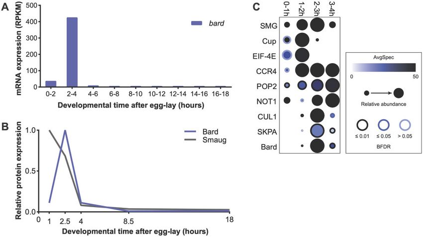
2   |   GENETICS, 2021, Vol. 00, No. 0

specific target proteins for ubiquitination and degradation                IP-MS experiments was w1118. Additional fly strains were ac-
through the 26S proteasome. Hundreds of E3 ubiquitin ligases               quired from other labs or from the Bloomington Drosophila Stock
have been identified across eukaryotes, belonging to several con-          Center (BDSC). The da-GAL4 driver PfGAL4-da.G32g (Wodarz et al.
served classes (reviewed in Morreale and Walden 2016; Zheng                1995) used for RNAi knockdown (BDSC #55851) was a gift from
and Shabek 2017).                                                          T. Harris. mCherry RNAi was used as control (gift from T. Hurd;
    The UPS has been implicated in several aspects of the develop-         BDSC #35785). For immunostaining experiments w1118; Df(3R)
mental transitions from meiotically arrested oocyte to embryo              BSC510/TM6C, Sb1 cu1 (BDSC #25014) was crossed with w1118;
and from maternal to zygotic control of development in                     DrMio/TM3, Pfw[þmC]¼GAL4-twi.Gg2.3, PfUAS-2xEGFPgAH2.3, Sb1
                                                                           Ser1 (BDSC #6663) to produce flies carrying Df(3R)BSC510 and the

                                                                                                                                                  Downloaded from https://academic.oup.com/genetics/advance-article/doi/10.1093/genetics/iyab177/6404591 by guest on 09 December 2021
Drosophila (Aviles-Pagan et al. 2020; Cao et al. 2020; Zavortink et al.
2020), C. elegans (Guven-Ozkan et al. 2008; Kisielnicka et al. 2018;       twi>EGFP balancer. Details on the generation of transgenic
Spike et al. 2018) and mouse (Yang et al. 2017). Recently, we              strains for bard RNAi and bard rescue constructs are given below.
showed that the UPS is involved in the clearance of SMG and its
co-factors during the MZT (Cao et al. 2020): Cup, TRAL and ME31B           Embryo collection
are targeted for degradation in the middle of the MZT by the C-ter-        Embryos were collected on apple juice agar plates supplemented
minal to LisH (CTLH) E3-ligase complex, while SMG is degraded              with yeast paste from cages of adult flies in 1-h intervals (2 h for
near the end of the MZT by the Skp/Cullin/F-box (SCF) E3-ligase            immunostaining) and aged to the desired time points at 25 C.
complex. The SCF complex is one of the best studied and most               Embryos were washed off the surface of plates with PBST
highly conserved E3 ubiquitin ligases, particularly known for its          (1 PBS, 0.1% Tween-20) and collected through a mesh to remove
role in cell cycle regulation (Bai et al. 1996). This complex includes     excess yeast. Embryos were dechorionated with cold 4% sodium
the scaffold protein Cul1, the ubiquitin-binding RING-domain pro-          hypochlorite for 1 min, rinsed with PBST to remove bleach, and
tein Roc1/Rbx1, and the adapter protein Skp1. The F-box is an ap-          placed on ice for further processing, described below.
proximately 50 amino-acid-long motif that serves as a site of
protein-protein interaction either in SCF E3 ubiquitin ligase com-         Immunoprecipitation and mass spectrometry
plexes or in other contexts (Kipreos and Pagano 2000). In SCF E3-          Embryos were collected from w1118 flies and aged to 0–1 h
ligase complexes, F-box proteins function as the substrate-binding         (i.e., processed immediately), 1–2, 2–3, or 3–4 h. Dechorionated
subunit; thus, the spatio-temporal regulation of F-box protein ex-         embryos were crushed in a minimal volume of lysis buffer
pression can provide specificity both for the specific substrate(s) that   (150 mM KCl, 20 mM HEPES-KOH pH 7.4, 1 mM MgCl2, 0.1% Triton
are cleared and for where and when this occurs. While at least 45          X-100, supplemented with protease inhibitors and 1 mM DTT),
F-box proteins have been identified in Drosophila, only a small subset     cleared by centrifugation for 15 min at 4 C and 20,000  g, and
of these has known substrates or binding sites (Dui et al. 2012).          stored at 80 C. Immediately prior to immunoprecipitation (IP),
    We have shown that, during the MZT, SMG physically inter-              protein concentration was measured by Bradford Assay
acts with core members of the SCF complex, as well as two F-box            (Bio-Rad), and samples were diluted to 15 mg/ml with lysis buffer.
proteins, Supernumerary limbs (SLMB) and CG14317 (Cao et al.               For each IP, 250 ml of diluted lysate was mixed with 350 mg/ml
2020). Deletion of the C-terminal region of SMG, which is essen-           RNase A, 2 ml of guinea pig anti-SMG antibody (Tadros et al. 2007)
tial for its clearance at the end of the MZT, abrogates interaction        or guinea pig normal serum as control, and 10 ml of Protein A
with both of these F-box proteins (Cao et al. 2020). SCFSLMB is a ho-      beads (Roche). IPs were incubated for 3 h at 4 C with end-over-
molog of the mammalian cell cycle regulator SCFb-TrCP and has              end rotation. Beads were washed four times with lysis buffer,
previously reported roles in ovary development and cell cycle reg-         twice with lysis buffer lacking Triton X-100, then transferred to
ulation in Drosophila (Heriche et al. 2003; Muzzopappa and                 new tubes and washed twice more with lysis buffer lacking
Wappner 2005). Knockdown of SLMB or core components of the                 Triton X-100 to remove all traces of detergent. Downstream
SCF complex in the early embryo results in stabilization of SMG            tryptic digest, sample preparation and HPLC/MS were the same
protein (Cao et al. 2020). However, these proteins are loaded              as our previously described FLAG IP-MS protocol (Cao et al. 2020).
maternally and persist well-beyond the MZT (Cao et al. 2020);
thus, their expression per se cannot explain the timing of SMG
                                                                           RNAi
clearance by the UPS.                                                      A transgenic construct designed to express a short hairpin RNA
    In contrast to the other SCF components, CG14317 protein and           against CG14317/bard was generated. A 21-nucleotide sequence
its cognate mRNA show an extremely temporally restricted ex-               mapping to CG14317/bard with no predicted off-target matches of
pression pattern that coincides with ZGA and the degradation of            up to 16 nucleotides in the Drosophila genome was cloned into a
SMG at the end of the MZT (Graveley et al. 2011; Casas-Vila et al.         microRNA scaffold in the pVALIUM22 vector (Ni et al. 2011), and
2017; Cao et al. 2020). These observations made CG14317 an ideal           injected into y1 Pfy[þt7.7]¼CaryIPgsu(Hw)attP8 v1 (BDSC #34769)
candidate to function as a timer for the precise temporal degra-           by Rainbow Transgenic Flies (Camarillo, CA). The shRNA sense-
dation of SMG. Here, we show that CG14317 is required for SMG              strand sequence was 50 -CGATCAGTTCGACAGTTGTGT-30 , and
protein degradation, and that its distinctive expression pattern           the antisense strand sequence was 50 -ACAACTGTCGAACTGAT
restricts the binding of the SCFCG14317 and SCFSLMB complexes              CGGT-30 . “Maternalþzygotic” knockdown was performed by
with SMG, to the end of the MZT. We name the CG14317 gene                  crossing females expressing the da-GAL4 ubiquitous driver with
bard and its encoded protein Bard, for Bard the Bowman’s role in           males expressing UAS-shRNA; F1 females were then crossed to
targeting Smaug for destruction (Tolkien 1937).                            males expressing UAS-shRNA, from which embryos were col-
                                                                           lected for RNAi assays. This method resulted in 28% depletion of
                                                                           bard mRNA (Figure 2A).
Materials and methods
Drosophila stocks and husbandry                                            Reverse transcription and qPCR
Drosophila melanogaster stocks were cultivated under standard              Total RNA was collected from 0 to 4 h embryos in 200 ml
laboratory conditions at 25 C. The “wild-type” strain used for            (approximately 10 volume) of TRI reagent (Sigma) following the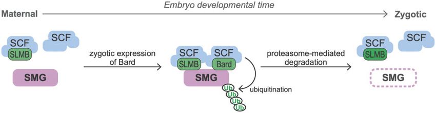
W. X. Cao et al.   |   3

manufacturer’s protocol. cDNA was synthesized from 1 mg of to-        washed 3  15 min with PBSTx at room temperature, then
tal RNA per sample using the Superscript IV reverse transcriptase     incubated in secondary antibodies (1:300 Cy3-conjugated donkey
kit (Invitrogen), and reactions were primed using random hexam-       anti-guinea pig [Jackson ImmunoResearch], 1:300 goat anti-
ers. Reactions containing single-stranded cDNA were diluted 1:20      mouse Alexa Fluor 488 [ThermoFisher]) for 1 h rocking at room
with ultra-pure water and used for quantification by qPCR. qPCR       temperature. Embryos were washed 5  10 min with PBSTx, and
was performed using the Sensifast SYBr PCR mix (Bioline) follow-      mounted in 2.5% DABCO, 70% glycerol in PBS. Details of antibod-
ing the manufacturer’s protocol. Each reaction used 5 ml of           ies used can be found in the Reagents Table. Images were col-
diluted cDNA, primed with gene-specific primers. Quantitative         lected using a Zeiss AxioSkop-2 MOT fluorescence microscope

                                                                                                                                               Downloaded from https://academic.oup.com/genetics/advance-article/doi/10.1093/genetics/iyab177/6404591 by guest on 09 December 2021
real-time PCR was performed on a Bio-Rad CFX384 Real-Time             and the QCapture Suite PLUS acquisition software. Average
System, and gene expression was analyzed using the Bio-Rad CFX        fluorescence intensity of each embryo was quantified in ImageJ.
manager. bard expression was averaged across three technical
replicates and normalized to the RpL32 control.                       Statistical analysis
                                                                      The ProHits software package (Liu et al. 2010) was used to perform
Western blotting                                                      peptide validation and protein interaction analysis. Proteins with
Dechorionated embryos were counted, then crushed using a              associated peptide counts were filtered for iProphet probability
microcentrifuge tube pestle in SDS-PAGE sample buffer at a con-       >0.95 and number of unique peptides 2 (Shteynberg et al. 2011).
centration of 1 embryo/ml and boiled for 2 min. Eight microliter      Significance Analysis of INTeractome (SAINT) was used to deter-
per sample was loaded in 6% SDS-PAGE and subsequently trans-          mine the probability of each interacting protein (Choi et al. 2011).
ferred to PVDF membrane. Blots were blocked at room tempera-          SAINTexpress was run on the ProHits interface with nburn ¼
ture with 2% nonfat milk in PBST (0.1% Tween-20) for 30 min, and      2000; niter ¼ 5000; lowMode ¼ 1; minFold ¼ 1; normalize ¼ 1; no
incubated at 4 C overnight with primary antibody (1:20,000           compression for bait (n ¼ 3) or control (n ¼ 2). Dot plot of the
guinea pig anti-SMG; 1:50,000 mouse anti-Tubulin [Sigma T5168])       SAINT analysis was generated through ProHits-viz (Knight et al.
diluted in blocking solution. Blots were washed for 3  10 min        2017). Detailed results of the SAINT analysis for all interactions
with PBST at room temperature with rocking, and incubated             are listed in Supplementary Table S1.
with 1:5000 HRP-conjugated secondary antibodies (Jackson                 All other graphing and statistical analyses were carried out
ImmunoResearch) in blocking solution at room temperature for          using GraphPad PRISM 8.3. Quantification of Western blots com-
1 h. Blots were washed again for 3  15 min with PBST, developed      paring SMG expression in control vs. bard RNAi across 3 biological
using Immobilon Luminata Crescendo Western HRP substrate              replicates were analyzed using the two-tailed Student’s t-test.
(Millipore), imaged using ImageLab (Biorad), and quantified using     Quantification of IF, comparing SMG expression in GFP(þ) vs
ImageJ. Details of the antibodies used can be found in the            GFP() embryos were performed for at least 10 embryos per cate-
Reagents Table.                                                       gory, and analyzed using the two-tailed Mann–Whitney U-test.

Transgenic rescue constructs
An approximately 7.8 kb genomic fragment containing CG14317/
                                                                      Results
bard, and extending 3.9 kb upstream and 2.3 kb downstream of          Bard is expressed and interacts with Smaug at
the transcript (diagrammed in Figure 3A), was PCR amplified           the end of the MZT
from w1118 genomic DNA using primers flanked with BamHI and           To compare the expression of Bard to that of SMG we used previ-
NotI restriction sites. This endogenous rescue construct was          ously published transcriptomic and proteomic datasets. From
inserted by restriction digest and ligation into the pCaSpeR4 clon-   modENCODE transcriptome data (Graveley et al. 2011), bard
ing vector containing an attB site (Tadros et al. 2007; Markstein     mRNA is present at very low levels in embryos 0–2 h after egg-lay
et al. 2008). For the 3xFLAG-tagged rescue construct, the PCR-        (38 RPKM), accumulates to very high levels by 2–4 h (427 RPKM),
amplified genomic fragment was first ligated into the pUC19           and decreases more than 40-fold by 4–6 h (to 10 RPKM;
backbone, and an N-terminal 3xFLAG coding sequence was                Figure 1A). The bard transcript is not detected at any other stage
inserted after the start codon by site-directed mutagenesis. The      during embryogenesis and is absent from ovaries. Thus, bard is a
3xFLAG-tagged rescue fragment was excised with BamHI and              strictly zygotic mRNA that is synthesized in a burst near the end
NotI, and cloned into pCaSpeR4 as described above. Rescue frag-       of the MZT and then is cleared from the embryo.
ments in pCaSpeR4 were integrated into an attP40 landing site on          Next, we plotted the expression pattern of the Bard and SMG
the second chromosome (Markstein et al. 2008) using the phiC31        proteins in the early embryo using our previously published em-
integrase method by Rainbow Transgenic Flies (Camarillo, CA),         bryo proteome study (Cao et al. 2020). The Bard protein expres-
and crossed to the third chromosome deficiency for rescue             sion pattern is almost identical to that of its cognate mRNA: Bard
experiments.                                                          is absent from the early embryo, accumulates rapidly toward
                                                                      the end of the MZT coincident with degradation of SMG, and is
Immunostaining                                                        rapidly cleared from the embryo shortly thereafter (Figure 1B).
Two- to four-hour old embryos were collected and dechorionated        An independent proteomic study is consistent with this Bard ex-
as described above. Dechorionated embryos were fixed and              pression profile (Casas-Vila et al. 2017).
permeabilized in 4% formaldehyde and heptane for 40 min, and              Given the temporal correlation between Bard accumulation
devitellinized by addition of methanol followed by vigorous shak-     and SMG protein degradation, we asked whether Bard and the
ing for 30 s. Fixed embryos were rehydrated by washing 4 times        other components of the SCF E3 ligase complex bind SMG specifi-
with PBSTx (1xPBS þ 0.1% Triton X-100) and blocked with 10% bo-       cally during this time window. To investigate the temporal inter-
vine serum albumin (BSA) in PBSTx for 1 h at room temperature.        action between SMG and SCFBard, we performed a time-course
Embryos were incubated at 4 C overnight with primary antibod-        immunoprecipitation followed by mass spectrometry (IP-MS) on
ies (1:1000 guinea pig anti-SMG and 1:200 mouse anti-GFP [Sigma       endogenous SMG over the first 4 h of embryo development. As a
#G6539]) diluted in 1% BSA in PBSTx, with rocking. Embryos were       control for the time-course we examined SMG’s interactions with
4 | GENETICS, 2021, Vol. 00, No. 0
Downloaded from https://academic.oup.com/genetics/advance-article/doi/10.1093/genetics/iyab177/6404591 by guest on 09 December 2021
Figure 1 bard mRNA and Bard protein are expressed in a narrow time window, coinciding with SCFBard binding to SMG and its clearance from the
embryo at the end of the MZT. (A) Developmental transcriptome (Graveley et al. 2011) captures bard transcript expression almost exclusively at 2–4 h
after egg-lay. (B) A developmental proteomic study (Cao et al. 2020) shows that Bard protein expression closely correlates with that of its cognate
transcript and is coincident with SMG protein clearance. (C) Dot-plot of developmental IP-MS experiments of SMG over the first 4 h of embryo
development for interactions with its co-regulators and the SCF complex. Average spectral counts, relative abundance normalized to control IP, and
BFDR of each significant interaction based on SAINT analysis are plotted. Raw data are in Supplementary Table S1. Note that SMG peptide abundance
relative to control IP captures over these timepoints, closely resembles total SMG levels in the embryo.
its translational co-repressor, Cup, which is an eIF-4E-binding required for binding of the SCF complex to SMG at the end of the
protein, as well as with eIF-4E itself. Cup is known to be cleared MZT.
by the middle of the MZT (Cao et al. 2020) and, as expected, in our
time-course, interactions were largely lost by 2–3 h (Figure 1C). Bard is required for degradation of SMG at the end
As a second control, we looked at SMG’s interactions with the of the MZT
deadenylase-complex components CCR4 (Twin), POP2 and NOT1, To determine whether Bard is required for the degradation of
which SMG recruits to destabilize its targets and which are SMG at the end of the MZT, we first knocked down bard tran-
expressed throughout the MZT (Semotok et al. 2005; Cao et al. scripts in the early embryo using RNAi and examined the effect
2020). These proteins showed their strongest interactions with on the temporal profile of SMG protein. To do so, we cloned an
SMG after 0–1 h, and continuing throughout the rest of the time- shRNA targeting bard mRNA into a UAS expression vector opti-
course (Figure 1C). These results agree with previous studies sug- mized for germline expression (see Materials and Methods).
gesting that SMG continues to function to direct maternal RNA Transgenic females carrying both the UAS-bard shRNA transgene
destabilization after the degradation of its translational co- and the ubiquitous daughterless-GAL4 driver express shRNA
repressors, and further support the hypothesis that there is a against bard during oogenesis, thus their embryos have mater-
“repression-to-degradation switch” in transcript regulation dur- nally supplied shRNA. By crossing these females to males
ing the MZT (Wang et al. 2017; Cao et al. 2020). We note that the expressing UAS-bard shRNA we maximized knockdown via
SMG protein we immunoprecipitated at the 3–4 h time-point is “maternal þ zygotic” (M þ Z) RNAi (Figure 2A; 28% knockdown
likely to derive in large part from the primordial germ cells was achieved). Despite the limited knockdown, we found that
(PGCs), where SMG protein persists at high levels and continues knockdown of bard in the embryo resulted in significant stabiliza-
to function in mRNA degradation (Siddiqui et al. 2012). tion of SMG protein (Figure 2, B and C). In the mCherry control
Confident that our time-course was accurate, we then knockdown, SMG was largely depleted by 3–4 h after egg-lay, as
assessed the interaction of SMG with the SCFBard subunits CUL1, expected. In contrast, in bard M þ Z shRNA knockdown embryos
SKPA, and Bard. As expected, Bard almost exclusively interacted SMG persisted at this time point, suggesting that depletion of
with SMG at 2–3 h (Figure 1C), during the time when Bard accu- Bard leads to stabilization of SMG protein.
mulates in the embryo and SMG is being degraded. Strikingly, To validate the role of Bard in SMG protein degradation, we
CUL1 and SKPA interaction with SMG was almost identical to made use of a chromosomal deficiency that deletes approxi-
that of Bard despite the fact that these proteins are maternally mately 115 kb from chromosome 3R, including the bard locus
loaded and present at constant levels throughout the MZT (Cao (Figure 3A). Df(3R)BSC510 deletes CG14317/bard, in addition to
et al. 2020). These results are consistent with the hypothesis that CG14316, CG7218, CG14315, CG14316, heartless, stripe; the noncod-
expression of the substrate-recognition F-box subunit, Bard, is ing RNAs CR45104 and CR45103; and two tRNAs (Cook et al. 2012).W. X. Cao et al. | 5
embryogenesis. Thus, we reasoned that the deficiency was un-
likely to affect early embryo development. In agreement with
this, we observed that the GFP() homozygous deficiency em-
bryos developed normally through the MZT and early gastrula-
tion, comparable to their GFP(þ) siblings (Figure 3, C–E).
Strikingly, while SMG protein in embryos heterozygous for
Df(3R)BSC510 or homozygous for the balancer chromosome was
depleted from the bulk cytoplasm by cellularization (i.e., prior to
Downloaded from https://academic.oup.com/genetics/advance-article/doi/10.1093/genetics/iyab177/6404591 by guest on 09 December 2021
gastrulation), homozygous deficiency embryos showed ubiqui-
tous and high SMG expression that persisted well beyond the on-
set of gastrulation (Figure 3, C and F). These results provide
further evidence that Bard is required for degradation of SMG at
the end of the MZT. We note that it was not possible in this ex-
periment to distinguish Df(3R)BSC510/Balancer from Balancer/
Balancer embryos since both genotypes were GFP(þ) (Figure 3B).
Thus, we were unable to assess whether there was partial stabili-
zation of SMG in the deficiency heterozygotes as might be
predicted from the knockdown experiment. We also note that
the stage assayed in this experiment (germband extended) is
later than that in the knockdown so the data are not directly
comparable.
Finally, to confirm Bard’s role in SMG clearance, we produced
two independent transgenic lines carrying, inserted on the sec-
ond chromosome, a small genomic region that includes bard but
none of the other genes deleted by Df(3R)BSC510 (Figure 3A).
Embryos expressing either Bard or an N-terminal FLAG-tagged
version of Bard under control of its endogenous regulatory ele-
ments, were sufficient to fully rescue the defect in SMG degrada-
tion observed in homozygous deficiency embryos (Figure 3, D–F).
Taken together, these data provide strong genetic evidence that
Bard targets SMG for clearance at the end of the Drosophila MZT.
Discussion
Bard was predicted to be an SCF component based on presence of
an F-box domain (Dui et al. 2012). However, it had no known sub-
strate and its function as SCF E3 ligase complex subunit in
Drosophila had not been established. Previously, we noted that the
Figure 2 RNAi knockdown of bard in the early embryo results in combination of Bard’s distinctive expression profile in the early
stabilization of SMG protein. (A) Maternal þ zygotic knockdown of bard embryo and its physical interaction with SMG supported a poten-
transcripts. Zero- to four-hour old embryos were collected from flies
tial role as an SCF component that might confer precise temporal
expressing UAS-shRNA directed against bard under control of the
daughterless-GAL4 driver (see Materials and Methods). There was 28% regulation on clearance of SMG at the end of the MZT (Cao et al.
depletion of bard mRNA relative to control knockdown with mCherry 2020). Here we have presented proteomic data that support a role
shRNA. Note: Protein expression could not be assayed due to lack of an for Bard as a “timer” for recruitment of SCF to SMG as well as ge-
antibody against Bard. (B) Western blot of embryos collected over the netic evidence that Bard is required for the degradation of SMG at
first 4 h after egg-lay (AEL) from mCherry control knockdown or bard
knockdown. The mCherry control is almost identical to the previously
the end of the MZT. An earlier study found that SMG is not de-
reported time course for wild type (cf. Figure 4, lanes 10–13 in Benoit graded in activated, unfertilized eggs, which undergo maternally
et al. 2009). SMG persists at high levels 3–4 h AEL when bard is knocked directed post-transcriptional processes (e.g., mRNA destabiliza-
down in the embryo by RNAi. (C) Quantification of SMG expression, tion) but do not undergo transcriptional activation of their ge-
normalized to the a-Tubulin loading control, across three biological
nome (Bashirullah et al. 1999; Benoit et al. 2009), leading to the
replicates. *P < 0.05, Student’s t-test.
hypothesis that zygotic transcription is required for SMG clear-
ance. Since bard mRNA and Bard protein are exclusively zygoti-
By generating a stock carrying this deficiency over a twist>EGFP
cally synthesized (Figure 1, A and B), our data provide a
balancer, we could easily distinguish by the absence of GFP, those
mechanistic basis for the dependence of SMG degradation on ac-
embryos that were homozygous for Df(3R)BSC510, thus lacking
tivation of zygotic transcription.
bard, from GFP-positive embryos that carried the balancer chro- There is now evidence that two F-box proteins—SLMB (Cao
mosome (Figure 3B). Other than bard, most of the genes deleted et al. 2020) and Bard (this study)—are required for degradation of
by this deficiency are not expressed in the embryo during the SMG at the end of the MZT. Although SLMB as well as the core
time period we were investigating. Exceptions are: the heartless components of the SCF complex (CUL1, SKPA, and ROC1a) are all
transcript, which is moderately expressed in the early embryo, expressed at relatively constant levels in the early embryo, here,
but Heartless protein is known to be expressed and function at a we have shown that the SCF complex does not interact with SMG
later stage in embryo development (Beiman et al. 1996); and until the end of the MZT, coinciding with accumulation of Bard in
CG7218, a maternally loaded mRNA with no known role in the embryo. The mammalian homolog of SLMB, the cell cycle6 | GENETICS, 2021, Vol. 00, No. 0
Downloaded from https://academic.oup.com/genetics/advance-article/doi/10.1093/genetics/iyab177/6404591 by guest on 09 December 2021
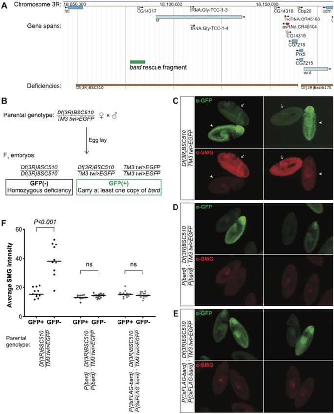
Figure 3 Bard is required for the degradation of SMG in the embryo at the end of the MZT. (A) JBrowse genome browser snapshot of the region deleted
by Df(3R)BSC510, which includes bard (CG14317) and several neighboring genes on Chromosome 3R. The bard rescue fragment used in panels D and E is
annotated in green. (B) Embryos were collected from flies carrying Df(3R)BSC510 over a twist (twi)>EGFP balancer as described in Materials and Methods.
Homozygous deficiency embryos could be distinguished from embryos carrying the balancer by lack of GFP staining. (C) Among embryos undergoing
gastrulation, those carrying the balancer chromosome show low levels of SMG expression (GFPþ, arrowheads) comparable to clearance of SMG in wild-
type, whereas homozygous deficiency embryos show significantly higher, ubiquitous SMG expression (GFP, arrows). This post-MZT persistence of
SMG expression is rescued with either: (D) a transgene expressing Bard, or (E) a transgene expressing 3FLAG-tagged Bard. (F) Quantification of SMG
intensities of embryos with the indicated genotypes, of which examples are shown in panels B–D; n > 10 for each group, ns: not significant, Mann–
Whitney U-test.W. X. Cao et al. | 7
Downloaded from https://academic.oup.com/genetics/advance-article/doi/10.1093/genetics/iyab177/6404591 by guest on 09 December 2021
Figure 4 Model of the temporal regulation of SMG protein degradation by SCFBard during the MZT. During the maternal phase of the MZT, although the
SCF core components and the F-box protein SLMB are present they do not bind SMG. Zygotic synthesis of Bard toward the end of the MZT promotes
binding to SMG of SCFBard and SCFSLMB. This triggers ubiquitin-mediated proteasomal degradation of SMG.
regulator b-TrCP, has been shown to function in heterodimeric Additional maternal proteins with a similar decay profile to
complexes with another F-box protein to increase the efficiency SMG include the histone H1 variant BigH1 and the cell cycle regu-
of the ubiquitination of its target protein (Tang et al. 2007). Thus, latory components APC7, Cyclin B, and Scrambled, among others
it is entirely possible here that SLMB and Bard function coopera- (Cao et al. 2020). Intriguingly, like SMG, both Scrambled and BigH1
tively to recruit the SCF complex to SMG for ubiquitination. Such have specific roles that are restricted to the MZT. Scrambled is re-
a model highlights Bard’s temporally restricted expression in the quired for actin organization during syncytial divisions in the
embryo as a timer for the action on SMG of the SCF E3-ligase early embryo but is not required for blastoderm cellularization or
complex as a whole (Figure 4). We note that, while SLMB was post-cellularization mitotic divisions (Stevenson et al. 2001).
found to interact with SMG in our previous IP-MS experiments BigH1 keeps the zygotic genome silent and aids in rapid nuclear
(Cao et al. 2020), it did not pass the threshold as a significant divisions in the early embryo, while clearance of BigH1 from the
interactor of SMG in our time-course IP-MS at any of the time chromatin and replacement with histone H1 at the end of the
points, possibly because of the small scale of these experiments MZT is required to permit ZGA (Pérez-Montero et al. 2013; Henn
due to the technical challenge of collecting sufficient material in et al. 2020). Thus, like SMG, precisely timed degradation of these
narrow time windows. Future IP-Western blot or IP-MS experi- proteins at the end of the MZT is important for an orderly devel-
ments may provide additional insights into the cooperative bind- opmental progression. Future experiments will reveal whether
ing model by asking, for example, whether Bard interacts with these proteins are also substrates of SCFBard.
SMG in the absence of SLMB and vice versa. Such experiments
would require appropriate mutations in both genes; currently no Data availability
mutations are available in the bard gene. New fly strains and plasmids generated in this article are avail-
Multiple mechanisms restrict SMG expression to the MZT. able upon request. Source data plotted in Figure 1A were
Although smg mRNA is expressed during oogenesis, it is kept extracted from FlyBase (flybase.org). Source data plotted in
translationally repressed until the Pan gu kinase complex Figure 1B were extracted from Supplementary Table S1 of our
relieves repression upon egg activation (Tadros et al. 2007). previous study (Cao et al. 2020). The full list of significant interac-
Misexpression of SMG in in the germline of ovaries results in fail- tors from SAINT analysis of time-course IP-MS experiments pre-
ure to progress through the stages of oogenesis (Semotok et al. sented in Figure 1C is provided in Supplementary Table S1. Raw
2005). SMG also needs to be cleared at the end of the MZT. mass spectrometry data used for these analyses have been
Persistence of SMG beyond the MZT leads to a reduction in the deposited to ProteomeXchange (MassIVE MSV000088199,
levels of zygotically expressed transcripts carrying cis-elements PXD028973) (ftp://massive.ucsd.edu/MSV000088199/).
that can be bound by SMG (Cao et al. 2020). By timing the clear- Supplementary material is available at GENETICS online.
ance of SMG, Bard thus plays a crucial role in the orderly hand-
over from maternal to zygotic control of the transcriptome.
SMG is not the only protein that is cleared at the end of the
Acknowledgments
MZT: Our previous proteomic analyses (Cao et al. 2020) identified During the course of this research, we made extensive use of
additional proteins that are known to have important roles in the FlyBase. We thank Dr. Craig Smibert and anonymous reviewers
early embryo and show a similar degradation profile to SMG, in- for constructive feedback about the manuscript.
cluding the germ plasm RNA-binding proteins Vasa, Oskar, and
Tudor. During the MZT, Bard is transcribed in the soma but not
Funding
the PGCs (see http://flyexpress.net/search/genes/CG14317/
images/BDGP/LDVO) (Konikoff et al. 2012). This is consistent with W.X.C. was supported in part by an Ontario Graduate
Scholarship and University of Toronto Open Scholarships. The re-
the fact that the PGCs are transcriptionally silent after they bud
search was funded by a Canadian Institutes of Health Research
from the posterior of the early embryo (Van Doren et al. 1998;
Project Grant to H.D.L. (PJT-159702).
Hanyu-Nakamura et al. 2008); budding occurs prior to Bard syn-
thesis in the bulk cytoplasm. We showed previously that SMG
persists in PGCs after it is cleared from the bulk cytoplasm Author contributions
(Siddiqui et al. 2012). We speculate that the presence of SMG, The project was designed by H.D.L. and W.X.C. The SMG IP-MS
Vasa, Oskar, and Tudor in the PGCs may, at least in part, be due time course was carried out by W.X.C. and S.L. under the supervi-
to the absence of Bard. sion of H.D.L and S.A., respectively. The RNAi experiments were8 | GENETICS, 2021, Vol. 00, No. 0
carried out by W.X.C. under the supervision of H.D.L. The bard de- Hanyu-Nakamura K, Sonobe-Nojima H, Tanigawa A, Lasko P,
ficiency and rescue experiments were carried out by W.X.C. and Nakamura A. 2008. Drosophila Pgc protein inhibits P-TEFb re-
A.K. under the supervision of H.D.L. W.X.C. wrote the first draft of cruitment to chromatin in primordial germ cells. Nature. 451:
the manuscript, which was revised by H.D.L. 730–733.
Hara M, Lourido S, Petrova B, Lou HJ, Von Stetina JR, et al. 2018.
Identification of PNG kinase substrates uncovers interactions
Conflicts of interest with the translational repressor TRAL in the oocyte-to-embryo
The authors declare that there is no conflict of interest. transition. eLife. 7:e33150.
Downloaded from https://academic.oup.com/genetics/advance-article/doi/10.1093/genetics/iyab177/6404591 by guest on 09 December 2021
Henn L, Szabo A, Imre L, Roman A, Abraham A, et al. 2020.
Alternative linker histone permits fast paced nuclear divisions in
Literature cited early Drosophila embryo. Nucleic Acids Res. 48:9007–9018.
Aviles-Pagan EE, Kang ASW, Orr-Weaver TL. 2020. Identification of Heriche JK, Ang D, Bier E, O’Farrell PH. 2003. Involvement of an
new regulators of the oocyte-to-embryo transition in Drosophila. SCFSlmb complex in timely elimination of E2F upon initiation of
G3 (Bethesda). 10:2989–2998. DNA replication in Drosophila. BMC Genet. 4:9.
Bai C, Sen P, Hofmann K, Ma L, Goebl M, et al. 1996. SKP1 connects Jeske M, Moritz B, Anders A, Wahle E. 2011. Smaug assembles an
cell cycle regulators to the ubiquitin proteolysis machinery ATP-dependent stable complex repressing nanos mRNA transla-
tion at multiple levels. EMBO J. 30:90–103.
through a novel motif, the F-box. Cell. 86:263–274.
Kipreos ET, Pagano M. 2000. The F-box protein family. Genome Biol.
Bashirullah A, Halsell SR, Cooperstock RL, Kloc M, Karaiskakis A, et
1:REVIEWS3002.
al. 1999. Joint action of two RNA degradation pathways controls
Kisielnicka E, Minasaki R, Eckmann CR. 2018. MAPK signaling cou-
the timing of maternal transcript elimination at the midblastula
ples SCF-mediated degradation of translational regulators to oo-
transition in Drosophila melanogaster. EMBO J. 18:2610–2620.
cyte meiotic progression. Proc Natl Acad Sci USA. 115:
Beiman M, Shilo BZ, Volk T. 1996. Heartless, a Drosophila FGF recep-
e2772–e2781.
tor homolog, is essential for cell migration and establishment of
Knight JDR, Choi H, Gupta GD, Pelletier L, Raught B, et al. 2017.
several mesodermal lineages. Genes Dev. 10:2993–3002.
ProHits-viz: a suite of web tools for visualizing interaction proteo-
Benoit B, He CH, Zhang F, Votruba SM, Tadros W, et al. 2009. An es-
mics data. Nat Methods. 14:645–646.
sential role for the RNA-binding protein Smaug during the
Konikoff CE, Karr TL, McCutchan M, Newfeld SJ, Kumar S. 2012.
Drosophila maternal-to-zygotic transition. Development. 136:
Comparison of embryonic expression within multigene families
923–932.
using the FlyExpress discovery platform reveals more spatial
Cao WX, Kabelitz S, Gupta M, Yeung E, Lin S, et al. 2020. Precise tem-
than temporal divergence. Dev Dyn. 241:150–160.
poral regulation of post-transcriptional repressors is required for
Liu G, Zhang J, Larsen B, Stark C, Breitkreutz A, et al. 2010. ProHits: in-
an orderly Drosophila maternal-to-zygotic transition. Cell Rep.
tegrated software for mass spectrometry-based interaction pro-
31:107783.
teomics. Nat Biotechnol. 28:1015–1017.
Casas-Vila N, Bluhm A, Sayols S, Dinges N, Dejung M, et al. 2017. The
Luo H, Li X, Claycomb JM, Lipshitz HD. 2016. The Smaug
developmental proteome of Drosophila melanogaster. Genome Res.
RNA-binding protein is essential for microRNA synthesis during
27:1273–1285.
the Drosophila maternal-to-zygotic transition. G3 (Bethesda). 6:
Chen L, Dumelie JG, Li X, Cheng MH, Yang Z, et al. 2014. Global regu-
3541–3551.
lation of mRNA translation and stability in the early Drosophila
Markstein M, Pitsouli C, Villalta C, Celniker SE, Perrimon N. 2008.
embryo by the Smaug RNA-binding protein. Genome Biol. 15:r4.
Exploiting position effects and the gypsy retrovirus insulator to
Choi H, Larsen B, Lin ZY, Breitkreutz A, Mellacheruvu D, et al. 2011.
engineer precisely expressed transgenes. Nat Genet. 40:476–483.
SAINT: probabilistic scoring of affinity purification-mass spec- Morreale FE, Walden H. 2016. Types of ubiquitin ligases. Cell. 165:
trometry data. Nat Methods. 8:70–73. 248.e241.
Cook RK, Christensen SJ, Deal JA, Coburn RA, Deal ME, et al. 2012. Muzzopappa M, Wappner P. 2005. Multiple roles of the F-box protein
The generation of chromosomal deletions to provide extensive Slimb in Drosophila egg chamber development. Development.
coverage and subdivision of the Drosophila melanogaster genome. 132:2561–2571.
Genome Biol. 13:r21. Nelson MR, Leidal AM, Smibert CA. 2004. Drosophila Cup is an
Dahanukar A, Walker JA, Wharton RP. 1999. Smaug, a novel eIF4E-binding protein that functions in Smaug-mediated transla-
RNA-binding protein that operates a translational switch in tional repression. EMBO J. 23:150–159.
Drosophila. Mol Cell. 4:209–218. Ni JQ, Zhou R, Czech B, Liu LP, Holderbaum L, et al. 2011. A
Dui W, Lu W, Ma J, Jiao R. 2012. A systematic phenotypic screen of genome-scale shRNA resource for transgenic RNAi in Drosophila.
F-box genes through a tissue-specific RNAi-based approach in Nat Methods. 8:405–407.
Drosophila. J Genet Genomics. 39:397–413. Pérez-Montero S, Carbonell A, Morán T, Vaquero A, Azorı́n F. 2013.
Götze M, Dufourt J, Ihling C, Rammelt C, Pierson S, et al. 2017. The embryonic linker histone H1 variant of Drosophila, dBigH1,
Translational repression of the Drosophila nanos mRNA involves regulates zygotic genome activation. Dev Cell. 26:578–590.
the RNA helicase Belle and RNA coating by Me31B and Trailer Pinder BD, Smibert CA. 2013. microRNA-independent recruitment of
hitch. RNA. 23:1552–1568. Argonaute 1 to nanos mRNA through the Smaug RNA-binding
Graveley BR, Brooks AN, Carlson JW, Duff MO, Landolin JM, et al. protein. EMBO Rep. 14:80–86.
2011. The developmental transcriptome of Drosophila mela- Ravid T, Hochstrasser M. 2008. Diversity of degradation signals in
nogaster. Nature. 471:473–479. the ubiquitin-proteasome system. Nat Rev Mol Cell Biol. 9:
Guven-Ozkan T, Nishi Y, Robertson SM, Lin R. 2008. Global transcrip- 679–690.
tional repression in C. elegans germline precursors by regulated Semotok JL, Cooperstock RL, Pinder BD, Vari HK, Lipshitz HD, et al.
sequestration of TAF-4. Cell. 135:149–160. 2005. Smaug recruits the CCR4/POP2/NOT deadenylase complexW. X. Cao et al. | 9
to trigger maternal transcript localization in the early Drosophila Tang X, Orlicky S, Lin Z, Willems A, Neculai D, et al. 2007. Suprafacial
embryo. Curr Biol. 15:284–294. orientation of the SCFCdc4 dimer accommodates multiple geom-
Semotok JL, Luo H, Cooperstock RL, Karaiskakis A, Vari HK, et al. etries for substrate ubiquitination. Cell. 129:1165–1176.
2008. Drosophila maternal Hsp83 mRNA destabilization is di- Tolkien JRR. 1937. The Hobbit. London: George Allen & Unwin.
rected by multiple SMAUG recognition elements in the open Van Doren M, Williamson AL, Lehmann R. 1998. Regulation of zy-
reading frame. Mol Cell Biol. 28:6757–6772. gotic gene expression in Drosophila primordial germ cells. Curr
Shteynberg D, Deutsch EW, Lam H, Eng JK, Sun Z, et al. 2011. Biol. 8:243–246.
iProphet: multi-level integrative analysis of shotgun proteomic Vastenhouw NL, Cao WX, Lipshitz HD. 2019. The
Downloaded from https://academic.oup.com/genetics/advance-article/doi/10.1093/genetics/iyab177/6404591 by guest on 09 December 2021
data improves peptide and protein identification rates and error maternal-to-zygotic transition revisited. Development. 146:
estimates. Mol Cell Proteomics. 10:M111.007690. dev161471.
Siddiqui NU, Li X, Luo H, Karaiskakis A, Hou H, et al. 2012. Wang M, Ly M, Lugowski A, Laver JD, Lipshitz HD, et al. 2017. ME31B
Genome-wide analysis of the maternal-to-zygotic transition in globally represses maternal mRNAs by two distinct mechanisms
Drosophila primordial germ cells. Genome Biol. 13:r11. during the Drosophila maternal-to-zygotic transition. eLife. 6:
Spike CA, Huelgas-Morales G, Tsukamoto T, Greenstein D. 2018. e27891.
Multiple mechanisms inactivate the LIN-41 RNA-binding protein Wodarz A, Hinz U, Engelbert M, Knust E. 1995. Expression of crumbs
to ensure a robust oocyte-to-embryo transition in Caenorhabditis confers apical character on plasma membrane domains of ecto-
elegans. Genetics. 210:1011–1037. dermal epithelia of Drosophila. Cell. 82:67–76.
Stevenson VA, Kramer J, Kuhn J, Theurkauf WE. 2001. Centrosomes Yang Y, Zhou C, Wang Y, Liu W, Liu C, et al. 2017. The E3 ubiquitin li-
and the Scrambled protein coordinate microtubule-independent gase RNF114 and TAB1 degradation are required for
actin reorganization. Nat Cell Biol. 3:68–75. maternal-to-zygotic transition. EMBO Rep. 18:205–216.
Tadros W, Goldman AL, Babak T, Menzies F, Vardy L, et al. 2007. Zavortink M, Rutt LN, Dzitoyeva S, Henriksen JC, Barrington C, et al.
SMAUG is a major regulator of maternal mRNA destabilization in 2020. The E2 Marie Kondo and the CTLH E3 ligase clear deposited
Drosophila and its translation is activated by the PAN GU kinase. RNA binding proteins during the maternal-to-zygotic transition.
Dev Cell. 12:143–155. Elife. 9:e53889.
Tadros W, Lipshitz HD. 2009. The maternal-to-zygotic transition: a Zheng N, Shabek N. 2017. Ubiquitin ligases: structure, function, and
play in two acts. Development. 136:3033–3042. regulation. Annu Rev Biochem. 86:129–157.
Communicating editor R. DuronioYou can also read


