University of Birmingham Consequences of cathepsin C inactivation for membrane exposure of proteinase 3, the target antigen in autoimmune vasculitis
←
→
Page content transcription
If your browser does not render page correctly, please read the page content below
University of Birmingham

Consequences of cathepsin C inactivation for
membrane exposure of proteinase 3, the target
antigen in autoimmune vasculitis
Seren, Seda; Rashed Abouzaid, Maha; Eulenberg-Gustavus, Claudia; Hirschfeld, Josefine;
Nasr Soliman, Hala; Jerke, Uwe; N'Guessan, Koffi; Dallet-Choisy, Sandrine; Lesner, Adam;
Lauritzen, Conni; Schacher, Beate; Eickholz, Peter; Nagy, Nikoletta; Szell, Marta; Croix,
Cécile; Viaud-Massuard, Marie-Claude; Al Farraj Aldosari, Abdullah; Ragunatha, Shivanna;
Ibrahim Mostafa, Mostafa; Giampieri, Francesca
DOI:
10.1074/jbc.RA118.001922
License:
None: All rights reserved

Document Version
Peer reviewed version

Citation for published version (Harvard):
Seren, S, Rashed Abouzaid, M, Eulenberg-Gustavus, C, Hirschfeld, J, Nasr Soliman, H, Jerke, U, N'Guessan,
K, Dallet-Choisy, S, Lesner, A, Lauritzen, C, Schacher, B, Eickholz, P, Nagy, N, Szell, M, Croix, C, Viaud-
Massuard, M-C, Al Farraj Aldosari, A, Ragunatha, S, Ibrahim Mostafa, M, Giampieri, F, Battino, M, Cornillier, H,
Lorette, G, Stephan, J-L, Goizet, C, Pedersen, J, Gauthier, F, Jenne, DE, Marchand-Adam, S, Chapple, IL,
Kettritz, R & Korkmaz, B 2018, 'Consequences of cathepsin C inactivation for membrane exposure of proteinase
3, the target antigen in autoimmune vasculitis', Journal of Biological Chemistry, vol. 293, no. 32, pp. 12415-
12428. https://doi.org/10.1074/jbc.RA118.001922

Link to publication on Research at Birmingham portal

Publisher Rights Statement:
This research was originally published in the Journal of Biological Chemistry. Seren et al., Consequences of cathepsin C inactivation for
membrane exposure of proteinase 3, the target antigen in autoimmune vasculitis, Journal of Biological Chemistry, Vol 293, pp. 12415-12428

General rights
Unless a licence is specified above, all rights (including copyright and moral rights) in this document are retained by the authors and/or the
copyright holders. The express permission of the copyright holder must be obtained for any use of this material other than for purposes
permitted by law.

•Users may freely distribute the URL that is used to identify this publication.
•Users may download and/or print one copy of the publication from the University of Birmingham research portal for the purpose of private
study or non-commercial research.
•User may use extracts from the document in line with the concept of ‘fair dealing’ under the Copyright, Designs and Patents Act 1988 (?)
•Users may not further distribute the material nor use it for the purposes of commercial gain.

Where a licence is displayed above, please note the terms and conditions of the licence govern your use of this document.

When citing, please reference the published version.
Take down policy
While the University of Birmingham exercises care and attention in making items available there are rare occasions when an item has been
uploaded in error or has been deemed to be commercially or otherwise sensitive.

If you believe that this is the case for this document, please contact UBIRA@lists.bham.ac.uk providing details and we will remove access to
the work immediately and investigate.
Download date: 06. Mar. 2022
Consequences of cathepsin C inactivation on membrane expression of proteinase 3, the target
                            antigen in autoimmune vasculitis

    Seda Seren1, Maha Rashed Abouzaid2#, Claudia Eulenberg-Gustavus3#, Josefine Hirschfeld4#,
    Hala Soliman5, Uwe Jerke3, Koffi N’Guessan1, Sandrine Dallet-Choisy1, Adam Lesner6, Conni
     Lauritzen7, Beate Schacher8, Peter Eickholz8, Nikoletta Nagy9, Marta Szell9, Cécile Croix10,
      Marie-Claude Viaud-Massuard10, Abdullah Al Farraj Aldosari11, Shivanna Ragunatha12,
     Mostafa Ibrahim Mostafa2, Francesca Giampieri13, Maurizio Battino13, Hélène Cornillier14,
     Gérard Lorette15, Jean-Louis Stephan16, Cyril Goizet17, John Pedersen7, Francis Gauthier1,
     Dieter E. Jenne18, Sylvain Marchand-Adam1, Iain L. Chapple4, Ralph Kettritz3,19 and Brice
                                            Korkmaz1*

#
    The authors contributed equally to this work

                Running title: Cathepsin C inactivation and membrane-bound proteinase 3

*To whom correspondence should be addressed: Brice Korkmaz, INSERM U-1100 “Centre d’Etude
des Pathologies Respiratoires (CEPR)”,Université François Rabelais, Faculté de Médecine, 10 Bld.
Tonnellé, 37032, Tours, France; e-mail: brice.korkmaz@inserm.fr; Tel: 0033 2 47 36 63 86
1
  INSERM U-1100, “Centre d’Etude des Pathologies Respiratoires” and Université François Rabelais,
Tours, France
2
  Department of Oro-Dental Genetics, National Research Centre, Cairo, Egypt
3
  Experimental and Clinical Research Center, Charité und Max-Delbrück-Centrum für Molekulare
Medizin in der Helmholtz-Gemeinschaft (MDC), Berlin, Germany
4
  Institute of Clinical Sciences, College of Medical and Dental Sciences, Periodontal Reseaech Group,
University of Birmingham, and Birmingham Community Health Trust, Edgbaston, Birmingham, UK
5
  Department of Clinical Genetics, National Research Centre, Egypt
6
  Faculty of Chemistry, University of Gdansk, Poland
7
  Unizyme Laboratories A/S, Hörsholm, Denmark
8
  Department of Periodontology, Johann Wolfgang Goethe-University Frankfurt, Frankfurt, Germany
9
  Department of Medical Genetics, University of Szeged, Szeged, Hungary
10
   UMR-CNRS 7292 “Génétique, Immunothérapie, Chimie et Cancer” and Université François
Rabelais, Tours, France
11
   Department of Prosthetic, College of Dentistry, King Saud University, Riyadh, Kingdom of Saudi
Arabia
12
   Department of Dermatology, Venereology, and Leprosy, ESIC Medical College and PGIMSR
Rajajinagar, Bengaluru, Karnataka, India
13
   Deptment Clinical Sciences, Università Politecnica delle Marche, Ancona, Italy
14
   Service de Dermatologie, Centre Hospitalier Universitaire de Tours, Université François Rabelais,
Tours, France.
15
   UMR-INRA1282 « Laboratoire de Virologie et Immunologie Moléculaires », Université François
Rabelais, Tours, France
16
   Service d'Hématologie Immunologie et Rhumatologie Pédiatrique, Centre Hospitalier Universitaire
de Saint-Etienne, Saint-Priest-en-Jarez, France
17
   INSERM-U1211, Neuropediatric and Neurogenetic department, MRGM laboratory, Pellegrin
Hospital and University, Bordeaux, France
18
   Comprehensive Pneumology Center, Institute of Lung Biology and Disease, German Center for
Lung Research (DZL), Munich and Max Planck Institute of Neurobiology, Planegg-Martinsried,
Germany

                                                   1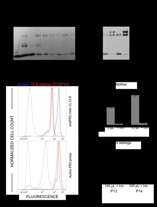
19
  Nephrology and Intensive Care Medicine, Charité-Universitätsmedizin, Berlin, Germany

Keywords: proteinase 3, cathepsin C, neutrophil, granulomatosis with polyangiitis, Papillon-Lefèvre
syndrome

ABSTRACT                                               target autoantigen in GPA is the neutrophil
                                       m
    Membrane-bound proteinase 3 (PR3 ) is the          serine protease (NSP) proteinase 3 (PR3) (EC
main target antigen of anti-neutrophil                 3.4.21.76) (3,4). GPA patients develop anti-
cytoplasmic autoantibodies (ANCA) in                   neutrophil cytoplasmic autoantibodies to PR3
granulomatosis with polyangiitis (GPA), a              (PR3-ANCA) that bind to membrane-bound
systemic small-vessel vasculitis. Binding of           PR3 (PR3m) on the neutrophil surface (5,6). The
ANCA to PR3m triggers neutrophil activation            membrane expression of PR3 is mediated by a
with the secretion of enzymatically active PR3         hydrophobic patch at the protease surface,
and related neutrophil serine proteases, thereby       which is not conserved in other related NSPs,
contributing to vascular damage. PR3 and               such as human neutrophil elastase (HNE),
related proteases are activated from proforms          cathepsin G (CG) and neutrophil serine protease
by the lysosomal cysteine protease cathepsin C         4 NSP-4 (7-9). Binding of PR3-ANCA to PR3m
(CatC) during neutrophil maturation. We                on cytokine-primed neutrophils induces cell
hypothesized that pharmacological inhibition of        activation resulting in neutrophil extracellular
CatC provides an effective measure to reduce           traps (NETs) production, and in granule protein
PR3m and has therefore implications as a novel         and superoxide release (10,11). Secreted active
therapeutic approach in GPA. We first studied          proteases, including PR3 and related NSPs,
PR3 in neutrophils from 21 patients with               exert proteolytic activity on endothelial cells
Papillon-Lefèvre syndrome (PLS), a genetic             thereby contributing to vascular necrosis (5,12).
form of CatC deficiency. PLS neutrophil lysates        NETs are known to be directly implicated in
showed a largely reduced, but still detectable         ANCA induction as well as endothelial damage
(0.5-4%) PR3 activity when compared to                 (13). There is no treatment for GPA that is based
healthy control cells. Despite extremely low           on disease-specific mechanisms and the current
levels of cellular PR3, the amount of                  protocols involve combined administration of
constitutive PR3m expressed on the surface of          steroids with either cyclophosphamide or
quiescent neutrophils, and the typical bimodal         rituximab (14,15). These standard treatments
membrane distribution pattern, was similar to          are associated with toxicity highlighting the
what was observed in healthy neutrophils.              need to develop novel, more specific
However, following cell activation, there was          therapeutic strategies (16).
no significant increase in the total amount of             Cathepsin C (CatC) (EC 3.4.14.1), also
PR3m on PLS neutrophils, whereas the total             known as dipeptidyl peptidase I, is a lysosomal
amount of PR3m on healthy neutrophils was              amino peptidase belonging to the papain
significantly increased. We then explored the          superfamily of cysteine peptidases (17). CatC
effect of pharmacological CatC inhibition on           catalyzes the cleavage of two residues from the
PR3 expression in normal neutrophils using a           N-termini of peptides and proteins. CatC, which
potent cell permeable CatC inhibitor and a             is ubiquitously expressed in mammals is
CD34+ hematopoietic stem cell model. Human             considered to be a major intracellular
CD34+ hematopoietic stem cells were treated            processing enzyme. High concentrations of
with the inhibitor during neutrophil                   CatC are detected in immune defense cells
differentiation over 10 days. We observed              including neutrophils, mast cells, lymphocytes
strong reductions in PR3m, cellular PR3 protein        and macrophages. CatC is the physiological
and proteolytic PR3 activity whereas neutrophil        activator of several immune cell-associated
differentiation was not compromised.                   serine proteases such as NSPs (18,19). NSPs are
                                                       synthetized as inactive zymogens containing a
                                                       di-propeptide in the myeloblast/promyelocyte
                                                       stage in the bone marrow (8,20). The proforms
   Granulomatosis with polyangiitis (GPA) is a
                                                       mature in this very early developmental stage,
systemic     small-vessel   vasculitis   most
                                                       induced by CatC, through the cleavage of the N-
commonly affecting the upper and lower
                                                       terminal di-propeptide. The cleavage of the di-
respiratory tract and kidneys (1,2). The main
                                                       propeptide by CatC results in a re-orientation

                                                   2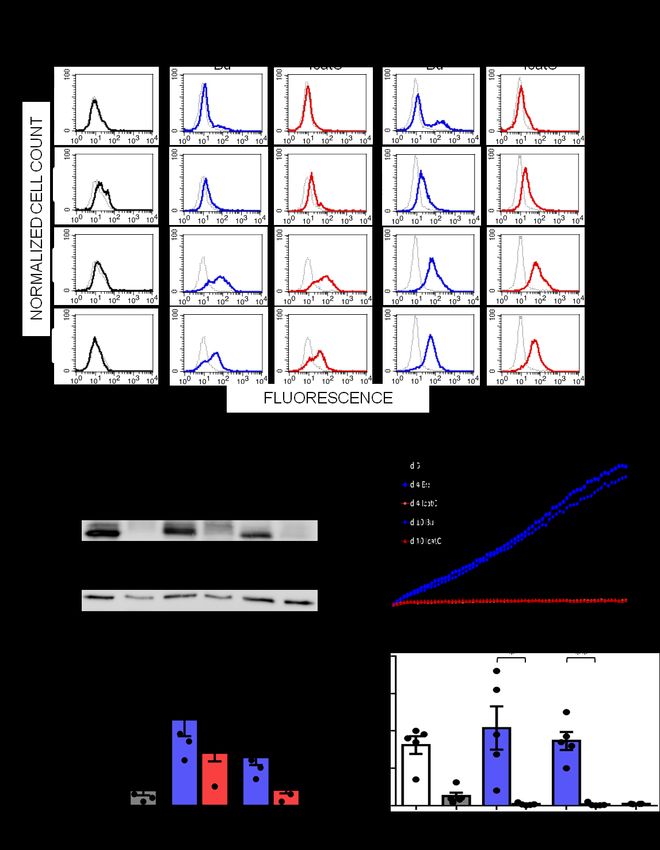
and remodeling of three surface loops within the        (32-34). Thus, it is conceivable that mimicking
activation domain of the protein and renders the        the genetic situation in PLS neutrophils by
S1 pocket of the active-site accessible to              pharmacological CatC inhibition in bone
substrates (21). After processing, the active           marrow precursor cells would provide an
proteases are stored in cytoplasmic granules.           attractive therapeutic strategy in GPA to
    CatC is synthetized as a 60-kDa single chain        eliminate     major      PR3-related     disease
pro-form containing an exclusion domain, a              mechanisms, including the PR3-ANCA
propeptide, a heavy chain and a light chain (22).       autoantigen itself. However, the effect of CatC
Pro-CatC which is a dimer, can be efficiently           inactivation on PR3 that is presented on the
activated by proteolysis with CatL and S in vitro       neutrophil surface where it becomes accessible
(22). The initial cleavages liberate the                to anti-PR3 antibodies is not known.
propeptide from the catalytic region.                       In this work, we investigated the
Subsequently, a further cleavage occurs                 consequences of CatC inactivation on
between the heavy chain and the light chain             membrane-expression of PR3. First, we
which form a papain-like structure (22,23). X-          quantified the residual proteolytic activity of
ray images of mature CatC structures revealed           CatC and PR3 in white blood cell (WBC)
that the exclusion domain, the heavy chain and          lysates or isolated neutrophils from PLS
the light chain are held together by non-covalent       patients. Second, we studied the membrane-
interactions (17). Mature CatC is a tetramer            expression of PR3 on PLS neutrophils. Finally,
formed by four identical monomers with their            we used a potent synthetic cell permeable nitrile
active site clefts fully solvent exposed. The           inhibitor to evaluate the effect of
presence of the exclusion domain blocks the             pharmacological CatC inhibition on membrane-
active site beyond the S2 pocket and it is              PR3 expression in normal neutrophils generated
responsible for the diaminopeptidase activity of        from human CD34+ progenitor cells.
CatC (17,24).
    Loss of function mutations in the CatC gene         Results
(CTSC) results in Papillon-Lefèvre syndrome             CatC in blood cells from PLS patients
(PLS) (OMIM: 245000) (25,26), a rare                        Blood samples were collected from 13 PLS
autosomal recessive disease affecting 1 to 4            patients from European, Asian and African
persons per million (27,28). PLS involves an            countries. PLS diagnosis was firmly established
aggressive pre-pubertal periodontitis, leading to       by genetic testing. These patients carried either
complete tooth loss in adolescence and                  premature stop codon, missense, nonsense or
palmoplantar keratoderma. More than 75                  frameshift mutations in their CTSC (Table I).
mutations have been identified in PLS, with             Blood from 8 additional patients with clinically
missense and nonsense mutations being the               suspected PLS was obtained. These patients
most frequent, but small deletions, insertions          showed typical symptoms of early-onset
and splice site mutations have also been                periodontitis and hyperkeratosis (Fig. 1A).
reported (29). The presumptive diagnosis of             WBC lysates from these patients and from
PLS can be made by clinical signs and                   healthy controls differed by their protein profile
symptoms, but confirmation requires CTSC                as observed by SDS-PAGE (Fig. 1B). CatC
sequencing. Analysis of urinary CatC in                 activity was assayed in peripheral WBC lysates
suspected patients can be also used as an early,        or in purified neutrophils using a CatC-selective
simple and easy diagnostic test (30). Roberts et        FRET substrate in the presence or absence of
al. (31) demonstrated a variety of neutrophil           the selective nitrile CatC inhibitor (L)-Thi-(L)-
defects in PLS patients, arising downstream of          Phe-CN. Strong CatC activity was observed in
the failure to activate NSPs by CatC. These             control neutrophils and was completely
functional defects included failure to produce          abrogated by the specific CatC inhibitor. In
NETs, reduced chemotaxis and exaggerated                contrast, we did not detect any CatC activity in
cytokine and reactive oxygen species release.           samples, from genetically and clinically
Pham et al. (19) also studied neutrophils from          diagnosed PLS patients (Table I and Fig. 1C),
PLS patients and observed that the loss of CatC         nor any CatC protein in cell lysates from PLS
activity was associated with strong reduction in        patients using a specific anti-CatC Ab (Fig.
the proteolytic activity of NSPs. In addition,          1D). CatC activity and the CatC antigen were
only very low protein amounts of PR3 and                also absent in the urine of all PLS patients,
related NSPs were detected in PLS neutrophils           unlike healthy controls (Fig. 1C, D). Thus, the

                                                    3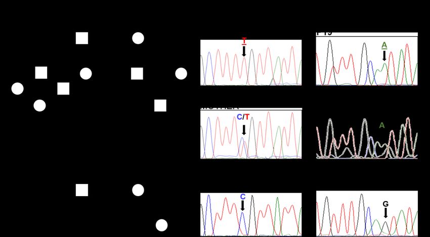
CatC deficiency of all 7 patients was confirmed Membrane surface expression of PR3 on
as described in (30) and the mutations were PLS neutrophils
identified by CTSC gene sequencing for some Since the membrane expression of PR3
of these patients (supp.Fig. 2, Table I). Once depends on the activation status of neutrophils,
CatC deficiency was clearly established, we we analyzed both quiescent neutrophils from
studied the fate of PR3 in samples from PLS two local patients (PLS 13 and PLS 14) 30 min
patients. after blood collection and neutrophils from
foreign blood samples collected 24-72 h before
Proteolytically active PR3 in blood cells from the analysis and thus inevitably activated during
PLS patients shipping. As a control for the latter conditions,
WB analysis of white blood cell or cells from either the parents or healthy
neutrophil lysates from PLS patients showed individuals of the patient’s country-of-origin
that low amounts of the PR3 antigen were still were collected at the same time. The flow
present in all PLS samples (Fig. 2A and cytometry analysis of PR3m using anti-PR3
supp.Fig. 4A). We checked that residual PR3 mAbs CLB12.8 showed that constitutive PR3m
was enzymatically active by incubating PLS was present in significant amounts on quiescent
samples with purified exogenous alpha-1- PLS neutrophils and showed the typical
proteinase inhibitor (1PI) (35) and observing bimodal pattern with low (PR3m(low)) and high
the appearance of an additional ~75 kDa band (PR3m(high)) subsets. After cell activation with a
corresponding to the irreversible complex calcium ionophore (A23187), we observed a
between active PR3 and its inhibitor (Fig. 2B). single homogeneous PR3 population but no
In contrast to mature PR3, inactive pro-PR3 did significant increase of the total amount of PR3m
not form any irreversible complex with 1PI at the surface of PLS neutrophils whereas the
(Fig. 3). We confirmed the presence of bimodal pattern was conserved on control cells
proteolytically active PR3 in permeabilized and the total amount of PR3 increased
PLS neutrophils using an activity-based probe significantly (Fig. 5). We also found a single
(Bt-PEG66-PYDA(O-C6H4-4-Cl)2) selective for PR3m-presenting neutrophil population in PLS
PR3 and a fluorescent streptavidin derivative to cells that were spontaneously and inevitably
reveal the formation of irreversible complexes activated during transit (experiments with cells
(Fig. 2C). We also measured PR3 activity in from 9 different PLS patients, Fig. 6 and
supernatants of PLS cells activated with the supp.Fig. 4). The PR3 mean fluorescence
A23187 calcium ionophore using ABZ- intensity of these PLS neutrophils was 26 ± 8 %
VAD(nor)VADYQ-EDDnp as a substrate (Fig. of the intensity found in controls. Thus, the
2D). PR3 activity in PLS cell supernatants was absence of CatC in PLS patients affected
about 1/20 that in controls cells and was almost constitutive PR3 expression at the surface of
totally abrogated by the PR3-specific inhibitor quiescent neutrophils only marginally but
Bt-PYDAP(O-C6H4-4-Cl)2. We found that 10 to largely reduced the PR3 antigen exposure on the
20 times more cell lysate proteins from PLS neutrophil surface of activated cells. Next, we
patients (2.5 to 5 µg/well) as compared to assessed whether an early treatment of
healthy controls (0.25 to 0.5 µg/well), were neutrophil precursor cells by a CatC inhibitor,
required to achieve similar PR3 activity values. would diminish PR3m expression, cellular PR3
Enzymatic activities in PLS cells and healthy amount and proteolytic activity.
control cells, measured on ABZ-
VAD(nor)VADYQ-EDDnp substrate, were Membrane surface expression of PR3 on
almost completely inhibited by the PR3 neutrophils generated from human CD34+
inhibitor (Fig. 4A, B, C). From these results, we progenitor cells in the presence of a CatC
estimated that blood cells of PLS patients inhibitor
contained from 0.5 to 4% of the PR3 activity in We differentiated human CD34+ HSC
healthy controls cells (Fig. 4D and Table I). isolated from umbilical cord blood into
Marginal, but still detectable activity of CG was neutrophils in the presence or absence of a
also observed in PLS samples using the potent cell permeable cyclopropyl nitrile CatC
appropriate selective FRET substrate (data not inhibitor (IcatC) (32). Expression of the
show). Next, we checked whether PR3 was neutrophil surface markers CD16, CD66b, and
present at the cell surface of PLS neutrophils. CD11b was assessed by flow cytometry during
the 10 day differentiation period and confirmed
4neutrophil differentiation (Fig. 7A). At day 10, pharmacological CatC inactivation on the fate a typical bimodal PR3m expression pattern was of soluble PR3 and PR3m. observed by flow cytometry with a PR3m- Because CatC is pathologically inactivated positive neutrophil subset of approximately 30- by gene mutations in PLS patients, we first used 40%. Importantly, CatC inhibition with the WBC lysates from 21 PLS, 17 of these patients pharmacological compound IcatC did not affect with established missense, frameshift or neutrophil differentiation, but eliminated the nonsense mutations, to investigate the fate of bimodal PR3m expression pattern leaving only cellular and membrane PR3. As expected, we marginal PR3m amounts on the cell surface observed neither CatC activity nor (Fig. 7A). The mean fluorescence intensity immunoreactive CatC protein in PLS cell values for PR3m were reduced to 17±5 % by lysates irrespective of the underlying CatC IcatC at day 10 (p
of pro-PR3 was degraded very early in PLS                consistent with a bimodal expression typically
neutrophil precursors. Our data are compatible           seen with blood neutrophils. Differentiation of
with the notion that a small amount of the pro-          CD34+ HSC into neutrophils in the presence of
PR3 can be early processed into an active                the CatC inhibitor IcatC did not alter the
protease by one or several aminopeptidase(s)             expression of the neutrophil surface markers
other than CatC. These enzymes remain to be              CD16b, CD66b and CD11b but resulted in
identified.                                              strong reduction of intracellular and membrane
    Human PR3 is expressed constitutively in a           PR3. Pharmacological CatC inhibition
bimodal manner with two populations of                   eliminated PR3 from normal neutrophils more
neutrophils presenting either high (PR3m(high))          effectively than mutated CatC in PLS
or low (PR3m(low)) amounts of the protease on            neutrophils. It is conceivable that additional
their surface (36,37). The level of PR3m on              aminopeptidases exist in blood neutrophils that
resting neutrophils and the percentage of PR3m           were absent in CD34+ HCS-derived neutrophils
expressing neutrophils is stable over time for a         or that the potent IcatC inhibitor inhibited CatC
given individual (5,37). In spite of the low PR3         together with additional proteases involved in
level in PLS cell lysates, we observed that PR3m         the activation of the pro-PR3. Our current
was present at the surface of resting PLS                observations in PLS cells and our recent work
neutrophils showing a typical bimodal                    reporting the complete disappearance of HNE
distribution similar to control neutrophils. This        in bone marrow cells from healthy donors
observation suggests that the expression of              pulse-chased in presence of IcatC supports the
constitutive PR3m on resting neutrophils is              latter hypothesis (32).
                                                             To conclude, we showed here that CatC is
independent of intracellular PR3 levels and
                                                         the major but not the unique pro-PR3
remains stable even when CatC is inactive.
                                                         processing protease in neutrophils since low
Activating cells from PLS patients with the
                                                         amounts of proteolytically active PR3 are still
calcium ionophore A23187 resulted in a PR3
                                                         present in neutrophils of CatC deficient
increase on the neutrophil surface but this
                                                         individuals. Treating CD34+ hematopoietic
increase was significantly smaller than that
                                                         stem cells with the CatC inhibitor ICatC
observed in control cells. Thus, the genetic
                                                         resulted in an almost total absence of
inactivation of CatC results in a dramatic
                                                         intracellular PR3 and PR3m in stem cells-
decrease of PR3 within intracellular granules
                                                         derived neutrophils. The elimination of the
but does not interfere with the constitutive
                                                         PR3-ANCA target antigen supports the notion
expression of proteolytically inactive PR3
                                                         that pharmacological CatC inhibition provides
(38,39) at the surface of quiescent PLS
                                                         an alternative therapeutic strategy for reducing
neutrophils. This data suggests a different
                                                         neutrophil-mediated vascular inflammation in
intracellular storage site and a different
                                                         auto-immune vasculitis. We previously showed
intracellular pathway for constitutive and
                                                         that a prolonged IcatC administration in the
induced PR3m. Unexpectedly, and in contrast to
                                                         macaque resulted in an almost complete
control cells, no bimodal PR3m expression
                                                         elimination of PR3 and NE (32). Unlike humans
pattern was observed in activated PLS cells.
                                                         however, macaques do not display constitutive
This observation was made in both
                                                         PR3 at the surface of their circulating
spontaneously activated cells during shipping
                                                         neutrophils and therefore cannot be used as a
and with a pharmacological compound. We
                                                         relevant model of GPA. Only clinical studies in
have no obvious explanation for this finding at
                                                         GPA patients will answer the question whether
the moment.
                                                         or not a CatC inhibitor may function as a PR3-
    We showed previously that a two-step
                                                         ANCA antigen suppressor.
amplification/differentiation protocol of human
CD34+ hematopoietic stem cells obtained from
umbilical cord blood results in differentiated           Experimental Procedures
neutrophils (40). We used this model system to              Blood collection- Blood samples were
investigate the production and the fate of PR3           collected from 21 PLS patients from European
in the presence of a CatC nitrile inhibitor, IcatC       countries (Germany, the UK, Italy, France,
(23). A subset of PR3m-positive cells was                Hungary), from Asian countries (India, Saudi
detectable by flow cytometry at day10 whereas            Arabia) and from Egypt. The 13 healthy
a second cell subset remained negative,                  volunteers were from France, India, Italy, Saudi
                                                         Arabia and Egypt. 2-15 mL peripheral blood

                                                     6
samples from healthy control donors and were separated from cell debris by
patients with PLS were collected into EDTA K2 centrifugation at 10,000 x g for 10 min. Soluble
preservative tubes by peripheral venipuncture. fractions were concentrated by ultrafiltration
Samples were taken giving informed consent. (Vivaspin (filtration threshold 10 kDa)) in some
Red blood cells lysis took place with 0.1 mM experiments. Proteins were assayed with a
EDTA, 10 mM KHCO3, 150 mM NH4Cl and bicinchoninic acid assay (BCA) (Thermo Fisher
white cells pelleted with centrifugation for 5 Scientific, Villebon sur Yvette, France).
min at 400 x g. The CatC activity in cell lysates was
measured spectrofluorometrically (Spectra Max
Blood neutrophil purification- Neutrophils Gemini EM) at 420 nm with or without the
were isolated by Percoll density centrifugation, nitrile inhibitor (L)-Thi-(L)-Phe-CN (23) (1 µM
employing two discontinuous gradients of final, 20 min incubation at 37°C) using Thi-
1.079 and 1.098, and purified by erythrocyte Ala(Mca)-Ser-Gly-Tyr(3-NO2)-NH2 (41) (20
lysis (0.83% NH4Cl containing 1% KHCO3, µM final) as selective fluorescence resonance
0.04% EDTA, and 0.25% BSA) previously energy transfer (FRET) substrate in 50 mM
described (31). Cells were then re-suspended in sodium acetate, 30 mM NaCl, 1 mM EDTA, 2
gPBS (phosphate-buffered saline) (1 mM mM DTT, pH 5.5 at 37°C. Mature human CatC
glucose) and cations (1 mM MgCl2, 1.5 mM was used as control (Unizyme Laboratories,
CaCl2). Cell viability was determined by Hørsholm, Denmark).
Trypan blue dye exclusion (typically 98%) and The PR3 activity in cell lysates was
cell purity by cytospin. measured at 420 nm with or without the PR3
inhibitor Ac-PYDAP(O-C6H6-4-Cl)2 (42) (0.5
Differentiation of CD34+ hematopoietic µM final, 20 min incubation at 37°C) using
stem cells from umbilical cord blood into ABZ-VAD(nor)VADYQ-EDDnp (20 µM final,
neutrophils- Umbilical cord blood samples Genecust, Dudelange, Luxembourg) as a
were taken giving informed consent. substrate in 50 mM HEPES buffer, 750 mM
Mononuclear cells were obtained from anti- NaCl, 0.05% NP40, pH 7.4 at 37°C. The CatG
coagulated cord blood by centrifugation over a activity was measured at 420 nm in 50 mM
LSM1077 (PAA, Pasching, Austria) gradient at HEPES buffer, 100 mM NaCl, 0.05% NP-40,
800 x g for 20 min. Cells were washed and pH 7.4 at 37°C, in the presence or not of 2 µM
stained using the CD34+ progenitor isolation kit antichymotrypsin, using ABZ-TPFSGQ-
(Miltenyi, Bergisch-Gladbach, Germany) and EDDnp (43) (20 µM final, Genecust,
sorted according to the manufacturer’s Dudelange, Luxembourg) as a substrate.
instructions. CD34+ cells were cultivated in
stem span serum free medium (Cell Systems, St. Western blotting- The pellet of purified
Katharinen, Germany) supplemented with blood neutrophils and WBC were directly lysed
Penicillin/Streptomycin, 100 ng/mL SCF, 20 in SDS sample buffer (25 mM Tris (pH 7), 10%
ng/ml TPO and 50 ng/mL FLT3-L (Peprotech, glycerol, 1% SDS, 10% 2-mercaptoethanol).
London, UK) for expansion. Neutrophil The pellet of CD34+ HSC or neutrophil-
differentiation was performed in RPMI with differentiated CD34+ HSC were lysed in sample
10% FCS, 10 ng/mL G-CSF (Peprotech), and buffer (20 mM Tris (pH 8,8), 138 mM NaCl,
either DMSO control or 1 µM IcatC. Medium 10% glycerol, 2 mM EDTA, 1%Triton-X-100,
was changed every other day. We PR3- 1% NP-40 and protease inhibitor mix). The total
phenotyped the neonatal neutrophils obtained protein concentration has been determined by
from the freshly harvested umbilical cords by the BCA (Thermo Fisher Scientific, Villebon
flow cytometry prior to the CD34+ HSC sur Yvette, France) or Bradford (Bio-Rad,
isolation. We selected only cord blood where Hercules, USA) assay.
the neonatal neutrophils showed a clear bimodal The proteins were separated on 10% or 12%
membrane PR3 pattern. SDS-polyacrylamide gel electrophoresis (SDS-
PAGE) under reducing and denaturing
Measurement of protease activities in cell conditions (7-50 µg of protein per lane). They
lysates- WBC, purified blood neutrophils, were transferred to a nitrocellulose (Hybond)-
CD34+ or neutrophil-differentiated CD34+ HSC Enhanced chemiluminescence (ECL)
were lysed in 50 mM HEPES buffer, 750 mM membrane at 4°C. Free sites on the membranes
NaCl, 0.05% NP-40, pH 7.4. Soluble fractions were blocked by incubation with 5% nonfat
7dried milk in PBS, 0.1% Tween for 90 min at (Santa Cruz Biotechnology, Heidelberg,
room temperature (RT). They were washed Germany) or the secondary antibody FITC-
twice with PBS, Tween 0.1% and incubated conjugated IgG1 Fab2 (DAK-GQ1, 5µg/mL)
overnight with a primary antibody (murine anti- (Dako, Hamburg, Germany). Dead cells were
human CatC antibody (Ab) directed against the stained with Viobility 405/520 Fixable Dye
heavy chain of CatC (Ab1, sc-747590) (1:1000, (1:200) (Miltenyi Biotec, Bergisch-Gladbach,
Santa Cruz Biotechnology, Heidelberg, Germany). The gating strategies used are
Germany (30)), goat anti-human CatC (Ab2, described in supp.Fig. 1. The compensation
EB11824) directed against the propeptide was performed using VenturiOne software.
(1:1000, Everest Biotech, Oxfordshire, UK
(30)), rabbit anti-PR3 Ab (ab133613) (1:1000, Genetic analysis-
Abcam, Cambridge, UK) (23)), rabbit anti- Extraction of genomic DNA (salting out
myeloperoxydase (MPO) heavy chain (1:500, procedure): Peripheral blood samples were
sc-16128-R, Santa Cruz Biotechnology, obtained from the patients and both parents (if
Heidelberg, Germany) followed by a specific available) after informed consent had been
secondary antibody (a sheep anti-mouse IgG given according to NRC guidelines. Genomic
secondary antibody (1:10000, A5906, Sigma- DNAs were prepared as previously described
Aldrich), a goat anti-rabbit IgG secondary by (44) with some additional modifications
antibody (1:10000, A9169, Sigma-Aldrich)). described by (45).
Membranes were washed (3 x 10 min) with
PBS, 0.1% Tween and the detection was PCR amplification of CTSC gene exons: For
performed by ECL system. analysis of CTSC mutations, eight different
specific amplifications using CTSC gene
Flow cytometry- WBC from PLS patients or specific primers carried out on the genomic
healthy controls were resuspended in PBS and DNA according to Toomes et al., (26), except
a blocking step was performed with 5% bovine for the newly developed primer pairs for exons
serum albumin (BSA), 2.5 mM EDTA in PBS 1 and the 5´ half of exon 7.
for 15 min at 4°C. Or WBC were fixed with 2%
paraformaldehyde and permeabilized with
0,5% Triton-X 100 in PBS and non-specific
binding sites were blocked with 5% BSA. Flow
cytometry analyses were performed using a
MACSQuant analyzer (Miltenyi Biotec,
Bergisch-Gladbach, Germany) and VenturiOne
software (Applied Cytometry, Sheffield, United
Kingdom). These analyses were performed
using the following Abs: V450-conjugated
CD14 (MφP9, 1:200), PE-conjugated CD3
(HIT3a, 1:200), PE-Cy™-conjugated CD11b
(M1/70, 1:100), APC-conjugated CD16 (3G8,
1:200), APC-H7-conjugated CD45 (2D1,
1:200) (BD Biosciences, Le Pont de Claix,
France), PerCP-Vio700-conjugated CD15
(VIMC6, 1:100) (Miltenyi Biotec, Bergisch-
Gladbach, Germany), FITC-conjugated IgG1 PCR was performed in a final volume of 25
(679.1Mc7, 1:20) (Dako, Hamburg, Germany), μL containing ~ 100 ng genomic DNA, MgCl2
FITC-conjugated CD16 (DJ130c, 1:20) (Dako, (1.5 mM), dNTP mixture (0.2 mM), Taq DNA
Hamburg, Germany), FITC-conjugated CD18 polymerase (2 U/μL), and 10 μM of each primer
(/E4, 1:20) (Beckmann Coulter, Krefeld, (MWG-BIOtech, Ebersberg, Germany).
Germany), FITC-conjugated CD66b (80H3, The amplification conditions were as
1:20) (Beckmann Coulter, Krefeld, Germany). follows: 2 min at 95°C for one cycle, followed
The PR3 was labelled with the primary mouse by 35 cycles of 30 s at 94°C, 30 s at the
mAb CLB12.8 (1:50) (Sanquin, Amsterdam, annealing temperature of the primers (53°C for
Netherlands) and the secondary antibody FITC- exons 7a and 7b, 54.5°C for exon2, 55.2°C for
conjugated anti-mouse IgG (sc-2010, 1:100) exons 1 and 6, 56.6°C for exon 3, 57.2°C for
8exon 4 and 58°C for exon 5), and 1 minute at using the ABI Prism Big Dye Terminator v3.1
72°C in a thermal cycler (Agilent Technologies Cycle Sequencing kit (Applied Biosystems) and
SureCycler 8800) (46). Five microliters aliquots the sequencing reaction products were
of the PCR products were analyzed by 2% separated on an ABI Prism 310 Genetic
agarose gel electrophoresis. Analyzer (Applied Biosystems). Alignment of
sequenced results used NCBI genomic
Mutation analysis: PCR products were purified sequence NG_008365.1 and reference cDNA
using the QIA Quick PCR Purification kit sequence NM_000348.3 for result
(Qiagene) followed by bidirectional sequenced interpretation.
9Acknowledgments: This work was supported by the “Ministère de l'Enseignement Supérieur et de la
Recherche”, the “Région Centre-Val de Loire” (Project BPCO-Lyse). This project has received funding
from the European Union’s Horizon 2020 research and innovation programme under grant agreement No
668036 (RELENT). Responsibility for the information and views set out in this study lies entirely with
the authors. BK acknowledges the “Alexandre von Humboldt Foundation” for a short term institutional
research training grant (2016, Comprehensive Pneumology Center, Munich). The authors thank Lise
Vanderlynden (INSERM U-1100) for technical assistance.
Conflicts of interest: The authors declare no competing financial interests.
Authorship contributions: Brice Korkmaz supervised the work. Brice Korkmaz, Ralph Kettritz and
Sylvain Marchand-Adam participated in the research design. Seda Seren, Maha Rashed Abouzaid,
Claudia Eulenberg-Gustavus, Josefine Hirschfeld, Hala Soliman, Uwe Jerke, Koffi N’Guessan, Sandrine
Dallet-Choisy, conducted the experiments. Brice Korkmaz, Ralph Kettritz, Iain Chapple, Sylvain
Marchand-Adam, Dieter E. Jenne, Francis Gauthier performed data analyses. All other authors
contributed samples or other essential material (chemical compounds, PLS bloods/urines). Brice
Korkmaz and Ralph Kettritz wrote the manuscript. All authors contributed to the writing and revision
processes of the manuscript.
10References
1. Millet, A., Pederzoli-Ribeil, M., Guillevin, L., Witko-Sarsat, V., and Mouthon, L. (2013)
Antineutrophil cytoplasmic antibody-associated vasculitides: is it time to split up the group?
Ann Rheum Dis 72, 1273-1279
2. Pagnoux, C. (2016) Updates in ANCA-associated vasculitis. Eur J Rheumatol 3, 122-133
3. Jenne, D. E., Tschopp, J., Ludemann, J., Utecht, B., and Gross, W. L. (1990) Wegener's
autoantigen decoded. Nature 346, 520
4. Thieblemont, N., Wright, H. L., Edwards, S. W., and Witko-Sarsat, V. (2016) Human
neutrophils in auto-immunity. Semin Immunol 28, 159-173
5. Kettritz, R. (2016) Neutral serine proteases of neutrophils. Immunol Rev 273, 232-248
6. Schonermarck, U., Csernok, E., and Gross, W. L. (2015) Pathogenesis of anti-neutrophil
cytoplasmic antibody-associated vasculitis: challenges and solutions 2014. Nephrol Dial
Transplant 30 Suppl 1, i46-52
7. Korkmaz, B., Kuhl, A., Bayat, B., Santoso, S., and Jenne, D. E. (2008) A hydrophobic patch
on proteinase 3, the target of autoantibodies in Wegener granulomatosis, mediates membrane
binding via NB1 receptors. J Biol Chem 283, 35976-35982
8. Korkmaz, B., Horwitz, M. S., Jenne, D. E., and Gauthier, F. (2010) Neutrophil elastase,
proteinase 3, and cathepsin G as therapeutic targets in human diseases. Pharmacol Rev 62,
726-759
9. Korkmaz, B., Moreau, T., and Gauthier, F. (2008) Neutrophil elastase, proteinase 3 and
cathepsin G: physicochemical properties, activity and physiopathological functions. Biochimie
90, 227-242
10. Kettritz, R. (2012) How anti-neutrophil cytoplasmic autoantibodies activate neutrophils. Clin
Exp Immunol 169, 220-228
11. Kessenbrock, K., Krumbholz, M., Schonermarck, U., Back, W., Gross, W. L., Werb, Z.,
Grone, H. J., Brinkmann, V., and Jenne, D. E. (2009) Netting neutrophils in autoimmune
small-vessel vasculitis. Nat Med 15, 623-625
12. Jerke, U., Hernandez, D. P., Beaudette, P., Korkmaz, B., Dittmar, G., and Kettritz, R. (2015)
Neutrophil serine proteases exert proteolytic activity on endothelial cells. Kidney Int 88, 764-
775
13. Schreiber, A., Rousselle, A., Becker, J. U., von Massenhausen, A., Linkermann, A., and
Kettritz, R. (2017) Necroptosis controls NET generation and mediates complement activation,
endothelial damage, and autoimmune vasculitis. Proc Natl Acad Sci U S A 114, E9618-E9625
14. Kallenberg, C. G. (2015) Pathogenesis and treatment of ANCA-associated vasculitides. Clin
Exp Rheumatol 33, S11-14
15. Yates, M., and Watts, R. (2017) ANCA-associated vasculitis. Clin Med (Lond) 17, 60-64
16. Chaigne, B., and Guillevin, L. (2016) New therapeutic approaches for ANCA-associated
vasculitides. Presse Med 45, e171-178
17. Turk, D., Janjic, V., Stern, I., Podobnik, M., Lamba, D., Dahl, S. W., Lauritzen, C., Pedersen,
J., Turk, V., and Turk, B. (2001) Structure of human dipeptidyl peptidase I (cathepsin C):
exclusion domain added to an endopeptidase framework creates the machine for activation of
granular serine proteases. EMBO J 20, 6570-6582
18. Adkison, A. M., Raptis, S. Z., Kelley, D. G., and Pham, C. T. (2002) Dipeptidyl peptidase I
activates neutrophil-derived serine proteases and regulates the development of acute
experimental arthritis. J Clin Invest 109, 363-371
19. Pham, C. T., Ivanovich, J. L., Raptis, S. Z., Zehnbauer, B., and Ley, T. J. (2004) Papillon-
Lefevre syndrome: correlating the molecular, cellular, and clinical consequences of cathepsin
C/dipeptidyl peptidase I deficiency in humans. J Immunol 173, 7277-7281
20. Korkmaz, B., Lesner, A., Guarino, C., Wysocka, M., Kellenberger, C., Watier, H., Specks, U.,
Gauthier, F., and Jenne, D. E. (2016) Inhibitors and Antibody Fragments as Potential Anti-
Inflammatory Therapeutics Targeting Neutrophil Proteinase 3 in Human Disease. Pharmacol
Rev 68, 603-630
1121. Jenne, D. E., and Kuhl, A. (2006) Production and applications of recombinant proteinase 3,
Wegener's autoantigen: problems and perspectives. Clin Nephrol 66, 153-159
22. Dahl, S. W., Halkier, T., Lauritzen, C., Dolenc, I., Pedersen, J., Turk, V., and Turk, B. (2001)
Human recombinant pro-dipeptidyl peptidase I (cathepsin C) can be activated by cathepsins L
and S but not by autocatalytic processing. Biochemistry 40, 1671-1678
23. Hamon, Y., Legowska, M., Herve, V., Dallet-Choisy, S., Marchand-Adam, S., Vanderlynden,
L., Demonte, M., Williams, R., Scott, C. J., Si-Tahar, M., Heuze-Vourc'h, N., Lalmanach, G.,
Jenne, D. E., Lesner, A., Gauthier, F., and Korkmaz, B. (2016) Neutrophilic Cathepsin C Is
Maturated by a Multistep Proteolytic Process and Secreted by Activated Cells during
Inflammatory Lung Diseases. J Biol Chem 291, 8486-8499
24. Molgaard, A., Arnau, J., Lauritzen, C., Larsen, S., Petersen, G., and Pedersen, J. (2007) The
crystal structure of human dipeptidyl peptidase I (cathepsin C) in complex with the inhibitor
Gly-Phe-CHN2. Biochem J 401, 645-650
25. Hart, T. C., Hart, P. S., Bowden, D. W., Michalec, M. D., Callison, S. A., Walker, S. J.,
Zhang, Y., and Firatli, E. (1999) Mutations of the cathepsin C gene are responsible for
Papillon-Lefevre syndrome. J Med Genet 36, 881-887
26. Toomes, C., James, J., Wood, A. J., Wu, C. L., McCormick, D., Lench, N., Hewitt, C.,
Moynihan, L., Roberts, E., Woods, C. G., Markham, A., Wong, M., Widmer, R., Ghaffar, K.
A., Pemberton, M., Hussein, I. R., Temtamy, S. A., Davies, R., Read, A. P., Sloan, P., Dixon,
M. J., and Thakker, N. S. (1999) Loss-of-function mutations in the cathepsin C gene result in
periodontal disease and palmoplantar keratosis. Nat Genet 23, 421-424
27. Gorlin, R. J., Sedano, H., and Anderson, V. E. (1964) The Syndrome of Palmar-Plantar
Hyperkeratosis and Premature Periodontal Destruction of the Teeth. A Clinical and Genetic
Analysis of the Papillon-Lef'evre Syndrome. J Pediatr 65, 895-908
28. Hart, T. C., and Shapira, L. (1994) Papillon-Lefevre syndrome. Periodontol 2000 6, 88-100
29. Nagy, N., Valyi, P., Csoma, Z., Sulak, A., Tripolszki, K., Farkas, K., Paschali, E., Papp, F.,
Toth, L., Fabos, B., Kemeny, L., Nagy, K., and Szell, M. (2014) CTSC and Papillon-Lefevre
syndrome: detection of recurrent mutations in Hungarian patients, a review of published
variants and database update. Mol Genet Genomic Med 2, 217-228
30. Hamon, Y., Legowska, M., Fergelot, P., Dallet-Choisy, S., Newell, L., Vanderlynden, L.,
Kord Valeshabad, A., Acrich, K., Kord, H., Charalampos, T., Morice-Picard, F., Surplice, I.,
Zoidakis, J., David, K., Vlahou, A., Ragunatha, S., Nagy, N., Farkas, K., Szell, M., Goizet, C.,
Schacher, B., Battino, M., Al Farraj Aldosari, A., Wang, X., Liu, Y., Marchand-Adam, S.,
Lesner, A., Kara, E., Korkmaz-Icoz, S., Moss, C., Eickholz, P., Taieb, A., Kavukcu, S., Jenne,
D. E., Gauthier, F., and Korkmaz, B. (2016) Analysis of urinary cathepsin C for diagnosing
Papillon-Lefevre syndrome. FEBS J 283, 498-509
31. Roberts, H., White, P., Dias, I., McKaig, S., Veeramachaneni, R., Thakker, N., Grant, M., and
Chapple, I. (2016) Characterization of neutrophil function in Papillon-Lefevre syndrome. J
Leukoc Biol 100, 433-444
32. Guarino, C., Hamon, Y., Croix, C., Lamort, A. S., Dallet-Choisy, S., Marchand-Adam, S.,
Lesner, A., Baranek, T., Viaud-Massuard, M. C., Lauritzen, C., Pedersen, J., Heuze-Vourc'h,
N., Si-Tahar, M., Firatli, E., Jenne, D. E., Gauthier, F., Horwitz, M. S., Borregaard, N., and
Korkmaz, B. (2017) Prolonged pharmacological inhibition of cathepsin C results in
elimination of neutrophil serine proteases. Biochem Pharmacol 131, 52-67
33. Perera, N. C., Wiesmuller, K. H., Larsen, M. T., Schacher, B., Eickholz, P., Borregaard, N.,
and Jenne, D. E. (2013) NSP4 is stored in azurophil granules and released by activated
neutrophils as active endoprotease with restricted specificity. J Immunol 191, 2700-2707
34. Sorensen, O. E., Clemmensen, S. N., Dahl, S. L., Ostergaard, O., Heegaard, N. H., Glenthoj,
A., Nielsen, F. C., and Borregaard, N. (2014) Papillon-Lefevre syndrome patient reveals
species-dependent requirements for neutrophil defenses. J Clin Invest 124, 4539-4548
35. Korkmaz, B., Poutrain, P., Hazouard, E., de Monte, M., Attucci, S., and Gauthier, F. L. (2005)
Competition between elastase and related proteases from human neutrophil for binding to
alpha1-protease inhibitor. Am J Respir Cell Mol Biol 32, 553-559
1236. Halbwachs-Mecarelli, L., Bessou, G., Lesavre, P., Lopez, S., and Witko-Sarsat, V. (1995)
Bimodal distribution of proteinase 3 (PR3) surface expression reflects a constitutive
heterogeneity in the polymorphonuclear neutrophil pool. FEBS Lett 374, 29-33
37. Schreiber, A., Busjahn, A., Luft, F. C., and Kettritz, R. (2003) Membrane expression of
proteinase 3 is genetically determined. J Am Soc Nephrol 14, 68-75
38. Korkmaz, B., Jaillet, J., Jourdan, M. L., Gauthier, A., Gauthier, F., and Attucci, S. (2009)
Catalytic activity and inhibition of wegener antigen proteinase 3 on the cell surface of human
polymorphonuclear neutrophils. J Biol Chem 284, 19896-19902
39. Korkmaz, B., Lesner, A., Letast, S., Mahdi, Y. K., Jourdan, M. L., Dallet-Choisy, S.,
Marchand-Adam, S., Kellenberger, C., Viaud-Massuard, M. C., Jenne, D. E., and Gauthier, F.
(2013) Neutrophil proteinase 3 and dipeptidyl peptidase I (cathepsin C) as pharmacological
targets in granulomatosis with polyangiitis (Wegener granulomatosis). Semin Immunopathol
35, 411-421
40. Schreiber, A., Otto, B., Ju, X., Zenke, M., Goebel, U., Luft, F. C., and Kettritz, R. (2005)
Membrane proteinase 3 expression in patients with Wegener's granulomatosis and in human
hematopoietic stem cell-derived neutrophils. J Am Soc Nephrol 16, 2216-2224
41. Legowska, M., Hamon, Y., Wojtysiak, A., Grzywa, R., Sienczyk, M., Burster, T., Korkmaz,
B., and Lesner, A. (2016) Development of the first internally-quenched fluorescent substrates
of human cathepsin C: The application in the enzyme detection in biological samples. Arch
Biochem Biophys 612, 91-102
42. Guarino, C., Legowska, M., Epinette, C., Kellenberger, C., Dallet-Choisy, S., Sienczyk, M.,
Gabant, G., Cadene, M., Zoidakis, J., Vlahou, A., Wysocka, M., Marchand-Adam, S., Jenne,
D. E., Lesner, A., Gauthier, F., and Korkmaz, B. (2014) New selective peptidyl
di(chlorophenyl) phosphonate esters for visualizing and blocking neutrophil proteinase 3 in
human diseases. J Biol Chem 289, 31777-31791
43. Attucci, S., Korkmaz, B., Juliano, L., Hazouard, E., Girardin, C., Brillard-Bourdet, M.,
Rehault, S., Anthonioz, P., and Gauthier, F. (2002) Measurement of free and membrane-bound
cathepsin G in human neutrophils using new sensitive fluorogenic substrates. Biochem J 366,
965-970
44. Miller, S. A., Dykes, D. D., and Polesky, H. F. (1988) A simple salting out procedure for
extracting DNA from human nucleated cells. Nucleic Acids Res 16, 1215
45. Essawi, M., Gad, Y. Z., el-Rouby, O., Temtamy, S. A., Sabour, Y. A., and el-Awady, M. K.
(1997) Molecular analysis of androgen resistance syndromes in Egyptian patients. Dis
Markers 13, 99-105
46. Selvaraju, V., Markandaya, M., Prasad, P. V., Sathyan, P., Sethuraman, G., Srivastava, S. C.,
Thakker, N., and Kumar, A. (2003) Mutation analysis of the cathepsin C gene in Indian
families with Papillon-Lefevre syndrome. BMC Med Genet 4, 5
47. Ragunatha, S., Ramesh, M., Anupama, P., Kapoor, M., and Bhat, M. (2015) Papillon-Lefevre
syndrome with homozygous nonsense mutation of cathepsin C gene presenting with late-onset
periodontitis. Pediatr Dermatol 32, 292-294
48. Soliman, H., ELdeen, G. H., and Mustafa, I. M. (2015) A novel nonsense mutation in
cathepsin C gene in an Egyptian patient presenting with Papillon–Lefèvre syndrome. Egypt J
Med Hum Genet 16, 387–392
49. Martinho, S., Levade, T., Fergelot, P., and Stephan, J. L. (2017) [Papillon-Lefevre syndrome:
A new case]. Arch Pediatr 24, 360-362
50. Bullon, P., Morillo, J. M., Thakker, N., Veeramachaneni, R., Quiles, J. L., Ramirez-Tortosa,
M. C., Jaramillo, R., and Battino, M. (2014) Confirmation of oxidative stress and fatty acid
disturbances in two further Papillon-Lefevre syndrome families with identification of a new
mutation. J Eur Acad Dermatol Venereol 28, 1049-1056
51. Jegot, G., Derache, C., Castella, S., Lahouassa, H., Pitois, E., Jourdan, M. L., Remold-
O'Donnell, E., Kellenberger, C., Gauthier, F., and Korkmaz, B. (2011) A substrate-based
approach to convert SerpinB1 into a specific inhibitor of proteinase 3, the Wegener's
granulomatosis autoantigen. FASEB J 25, 3019-3031
1352. Dau, T., Sarker, R. S., Yildirim, A. O., Eickelberg, O., and Jenne, D. E. (2015)
Autoprocessing of neutrophil elastase near its active site reduces the efficiency of natural and
synthetic elastase inhibitors. Nat Commun 6, 6722
14FOOTNOTES
The abbreviations used are: 1PI, alpha-1-proteinase inhibitor; Ab, antibody; ABZ, ortho-aminobenzoic
acid; ANCA, anti-neutrophil cytoplasmic autoantibody; BCA, bicinchoninic acid; Bt, biotin; CatC,
cathepsin C; CG; cathepsin G; DMSO, dimethyl sulfoxide; ECL, enhanced chemiluminescence;
EDDnp, N-(2.4-dinitrophenyl)ethylenediamine; FRET, fluorescence resonance energy transfer; GPA,
granulomatosis with polyangiitis; HBSS, Hank’s balanced salt buffer; HNE, human neutrophil elastase;
HSC, hematopoietic stem cell; MPO, myeloperoxidase; NSP; neutrophil serine protease; PBS,
phosphate-buffered saline; PEG, polyethlene glycol; PLS, Papillon-Lefèvre syndrome; PMN,
polymorphonuclear neutrophil; PR3, proteinase 3; PR3m, membrane-bound PR3; RT, room temperature;
SDS-PAGE, sodium dodecyl sulfate polyacrylamide gel electrophoresis; WBC, white blood.
15TABLE I
Patient’s informations
Patients Age Gender Ethnicity CatC mutation CatC activity PR3 activity
(Years) (%)
1 18 M Indian c.912C>A (p.Y304X)a Not detectable 4.3
(India) nonsense
2 12 F Egyptian c.711G>A (p.W237X)b Not detectable 3.9*
(Egypte) nonsense
3 8 F Turkish c.628C>T (p.Arg210X)c Not detectable 1.8
(France) c.1286G>A (p.Trp429X)
Compound heterozygous
nonsense
4 44 F Italian c.1141delC Not detectable 1.8
(Italy) (p.L381fsX393)d
frameshift
5 20 M Saudi Arabian c.815G>C (p.R272P)e Not detectable 2.1*
(Saudi Arabia) missense
6 27 M Saudi Arabian c.815G>C (p.R272P)e Not detectable Not tested
(Saudi Arabia) missense
7 5 M Pakistanian c.815G>C (p.R272P)‡ Not detectable 0.75
(UK) missense
8 4 M Pakistanian c.815G>C (p.R272P)‡ Not detectable 1.8**
(UK) missense
9 16 M Pakistanian c.815G>C (p.R272P)‡ Not detectable 0.63**
(UK) missense
10 11 M Pakistanian c.815G>C (p.R272P)‡ Not detectable 0.50
(UK) missense
11 16 F Pakistanian c.815G>C (p.R272P)‡ Not detectable 0.58**
(UK) missense
12 14 M British c.415G>A (p.G139R)‡ Not detectable 0.50
caucasian c.1280A>C (p.N427T)
(UK) Compound heterozygous
missense
13 17 F Moroccan c.757G>A (p.A253I)# Not detectable 1.7
(France) missense
14 11 M Moroccan c.757G>A (p.A253I)# Not detectable 2.0
(France) missense
15 13 F Egyptian Not identified Not detectable 1.1**
(Egypte)
16 7 M Nubian c.1015C>T (p.R339C)# Not detectable 0.8*
(Egypte) missense
17 12 F Nubian c.1015C>T (p.R339C) Not detectable 0.9*
(Egypte) suspected
18 12 M Egyptian Not identified Not detectable 1.3*
(Egypte)
19 12 M Egyptian a splice site mutation in Not detectable 1.5*
(Egypte) intron 3 IVS3-1G A
20 17 M Egyptian Not identified Not detectable 2.1
(Egypte)
21 13 M Egyptian Not identified Not detectable 2.5
(Egypte)
#
CatC mutations identified in this work. ‡Identified by Professor N.S Thakkar, Academic Unit of
Medical Genetics, University of Manchester, Manchester, UK. The mutation carried by patients 13 and
14 were determined as in Hamon et al., 2016 (30)
a
Ragunatha et al., 2015 (47)
16b
Soliman et al., 2015 (48)
c
Martinho et al., 2017 (49)
d
Bullon et al., 2014 (50)
e
Hamon et al., 2016 (30)
Patient 13 and patient 14 are siblings
Patient 16 and patient 17 are siblings
Patient 2 and patient 15 are cousins
* Purified neutrophil lysates
** WBC lysates
The Nubian ethnicity people are also North African (residing upper Egypt at the borders with Sudan)
but they have characteristic features of dark skin and African facial features.
17Figure 1. CatC in biological samples of PLS patients. (A) Characteristic dental and palmoplantar
features of PLS (patient 18). Photos show early loss of teeth and hyperkeratosis of the palms and soles.
(B) Neutrophil and WBC lysates from PLS patients and from healthy controls, lysed in 50 mM HEPES
buffer, 750 mM NaCl, 0.05% NP40, pH 7.4 during 5 min at RT and analyzed by SDS-PAGE (12%)
/silver staining under reducing conditions (10 µg/lane) strongly differ by their protein profile. (C)
Measurement of CatC activity in WBC lysates (10 µg of protein) (Top) and concentrated urines (Bottom)
in the presence or not of a selective CatC inhibitor. The residual proteolytic activity was not inhibited
by the CatC inhibitor which demonstrates the absence of CatC activity in PLS samples. (D) Western-
blot analysis of WBC lysates (10 µg of protein) and concentrated urines of PLS samples and controls
using anti-CatC antibodies shows the absence of the CatC heavy chain in all PLS samples. The urines
were collected and analyzed as in Hamon et al., 2016 (30). C: control, P: PLS patient, FU: fluorescence
unit.
18Figure 2. Active PR3 in PLS blood samples. (A) Western-blotting of WBC lysates (10 µg of protein)
from PLS and healthy controls using anti-PR3 antibodies: low amounts of PR3 are present in PLS
samples. (B) Western-blotting of PLS white blood cells lysates (10 µg of protein) incubated with 1PI
(5 µM) in 50 mM HEPES buffer, 750 mM NaCl, 0.05% NP40, pH 7.4 during 3 h at 37°C. The de novo
formation of irreversible 1PI-PR3 complexes of about 75 kDa reveals that PR3 is proteolytically active
in spite of the absence of active CatC. (C) (Top) Flow cytometry analysis of the expression of PR3 in
permeabilized PLS neutrophils. Using anti-PR3 antibodies, a lesser fluorescence is observed in
permeabilized neutrophils from two PLS siblings (P13 and P14) (red) as compared with their mother
used here as a control (blue). The grey peak corresponds to the isotype control. (Bottom). The use of
PR3 activity probe Biotin-PEG66-PYDA(O-C6H4-4-Cl)2 as in Guarino et al., (42) shows that the residual
PLS PR3 is enzymatically active. The gray peek indicates the fluorescence of permeabilized neutrophils
incubated with streptavidin-Alexa Fluor488. The dotted gray peak corresponds to the
(auto)fluorescence of permeabilized neutrophils. (D) PR3 activity in supernatants of calcium ionophore
A23187 (Sigma-Aldrich, St. Quentin Fallavier, France) activated WBC in the presence or absence of
the specific PR3 inhibitor Ac-PYDA(O-C6H4-4-Cl)2. PR3 activity in PLS cell supernatants is about 1/20
of that in control cells and is almost totally inhibited in the presence of the selective PR3 inhibitor. C:
control, Inh: inhibitor, P: PLS patient, FU: fluorescence unit.
19Figure 3. Irreversible complex formation analysis of pro-PR3 and pro-HNE with α1PI. (A)
Recombinant pro-PR3 (0.8 µM), mature PR3 (0.8 µM) produced and purified as in (51) and (B) pro-
HNE (1 µM), HNE (3 µM) produced and purified as in (52) were incubated with recombinant α1PI (5
µM) in 50 mM HEPES buffer, 750 mM NaCl, 0.05% NP40, pH 7.4 at 37°C, and then analyzed by SDS-
PAGE and silver staining. The formation of stable covalent complexes of about 75 kDa was observed
with pro-HNE but not with pro-PR3 as visualized in 15% SDS-PAGE under reducing and denaturing
conditions.
20Figure 4. PR3 activity in PLS blood samples. PR3 activities in purified neutrophil (A, Left) or in WBC
lysates (A, Right). PR3 activities were measured with the selective substrate ABZ-VAD(nor)VADYQ-
EDDnp. Samples were also incubated with the selective PR3 inhibitor Bt-PYDA(O-C6H4-4-Cl)2 to
distinguish PR3 activity and nonspecific signal (B,C). Low levels of active PR3 were found in PLS cell
lysates (5µg) compared to control cell lysates (0,25µg). (D) Percentage of PR3 activity in neutrophils
and whole blood samples. We calculated the percentage of PR3 activity in purified neutrophils and in
WBC of PLS patients (n=20) compared to healthy controls cells (n=11). We estimated that PLS cells
contained 0.5 to 4 % of PR3 activities compared to healthy cells. Similar results were found in three
independent experiments. C: control, Inh: inhibitor, P: PLS patient, FU: fluorescence unit.
21Figure 5. Membrane expression of PR3 on quiescent and chemically activated PLS neutrophils. WBC
from PLS siblings (local patients P13 and P14) and their mother were activated using calcium ionophore
(A23187) 30 min after blood collection. Both viable quiescent PLS (red dotted line) and control (blue
dotted line) cells showed expression of PR3 on their surface. After chemical activation (continuous
lines), membrane PR3 was largely increased on control cells while PR3 on PLS neutrophils was almost
the same as on quiescent cells. We used Viobility 405/452 Fixable Dye to discriminate between live and
apoptotic/dead cells. Flow cytometry revealed 81±5% viable neutrophils in all samples. No statistically
significant difference between quiescent and activated neutrophils was observed (t test). P: PLS patient.
Numbers indicate mean fluorescent intensity values.
22You can also read