Nuclear condensates of p300 formed though the structured catalytic core can act as a storage pool of p300 with reduced HAT activity - Nature
←
→
Page content transcription
If your browser does not render page correctly, please read the page content below
ARTICLE
                  https://doi.org/10.1038/s41467-021-24950-8                 OPEN

                  Nuclear condensates of p300 formed though the
                  structured catalytic core can act as a storage pool
                  of p300 with reduced HAT activity
                  Yi Zhang 1, Kyle Brown2, Yucong Yu3, Ziad Ibrahim4, Mohamad Zandian 1, Hongwen Xuan3,
                  Steven Ingersoll2, Thomas Lee5, Christopher C. Ebmeier 5, Jiuyang Liu1, Daniel Panne4, Xiaobing Shi                                             3,

                  Xiaojun Ren2 & Tatiana G. Kutateladze 1 ✉
1234567890():,;

                  The transcriptional co-activator and acetyltransferase p300 is required for fundamental
                  cellular processes, including differentiation and growth. Here, we report that p300 forms
                  phase separated condensates in the cell nucleus. The phase separation ability of p300 is
                  regulated by autoacetylation and relies on its catalytic core components, including the his-
                  tone acetyltransferase (HAT) domain, the autoinhibition loop, and bromodomain. p300
                  condensates sequester chromatin components, such as histone H3 tail and DNA, and are
                  amplified through binding of p300 to the nucleosome. The catalytic HAT activity of p300 is
                  decreased due to occlusion of the active site in the phase separated droplets, a large portion
                  of which co-localizes with chromatin regions enriched in H3K27me3. Our findings suggest a
                  model in which p300 condensates can act as a storage pool of the protein with reduced HAT
                  activity, allowing p300 to be compartmentalized and concentrated at poised or repressed
                  chromatin regions.

                  1 Department of Pharmacology, University of Colorado School of Medicine, Aurora, CO, USA. 2 Department of Chemistry, University of Colorado, Denver, CO,

                  USA. 3 Center for Epigenetics, Van Andel Research Institute, Grand Rapids, MI, USA. 4 Department of Molecular and Cell Biology, Leicester Institute of
                  Structural and Chemical Biology, University of Leicester, Leicester, UK. 5 Department of Biochemistry, University of Colorado, Boulder, CO, USA.
                  ✉email: tatiana.kutateladze@cuanschutz.edu

                  NATURE COMMUNICATIONS | (2021)12:4618 | https://doi.org/10.1038/s41467-021-24950-8 | www.nature.com/naturecommunications                              1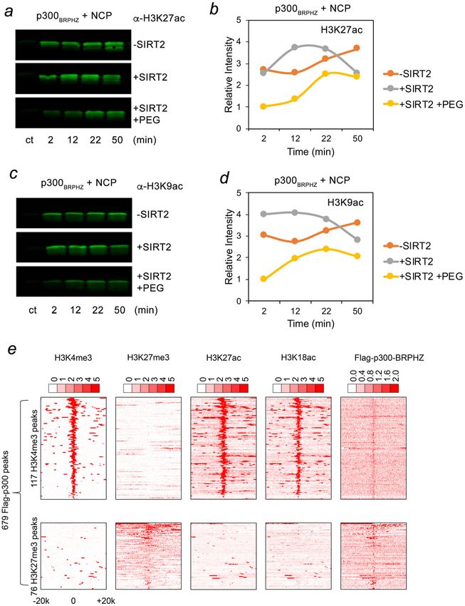
ARTICLE                                                                 NATURE COMMUNICATIONS | https://doi.org/10.1038/s41467-021-24950-8

F
      ormation of condensates in the cell nucleus has been shown             full-length p300 (YFP-p300FL) and monitored the localization of
      to drive the assembly of nuclear bodies, Cajal bodies,                 YFP-p300FL in live cells by fluorescence microscopy (Fig. 1b–d).
      nucleoli, and speckles1,2. These membraneless compart-                 In the majority of cells assayed, YFP-p300FL was somewhat dif-
ments are produced via the liquid–liquid phase separation (LLPS)             fusely distributed throughout the nucleus, but 15–30% of cells
mechanisms and depend on weak, multivalent interactions                      displayed nuclear speckles various in size (Fig. 1b). These data
involving intrinsically disordered regions (IDRs) of                         indicate the presence of discrete YFP-p300FL compartments with
biomolecules3–5. Several nuclear proteins and chromatin reg-                 elevated protein concentration and are in line with previous
ulators, including HP1, BRD4, CBX, and MORC3, have been                      reports showing that both endogenous and expressed p300 (and
shown to form condensates with their phase separation ability                its homolog CBP) form dynamic nuclear bodies34–37.
being necessary for biological functions and subnuclear localiza-               To determine whether p300 is mobile in the speckles, we
tion of these proteins6–16.                                                  measured the diffusion kinetics of selected p300 puncta by
   The transcriptional co-activator p300 is an acetyltransferase             fluorescence recovery after photobleaching (FRAP) experiments.
that is frequently dysregulated in disease, particularly cancer.             For each selected region, the laser beam was applied after two
p300 associates with over 400 binding partners, including tran-              initial scans, and cell images were collected at 10 s intervals for a
scription factors, DNA-binding proteins and subunits of the RNA              duration of 280 s (Fig. 1c, d). The experiments were performed on
polymerase II complex, to form activation complexes and facil-               seven cells, and changes in the fluorescence signal of each bleached
itate transcription17–21. p300 acetylates histones and a number of           region were analyzed. The averaged signal intensity, which was
essential nonhistone proteins, notably p53, E2F1, and androgen               normalized and plotted (Fig. 1c), showed a ~60% recovery of the
and estrogen receptors22–24. The highly selective acetylation of             bleached regions’ fluorescence with a half-life (t1/2) of 59 s.
the H3K27 and H3K18 sites by p300 requires a cooperative action              Because the photobleaching irreversibly abolishes fluorescence, the
of its two functional domains: acetyllysine-binding bromodomain              recovery of the signal implies that YFP-p300FL readily moves from
(BD) and the H3 tail-binding ZZ domain25.                                    unbleached regions to the bleached region. The fast signal
   p300 and its close homolog CBP share the same domain                      recovery, and therefore the fast diffusion of p300, suggest that
architecture, consisting of several conserved modules, including             YFP-p300FL speckles are viscous liquid droplets characterized by
the catalytic histone acetyltransferase (HAT) domain. Located in             rapid protein exchange with the surrounding environment.
the middle of the protein, the HAT domain is surrounded by BD,               Collectively, these results demonstrate that full length p300 forms
a RING finger, and a plant homeodomain finger (PHD) from one                   dynamic nuclear condensates with liquid/gel-like properties.
side and the ZZ domain from another side that together comprise
the catalytic core of p300 (Fig. 1a). The p300 catalytic activity is
                                                                             The catalytic core of p300 phase separates into liquid droplets
regulated through acetylation/deacetylation of a long loop in the
                                                                             in cells and in vitro. The middle part of p300, consisting of the
HAT domain, termed the autoinhibitory loop (AIL), which in a
                                                                             BD followed by the RING, PHD, HAT, and ZZ domains, com-
hypoacetylated form impedes the acetyltransferase activity of
                                                                             prises the catalytic core that we postulated might be involved in
p300 but releases the inhibition upon hyper autoacetylation17,26.
                                                                             the phase separation process. To test this idea, we generated YFP-
Structural studies of the p300 region encompassing BD, RING,
                                                                             tagged truncated p300 construct (aa 1024–1830 of p300), which
PHD and the HAT domain suggest an additional layer of p300
                                                                             in addition to the catalytic core contains a TAZ2 domain and is
regulation through the RING finger that contributes to the
                                                                             referred to as p300BRPHZT. We have previously shown that
inhibition by occluding the HAT active site27. Furthermore, the
                                                                             p300BRPHZT associates with chromatin in vitro and in vivo
BD–RING–PHD–HAT region of p300 has been shown to form a
                                                                             comparably to full-length p30025. The YFP-p300BRPHZT protein
dimer, in which the hypoacetylated form of AIL of one molecule
                                                                             was produced using a doxycycline (DOX) inducible pTripZ
inserts in the active site of the HAT domain from another
                                                                             vector, and expression of p300BRPHZT was visualized in live HeLa
molecule and thus undergoes ‘in trans’ autoacetylation, a reaction
                                                                             cells. Much like the full-length protein, YFP-p300BRPHZT localized
that is controlled by signal-dependent transcription factor
                                                                             primarily to the nucleus and formed foci of various sizes (Sup-
dimerization28. Genome-wide-mapping analysis reveals that
                                                                             plementary Fig. 1). Furthermore, FRAP experiments showed a
p300/CBP generally occupies transcriptionally active chromatin
                                                                             ~80% fluorescence recovery with t1/2 = 29 s, indicating that YFP-
marked by the active H3K27ac modification, however a large
                                                                             p300BRPHZT and full length YFP-p300 have similar capabilities to
number of silent or poised genes marked by the repressive
                                                                             form dynamic condensates in cells (Fig. 1e).
H3K27me3 modification and bound by p300/CBP has also been
                                                                                The cell nucleus is a highly crowded and viscous compartment
identified25,29–33. In cells, p300/CBP is ubiquitously expressed
                                                                             packed with DNA, histones, and a multitude of nuclear proteins.
and can form discrete foci in the nucleus and localize to PML
                                                                             To mimic molecular crowding, polyethylene glycol (PEG) is
nuclear bodies34–37.
                                                                             frequently used in vitro. Addition of PEG (PEG3350) to a
   In this study, we show that the structured catalytic core of p300
                                                                             solution containing the purified catalytic core, p300BRPHZ (aa
undergoes phase separation and forms liquid condensates in cells
                                                                             1035–1720 of p300), rapidly induced cloudiness in the originally
and in vitro. The acetylation state of p300 modulates its ability to
                                                                             clear solution, indicative of the conversion to a heterogenous
phase separate through attenuating intermolecular interactions
                                                                             suspension (described in detail below). Under a microscope, we
involving the autoinhibition loop, the HAT domain, and BD. Our
                                                                             observed spherical droplets of various sizes, suggesting the
data suggest a model for compartmentalization and concentration
                                                                             formation of liquid–liquid phase-separated condensates of the
of a pool of p300 with reduced catalytic activity at poised or
                                                                             catalytic core of p300 (Fig. 1f). About 35% of p300BRPHZ
repressed chromatin regions through the formation of phase-
                                                                             remained in the supernatant after removing the droplets by
separated condensates.
                                                                             centrifugation (Supplementary Fig. 2).

Results and discussion                                                       Self-acetylation of p300 impairs its ability to phase separate.
p300 forms dynamic condensates in cells. p300 is highly                      We found that different batches of purified p300BRPHZ showed
abundant in the cell nucleus and can shuttle between the                     variable ability to form droplets under otherwise identical con-
nucleoplasmic and cytoplasmic fractions38–40. To characterize the            ditions (Figs. 1f and 2a, b). Recombinantly expressed p300 is
distribution of p300, we transfected HeLa cells with YFP-tagged              auto-acetylated to varying degrees26,41. We, therefore, assessed

2                NATURE COMMUNICATIONS | (2021)12:4618 | https://doi.org/10.1038/s41467-021-24950-8 | www.nature.com/naturecommunications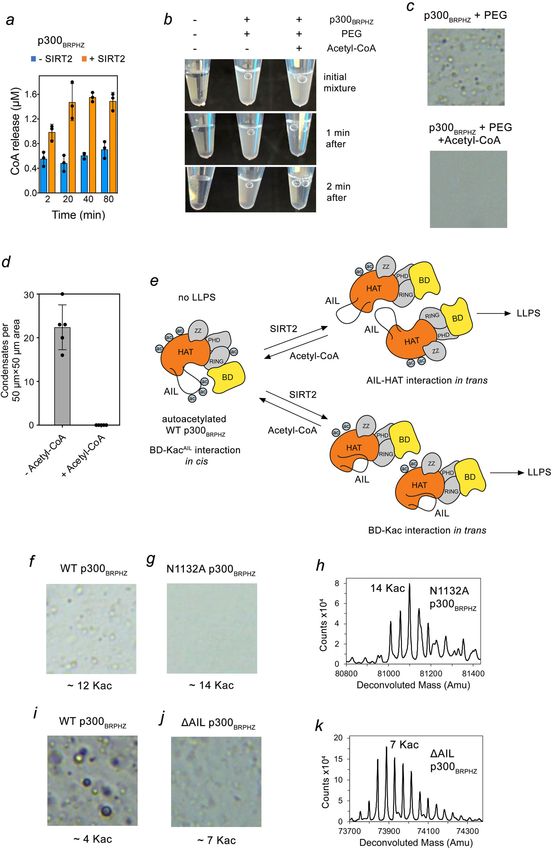
NATURE COMMUNICATIONS | https://doi.org/10.1038/s41467-021-24950-8                                                                     ARTICLE

Fig. 1 p300 phase separates to condensates in living cells. a Schematic of p300FL and p300BRPHZ. b Representative images of HeLa cells expressing YFP-
p300FL. Scale bar, 5 µm. c FRAP curve of YFP-p300FL was obtained from averaging data from six HeLa cells. Error bars represent SEM. n = 6 cells examined
over three independent experiments. d Representative FRAP images of YFP-p300FL expressed in HeLa cells. The images were taken before and after
photobleaching at indicated time points. The bleached condensate is indicated by a white arrowhead. Scale bar, 5 µm. e FRAP curve of YFP-p300BRPHZT
condensates in HeLa cells. The FRAP curve was obtained from averaging data from four cells. Error bars represent SEM. f A representative image of a
sample (from at least three replicates) containing 13 µM p300BRPHZ and 12% PEG3350 (PEG) in a 100 µm × 100 µm square region under a microscope.

the self-acetylation levels of two different batches of purified                 acetyl group. Compared with its theoretical mass of 80,548.5 Da,
p300BRPHZ by liquid chromatography–mass spectrometry                            recombinantly expressed p300BRPHZ from purification 1 was
(LC–MS). Acetylation levels of p300BRPHZ were highly hetero-                    acetylated on 13–25 lysine residues, with a median of 16 acety-
geneous, containing a series of species with discrete molecular                 lated lysines (Fig. 2c, d). The sequence of p300BRPHZ contains 60
mass. For example, purification 1 contained p300BRPHZ with                       lysine residues, indicating that ~20–40% of lysine residues were
molecular mass ranging from ~81 to 81.7 kDa (Fig. 2a, c, and d).                acetylated. The p300BRPHZ protein from purification 2 had on
The increment of each mass peak was ~42 Da, the mass of one                     average 26 acetylated lysines. p300BRPHZ with the lower

NATURE COMMUNICATIONS | (2021)12:4618 | https://doi.org/10.1038/s41467-021-24950-8 | www.nature.com/naturecommunications                              3
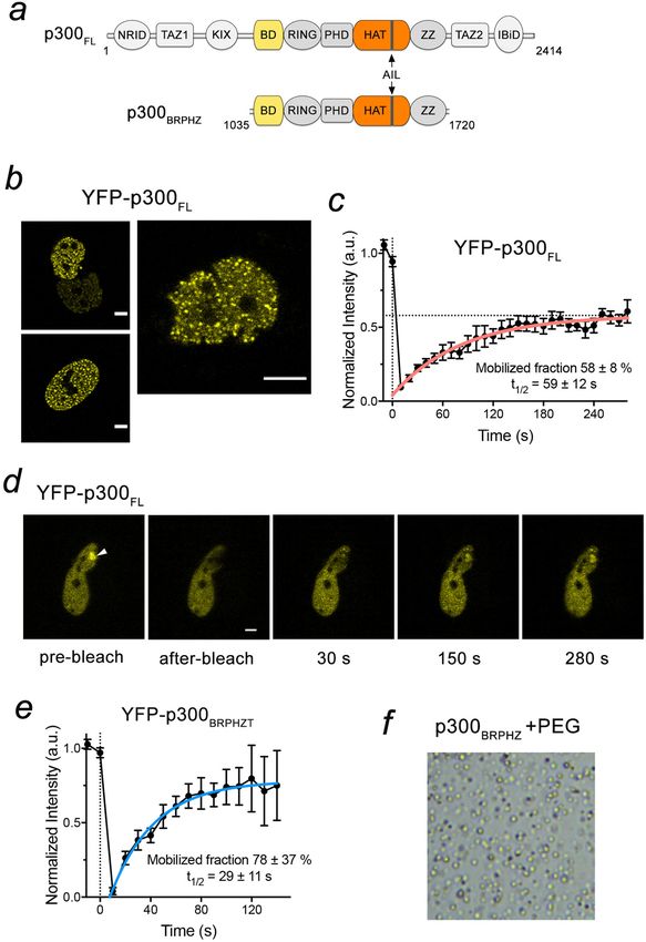
ARTICLE                                                                 NATURE COMMUNICATIONS | https://doi.org/10.1038/s41467-021-24950-8

acetylation level (purification 1) visibly formed condensates in the             We further examined whether acetylation levels modulate the
presence of PEG (Fig. 2a), while p300BRPHZ with the higher                   condensation behavior of p300BRPHZ. The NAD+-dependent
acetylation level (purification 2) showed almost no droplet for-              histone deacetylase SIRT2 has been shown to selectively and
mation (Fig. 2b). Together, these results suggest a negative cor-            uniquely deacetylate p300 in cells and in vitro42. We incubated
relation between the acetylation level of p300 and its ability to            p300BRPHZ with SIRT2 and NAD+ and then purified the protein
form condensates in vitro.                                                   by size-exclusion chromatography. This reduced the number of

4                NATURE COMMUNICATIONS | (2021)12:4618 | https://doi.org/10.1038/s41467-021-24950-8 | www.nature.com/naturecommunications
NATURE COMMUNICATIONS | https://doi.org/10.1038/s41467-021-24950-8 ARTICLE
Fig. 2 p300BRPHZ is heterogeneously acetylated and SIRT2-induced deacetylation promotes phase separation of p300BRPHZ. a, b Representative images
of two independently expressed and purified p300BRPHZ samples containing 13 µM protein and 12% PEG in a 50 µm × 50 µm square region on cover slides
under a microscope. c, d Liquid chromatography–mass spectrometry (LC–MS) analysis of p300BRPHZ shown in (a). A table of peak mass and the
corresponding number of acetylated lysine residues identified by mass spectrometry analysis are shown in (c) and (d). e, f Representative images of
p300BRPHZ samples shown in (a) and (b) were treated with the deacetylase SIRT2. g, h LC–MS analysis of p300BRPHZ shown in (e). A table of peak mass
and the corresponding number of acetylated lysine residues identified by mass spectrometry analysis are shown in (g) and (h). i Quantification of droplets
counted in images acquired for the samples in (a, b, e, f). The number of droplets was counted in five non-overlapping 50 µm × 50 µm square regions and
the mean value was plotted. Error bars represent SD. j Acetylated sites in p300BRPHZ identified by LC–MS/MS. Vertical bars indicate differences in signal
intensities of identical tryptic peptides derived from untreated vs. SIRT2-treated samples on the log2 scale. The highest intensities exceeding the plot were
essentially qualitative, as they were only present in the untreated sample. A schematic representation of p300BRPHZ is shown above. Source data are
provided in the Source Data file.
acetylated lysines in p300BRPHZ (purification 1) from a median of catalytic core (incapable of phase separation) to the hypoacety-
16 to a median of 4 (Fig. 2c, d, g, h). The same treatment of lated p300 catalytic core (capable of phase separation), which rely
p300BRPHZ (purification 2) reduced the acetylation level from a on intermolecular ‘in trans’ HAT-AIL or BD–acetyl–lysine (Kac)
median of 26 to 12 acetylated lysines. Generally, lower acetylation interactions (Fig. 3e). To test these possibilities, we examined the
levels were correlated with an increased propensity to form role of the BD and AIL in the formation of condensates. We used
droplets (Fig. 2i). a deletion of the entire autoinhibitory loop (ΔAIL) in p300BRPHZ
To identify regions in p300 affected by SIRT2 treatment, we or a mutation N1132A, which was previously shown to abrogate
analyzed tryptic peptides derived from untreated and SIRT2- acetyllysine binding of the BD27.
treated p300BRPHZ using liquid chromatography–tandem mass Compared to the WT p300BRPHZ protein that contained on
spectrometry (LC–MS/MS). 49 out of 60 lysine residues were average ~12 Kac sites and readily underwent phase separation
acetylated in untreated p300BRPHZ. Comparison of the signal (Fig. 3f), the N1132A mutant did not form PEG-induced droplets,
intensities of peptides revealed substantial differences in acetyla- despite having a similar acetylation level (~14 Kac sites)
tion levels of untreated and SIRT2-treated p300BRPHZ (Fig. 2j). (Fig. 3f–h). Likewise, the ΔAIL p300BRPHZ mutant with ~7 Kac
The most notable changes were observed for K1551, K1554, sites formed less visible condensates than the WT protein with ~4
K1555, K1557, and K1560 located in the AIL, as acetylation of Kac sites (Fig. 3i–k). We note that although the entire AIL was
these lysine residues was not detectable in the SIRT2-treated deleted, the ΔAIL p300BRPHZ mutant was still acetylated at 7
p300BRPHZ sample. Another AIL residue, K1568, also displayed a lysine residues located outside this loop in the
considerable reduction in signal intensity upon SIRT2 treatment. BD–RING–PHD–HAT–ZZ region. These data suggest that both
In contrast, signal intensity for lysine residues located in other HAT–AIL and BD–Kac-binding mechanisms contribute to
regions of p300BRPHZ was reduced to a substantially lesser degree. p300BRPHZ phase separation. In a hyper-acetylated form where
These data corroborate the idea that p300 self-acetylates a wide both the AIL and other regions of p300BRPHZ are acetylated, BD
range of lysine residues spanning the catalytic may favor intramolecular ‘in cis’ contacts with the acetylated AIL,
BD–RING–PHD–HAT–ZZ core, and that the auto-inhibitory limiting the probability of intermolecular interactions and leading
loop undergoes fast deacetylation, whereas deacetylation of other to a diffused distribution of p300BRPHZ. Upon SIRT2-treatment of
regions of p300BRPHZ occurs much slower. WT p300BRPHZ, deacetylation of the AIL results in the release of
Because our data suggest that acetylation of p300 negatively BD, allowing intermolecular ‘in trans’ BD–Kac interactions and
regulates its ability to phase separate, we reasoned that acetylation both intra- and intermolecular HAT–AIL interactions (Fig. 3e).
should decrease or even eliminate the PEG-induced p300BRPHZ In support of this model, SIRT2 treatment of N1132A
droplet formation. To test this, we first measured the HAT p300BRPHZ, which did not originally phase separate (Fig. 4a, left
activity of untreated and SIRT2-treated p300BRPHZ in the panel), led to the condensate formation (Fig. 4a, right panel, and
presence of acetyl-CoA by monitoring the release of the CoA Fig. 4b), suggesting that deacetylation of AIL promotes phase
product over time. As expected, SIRT2-treated p300BRPHZ separation through the HAT–AIL interaction (Fig. 4c). Further-
produced more CoA than untreated p300BRPHZ, due to the more, both SIRT2-treated or untreated ΔAIL p300BRPHZ formed
higher number of unmodified lysine residues (substrates) present droplets, although to a lesser degree compared to the WT protein,
in SIRT2-treated p300BRPHZ (Fig. 3a). The acetylation reaction reinforcing the role of the BD–Kac interaction in promoting
was fast and completed before 20 min, in keeping with the phase separation (Fig. 4d–f). Moreover, the catalytically impaired
previously reported activity of the p300 HAT domain41. Adding mutant D1399A p300BRPHZ showed the formation of the droplets
acetyl-CoA to a SIRT2-treated p300BRPHZ/PEG suspension led to through the HAT–AIL interaction, since no self-acetylation
a decrease in the cloudiness of the sample within 1 min (Fig. 3b), occurred in this mutant (Fig. 4g, h, and Supplementary Fig. 2b,
and liquid droplets were no longer visible under the microscope c). The SIRT2-dependent phase separation was also observed in
(Fig. 3c, d, and Supplementary Fig. 3a), indicating a disruption of the p300HZ construct containing only the HAT and ZZ domains
LLPS due to the HAT reaction. Collectively, these data suggest (Fig. 4i, j), whereas further deletion of AIL completely abolished
that auto-acetylation of the AIL decreases the formation of droplet formation, regardless of the SIRT2 treatment (Fig. 4k, l).
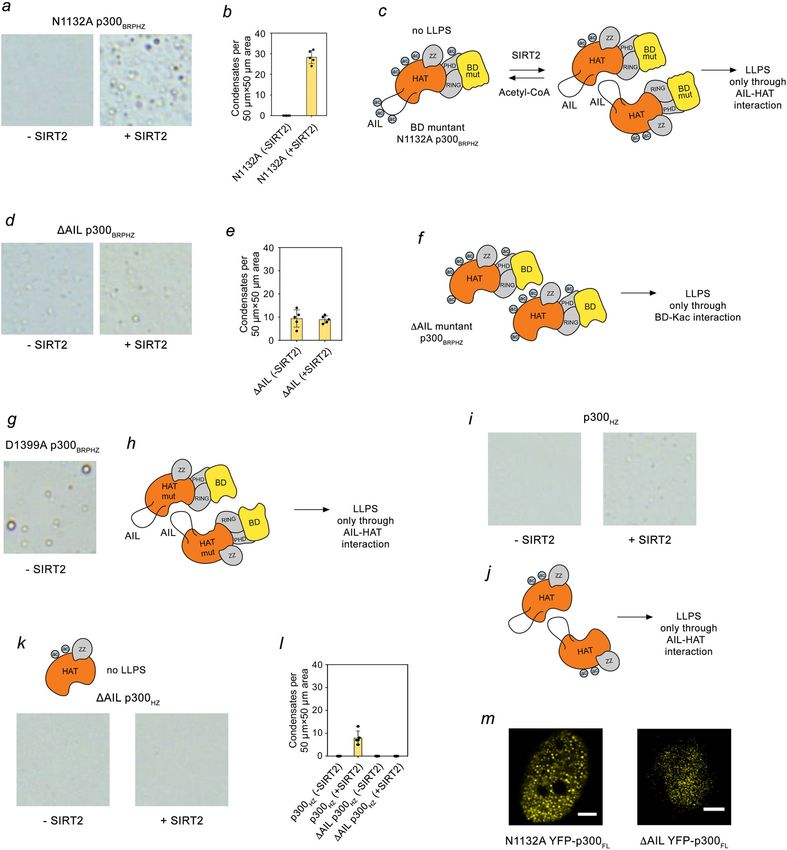
p300BRPHZ condensates. Lastly, both N1132A and ΔAIL mutants of YFP-p300FL formed
droplets in HeLa cells, confirming that full-length p300 can phase
Phase separation of p300 relies on both BD and AIL. Weak and separate through either HAT–AIL interaction in N1132A
often nonspecific multivalent interactions are believed to drive YFP–p300FL or BD–acetyl–lysine interaction in ΔAIL
phase separation1,4,7,8. The p300 HAT domain has been reported YFP–p300FL (Fig. 4m).
to associate with the hypoacetylated AIL from another p300 To further confirm our model, we performed small-angle X-ray
molecule28 and BD of CBP was shown to bind the AIL peptide scattering (SAXS) experiments on purified WT p300340–2094 and
acetylated at K159643. Accordingly, we envisage two distinct ΔAIL p300340–2094 proteins at various time points before and after
mechanisms for the transition of the hyperacetylated p300 incubating with acetyl-CoA (Fig. 5a). SAXS experiments provide
NATURE COMMUNICATIONS | (2021)12:4618 | https://doi.org/10.1038/s41467-021-24950-8 | www.nature.com/naturecommunications 5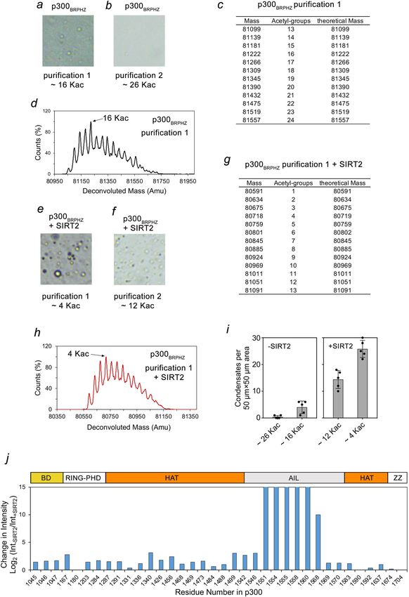
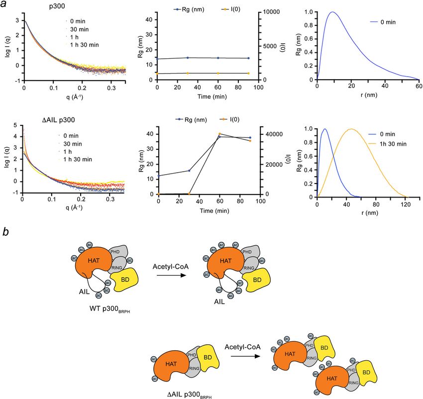
ARTICLE                                                                 NATURE COMMUNICATIONS | https://doi.org/10.1038/s41467-021-24950-8

information about the protein’s size and shape in solution. For              a maximum dimension of ~125 nm (Fig. 5a, bottom panel). These
WT p300340–2094, autoacetylation led to little changes in the SAXS           droplets likely arise through ‘in trans’ engagement of the BD with
profile: the Rg value remained unchanged at ~10 nm, in                        acetylated lysine residues outside AIL (Fig. 5b, bottom panel).
agreement with monomeric p300, possibly because the acetylated
AIL engages the BD ‘in cis’ (Fig. 5b, top panel). In contrast, for
ΔAIL p300340–2094, the Rg value increased over time (to ~35 nm)              p300 condensates sequester nucleosomal substrates. Besides
after autoacetylation for 1.5 h resulting in spherical droplets with         auto-acetylation, p300 catalyzes acetylation of lysine residues of

6                NATURE COMMUNICATIONS | (2021)12:4618 | https://doi.org/10.1038/s41467-021-24950-8 | www.nature.com/naturecommunications
NATURE COMMUNICATIONS | https://doi.org/10.1038/s41467-021-24950-8                                                                    ARTICLE

Fig. 3 p300BRPHZ phase separation requires both AIL and BD. a HAT activity of untreated p300BRPHZ (blue) and SIRT2-treated p300BRPHZ (orange)
measured by a fluorometric assay. Reactions were started by the addition of acetyl-CoA and quenched by flash-freeze at indicated time points. Data are
presented as mean values ± SD; error bars represent SD from triplicate measurements. b Phase separation of SIRT2-treated p300BRPHZ. The reaction
mixture contained 10 μM p300BRPHZ and 12% PEG. Addition of Acetyl-CoA led to the disassembly of the p300BRPHZ droplets. c Representative images of
p300BRPHZ samples from (k) after incubation for 2 min under a microscope. d Quantification of droplets counted in images acquired for the sample in (c).
The number of droplets was counted in five non-overlapping 50 µm × 50 µm square regions and the mean value was plotted. Error bar represents SD. e
Schematics of the p300BRPHZ phase separation mechanisms that can occur simultaneously. Multivalent ‘in trans’ interactions between HAT and
deacetylated AIL and/or BD and acetylated lysines outside AIL can promote the formation of condensates. f, g Representative images of WT p300BRPHZ (f)
and N1132A mutant (g) samples (from at least three replicates) containing 13 µM protein and 12% PEG in a 50 µm × 50 µm square region on cover slides
under a microscope. h LC–MS analysis of N1132A p300BRPHZ shown in (g). i, j Representative images of WT p300BRPHZ (i) and ΔAIL mutant (j) samples
(from at least three replicates) containing 13 µM protein and 12% PEG in a 50 µm × 50 µm square region on cover slides under a microscope. k LC–MS
analysis of ΔAIL p300BRPHZ shown in (j). Source data are provided in the Source Data file.

NATURE COMMUNICATIONS | (2021)12:4618 | https://doi.org/10.1038/s41467-021-24950-8 | www.nature.com/naturecommunications                             7
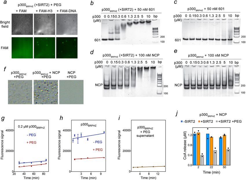
ARTICLE                                                                  NATURE COMMUNICATIONS | https://doi.org/10.1038/s41467-021-24950-8

Fig. 4 The molecular mechanisms of p300BRPHZ phase separation. a Representative images of untreated or SIRT2-deacetylated N1132A p300BRPHZ
samples containing 13 µM protein and 12% PEG in a 50 µm × 50 µm square region on cover slides under the microscope. b Quantification of droplets
counted in images acquired for the sample in (a). The number of droplets was counted in five non-overlapping 50 µm × 50 µm square regions and the mean
value was plotted. Error bar represents SD. c Schematic of the N1132A p300BRPHZ phase separation via the intermolecular interaction between the HAT
domain and deacetylated AIL. d Representative images of untreated or SIRT2-deacetylated ΔAIL p300BRPHZ samples containing 13 µM protein and 12%
PEG in a 50 µm × 50 µm square region on cover slides under a microscope. e Quantification of droplets counted in images acquired for the sample in (d).
The number of droplets was counted in five non-overlapping 50 µm × 50 µm square regions and the mean value was plotted. Error bar represents SD. f
Schematic of the ΔAIL p300BRPHZ phase separation via the intermolecular interaction between BD and acetylated lysines outside AIL. g A representative
image of purified D1399A p300BRPHZ sample (from at least three replicates) containing 13 µM protein and 12% PEG in a 50 µm × 50 µm square region on
cover slides under the microscope. h Schematic of the catalytically inactive D1399A p300BRPHZ phase separation via the intermolecular interaction
between the HAT domain and unmodified AIL. i Representative images of untreated or SIRT2-deacetylated WT p300HZ samples containing 13 µM protein
and 12% PEG in a 50 µm × 50 µm square region on cover slides under the microscope. j Schematic of the p300HZ phase separation via the intermolecular
interaction between the HAT domain and deacetylated AIL. k Representative images of untreated or SIRT2-deacetylated ΔAIL p300HZ samples containing
13 µM protein and 12% PEG in a 50 µm × 50 µm square region on cover slides under the microscope. l Quantification of droplets counted in images
acquired for the samples in (i, k). The number of droplets was counted in five non-overlapping 50 µm × 50 µm square regions and the mean value was
plotted. Data are presented as mean values ± SD; the error bar represents SD. m Representative images of HeLa cells (from at least three replicates)
expressing N1132A and ΔAIL mutants of YFP-p300FL. Scale bar, 5 µm.

histone proteins in nucleosomes, particularly at H3K18 and                    condition (Fig. 6f). These data imply that even when the AIL
H3K27 sites22,23,25. We, therefore, tested whether p300 droplets              binds to DNA and is, therefore, less available to contribute to the
could concentrate nucleosomal components, such as DNA and                     droplet formation through the HAT–AIL mechanism, the
histone tails. We monitored the recruitment of fluorescein                     intramolecular association through the BD–Kac mechanism is
(FAM)-labeled histone H3 tail (FAM-H3, residues 1–12 of H3)                   sufficient to maintain phase separation.
and FAM-labeled 37 bp double-stranded DNA (FAM-DNA) to
p300BRPHZ droplets using confocal microscopy (Fig. 6a and                     Catalytic HAT activity of p300 is decreased in condensates. We
Supplementary Fig. 3b). The p300 condensates exhibited bright                 examined the effect of phase separation on enzymatic activity and
fluorescence when incubated with FAM-DNA or FAM-H3, but                        histone substrate selectivity of p300BRPHZ. Because phase
not when incubated with the control FAM. The recruitment of                   separation conditions can promote enzymatic reactions owing to
histone H3 tail to condensates was due to binding of the ZZ                   higher local concentrations of both the enzyme and substrate44,
domain of p300 to the unmodified H3 tail25, however, p300 and                  we initially thought that p300 HAT activity would also be ele-
CBP were thought to not contact DNA themselves34. We noticed                  vated due to increased local concentration of p300BRPHZ and the
lower background fluorescence when p300 condensates were                       substrate, either p300BRPHZ and/or NCP inside the droplets. On
incubated with FAM-DNA compared to FAM-H3, which sug-                         the other hand, the intermolecular AIL–HAT interaction, which
gests that p300BRPHZ concentrates better and therefore binds                  leads to the formation of p300 condensates, physically blocks the
stronger to DNA than to the histone H31–12 peptide. Overall,                  active site of the HAT domain and thus would lead to a reduction
these results demonstrate that p300BRPHZ condensates can                      in the enzymatic activity, similar to what was observed for the
sequester nucleosomal components like histone H3 tail and DNA                 TOR kinase45. In addition to sterically occluding the
from the surrounding environment.                                             substrate–enzyme complex formation, the crowed viscous envir-
   Can p300 bind DNA and is this reaction mediated by                         onment in condensates slows diffusion of substrates and pro-
p300 self-acetylation? We tested the association of untreated                 ducts, which results in the reduced catalytic activity of
and SIRT2-treated p300BRPHZ with 601 DNA (147 bp) by                          enzymes46,47.
electrophoretic mobility shift assay (EMSA). The SIRT2-treated                   To compare the HAT activity in solution and phase-separated
p300BRPHZ, containing on average 4 Kac outside the AIL, strongly              droplets, we first monitored the self-acetylation of p300BRPHZ. As
bound to 601 DNA with an apparent Kd of ~0.3 μM (Fig. 6b). In                 shown in Fig. 6g, the addition of PEG to the SIRT2-treated
contrast, untreated p300BRPHZ, which contains on average ~16                  p300BRPHZ, which does not phase separate because of low protein
Kac including in the AIL, showed much weaker binding to 601                   concentration (0.2 μM), had essentially no effect on the change in
DNA in the same condition (Fig. 6c). DNA binding of p300BRPHZ,                the fluorescence signal (due to CoA release) over time, suggesting
therefore, is directly regulated by its acetylation level, as                 that the presence of PEG did not alter the catalytic activity of
acetylation neutralizes the positive charge of the unmodified                  p300BRPHZ (Fig. 6g). In contrast, the change in fluorescence was
lysine side chain, leading to a decrease in binding to the                    slower in the PEG-induced phase-separated suspension (Fig. 6h,
negatively charged DNA. We also found that auto-acetylation                   red line) or in the supernatant after the droplets were spun down
regulates p300BRPHZ association with nucleosomes. While SIRT2-                (Fig. 6i) compared to the change in fluorescence in the solution
treated p300BRPHZ bound to the reconstituted nucleosome core                  without droplets (Fig. 6h, blue line). These data indicate that
particle (NCP) with a low micromolar Kd in EMSA assays                        autoacetylation is inhibited when p300BRPHZ forms condensates.
(Fig. 6d), the association of untreated p300BRPHZ with NCP was                   We next measured the acetyltransferase activity of p300BRPHZ
notably compromised (Fig. 6e). Collectively, these data suggest               on reconstituted nucleosomes in homogenous solutions and
that the phase transition and nucleosome binding functions of                 under the phase separation condition. SIRT2-treated (containing
p300BRPHZ are linked to and regulated by its auto-acetylation.                ~12 Kac) p300BRPHZ and untreated (containing ~26 Kac)
   The results described above may suggest that DNA and the                   p300BRPHZ were mixed with NCP at a 1:1 ratio, and after the
HAT domain compete for the same hypoacetylated AIL, which                     addition of acetyl-CoA, the CoA release was monitored at
could lead to a decrease in phase separation ability of p300BRPHZ.            indicated time points. The reactions with NCP as a substrate
However, the addition of an equimolar amount of NCP to                        (Fig. 6j, blue and orange bars, and Supplementary Fig. 4a) were
p300BRPHZ stimulated the formation of more and larger droplets,               completed within 2 min for both SIRT2-treated and untreated
whereas NCPs alone did not phase separate under the same                      p300BRPHZ and were much faster than p300BRPHZ autoacetylation

8                 NATURE COMMUNICATIONS | (2021)12:4618 | https://doi.org/10.1038/s41467-021-24950-8 | www.nature.com/naturecommunications
NATURE COMMUNICATIONS | https://doi.org/10.1038/s41467-021-24950-8                                                                 ARTICLE

Fig. 5 ΔAIL p300340–2094 interacts ‘in trans’ in solution. a Small angle X-ray scattering (SAXS) data collected for WT (upper panel) and ΔAIL (lower
panel) p300340–2094 upon addition of acetyl-CoA at indicated time points including 0 min (blue) and 90 min (orange). SAXS intensity (left), Rg plots
(middle), and P(r) functions (right) determined from the experimental scattering data were shown. b Schematics of the WT and ΔAIL p300BRPH
intramolecular and intermolecular interactions between BD and acetylated lysines.

(Fig. 3a). The HAT reaction was also carried out in a suspension                again, the rate of NCP acetylation was decreased (Fig. 7b, d,
of PEG-induced droplets of SIRT2-treated p300BRPHZ and NCP.                     yellow line). These data further substantiate a reduction in the
Again, as in the case of autoacetylation (Supplementary Fig. 4b,                catalytic activity of p300BRPHZ upon formation of the
gray bars), the apparent HAT activity of p300BRPHZ in the phase-                condensates.
separated condensates was decreased compared to the HAT
activity of p300BRPHZ in solution (Fig. 6j, gray bars).
   To assess the histone substrate selectivity of p300BRPHZ, we                 p300 condensates preferably localize to chromatin regions
monitored acetylation of NCP by western blot using antibodies                   marked by H3K27me3. p300 often binds to enhancers and
against H3K27ac, H3K9ac, and H3K4ac (Fig. 7a–d and                              promoters enriched in active mono- and trimethylated H3K4
Supplementary Fig. 5). We found that either SIRT2-treated or                    (H3K4me1 and H3K4me3, respectively) marks, acetylating his-
untreated p300BRPHZ robustly acetylates H3K27 in NCP but                        tones, and stimulating gene transcription. However, a pool of
produces H3K9ac and H3K4ac to a lesser degree, which is in                      p300/CBP with a suppressed HAT activity has been shown to
agreement with our previous findings25 and with the data shown                   associate with poised and silent genomic regions marked by the
in Fig. 6j—the reaction was fast and completed in 2 min. The                    repressive modification H3K27me325,29–31,48,49. Indeed, ChIP-
selectivity of p300BRPHZ toward the H3K27 site was conserved in                 seq analysis of the H1299 cells expressing Flag-tagged WT
the suspension of SIRT2-treated p300BRPHZ droplets, however,                    p300BRPHZT identified 679 p300BRPHZT binding sites, and of these,

NATURE COMMUNICATIONS | (2021)12:4618 | https://doi.org/10.1038/s41467-021-24950-8 | www.nature.com/naturecommunications                          9
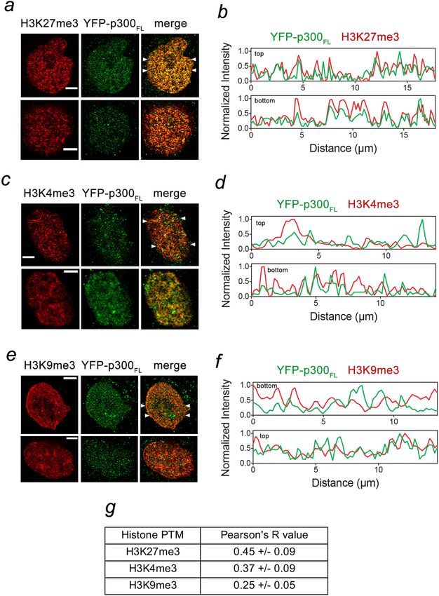
ARTICLE                                                                   NATURE COMMUNICATIONS | https://doi.org/10.1038/s41467-021-24950-8

Fig. 6 Acetylation regulates the association of p300 with DNA and the nucleosome. a Representative confocal images of phase-separated SIRT2-treated
p300BRPHZ condensates with FAM control (left), FAM-labeled H31–12 peptide (middle), and FAM-labeled DNA (right). A 50 µm × 50 µm square region is
shown for each sample. b, c EMSA of 147-bp 601 DNA incubated with increasing amounts of SIRT2-treated (b) and untreated (c) p300BRPHZ. DNA and
protein concentrations are shown above the gel image. d, e EMSA of the reconstituted nucleosome incubated with increasing amounts of SIRT2-treated (d)
and untreated (e) p300BRPHZ. Nucleosome core particle (NCP) and protein concentrations are shown above the gel image. f Phase separation of SIRT2-
treated p300BRPHZ alone (left), p300BRPHZ incubated with NCP (middle) and NCP alone (right) in a buffer containing 12% PEG. A 50 µm × 50 µm square
region is shown for each sample. Experiments in a–f were performed in at least two replicates. g HAT activity of diluted (0.2 μM) (g) and concentrated (10
μM) (h) SIRT2-deacetylated p300BRPHZ ± PEG was measured by a fluorometric assay. i HAT activity of SIRT2-deacetylated p300BRPHZ in supernatant
measured by a fluorometric assay. Droplets in the phase-separated p300BRPHZ sample were spun down for 10 min, and the supernatant was transferred to
another tube. For all experiments in (g–i), reactions were quenched by adding 6 M guanidine hydrochloride. The relative reaction rates (slope values) are
0.020 (−PEG) and 0.027 (+PEG) in (g), 0.27 (−PEG) and 0.09 (+PEG) in (h), and 0.035 in (i). Data in g, h are presented as mean values ± SD; error bars
represent SD from triplicate measurements. j HAT activity of untreated p300BRPHZ (blue), SIRT2-treated p300BRPHZ (orange), and SIRT2-treated phase-
separated p300BRPHZ droplet suspension (gray) on NCP measured by a fluorometric assay. Data are presented as mean values from duplicate
measurements.

117 p300BRPHZT binding sites were also enriched in H3K27ac and                 pool of p300 condensates has a preference to localize to the
H3K18ac—the primary products of acetylation by p300 (Fig. 7e).                 chromatin regions containing transcriptionally repressive
76 p300BRPHZT binding sites however were enriched in                           H3K27me3 modification as compared to the regions containing
H3K27me3. The notable absence of acetylation of H3K18 at these                 transcriptionally active modification H3K4me3 or the hetero-
sites suggests that the HAT activity of p300BRPHZT is decreased                chromatin mark H3K9me3.
when p300 colocalizes with H3K27me3.                                              It is becoming increasingly clear that the LLPS phenomenon
   Because the HAT activity of p300 is decreased in the phase-                 and the assembly of membraneless condensates by biological
separated condensates, we examined whether the condensates                     macromolecules in the nucleus play a crucial role in numerous
could select for chromatin modifications in HeLa cells using                    cellular processes. Formation of condensates allows for efficient
immunofluorescence. As shown in Fig. 8, the YFP-p300FL                          separation of the nuclear compartments in a spatiotemporal
condensates co-localize to a higher degree with the regions                    manner and/or concentration of the macromolecules to regulate,
enriched in H3K27me3 with a Pearson’s correlation coefficient                   activate or reduce their functions. Although the phase separation
(Pearson’s R) of 0.45 ± 0.09, but co-localization with H3K4me3                 mechanisms in the nucleus are currently a subject of intense
was less pronounced (Pearson’s R of 0.37 ± 0.09), and even                     studies, multivalent weak contacts involving IDRs of macro-
lower level of co-localization was observed with H3K9me3                       molecules have been widely acknowledged as a driving force for
(Pearson’s R of 0.25 ± 0.05). Together, these data suggest that a              the formation of biomolecular condensates. In this work, we show

10                 NATURE COMMUNICATIONS | (2021)12:4618 | https://doi.org/10.1038/s41467-021-24950-8 | www.nature.com/naturecommunications
NATURE COMMUNICATIONS | https://doi.org/10.1038/s41467-021-24950-8                                                                   ARTICLE

Fig. 7 A pool of p300 co-localizes with H3K27me3. a–d H3K27ac and H3K9ac western blot analysis of the reaction mixtures containing p300BRPHZ and an
equimolar amount of NCP as a substrate. Reactions were quenched by rapid freezing and the addition of SDS-loading buffer at indicated time points. The
intensity of bands is quantified, normalized to the +SIRT2/+PEG band at 2 min, and shown in (b) and (d). e Heatmaps of H3K4me3, H3K27me3, H3K27ac,
H3K18ac, and Flag-p300BRPHZT, ChIP-seq signals centered on p300-H3K4me3 and p300-H3K27me3 binding sites in a ±20 kb window in H1299 cells
stably expressing FLAG-p300BRPHZT. Heatmaps are ranked by H3K4me3 (upper panel) or H3K27me3 (lower panel). The color keys represent ChIP-seq
densities normalized to total reads.

that the major human acetyltransferase p300 forms liquid                        often associated with transcriptional activation and occupies gene
condensates in the nucleus and this function depends rather on                  promoters and enhancer elements but has also been reported to
the structured catalytic core of p300, including the HAT domain                 localize to the repressive sites, particularly those enriched in
and its AIL, and BD. We demonstrate that hyperacetylation of the                H3K27me329,30. These sites are characterized by overall low
p300 catalytic core, particularly of AIL, decreases the phase                   acetylation of histones, and since H3K27me3 does not preclude
separation ability, and that p300 utilizes two distinct molecular               binding of p300/CBP to the H3 tail, it was proposed that the HAT
mechanisms to assemble the condensates, which rely on                           activity is blocked at such sites31,49. The mechanism of this
intermolecular ‘in trans’ HAT–AIL and BD–(Kac outside AIL)                      blockage remains unclear. A few concepts have been put forth to
interactions. Furthermore, we found that the catalytic HAT                      explain the decrease in the p300/CBP catalytic activity, especially
activity of p300 is decreased in the phase-separated droplets,                  on histone lysine residues other than H3K27, as methylation of
which is likely due to steric blocking of the HAT active site.                  H3K27 obviously prevents its acetylation. These include regula-
   Our data suggest a model for compartmentalization and                        tion through phosphorylation or SUMOylation of p300/CBP34,50
concentration of p300 with reduced catalytic activity. p300/CBP is              and rapid degradation of p300. Our finding that a large portion of

NATURE COMMUNICATIONS | (2021)12:4618 | https://doi.org/10.1038/s41467-021-24950-8 | www.nature.com/naturecommunications                           11
ARTICLE                                                                   NATURE COMMUNICATIONS | https://doi.org/10.1038/s41467-021-24950-8

Fig. 8 p300 condensates show preference for co-localization with H3K27me3. a–f Nuclear localization of the YFP-p300FL condensates with indicated
posttranslational histone modifications (PTMs) in HeLa cells were examined by immunofluorescence. The cells were fixed and probed with anti-
H3K27me3 (a), anti-H3K4me3 (c), and anti-H3K9me3 (e) antibodies. Representative images are shown in the left panels: green, YFP-p300FL; red, PTMs;
yellow, overlap of fluorescence signals of YFP-p300FL and PTMs. The ImageJ software was used to plot fluorescence signal intensities of YFP-p300FL and
PTMs in the nuclei along the line (from left to right) indicated by white arrowheads (b, d, f). Scale bar, 5 µm. g A table of Pearson’s R values calculated
using the entire cell nuclei (n = 3 cells examined for each mark). Data are presented as mean values ± SD. Pearson’s R values range between +1 (perfect
positive correlation) and −1 (inverse correlation).

p300 condensates co-localizes with chromatin regions enriched in               enzymatic activity through phase transition has been reported for
H3K27me3 suggests an alternative mechanism. The p300                           the target of rapamycin (TOR) serine/threonine protein kinase in
condensates can act as a storage pool of the protein with reduced              the TORC1 complex45. The formation of the TORC1 foci results
HAT activity, allowing p300 and possibly other elements of the                 in steric occlusion of the TOR active site, subsequently leading to
transcriptional machinery to be compartmentalized and concen-                  the inhibition of the catalytic activity45. To better understand the
trated at the repressed chromatin sites. The formation of p300                 p300-dependent activation of gene transcription, it will be
condensates through blocking the catalytic site of the HAT                     essential in future studies to delineate and visualize by single-
domain provides a mechanism by which the enzymatic activity of                 molecule imaging51 the precise contacts between p300 and the
p300 can be downregulated. Similar downregulation of the                       repressed chromatin regions.

12                 NATURE COMMUNICATIONS | (2021)12:4618 | https://doi.org/10.1038/s41467-021-24950-8 | www.nature.com/naturecommunications
NATURE COMMUNICATIONS | https://doi.org/10.1038/s41467-021-24950-8                                                                                       ARTICLE

Methods                                                                                 µm × 250 mm) and gradient eluted from 2% to 40% acetonitrile over 40 min at 0.3
Cell culture, transfection, and imaging. WT full-length YFP-p300 plasmids were          μL/min using a Thermo Ultimate 3000 UPLC (Thermo Scientific). Peptides were
transfected into HeLa cells in a 3.5-cm-diameter tissue culture dish by Lipofecta-      detected with a Thermo Q-Exactive HF-X mass spectrometer (Thermo Scientific)
mine 3000 (Life Technology, L3000-075) using manufacturer instructions. The             scanning MS1 spectra at 120,000 resolution from 380 to 1580m/z with a 45 ms fill
cells were cultured in DMEM (company) supplemented with 10% fetal bovine                time and 3E6 AGC target. The top 12 most intense peaks were isolated with a
serum (company, need double-check) for 36 h. The cell culture medium was                1.4m/z window with a 100 ms fill time and 1E6 AGC target and 27% HCD collision
replaced with the live-cell imaging medium and maintained at 37 °C using a heater       energy for MS2 spectra collected at 15,000 resolution. Dynamic exclusion was
controller. The p300 BD–Ring–PHD–HAT–ZZ_TAZ2 region (BRPHZT, aa                         enabled for 5 s. MS data raw files were searched against the single Uniprot sequence
1024–1830) was cloned into a pTripZ-YFP vector. To establish a stable cell line,        for EP300 (Uniprot accession number Q09472) using Maxquant 1.6.14.0 with
viral titer harvested from HEK293T cells was used to infect HeLa cells with fusion      cysteine carbamidomethylation as a fixed modification, while methionine oxidation
gene. HEK293T cells were seeded in a 10-cm dish to reach 90–100% confluency the          and protein N-terminal and lysine side chain acetylation were set as variable
following day. The cells were then transfected using calcium phosphate pre-             modifications. The mass tolerances for the database search were 4.5 ppm for the
cipitation (21.0 µg psPAX2, 10.5, 21.0 µg of pTripZ-YFP-p300, 250 mM CaCl2, and         precursors and 20 ppm for the MS2 fragment ions, the minimum peptide length
1×Hank’s balanced salt solution) and incubated for 12 h at 37 °C and 5% CO2. Cells      was 7 residues with no additional applied score cutoffs. Peptide and protein level
were washed twice with culture medium and then incubated in culture medium for          FDR was set at 0.01. An intensity cut-off of 50 million counts was applied to the
48 h. Viral titer was harvested from HEK293T cell culture medium and was spun at        untreated sample prior to plotting the change in intensity in Fig. 2j.
1000 × g for 5 min to remove any cell debris. This was added to a single-cell
suspension of HeLa cells supplemented with 8 µg/mL Polybrene (SIGMA, H9268)
                                                                                        In vitro condensate formation. All in vitro condensate formation assays were
and was mixed. Solution of virus and HeLa cells was plated evenly into a 10-cm
                                                                                        performed in a buffer containing 15 mM Tris–HCl (pH 7.5), 150 mM NaCl and 2
dish and incubated for 12 h. Culture medium of infected cells was then replaced,
                                                                                        mM DTT unless otherwise stated. All samples were prepared on ice and incubated
and cells were maintained.
                                                                                        for ~5 min before imaging on siliconized glass cover slides (Hampton). For WT
                                                                                        and mutant p300BRPHZ and p300HZ alone, 13 μM protein samples were parallelly
FRAP experiments. FRAP imaging was performed using a Zeiss LSM 700                      prepared with or without 12% (w/v) PEG 3350. For the condensate formation assay
Observer as described previously16. Briefly, two images were taken before photo-         with reconstituted nucleosomes, 6.4 μM SIRT2-treated p300BRPHZ and an equal
bleaching, and 15–30 images were taken with 10 s intervals immediately after            amount of NCP were incubated in a buffer containing 10 mM Tris 7.5, 60 mM
photobleaching. The images were analyzed using ImageJ. Fluorescence intensities         NaCl, 2 mM DTT, and 12% (w/v) PEG350. Microscopy of the droplets was done
were normalized to the signal before photobleaching to obtain the fluorescence           using an M150C-I microscope (AmScope) equipped with a ×10 objective and an
recovery.                                                                               MD35 digital camera (AmScope). A microscope camera calibration slide (OMAX,
                                                                                        0.01 mm) was used to determine the scale. A 50 μm × 50 μm square area was
                                                                                        selected as a representative image for each sample. The number of condensates was
Protein expression and purification. The construct of human full-length p300             also counted in a 50 μm × 50 μm square area. Five non-overlapping square regions
protein-containing BD–RING–PHD–HAT–ZZ region (BRPHZ, amino acids                        were counted for each sample and plotted. Experiments were repeated in at least
1035–1720) was cloned in the pGEX-6P-1 vector and expressed in BL21 (RIL) cells         three batches of purified and SIRT2-treated p300 proteins.
as previously described25. Protein production was induced with 0.1 mM IPTG and              To prepare p300 condensates that concentrate DNA or H3, 13 μM SIRT2-
cultured overnight at 16 °C in Luria broth (LB) medium. The GST-tagged proteins         treated p300 was mixed with either 6 μM FAM, 6 μM FAM-labeled 37 bp dsDNA,
were purified on Pierce glutathione agarose beads (Thermo-Fisher) in 20 mM               or 6 μM FAM-labeled histone H3 tail (aa 1–12). Confocal images were acquired on
Tris–HCl (pH 7.5), 500 mM NaCl, and 3 mM DTT. The GST tag was cleaved                   a Zeiss Observer.Z1 inverted microscope using a ×40 oil objective and digitally
overnight at 4 °C with PreScission proteases. The SIRT2 (38–356) construct fused        captured. For the excitation of FAM, a 488 nm laser was used. Images were
with His6x-SUMO was obtained from Addgene (addgene ID:102622). SIRT2                    processed and presented using ImageJ and Photoshop.
protein was expressed and purified using a standard protocol. In brief, the cells
were disrupted by sonication, and the cell lysate was spun down to remove the
debris. The supernatant was loaded to a nickel column (Histrap, GE Healthcare)          Nucleosome reconstitution. The poly-cistronic vector of Xenopus laevis Histone
pre-equilibrated with a buffer containing 50 mM Tris–HCl pH 7.5 and 500 mM              octamers was obtained from the Jean-Francois Couture lab. A His-tag and a TEV
NaCl. The SIRT2 protein was eluted with a 0–500 mM linear gradient of imidazole.        cleavage site were introduced before histone H3 by mutagenesis. Expression and
The deacetylation reaction was carried out by incubating purified p300 WT and            purification of histone octamers were performed as reported previously53. The His-
mutant proteins with SIRT2 overnight in the cold room in a buffer containing            tag before histone H3 was cleaved to expose the Ala1 residue using TEV protease.
20–50 mM Tris (pH 7.5), 300 mM NaCl, 5 mM MgCl2, 2 mM NAD, and 2 mM                     601 DNA was prepared as previously described54. NCPs were reconstituted by
DTT. Deacetylated p300 proteins were further purified by size-exclusion chro-            combining octamer with 1.1× excess DNA and performing slow salt dialysis.
matography and concentrated in Millipore concentrators.                                 Reconstituted nucleosomes were further purified by size-exclusion chromatography
                                                                                        and concentrated in Millipore concentrators.
Trypsin digestion of acetylated p300 proteins. Purified p300BRPHZ (40–50 µg)
with or without SIRT2 treatment was denatured, reduced, and alkylated in 5% (w/         Acetyltransferase assays. Purified untreated and SIRT2-treated p300BRPHZ
v) sodium dodecyl sulfate (SDS), 10 mM tris (2-carboxyethyl) phosphine hydro-           proteins were buffer exchanged into reaction buffer containing 10 mM Tris–HCl
chloride (TCEP–HCl), 40 mM 2-chloroacetamide, 50 mM Tris pH 8.5 and boiled at           (pH 7.5) and 60 mM NaCl. For auto-acetylation experiments in Fig. 3a–c, 10 μM
95 °C for 10 min. Samples were prepared for mass spectrometry analyses using the        p300 protein was incubated in HAT reaction buffer (10 mM Tris pH 7.5 and 60
SP3 method52. Carboxylate-functionalized speedbeads (GE Life Sciences) were             mM NaCl) in the absence or presence of 12% (w/v) PEG 3350. For histone acetyl-
added to protein samples. Acetonitrile was added to 80% (v/v) to precipitate            transferase assays in Fig. 6j, 6.4 μM p300 protein, and 6.4 μM reconstituted NCP
protein and bind it to the beads. The protein-bound beads were washed twice with        were incubated in HAT reaction buffer (10 mM Tris pH 7.5 and 60 mM NaCl).
80% (v/v) ethanol and twice with 100% acetonitrile. Lys-C/Trypsin mix (Promega)         Reactions were started by adding 0.5 mM acetyl-CoA at room temperature and
was added for 1:50 protease to protein ratio in 50 mM Tris pH 8.5 and incubated         quenched by flash-freeze at indicated time points. For each time point, 8 μl reaction
rotating at 37 °C overnight. To clean up tryptic peptides, acetonitrile was added to    mixture was diluted to 200 μl assay buffer and immediately heated to 95 °C for 5
95% (v/v) to precipitate and bind peptides to the beads. One wash with 100%             min to inactivate p300. The samples were then applied to an ultrafiltration device
acetonitrile was performed and tryptic peptides were eluted twice with 1% (v/v)         (10k cut-off) to collect flow-through for CoA quantification by the fluorometric
trifluoroacetic acid (TFA), 3% (v/v) acetonitrile in water. Eluate was dried using a     assay kit (Abcam, 138889). The kit utilizes a fluorogenic green indicator that
speed-vac rotatory evaporator.                                                          became strongly fluorescent upon reacting with the –SH group in CoA. The
                                                                                        fluorescence signals in samples collected from the reactions were measured
                                                                                        according to the product manual using a 96-well microplate reader. For measuring
Liquid chromatography and tandem mass spectrometry (LC–MS/MS) analy-
                                                                                        the HAT activity of p300 (Fig. 6g–i) in the diluted solution, droplet mixture, and
sis. For estimations of intact protein masses, untreated and SIRT2-treated WT and       supernatant, reactions were quenched by mixing with a denaturing buffer con-
mutant p300 protein samples were resolved using a Waters AQCUITY UPLC.                  taining 20 mM Tris–HCl (pH 7.5), 150 mM NaCl and 6 M guanidine–HCl at
Proteins were diluted to final concentration of 0.3 µg/µL using Buffer A (0.1%           indicated points. All experiments were performed in triplicates.
formic acid in water), of which 3 µg of protein was loaded onto a Waters                    To detect histone H3 acetylation at specific lysine sites, reactions were quenched
ACQUITY UPLC Protein BEH C4 Column (300 Å, 1.7 µm, 2.1 mm × 100 mm).                    by flash-freezing in liquid nitrogen and then analyzed by SDS–PAGE and western
Salts were removed with 3% Buffer B (0.1% formic acid in acetonitrile) at 0.2 mL/       blot analysis. Western blot results were quantified by LI-COR Odyssey System
min for 3 min and proteins were eluted using a linear gradient of Buffer B from 3%      using the following antibodies: anti-H3K4ac (ab176799, 1:1000), anti-H3K9ac
to 80% in 4 min at 0.2 mL/min. The UPLC was coupled directly with a Synapt G2           (ab4441, 1:1000), and anti-H3K27ac (ab177178, 1:1000) from Abcam, and anti-
HDMS qTOF mass spectrometer scanning 400–2500m/z. Charge deconvolution                  H3K18ac (39755, 1:1000) from Active Motif.
was performed using Waters MaxEnt software.
    For acetylation sites analysis, the trypsinized peptides were resuspended in 0.1%
TFA, 3% acetonitrile in water, of which 1 picomole of the peptides for each sample      SAXS analysis. X-ray scattering data were collected at the Bio-SAXS beamline
was directly injected onto a Waters M-class column (1.7 µm, 120 A, rpC18, 75            (BM29) of the European Synchrotron Radiation Facility. Data were collected with a

NATURE COMMUNICATIONS | (2021)12:4618 | https://doi.org/10.1038/s41467-021-24950-8 | www.nature.com/naturecommunications                                                  13
ARTICLE NATURE COMMUNICATIONS | https://doi.org/10.1038/s41467-021-24950-8
photon-counting Pilatus 1 M detector at a sample-detector distance of 2.86 m, a 6. Sabari, B. R. et al. Coactivator condensation at super-enhancers links phase
wavelength of λ = 0.991 Å, and an exposure time of 1 second/frame. A momentum separation and gene control. Science 361, eaar3958 (2018).
transfer range of 0.008–0.47 Å−1 was covered (q = 4πsin θ/λ, where θ is the scat- 7. Larson, A. G. et al. Liquid droplet formation by HP1alpha suggests a role for
tering angle and λ the X-ray wavelength). A time-resolved SAXS experiment was phase separation in heterochromatin. Nature 547, 236–240 (2017).
performed with the p300 (aa 324–2094) and ΔAIL p300 constructs, produced as 8. Strom, A. R. et al. Phase separation drives heterochromatin domain formation.
described55, during auto-acetylation. 150 µL of the reaction mixtures in buffer (20 Nature 547, 241–245 (2017).
mM HEPES pH7.0, 500 mM NaCl, 5 µM ZnCl2, 0.5 mM TCEP) were prepared and 9. Hnisz, D., Shrinivas, K., Young, R. A., Chakraborty, A. K. & Sharp, P. A. A
pipetted in the thermostated sample holder tube already adjusted to 30 °C. 2 mM phase separation model for transcriptional control. Cell 169, 13–23 (2017).
acetyl-CoA was added to each reaction tube and in the background, buffer to start 10. Boija, A. et al. Transcription factors activate genes through the phase-
the reaction. Four scattering curves were recorded on each sample at 0, 30, 60, and separation capacity of their activation domains. Cell 175, 1842–1855e1816
90 min. Rg and I(0) values were obtained from the Guinier approximation Rg < 1.3 (2018).
using Primus56. Distance distribution functions p(r) were computed from the 11. Zhang, Y. et al. MORC3 forms nuclear condensates through phase separation.
entire scattering curve using GNOM56. iScience 17, 182–189 (2019).
12. Plys, A. J. et al. Phase separation of Polycomb-repressive complex 1 is
EMSA with DNA and nucleosomes. EMSA experiments were performed essentially governed by a charged disordered region of CBX2. Genes Dev. 33, 799–813
as described57. Briefly, increasing amounts of p300 were incubated with 147 bp 601 (2019).
DNA (50 nM) in a DNA binding buffer containing 20 mM Tris–HCl (pH 7.5), 50 13. Cho, W. K. et al. Mediator and RNA polymerase II clusters associate in
mM NaCl, and 2 mM DTT for 5 min. The reaction mixtures were loaded on 5% transcription-dependent condensates. Science 361, 412–415 (2018).
polyacrylamide gels, and electrophoresis was performed in 0.2 × TBE buffer at 100 V 14. Nair, S. J. et al. Phase separation of ligand-activated enhancers licenses
for 1.5 h on ice. For EMSA experiments with reconstituted nucleosomes (100 nM), the cooperative chromosomal enhancer assembly. Nat. Struct. Mol. Biol. 26,
NaCl concentration in binding buffer was increased to 75 mM, and electrophoresis 193–203 (2019).
was performed in 0.2 × TB buffer. Gels were stained with SYBR Gold (Invitrogen). 15. Boehning, M. et al. RNA polymerase II clustering through carboxy-terminal
domain phase separation. Nat. Struct. Mol. Biol. 25, 833–840 (2018).
ChIP-seq analysis. Flag-p300BRPHZT, H3K18ac, and H3K27ac ChIP-seq reads 16. Tatavosian, R. et al. Nuclear condensates of the Polycomb protein chromobox
were obtained from GSE109591; H3K4me3 and H3K27me3 ChIP-seq reads were 2 (CBX2) assemble through phase separation. J. Biol. Chem. 294, 1451–1463
from GSE81322. ChIP-seq raw reads were mapped to the hg38 genome by hisat2 (2019).
(v2.1.0) with no-spliced-alignment, -k 1. H3K4me3 and H3K27me3 peaks were 17. Liu, X. et al. The structural basis of protein acetylation by the p300/CBP
called by macs2 callpeak (v2.1.2) with broad parameter. Flag-p300BRPHZT peaks transcriptional coactivator. Nature 451, 846–850 (2008).
were converted to hg38 by UCSC liftOver. Heatmaps were generated by danpos 18. Ogryzko, V. V., Schiltz, R. L., Russanova, V., Howard, B. H. & Nakatani, Y.
(v2.2.2) with 200 bp bin size and visualized by TreeView (v1.1.6). The transcriptional coactivators p300 and CBP are histone acetyltransferases.
Cell 87, 953–959 (1996).
Immunofluorescence. HeLa cells were cultured in a six-well plate with 22-mm 19. Wang, Z. et al. Genome-wide mapping of HATs and HDACs reveals distinct
coverslips. YFP-p300 plasmids were transfected into HeLa cells by Lipofectamine functions in active and inactive genes. Cell 138, 1019–1031 (2009).
3000 (Life Technology, L3000-075) according to manufacturer instructions. One 20. Dancy, B. M. & Cole, P. A. Protein lysine acetylation by p300/CBP. Chem. Rev.
day after transfection, cells were fixed by 1% paraformaldehyde and then per- 115, 2419–2452 (2015).
meabilized with 0.2% Triton X-100. After blocking with 3% goat serum and 3% 21. Goodman, R. H. & Smolik, S. CBP/p300 in cell growth, transformation, and
BSA, cells were incubated, respectively, with pairs of primary antibodies: anti-GFP development. Genes Dev. 14, 1553–1577 (2000).
(Life Technologies; A-11120; 1:400 dilution) and anti-H3K27me3 (Millipore; 07- 22. Jin, Q. et al. Distinct roles of GCN5/PCAF-mediated H3K9ac and CBP/p300-
449; 1:200 dilution), anti-GFP (Life Technologies; A-11120; 1:400 dilution) and mediated H3K18/27ac in nuclear receptor transactivation. EMBO J. 30,
anti-H3K9me3 (Upstate; 07-442; 1:200 dilution), and anti-GFP (Life Technologies; 249–262 (2011).
A-11120; 1:400 dilution) and anti-H3K4me3 (Novus; NB21-1023B; 1:200 dilution), 23. Tang, Z. et al. SET1 and p300 act synergistically, through coupled histone
for two hours. After washing, cells were incubated with a pair of secondary anti- modifications, in transcriptional activation by p53. Cell 154, 297–310 (2013).
bodies: Alexa Fluor 488-labeled goat anti-mouse and Alexa Fluor 568-labeled goat 24. Gu, W. & Roeder, R. G. Activation of p53 sequence-specific DNA binding by
anti-rabbit, for two hours, and then were mounted by using ProLong Antifade acetylation of the p53 C-terminal domain. Cell 90, 595–606 (1997).
reagents (Life Technologies; P7481). Line plots were made using the plot profile 25. Zhang, Y. et al. The ZZ domain of p300 mediates specificity of the adjacent
feature of ImageJ. Pearson coefficient correlation for each pair of immuno- HAT domain for histone H3. Nat. Struct. Mol. Biol. 25, 841–849 (2018).
fluorescence images was calculated using the Coloc 2 plugin in ImageJ after the 26. Thompson, P. R. et al. Regulation of the p300 HAT domain via a novel
selection of the entire cell nuclei as the regions of specific interest. activation loop. Nat. Struct. Mol. Biol. 11, 308–315 (2004).
27. Delvecchio, M., Gaucher, J., Aguilar-Gurrieri, C., Ortega, E. & Panne, D.
Structure of the p300 catalytic core and implications for chromatin targeting
Data availability and HAT regulation. Nat. Struct. Mol. Biol. 20, 1040–1046 (2013).
All relevant data supporting the key findings of this study are provided in
28. Ortega, E. et al. Transcription factor dimerization activates the p300
the Supplementary Information and Source Data files or from the corresponding author
acetyltransferase. Nature 562, 538–544 (2018).
upon reasonable request. The mass spec data generated in this study have been deposited
29. Rada-Iglesias, A. et al. A unique chromatin signature uncovers early
to the PRIDE database under the accession number PXD026898. The publicly available
developmental enhancers in humans. Nature 470, 279–283 (2011).
ChIP-seq data analyzed in this study are available from Gene Expression Omnibus under
30. Zentner, G. E., Tesar, P. J. & Scacheri, P. C. Epigenetic signatures distinguish
accession codes GSE109591 and GSE81322. Source data are provided with this paper.
multiple classes of enhancers with distinct cellular functions. Genome Res. 21,
1273–1283 (2011).
Received: 8 March 2021; Accepted: 12 July 2021; 31. Holmqvist, P. H. & Mannervik, M. Genomic occupancy of the transcriptional
co-activators p300 and CBP. Transcription 4, 18–23 (2013).
32. Bedford, D. C., Kasper, L. H., Fukuyama, T. & Brindle, P. K. Target gene
context influences the transcriptional requirement for the KAT3 family of
CBP and p300 histone acetyltransferases. Epigenetics 5, 9–15 (2010).
33. Kasper, L. H., Qu, C., Obenauer, J. C., McGoldrick, D. J. & Brindle, P. K.
Genome-wide and single-cell analyses reveal a context dependent relationship
References
between CBP recruitment and gene expression. Nucleic Acids Res. 42,
1. Shin, Y. et al. Liquid nuclear condensates mechanically sense and restructure
11363–11382 (2014).
the genome. Cell 175, 1481–1491 (2018). e1413.
2. Brangwynne, C. P., Mitchison, T. J. & Hyman, A. A. Active liquid-like 34. Girdwood, D. et al. P300 transcriptional repression is mediated by SUMO
modification. Mol. Cell 11, 1043–1054 (2003).
behavior of nucleoli determines their size and shape in Xenopus laevis oocytes.
35. Boisvert, F. M., Kruhlak, M. J., Box, A. K., Hendzel, M. J. & Bazett-Jones, D. P.
Proc. Natl Acad. Sci. USA 108, 4334–4339 (2011).
The transcription coactivator CBP is a dynamic component of the
3. Li, P. et al. Phase transitions in the assembly of multivalent signalling proteins.
promyelocytic leukemia nuclear body. J. Cell Biol. 152, 1099–1106 (2001).
Nature 483, 336–340 (2012).
4. Banani, S. F., Lee, H. O., Hyman, A. A. & Rosen, M. K. Biomolecular 36. LaMorte, V. J., Dyck, J. A., Ochs, R. L. & Evans, R. M. Localization of nascent
RNA and CREB binding protein with the PML-containing nuclear body. Proc.
condensates: organizers of cellular biochemistry. Nat. Rev. Mol. Cell Biol. 18,
Natl Acad. Sci. USA 95, 4991–4996 (1998).
285–298 (2017).
37. McManus, K. J. & Hendzel, M. J. Quantitative analysis of CBP- and P300-
5. Gibson, B. A. et al. Organization of chromatin by intrinsic and regulated phase
separation. Cell 179, 470–484 (2019). e421. induced histone acetylations in vivo using native chromatin. Mol. Cell. Biol.
23, 7611–7627 (2003).
14 NATURE COMMUNICATIONS | (2021)12:4618 | https://doi.org/10.1038/s41467-021-24950-8 | www.nature.com/naturecommunicationsYou can also read