Urogenital System of the Spotted Hyena (Crocuta crocuta Erxleben): A Functional Histological Study
←
→
Page content transcription
If your browser does not render page correctly, please read the page content below
JOURNAL OF MORPHOLOGY 256:205–218 (2003)

Urogenital System of the Spotted Hyena
(Crocuta crocuta Erxleben):
A Functional Histological Study
Gerald R. Cunha,1* Yuzhuo Wang,1 Ned J. Place,2 Wenhui Liu,3 Larry Baskin,3 and
Stephen E. Glickman2
1
  Department of Anatomy, University of California, San Francisco, California 94143
2
  Department of Psychology, University of California, Berkeley, California 94720
3
  Department of Urology, University of California, San Francisco, California 94143

ABSTRACT The unique urogenital anatomy and histol-              glans clitoris, which appear to account for the “partially-
ogy of female spotted hyenas (Crocuta crocuta Erxleben)         locking” of the male into the female during the late stages
was reexamined to identify adaptations of “structure” that      of a mating sequence. Taken together, it is evident that
enable/facilitate urination, mating, and parturition            the unusual sexual behaviors of the male and female
through the clitoris. Unusual features of penile anatomy        spotted hyenas are facilitated by unique structural modi-
required for meeting ceremonies and successful mating           fications of the relevant organs. J. Morphol. 256:205–218,
through a clitoral point of insertion were also examined.       2003. © 2003 Wiley-Liss, Inc.
As reported previously, the upper urogenital tract of the
female spotted hyena is typical of other carnivores and         KEY WORDS: spotted hyena; female urogenital tract;
consists of the oviducts, uterine horns, uterine body, and      male urogenital tract; clitoris; penis
vagina. An anatomically defined cervix is absent, even
though a histologically defined transition zone between
the uterine body and vagina was demonstrated. Adaptive
features of the upper genital tract were a helical-shaped          The unique urogenital anatomy of the female
uterine cavity, extensive smooth muscle in the uterus and       spotted hyena has drawn human attention for sev-
vagina, and a newly discovered submucosal mucous uro-           eral thousand years to ideas about hermaphroditism
genital gland (SMUG) located immediately caudal to the          and/or the ability of these animals to change sex
vagina. The extensive smooth muscle facilitates the expul-      from year to year, commonly expressed in Western
sion of the large pups at parturition through the recurved      writings well into the last century (Glickman, 1995).
birth canal. Secretions of the SMUG provide lubrication         Female spotted hyenas (Crocuta crocuta) have no
and protection for the urogenital mucosa during mating
                                                                external vagina, as the labia have fused during fetal
and parturition. Two types of “erections” are suggested by
behavioral observations: the common hemodynamic erec-           life to form a pseudo-scrotum (Fig. 1). The clitoris
tion required for insertion and thrusting by the male, and      has developed until it is the approximate size and
phallic “flipping” that commonly occurs earlier in the mat-      shape of the male penis and is traversed by a central
ing sequence and is sometimes seen during meeting cere-         urogenital canal, which serves as a common pas-
monies. Phallic “flipping” appears to be accomplished by         sageway for urinary and reproductive functions. The
the coordinated contractions of the large ischiocavernosus      clitoris also has erectile capabilities roughly equiv-
and retractor muscles acting on the semirigid organ. The        alent to those of the male (Fig. 2). In considering the
extremely thick tunica albuginea and interstitial collagen      functional aspects of the urogenital anatomy of
of the common corporal body of the penis and clitoris gives
                                                                the female spotted hyena, three unique aspects of
the flaccid phallus some degree of rigidity even in the
resting state in males and nulliparous females. Phallic         the system must be kept in mind: 1) the female
“flipping” implies a hinge region in which flexibility is the     spotted hyena receives the male during mating
key feature. Such a proximal hinge region of the male and       through the clitoral meatus and the clitoral portion
female phallus was defined and was notable for its dimin-        of the urogenital canal (Fig. 3); 2) the female spotted
ished collagen content. The urogenital sinus traversing
the clitoris was specialized for distensibility, thus facili-
tating receipt of the penis during mating and for passage
                                                                  Contract grant sponsor: NIH; Contract grant number: MH-39917.
of the infant to the tip of the glans clitoris, where it
emerges at parturition. The morphology of the glans penis
                                                                   *Correspondence to: G.R. Cunha, Department of Anatomy, Univer-
is notable for the tapered common corporal body that            sity of California, 3rd and Parnassus, San Francisco, CA 94143.
extends to the distal tip of the glans. This adaptation is      E-mail: grcunha@itsa.ucsf.edu
suggested to be required for a clitoral (as opposed to a
vaginal) point of insertion during mating. Finally, addi-
tional segments of erectile tissue devoid of a thick collag-
enous capsule were demonstrated in the glans penis and          DOI: 10.1002/jmor.10085

© 2003 WILEY-LISS, INC.
206                                           G.R. CUNHA ET AL.

                                                                                        Fig. 1. Abdominal region of
                                                                                     an adult female spotted hyena
                                                                                     (Crocuta crocuta). Note the pres-
                                                                                     ence of a pseudo-scrotum (ar-
                                                                                     rowheads) and the absence of an
                                                                                     external vagina. The clitoris is
                                                                                     in its nonerect retracted state.
                                                                                     The glans (large arrow) has been
                                                                                     extracted by the investigator
                                                                                     from its normal internal re-
                                                                                     tracted position (compare with
                                                                                     the erect clitoris in Fig. 2). Note
                                                                                     the nipples (small arrows).
                                                                                        Fig. 2. Prepubertal female
                                                                                     spotted hyena (Crocuta crocuta)
                                                                                     with an erect clitoris (arrow).
                                                                                        Fig. 3. Mating pair of the
                                                                                     spotted hyena (Crocuta crocuta).
                                                                                     The male has achieved insertion
                                                                                     into the anteriorly located clito-
                                                                                     ral meatus. The anterior posi-
                                                                                     tion of the clitoral meatus re-
                                                                                     quires an acute angle of
                                                                                     approach for the erect penis. The
                                                                                     white arrow points to the area of
                                                                                     the pseudo-scrotum (not shown,
                                                                                     see Fig. 1) and indicates the an-
                                                                                     gle of approach if the female of
                                                                                     this species had an external vag-
                                                                                     inal opening.
                                                                                        Fig. 4. Toward the end of an
                                                                                     extended period of labor, a fetal
                                                                                     hyena fills and stretches the cli-
                                                                                     toris (outlined by white dots) of a
                                                                                     primiparous female. The clitoral
                                                                                     meatus (large arrow) will even-
                                                                                     tually tear and permit birth to
                                                                                     occur. Subsequent deliveries,
                                                                                     through the stretched and torn
                                                                                     clitoral meatus, are much more
                                                                                     rapid. Note nipples (small ar-
                                                                                     rows).
                                                                                        Fig. 5. Entire reproductive
                                                                                     tract of the female spotted hyena
                                                                                     (Crocuta crocuta). Ovaries and
                                                                                     oviducts are encased in the ovar-
                                                                                     ian fat pad.
                                                                                        Fig. 6. Oviduct of the spotted
                                                                                     hyena (Crocuta crocuta). Note
                                                                                     complex mucosal folds, muscula-
                                                                                     ture (a, arrows), blood vessels
                                                                                     (b,c, arrowheads), ciliated (c, ar-
                                                                                     rows), and nonciliated epithelial
                                                                                     cells.

hyena gives birth to relatively large (1.1–1.6 kg)         tion clearly demonstrated that the gross “masculin-
precocial infants via a tortuous “recurved” passage-       ization” of the female hyena was limited to the ex-
way, with the fetus emerging through the urogenital        ternal genitalia, with the internal urogenital system
meatus at the tip of the glans clitoris (Fig. 4); and 3)   displaying the essential morphological features of a
as described below, both female and male hyenas            typical female mammal. More than 60 years later,
frequently engage in ritualized nonsexual “meeting         Matthews (1939) published a classic monograph on
ceremonies,” involving olfactory and gustatory in-         the unusual urogenital anatomy of this unique spe-
spection of the erect clitoris or penis.                   cies. For the first time, histological material was
  Watson (1877) provided the first modern anatom-           presented. Matthews reported the existence of a
ical description of the spotted hyena urogenital sys-      true vagina, interposed between the uterus and the
tem, supplemented with a set of fine half-tone illus-       clitoris, and supplied novel information regarding
trations. This work should have put to rest all            the arrangement of the corpora cavernosum, the
discussions of hermaphroditism, as Watson’s dissec-        corpus spongiosum, and positioning of various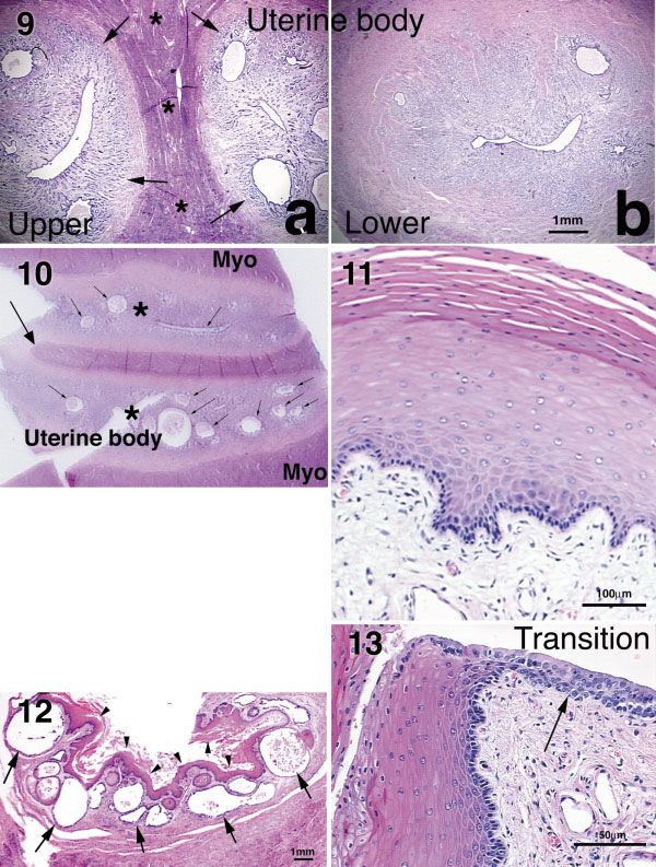
FUNCTIONAL ANATOMY OF THE SPOTTED HYENA                                           207
glands and muscles with regard to the central uro-       man et al., 1992, 1998; Drea et al., 1998; Licht et al.,
genital canal. Neaves et al. (1980) added significant     1998). In addition, the tissues of the retracted clito-
additional information regarding penile/clitoral sex-    ris would have to be firmly “stabilized.” Neaves et al.
ual dimorphism and expanded upon Matthew’s dis-          (1980) hypothesized that this stabilization is depen-
cussion of the unusual functional problems pre-          dent on the substantial retractor muscles that
sented by hyena urogenital morphology.                   course through the clitoris. Finally, Schneider
  However, over the past several decades a much          (1952) noted, and we have confirmed, that during
more detailed picture has emerged with regard to         the late stages of a mating sequence the male is
the functional demands placed on both female and         “partially locked” to the female. This “partial-lock”
male hyenas by the unique anatomical features of         can be broken, in contrast with that of a dog; and it
female spotted hyenas. A major goal of the present       is apparently the swelling of the distal glans penis
article is a reexamination of urogenital anatomy of      and clitoris, rather than the proximal bulb at the
the spotted hyena in functional terms. Specifically,      base of the shaft, that is responsible for retention of
we focus on the adaptive nature of “structure,”          the male in the female.
which enables/facilitates urination, mating, and            In this study we have paid particular attention to
parturition through the clitoris. We also note some      the manner in which tissue arrangements in the
unusual features of penile anatomy that are re-          female facilitate retraction and stabilization of the
quired for successful mating through a clitoral, as      clitoris, as well as permitting expansion of the glans
contrasted with the more typical vaginal, point of       to accommodate the male. We have also considered
insertion.                                               the structural features of the male that permit “flip-
                                                         ping” of the semierect phallus during the early
Functional Considerations: Urination,                    stages of mating, followed by extreme rigidity dur-
Mating, Parturition, and Meeting                         ing thrusting, and expansion of the eitoral glans,
Ceremonies                                               which is associated with the “semi-lock” that occurs
                                                         during the later stages of the mating sequence.
   Urination through the tip of the clitoris poses no       Parturition requires that large (i.e., 1.1–1.6 kg)
special difficulties and is found in other mammalian      precocial fetuses descend from a uterine horn and,
species, e.g., European moles (Matthews, 1935) and       after filling the clitoris, emerge from the urogenital
some prosimians (Drea et al., 1999), although there      meatus of the glans clitoris (Frank et al., 1995).
are some interesting mechanistic questions posed by      Neaves et al. (1980) noted a number of sexually
linkage of the urinary and reproductive systems in a     dimorphic features of clitoral/penile morphology
common canal in female hyenas. A detailed morpho-        that would serve to facilitate mating and delivery of
logical account is the base from which any physio-       young. In particular, they suggested that the ventral
logical analysis must proceed. Accordingly, we have      placement of the urogenital canal within the clitoris,
made a special effort to provide a detailed descrip-     and the absence of a surrounding corpus spongio-
tion of the confluence between the urethra and re-        sum, would permit the expansion of the urogenital
productive tract as they join to form the urogenital     canal to accommodate the male during mating and
sinus that traverses the shaft of the clitoris.          the fetus during parturition. However, this special-
   Anatomical obstacles do appear with regard to         ization is perhaps not sufficient to solve the problem.
mating, as the male has to achieve entry through a       The fetus moves along an exceptionally tortuous
small target, placed in a position that is consider-     route, first following a caudal-ventral path from the
ably anterior to the normal location of an external      uterus through the bony pelvic outlet, and then
vagina. A description of hyena mating behavior by        making a sharp turn in an anterior direction to
Schneider (1952) is in general accord with our own       traverse the clitoral canal to emerge through the
observations. The female hyena completely retracts       meatus of the glans clitoris (Frank et al., 1995).
the clitoris during mating and stands in near-              As noted above, estrogens enhance the size and
immobile fashion, while the male assumes an up-          elasticity of the urogenital meatus. Plasma relaxin
right posture and clasps the sides of the female with    concentrations also increase markedly during the
his forepaws just in front of her hindlegs. He then      final stages of gestation; and this hormone could
“flips” the semierect penis towards the abdomen of        well synergize with estrogens to facilitate delivery
the female, until the glans penis penetrates the uro-    (Steinetz et al., 1997). However, we now understand
genital meatus of the female. Thrusting begins after     that, despite the size and elasticity of the meatus at
intromission of the erect penis into the retracted       term, the urogenital meatus has to tear in order to
clitoris. As Neaves et al. (1980) suggest, this se-      permit delivery in a primiparous female. In our cap-
quence could not proceed unless the opening of the       tive colony, approximately 60% of first-births re-
urogenital meatus, and the clitoral portion of the       sulted in stillborn cubs, presumably because the
urogenital canal, had sufficient size and elasticity to   placenta detached and the cubs became anoxic dur-
accommodate the male. It is apparent that postnatal      ing the hours of labor that commonly follow detach-
ovarian secretions (estrogens in particular) play a      ment of the placenta, but precede delivery in a nul-
critical role in the development of a large clitoral     liparous female (Frank et al., 1995). Once again,
canal with a highly elastic urogenital meatus (Glick-    particular attention has been directed toward the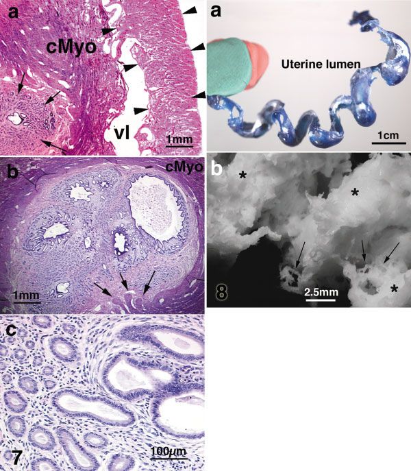
208 G.R. CUNHA ET AL.
TABLE 1. Sex, age, and reproductive status of the seven spotted were removed after the animals were euthanized as part of a
hyenas used for histological study larger study. The animals were 10 years old; both were nullipa-
rous. Each animal was immobilized with ketamine and xylazine
Hyena number Sex Age Reproductive state administered by blow-dart. Animals were then intubated and
transferred to the Department of Anatomy at the University of
1 F 10 year nulliparous/mated California, Davis, under general anesthesia. The external jugular
2 F 10 year virgin vein was incised for exsanguination and infusion of 10% formalin.
3 F 13 year multiparous After adequate fixation, the reproductive tracts were removed as
4 M 16 year mature an intact unit from the oviducts to the prepuce of the clitoris and
5 M 17 year mature placed in 10% formalin. A multiparous female, age 13 years, had
6 F fetus 96 day — a ovohysterectomy while under general anesthesia. This female
7 M fetus 96 day — was similarly immobilized and transferred to a surgical suite on
the UC, Berkeley, campus. The oviducts, uterine horns, and up-
per uterine corpus were removed and placed in 4% paraformal-
dehyde.
arrangement of tissues in the female that permit the The urogenital systems of two adult male hyenas, ages 16 and
unusual birth process to proceed. For example, we 17 years, were also removed and examined. Animals were immo-
bilized and transferred to a necropsy suite on the UC, Berkeley,
were interested in reexamining the distribution of campus. Shortly after a lethal injection of sodium pentobarbital,
urogenital musculature, with a view to understand- the internal and external reproductive tracts were rapidly dis-
ing the exceptional peristaltic actions required to sected and placed in 4% paraformaldehyde. One of these male
move, and ultimately expel, the fetus from the hyenas was euthanized because of disseminated lymphoma. The
lymphoma did not appear to grossly infiltrate the urogenital
uterus through the tip of the clitoris. A special effort system.
was also made to identify the distribution of local Lastly, the urogenital systems from a male and a female fetus,
glandular structures that would be expected to se- estimated gestational age 96 days of a 110-day gestation, were
crete lubricating agents, facilitating the passage of collected after a fetectomy was performed via a cesarean section
the fetus, and to examine the structure of the clitoral under general anesthesia. The pregnant female had been immo-
bilized as above and transferred to a surgical suite on the UC,
shaft and glans, with regard to playing the dual Berkeley, campus. The fetuses were euthanized by decapitation
roles of expansion during parturition and erection as part of a larger study. The urogenital tracts were rapidly
during “meeting ceremonies.” dissected and placed in 4% paraformaldehyde. All tissues were
Female and male spotted hyenas routinely display transported to UC, San Francisco, for anatomical and histological
analysis.
erections in nonsexual contexts when participating
in “meeting ceremonies.” Such ceremonies, which
are displayed by male and female hyenas, involve
two hyenas standing side-by-side, head to tail, and Anatomical Methods
inspecting one another’s external genitalia. These After fixation as described above, the entire genital tract was
“meeting ceremonies” are common events for hyenas dissected en block and photographed. The adult genital tracts
in nature, occurring when meeting after a period of were sectioned transversely into 1–2 cm segments represent-
ing all of the organs (Fig. 5). Individual segments were stored
separation and during periods of excitement or ten- in 70% ethanol and either photographed or scanned on an
sion. Hyena etiquette requires that the subordinate Epson G810A flat-bed scanner. The actual confluence of the
hyena initiate the ceremony by presenting its erect urethra and the genital tract was identified by dissection. After
genitalia for inspection by the dominant animal photography of the cranial or caudal surfaces of each trans-
verse block, 2–3-mm thick sections were excised from each
(Kruuk, 1972; East et al., 1993). In our colony, meet- segment and subsequently embedded in paraffin. Alterna-
ing ceremony erections often occur in less than 5 sec tively, a segment was bisected in the midsagittal or a coronal
(Krusko et al., 1988), with the organ capable of re- plane and then a 2–3-mm thick section was excised and em-
verting to a flaccid state and retracting into the bedded in paraffin. Six m-thick sections were prepared from
abdomen within a similar time interval. Kruuk the thick sections and stained with hematoxylin and eosin.
Masson’s trichrome stain was used to localize collagen in the
(1972) speculated that the social benefits accruing
from participation in meeting ceremonies were the
driving force in selection of the masculinized clitoral
morphology of the spotted hyena. Our focus was on TABLE 2. Animals used in specific figures
the possible existence of structural mechanism(s) in Figure numbers Hyena number
spotted hyenas that would permit rapid erection and
equally rapid detumescence/retraction of the clitoris 1–4 Animals other than those listed in
Table 1
or penis. 5–7 #1
8a #3
8b #2
MATERIALS AND METHODS 9–16 #1
Animals and Tissue Specimens 17a–b #1
17c #6
Spotted hyenas, Crocuta crocuta Erxleben, examined in this 18a #1
study were maintained at the Field Station for Behavioral Re- 18c–d #4
search of the University of California, Berkeley, CA. The internal 19 #5
and external reproductive tracts of seven animals were examined. 20 #7
Table 1 gives the ages and reproductive history of the animals 21 #2
used in this study. Table 2 is a cross-list of specific animals used 22 #5
in specific figures. Reproductive tracts of two adult female hyenas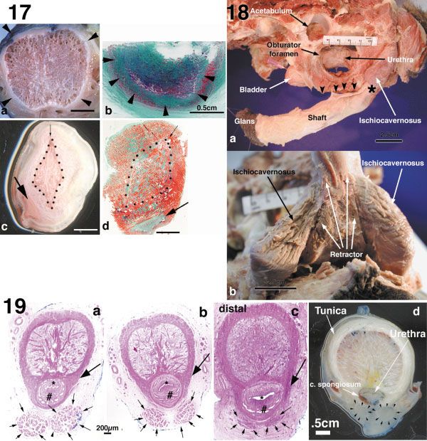
FUNCTIONAL ANATOMY OF THE SPOTTED HYENA                                                   209
corporal body as described previously (Lillie, 1965). Briefly,         loose connective tissue. The oviducts merge with the
sections were dewaxed, rehydrated, and postfixed in Bouin’s            uterine horns at the utero–tubule junction.
solution overnight at room temperature. After rinsing in tap
water, sections were stained with Weigert’s hematoxylin for 10           The uterine horns are about 7– 8 cm in length in
min and Biebrich scarlet and acid fuchsin for 1 min, respec-          the nonpregnant female. The wall of each uterine
tively. After a tap-water rinse, the sections were treated with       horn contains outer longitudinal and inner circular
1% phosphotungstic acid for 15 min, stained with 2% light             myometrial layers with an intervening vascular
green for 1 min, rinsed in water, fixed with 1% acetic acid for
3 min, and then dehydrated for mounting.
                                                                      layer (Fig. 7a). The circular myometrial layer sur-
  Uterine luminal casts were prepared using Batson’s No. 17           rounds the endometrial stroma (Fig. 7a,b). The in-
plastic replica and corrosion kit (Polysciences, Warrington, PA).     ner circular and outer longitudinal myometrial lay-
Briefly, the liquid plastic was catalyzed and perfused into the        ers are composed of a complex weave of smooth
lumen of fixed or unfixed uterine horns using a catheter and a 10       muscle bundles with little intercellular space or con-
ml syringe. After polymerization of the plastic, the uterine tissue
was digested with 1N potassium hydroxide. The resultant uterine       nective tissue between muscle bundles (Fig. 7a,b).
luminal cast was photographed in several orientations.                At the endometrial stroma/myometrial interface in-
                                                                      dividual smooth muscle bundles sometimes project
                                                                      into the endometrial stroma (Fig. 7b). The inner
RESULTS                                                               circular myometrial layer is separated from the
   Gross Anatomy: An Overview. Figure 5, a pho-                       outer longitudinal myometrial layer by a thick, well-
tograph of the female urogenital tract of the spotted                 defined vascular layer that contains large blood ves-
hyena, confirms most aspects of the line drawing in                    sels, loose connective tissue, and slender bundles of
Matthews (1939) (Fig. 12, page 19). The ovary and                     smooth muscle (Fig. 7a).
oviduct are embedded within the extensive ovarian                        Transverse sections of the uterine horns revealed
fat pad and thus are not visible without further                      3–5 large epithelial-lined luminal spaces (Fig. 7b).
dissection. Emerging from the ovarian fat pad are                     The diameters of individual uterine lumina are vari-
the uterine horns that fuse caudally to form the                      able, but in many cases sufficiently similar in size
uterine body. The uterine body, cervix and the va-                    that it was impossible to identify the actual main
gina extend caudally from the junction of the uterine                 uterine channel. Each lumen is lined with a typical
horns to the confluence with the urethra. These fe-                    simple columnar uterine epithelium and has its own
male genital organs are not discernable by gross                      constellation of associated uterine glands (Fig. 7b).
anatomical features. About midway along the uro-                      Uterine glands lined with a simple columnar non-
genital tract the two corpora cavernosa attach lat-                   ciliated epithelium projected radially from the uter-
erally to the pubic rami. At their bony attachments                   ine lumen into the endometrial stroma (Fig. 7b,c).
relatively large ischiocavernosus muscles cover the                   The uterine glands are generally straight and un-
corpora. The large bulbourethral glands lie within                    branched, although a subset of the glands is
the pelvic cavity immediately cranial to the attach-                  branched. The uterine glands are embedded in an
ment of the corporal bodies to the pubic rami. At its                 endometrial stroma containing fibroblasts and
approximate midpoint the urogenital tract under-                      many small-diameter blood vessels.
goes an almost 180 degree turn so that the tip of the                    Plastic corrosion casts were prepared to reveal the
clitoris points ventral-cranial (compare with the                     complexity of the uterine lumen. Plastic luminal casts
male genital tract, Fig. 18). The urinary bladder and                 of fresh specimens demonstrated that the uterine lu-
urethra lie ventral to the uterine body, cervix and                   men is not a single straight tube, but instead is coiled
vagina. The urethra runs caudally on the ventral                      into a helix (Fig. 8a). For this reason the length of the
aspect of the genital tract as a grossly defined struc-                uterine lumen is much longer (cranial to caudal) than
ture before merging with it to form the urogenital                    the external length of the uterine horn. The coiled
sinus. Thus, the angle of the intersection of the                     nature of the uterine lumen explains in part the fact
urethra with the urogenital tract is low, a fact ver-                 that multiple uterine lumina can be seen in a trans-
ified by histological analysis.                                        verse section. However, given the geometry of the he-
                                                                      lical uterine lumen, transverse sections could poten-
Histology of the Internal Female                                      tially contain only 2–3 separate lumina. Corrosion
Urogenital System                                                     casting of fixed uterine horns revealed luminal diver-
                                                                      ticula extending peripherally from the main uterine
   The ovary is surrounded by an extensive ovarian                    lumen (Fig. 8b). This complex organization of the lu-
fat pad containing the ovarian bursa and the ovi-                     minal cavity of the uterine horn was also corroborated
duct. The histology of the ovary will be the subject of               by examination of thick (1 mm) serial sections of the
a separate article. The oviducts are coiled and con-                  uterine horn (not illustrated).
tain a highly folded mucosa lined by a typical ovi-                      The two uterine horns merge to form the uterine
ductal epithelium composed of both ciliated and non-                  body. Initially, the right and left endometrial stro-
ciliated epithelial cells (Fig. 6). Individual epithelial             mas and associated uterine lumina are separated by
folds projecting into the lumen of the oviduct have a                 a central myometrial septum within the uterine
core of loose connective tissue and are highly vascu-                 body (Fig. 9a). This midline myometrial septum
lar. Deep to the mucosa is a muscular layer contain-                  maintains a constant thickness down to its blunt
ing slender bundles of smooth muscle embedded in                      termination within the uterine body (Fig. 10). Cau-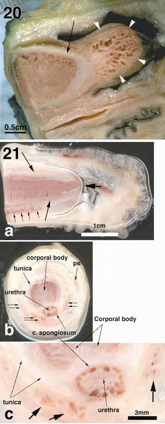
210                                         G.R. CUNHA ET AL.

                                                                                      Fig. 7. Transverse sections
                                                                                    through the uterine horn of a
                                                                                    spotted hyena (Crocuta crocuta).
                                                                                    a: Low-magnification view sur-
                                                                                    vey showing the endometrium
                                                                                    containing glands (arrows), the
                                                                                    circular myometrium (cMyo),
                                                                                    the vascular layer (VL), and the
                                                                                    longitudinal myometrium (ar-
                                                                                    rowheads). b: Endometrium and
                                                                                    circular myometrium (cMyo).
                                                                                    Note multiple uterine lumina
                                                                                    and associated uterine glands
                                                                                    embedded in the lightly stained
                                                                                    endometrial stroma. Individual
                                                                                    strands of myometrial bundles
                                                                                    (arrows) are seen at the endome-
                                                                                    trial stroma/myometrial inter-
                                                                                    face. c: High magnification of
                                                                                    uterine glands.

                                                                                      Fig. 8. Plastic casts of the lu-
                                                                                    men of the uterine horn of the
                                                                                    spotted hyena (Crocuta crocuta).
                                                                                    a: Liquid plastic was injected
                                                                                    into a fresh unfixed adult uter-
                                                                                    ine horn as described in Materi-
                                                                                    als and Methods. Under these
                                                                                    conditions the uterine glands
                                                                                    are not filled and the high pres-
                                                                                    sure at injection obliterates the
                                                                                    luminal evaginations. Note the
                                                                                    helical organization of the uter-
                                                                                    ine lumen. The red and green
                                                                                    material (left) is modeling clay
                                                                                    used to mount the plastic cast.
                                                                                    b: Liquid plastic was injected
                                                                                    into the lumen of a fixed uterine
                                                                                    horn. Adjacent turns of the helix
                                                                                    can be seen (*). Note the compli-
                                                                                    cated luminal projections and
                                                                                    uterine glands (arrows). Most of
                                                                                    the uterine glands were removed
                                                                                    to reveal the larger features.

dal to the central myometrial septum the two uter-       lium the stroma is primarily fibroblastic. At a
ine lumina fused into a single midline uterine cavity    deeper plane smooth muscle bundles are sepa-
surrounded by a constellation of uterine glands (Fig.    rated by connective tissue septa.
9b). It is important to note that the smooth muscle of      Between the vagina and the uterine body is the
the uterine horns and uterine body (myometrium)          cervical zone, which is not demarcated in any way by
constitute the bulk of the uterus. This continuous       gross external features. Thus, the uterine body, cervix,
muscle mass extends caudally into the fibromuscu-         and vagina cannot be distinguished grossly, even
lar wall of the cervix and vagina and provides the       though histological features clearly distinguish these
force required to expel the pups during delivery.        organs. The cervix is a transition zone, an area in
  In agreement with Matthews (1939), the spotted         which the stratified squamous cornified epithelium of
hyena has a histologically recognizable vagina.          the vagina coexists with large multiple cystic lumina
The state of vaginal epithelial differentiation          lined with simple columnar epithelial cells (Fig. 12).
surely varies throughout the estrous cycle. In the       These cystic lumina clearly define this region as dis-
females examined in this study a thickened corni-        tinct from the uterus above and the vagina below. True
fied epithelium was observed having the classical         uterine glands were not observed in the cervical zone.
histodifferentiation of vaginal epithelium in es-        The size of the cervical zone was difficult to discern,
trus (Fig. 11). The basal aspect of the vaginal          but is rather small, perhaps only a few millimeters
epithelium has an undulating surface with numer-         from cranial to caudal.
ous stubby papillae projecting into the underlying          Caudal to the vagina the stratified squamous vag-
stroma. The stroma contains many blood vessels of        inal epithelium changes abruptly to a urethral-like
variable size. In the vicinity of the vaginal epithe-    epithelium about 1–2 cm above the confluence of the
FUNCTIONAL ANATOMY OF THE SPOTTED HYENA 211 Fig. 9. Transverse sections of the uterine body of the spotted hyena (Crocuta crocuta). a: The upper cranial segment of the uterine body shows the central myometrial septum (*) separat- ing the right and left endometria (arrows), each containing the multiple uterine lumina. b: This transverse section is more cau- dal (lower) where the two endo- metria and associated uterine lumina have fused. Fig. 10. Coronal section through the uterine body of the spotted hyena (Crocuta crocuta). Note that the central fused myo- metrial septum terminates cau- dally (large arrow) as a blunt projection of muscle tissue sepa- rating the right and left endome- trial stromas (*) containing mul- tiple lumina (small arrows). Both endometrial stromas are in turn surrounded by myome- trium (Myo). Fig. 11. Section of the vagina of an adult spotted hyena (Cro- cuta crocuta). Note that the epi- thelium is very thick, indicative of the proestrous/estrous state. Fig. 12. The cervix of the spotted hyena (Crocuta crocuta) is a zone containing two types of epithelium: stratified squamous vaginal epithelium (arrowheads) and simple columnar epithelium (arrows) organized into cystic structures. Fig. 13. Transition zone be- tween the thick cornified vaginal epithelium (left) and the thin urethral-like epithelium (arrow) of the spotted hyena (Crocuta crocuta). genital tract and the urethra (Fig. 13). This is not recognizable externally. In contrast, Bartho- urethral-like epithelium is about 4 –5 cell layers in lin’s glands (bulbourethral glands, BUG) are recog- thickness. Apical cells are cuboidal to low columnar. nizable as large glands projecting laterally from the The most characteristic feature of this last segment urogenital tract just cranial to the ischiocavernosus of the genital tract before the confluence with the muscles (Fig. 5). Both the SMUG and the BUG are urethra is a submucosal mucous urogenital gland mucus-secreting glands. (SMUG) not previously reported (Fig. 14). The cra- In the female hyena the body of the bladder tapers nial aspect of the SMUG is located in the lower gradually to become the urethra, and thus a distinct segment of the genital tract near the junction be- bladder neck is not present (Fig. 5). The urethra is tween the vaginal and the urethral-like epithelium. lined by a typical urethral epithelium and extends The SMUG extends caudally over a linear distance caudally, first separated from the genital tract, but of about 2–3 cm to slightly below the confluence of eventually fusing externally with the genital tract in the urethra with the genital tract. The SMUG has the region of the vagina. The angle of intersection of multiple ducts that empty into the urogenital tract. the urethra and the vagina is low (⬃20°). For this The SMUG is confined to the submucosal tissue and reason the internal confluence of the lumina of the
212 G.R. CUNHA ET AL.
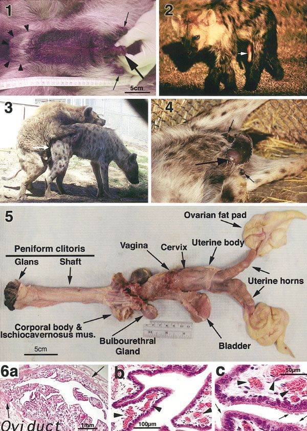
Fig. 14. Submucosal mucous
urogenital gland (SMUG) of the
spotted hyena (Crocuta crocuta).
a: Low magnification showing
the SMUG (large arrows) and
the mucosa (small arrows)
b: High magnification showing
mucous cells.
Fig. 15. a: Confluence of the
urethra with the urogenital
tract to form the urogenital si-
nus (large arrow) of an adult fe-
male spotted hyena (Crocuta
crocuta). b: The genital tract at
the level of arrow. c: The urethra
at the level of the arrow. Both of
these epithelia are similar.
Fig. 16. Transverse sections
of the clitoris of the adult (a,b)
and a near-term spotted hyena
(Crocuta crocuta) fetus (c).
a: Thick section of the root of the
clitoris near the attachment of
the corporal bodies (*) to the pu-
bic rami. The two corporal bod-
ies approach each other in the
midline and are covered by the
large ischiocavernosus muscles
(diamonds) and the retractor
muscles (double arrows). Note
the thick tunica albuginea sur-
rounding erectile tissue of the
corporal bodies. Ventral to the
corporal bodies is the urogenital
sinus (UGS). b: Thick section
through the midshaft of the
adult clitoris. The corporal body
is surrounded by the tunica al-
buginea. Immediately ventral-
lateral to the corporal body (b)
are the retractor muscles (ar-
rows). The most ventral structure
is the urogenital sinus (b), which
has a large collapsed folded wall
(arrowheads). c: Histological sec-
tion of the midshaft of the clitoris
of a near-term fetus. The corporal
body is surrounded by a thick tu-
nica albuginea demarcated by ar-
rowheads. The retractor muscles
(outlined by dots) are ventral-
lateral to the corporal body. The
urogenital sinus (UGS) is the
most ventral structure, having a
highly folded contour.
urethra and the genital tract occurs about 2 cm caudal the direction of the urogenital sinus changes about
to the external confluence of these gross structures. 180° as it traverses the peniform clitoris. As the
A true urogenital sinus forms at the confluence of urogenital sinus curves ventrally through the pelvic
the reproductive tract and the urethra (Fig. 15a). outlet, the right and left corpora cavernosa and as-
The epithelium of the urethra and the lower genital sociated ischiocavernosus muscles merge with the
tract are similar at and above the confluence of the tubular urogenital sinus to give bulk to the clitoris.
reproductive tract and urethra. Both epithelia ex-
hibit features of urethral epithelium (Fig. 15b,c).
External Genitalia of Female and Male
Once the urogenital sinus is formed at the conflu-
Hyenas: Similarities and Differences
ence, it extends caudally within the shaft of the
clitoris, which passes dorsal to the symphysis pubis The shaft of the clitoris is formed by a single large
and then curves ventrally and cranially. In this way corporal body formed by the fusion of the right andFUNCTIONAL ANATOMY OF THE SPOTTED HYENA 213
left corpora cavernosa near their proximal attach- that extend distally along the ventral aspect of the
ment to the ischiopubic rami where the corporal common corporal to insert along the tunica albu-
bodies are covered by exceedingly well-developed ginea (Figs. 18b, 19). Given the bulk of the ischio-
ischiocavernosus muscles (Fig. 16a). These two cor- cavernosus and retractor muscles and the geometry
poral bodies, each surrounded by thick tunica albu- of their attachments, it is likely that coordinated
ginea, meet and fuse in the midline near the pubic contraction of these muscles elicits the “phallic flip-
symphysis (Fig. 16b,c). The initial fusion of the two ping” that occurs during mating and during “erec-
corpora cavernosa is represented as a partial or com- tion” in meeting ceremonies in both male and female
plete midline connective tissue septum within the hyenas. The dramatic movements of the penis (and
common corporal body (Fig. 16b,c). However, pro- clitoris) in the nonerect state means that a flexible
ceeding distally, this midline septum gradually dis- “hinge” region must exist. This is located proximally
appears, and thus a single common corporal body is just distal to the bony attachment of the corpora and
defined externally by the tunica albuginea (Fig. ischiocavernosus muscles (asterisk in Fig. 18a). In
17a). The tunica albuginea is a particularly thick the hinge region the diameter of the common corpo-
sleeve of collagen fibers (Figs. 16a– c, 17a). From the ral body is considerably less than that distally. More
inner aspect of the tunica albuginea, thick collagen importantly, while the distal aspect of the corporal
bundles extend and intersect throughout the sub- body is stiff and noncompressible, even in the flaccid
stance of the corporal body (Fig. 17a,b). The abun- state, the hinge region is flexible and easily com-
dance of collagen within the corporal body gives the pressible. This difference in rigidity of the hinge vs.
shaft of the clitoris (and penis) a certain degree of the midshaft regions is associated with dramatic
rigidity even in the resting state. Immediately differences in cellularity and collagen content in
ventral-lateral to the corporal body of the clitoris are these two regions. Surface scans of the midshaft
the paired retractor muscles (Fig. 16b,c). These mus- suggest an abundance of collagen in the tunica and
cles are attached proximally to the pubic rami, run throughout the corporal body (Figs. 17a, 19d). This
longitudinally ventral to the corporal body, and at- abundance of collagen in the phallic midshaft was
tach distally along the tunica albuginea. In the cli- confirmed by Masson’s trichrome staining (Fig. 17b).
toris the urogenital sinus is located ventral to both In contrast, the hinge region is more cellular and
the corporal body and the paired retractor muscles.
contains considerably less collagen, as judged in sur-
The thin wall of the female urogenital sinus has a
face scans and in sections stained with Masson’s
collapsed and highly infolded contour (Fig. 16). For
trichrome (Fig. 17c,d). Also, the directionality of the
these reasons the lumen of the female urogenital
cellular and collagenous elements is distinctly dif-
sinus is highly distensible to accommodate the penis
ferent in the midshaft and hinge regions (Figs. 17,
during mating and for delivery of the cubs at partu-
rition. 19d).
Certain aspects of the anatomy of the phallus in In both sexes the midline corporal body of the
males and females are distinctly different, reflecting penis (and clitoris) is surrounded by an exceedingly
the different functional demands of the penis vs. the thick tunica albuginea (Figs. 17a, 19d). From the
peniform clitoris even though certain features are inner aspect of the tunica albuginea, thick collagen
shared in common. Euthanasia of an adult male bundles extend and intersect throughout the sub-
spotted hyena provided the opportunity to dissect stance of the corporal body in both sexes (Fig. 17a).
the intact pelvis and examine the spatial arrange- This gives the phallic shaft some degree of rigidity
ment of urogenital tract, its bony attachments, and even in the flaccid state in both sexes. In the mid-
associated muscles, which are similar in both the shaft the shape and diameter of the corporal body is
male and female. Figure 18a shows the bladder and similar in males and females, even though other
urethra extending caudally within the pelvis and features at midshaft distinguish the male and fe-
passing through the pelvic outlet. Not seen are the male phallus. One difference is the position of the
bilobar prostate and BUGs, which also lie within the retractor muscles. In females the retractor muscles
pelvic cavity. As the urethra passes caudally lie immediately ventral-lateral to the corporal body
through the pelvic outlet, the two corporal bodies and dorsal to the urogenital sinus (Fig. 16b,c). In
attached to the pubic rami approach each other in males the retractor muscles lie immediately ventral
the midline and fuse to form the common corporal to the urethra (Fig. 19). In females the cavity of the
body. The urogenital tract then turns almost 180° so urogenital sinus is voluminous, having a collapsed,
that the tip of the penis (also true for the clitoris) highly infolded thin wall. Erectile tissue and associ-
points ventral-cranially. In both sexes extremely ated tunica albuginea do not surround the urogeni-
large ischiocavernosus muscles (Fig. 18a,b) cover tal sinus in females (Fig. 16b,c). In contrast, in
the corporal bodies at their bony attachments. These males the cavity of the urogenital sinus (urethra) is
muscles arise from the pubic rami and insert on the small, oval-shaped, and is surrounded by blood-filled
proximal aspects of the corpora cavernosa as they spaces that constitute the corpus spongiosum (Fig.
are fusing to form the common corporal body. Lying 19). The corpus spongiosum is in turn demarcated
between the ischiocavernosus muscles and also aris- peripherally by a connective capsule (Fig. 19a– c). It
ing from the pubic rami are the retractor muscles should be noted that this sexual dimorphism devel-214 G.R. CUNHA ET AL. Fig. 17. Transverse sections through the adult peniform clitoris at midshaft (a,b) and the hinge region (c,d) of the spotted hyena (Crocuta crocuta). a: Scan of the female phallus at midshaft; note the thick tunica albuginea (arrowheads) and the intersecting network of coarse collagen fibers emerging from the inner aspect of the tunica albuginea and intersecting throughout the substance of the common corporal body. Not shown in (a,b) is the urogenital sinus, which is ventral to the tunica albuginea. The male corporal body at midshaft gave an identical pattern (see Fig. 19d). b: Thin section of the female phallus at midshaft stained with Masson’s trichrome; note that the tunica albuginea (arrowheads) and most of the common erectile body consist of green-stained collagen fibers. c: Scan of the hinge region of the clitoris; note the higher degree of cellularity and corresponding reduction of collagen fibers within the tunica and the common erectile body. The male hinge region gave an identical pattern. d: Thin section of the hinge region of the clitoris stained with Masson’s trichrome; note that the tunica albuginea is not defined by collagen fibers. Instead, the hinge region is notable for its general paucity of green-stained collagen fibers in the common erectile body. Finally, in (c,d) note the dorsal-ventral directionality of cellular and collagenous elements (small arrows). The common corporal body in (c,d) is demarcated by black dots. The large arrows in (c,d) denote the urethra. Scale bars ⫽ 0.5 cm. Fig. 18. Dissection of the pelvis and genital tract of the male spotted hyena (Crocuta crocuta). a: The bladder is partially covered by the pelvic bones. The urethra can be seen through the obturator foramen, which in life is covered by muscle. Attached to the inferior pubic rami are the ishiocavernosus muscles, which arise from the inferior pubic rami and insert into the corporal bodies. Note that the urogenital tract makes an almost 180° turn as it passes caudally out of the pelvis. Most of the shaft of the penis is thick, but the proximal portion of the shaft (*), called the hinge region, just distal to the attachment of the ishiocavernosus muscles is thinner (and more flexible) than the distal shaft of the penis. Arrowheads indicate the pubic symphysis. b: View of the root of the penis showing the ishiocavernosus muscles, which cover the corpora cavernosa at their bony attachments. Note the retractor muscle medial to the ishiocavernosus muscles extending distally along the ventral aspect of the shaft of the penis. Fig. 19. Transverse sections through the penis of the spotted hyena (Crocuta crocuta). a– c: Transverse sections of the penis of a near-term male fetus beginning proximally (a) and progressing distally (b-c). Note in (a– c) the common corporal body, the urethra (*), the corpus spongiosum (#) surrounded by the thick connective tissue layer, and the tunica albuginea (large arrows in a– c). The retractor muscles in (small arrows a– c) are ventral to the corpus spongiosum and in (c) insert into the tunica albuginea. d: Thick transverse section through the adult penis at midshaft. Note the thick tunica albuginea and the intersecting network of coarse collagen fibers emerging from the inner aspect of the tunica albuginea and extending throughout the substance of the common corporal body. Ventral to the common corporal body is the urethra, surrounded by the corpus spongiosum. Note the position of the retractor muscles (arrowheads) ventral to the urethra.
FUNCTIONAL ANATOMY OF THE SPOTTED HYENA                                           215
                    ops prenatally and is certainly evident in near-term
                    male and female fetuses (compare Figs. 16c, 19a– c).
                       The epithelium lining the urogenital sinus
                    traverses the clitoris and penis to emerge at the
                    glans. This epithelium is a typical urethral epithe-
                    lium in both sexes and does not change along the
                    shaft of the phallus until the external meatus, where
                    the urethral epithelium undergoes a transition to an
                    epidermis that extends onto the surface of the glans.
                    Distally the phallus in both sexes terminates at the
                    glans where the common corporal body ends in both
                    sexes. The shape and diameter of the distal aspect of
                    the common corporal body in the glans is profoundly
                    different in males and females. In females the com-
                    mon corporal body decreases only slightly in diam-
                    eter and ends bluntly in the glans (Fig. 20). In the
                    clitoris a separate erectile body (hyena urogenital
                    glanular extension, HUGE) forms most of the sub-
                    stance of the glans and is located distal to the com-
                    mon corporal body. The HUGE is not encapsulated
                    by a tunica albuginea (Fig. 20). This means that
                    engorgement with blood should result in expansion
                    of the clitoral glans. In males the common corporal
                    body tapers substantially within the glans, where its
                    diameter is only a fraction of that seen at midshaft
                    (Fig. 21a). In the male the tapered common corporal
                    body extends to the most distal aspect of the glans,
                    terminating immediately subjacent to the mucosa of
                    the tip of the glans penis (Fig. 21a). The tip of the
                    penis is not adorned with a separate distal erectile
                    body, as is the case for the female. However, the
                    proximal region of the penile glans does contain
                    erectile tissue, located ventral and lateral to the
                    urethra and the corpus spongiosum (Fig. 21b,c).
                    These latter erectile tissues are not encapsulated
                    and thus may expand during erection.

                       Fig. 20. Midsagittal section of the glans of the peniform clito-
                    ris of an adult female spotted hyena (Crocuta crocuta). Note the
                    common corporal body surrounded by the thick tunica albuginea
                    (arrow) terminates bluntly. The tip of the glans is adorned by a
                    separate erectile body (HUGE) not surrounded by a dense con-
                    nective capsule (white arrowheads).

                      Fig. 21. a: Midsagittal thick section through the adult glans
                    penis of the spotted hyena (Crocuta crocuta). Note that the com-
                    mon corporal body tapers to a narrow termination and is sur-
                    rounded by the thick tunica albuginea (large arrows). Separate
                    erectile tissue is ventral to the common corporal body and is not
                    surrounded by the tunica albuginea (small arrows). b: Transverse
                    section immediately proximal to the cut surface in (a); note the
                    common corporal body surrounded by the thick tunica albuginea.
                    The urethra is surrounded by the corpus spongiosum. Additional
                    erectile tissue is located ventral and lateral to the corpus spon-
                    giosum (small double arrows). c: Higher magnification of (b); note
                    the corpus spongiosum and additional erectile tissue (large ar-
                    rows), which are ventral and lateral to the corpus spongiosum.
                    This additional erectile tissue is not surrounded by a definite
                    connective tissue capsule.
216 G.R. CUNHA ET AL.
DISCUSSION protecting the urethral epithelium not only during
parturition, but during copulation as well. Intromis-
The detailed histological findings of the present sion appears to be traumatic to the urethral epithe-
study complement and clarify previous anatomical lium of the female, as postcoital bleeding has fre-
descriptions (Watson, 1877; Matthews, 1939; Davis quently been observed, even in multiparous females
and Story, 1949; Wells, 1968; Neaves et al., 1980) (Coscia, Weldele, Frank, and Glickman, unpublished
regarding the urogenital system of the spotted hy- observations). The vagina may also prove important
ena. Not since Matthew’s treatise in 1939 has a for sperm capacitation; this process may be delayed in
study followed the intricacies of the spotted hyena’s hyenas as sperm initially travel through a canal lined
reproductive tract from the oviduct to the glans cli- with a urethral epithelium. The secretory products
toris. As such, we have described the histological and histology of cervical glands, SMUG and BUG may
transitions from an upper reproductive tract that is vary with the estrous cycle as Matthews (1939) de-
fairly typical of female carnivores, to the highly mas- scribed with regards to the vaginal epithelium.
culinized lower genital tract that is unique to the Changes in pH and viscosity of these secretions may
female spotted hyena. prove important as the sperm begin the journey
From an external view, the upper reproductive through the tortuous reproductive tract of the female
tract appears to be essentially unremarkable. How- spotted hyena.
ever, the detailed histology in the present study has The embryological origin of the upper vagina in
revealed some interesting findings not reported by spotted hyenas is unknown, but is presumed to have
other investigators (Matthews, 1939). The helical differentiated from the Müllerian ducts (Cunha,
configuration of the uterine lumen, as demonstrated 1975). Vaginal tissue derived from either the Mül-
by the corrosion casts, adds greater length and sur- lerian ducts or the urogenital sinus is indistinguish-
face area compared to a straight endometrial cavity. able in adult mammals (Kurita et al., 2001); thus,
Sokolowski et al. (1973) noted a “corkscrew” appear- future research on the development of the vagina in
ance to the uterus during metestrus (early luteal spotted hyenas awaits embryological studies. Re-
phase) in beagle bitches. This “corkscrewing” only search to determine the temporal and spatial ex-
occurs shortly after ovulation, perhaps under the pression of the estrogen and androgen receptors in
influence of high progesterone levels, and is appar- the genital tract of the hyena fetus is planned. This
ent when the uterus is inspected as an intact gross type of study should provide insights into the devel-
specimen (Sokolowski et al., 1973). The helical con- opment of a female genital tract whose upper zones
figuration of the hyena uterus was not evident on are feminized and whose lower zones are masculin-
gross inspection; rather, the presence of multiple ized (Shapiro et al., 2000).
endometrial lumina on histological cross-section led A set of functional issues, concerning mating, par-
us to perform the corrosion casts. The diverticula turition, and meeting ceremonies, was raised in the
that protrude from the main endometrial canal also Introduction to this paper. A corresponding cluster
contribute to the appearance of multiple uterine lu- of morphological features was subsequently identi-
mina. We considered the possibility that this finding fied that relate to each of these events. With regard
might be an artifact of prolonged nulliparity and to parturition, the various muscles identified in the
pathology associated with persistent estrus. The uterine and vaginal passageways would be required
presence of these histological findings in the uterus for moving a large fetal hyena through the complex
from a multiparous female argues against this hy- passageway that results in birth. Multiple glands,
pothesis. The functional significance of the hyena’s identified in the female uterine and vaginal canals
uterine morphology is unknown. The placenta of the (SMUG and BUG), would also facilitate movement
spotted hyena is unique among carnivores in that of the fetus during parturition by providing lubrica-
the hyena placenta is hemochorial and villous, while tion. Both mating and parturition require that the
the placentae of all other carnivores studied are clitoris expand, either to accommodate the male, or
endotheliochorial and labyrinthine (Amoroso, 1959; to permit passage of the fetus; albeit the expansion
Wynn and Amoroso, 1964). Whether the idiosyncra- required during parturition is much greater than
sies of the spotted hyena endometrium and placenta that during mating. Neaves et al. (1980) observed
are related awaits further study. that the urethra and the urogenital canal are differ-
The transition from the uterine corpus to the lower entially embedded within the penis and the clitoris,
urogenital tract had not been described in detail before respectively, and that these differences permit the
the present study. Matthews (1939) improved on the expansion of the clitoris when the female gives birth.
description by Watson (1877) by identifying a vagina, In particular, they noted that the urethra of the
but an intervening cervix was not described until male is surrounded by a corpus spongiosum limiting
Wells (1968), and then only briefly. Additionally, sub- expansion, while the urogenital canal of the female
mucosal mucous urogenital glands (SMUG), never be- spotted hyena is encased in loose connective tissue,
fore described, are present in the genital tract as the facilitating such expansion. We agree with these
transition from a vaginal to urethral-like epithelium observations, as well as with their conclusions re-
occurs. The products of the cervical and submucosal garding the potential role of the retractor muscles in
glands may prove to be important for lubricating and retaining and stabilizing the clitoris for receipt ofFUNCTIONAL ANATOMY OF THE SPOTTED HYENA 217
the male during mating. In addition, we note that genitalia of spotted hyenas at birth are due to
the extensive infolding of the urogenital sinus in the greater androgenic activity in males. That is, the
female would also serve to facilitate such expansion, external morphology of the penis, and associated
while the broad distribution of collagen within the external musculature, as well as Onuf’s nucleus in
corporal body of the clitoris could provide an appro- the spinal cord, assumes the feminine phenotype
priate target for the actions of relaxin. This peptide following anti-androgen treatment (Forger et al.,
hormone potentially contributes to the elasticity of 1996; Drea et al., 1998). It remains to be determined
clitoral tissues and peaks during the period imme- whether the internal structure of the penis is also
diately preceding birth in the female spotted hyena anatomically feminized by such treatment (i.e., po-
(Steinetz et al., 1997). sition of the retractor muscles and morphology of the
In males and females, the existence of a “hinge glans). Although we can analyze the mechanisms
region” near the proximal attachment of the phallus that produce the uniquely synchronized urogenital
to the pubic bones, and the mode of attachment of adaptations of this species, it remains difficult to
the ishiocavernosus and retractor muscles, facilitate visualize the sequence of evolutionary events that
the “flipping” phase of the mating sequence. The produced this unusual co-adapted system.
presence of a thick collagenous tunica completely
enclosing the shaft of the penis would result in the
rigidity required for thrusting, while the absence of LITERATURE CITED
such a restrictive outer layer in the glans permits
expansion of that tissue and serves to retain the Amoroso E. 1959. Comparative anatomy of the placenta. Ann NY
penis in the female during the semi-locked terminal Acad Sci 75:855– 872.
Cunha GR. 1975. The dual origin of vaginal epithelium. Am J
portion of the mating sequence. The tapered shape Anat 143:387–392.
of the glans penis and its corporal body (which con- Davis D, Story H. 1949. The female external genitalia of the
trasts with the rounded contour of the glans clitoris) spotted hyena. Field Zool 31:277–283.
would also facilitate entry of the male during mat- Drea C, Weldele M, Forger N, Coscia E, Frank L, Licht P, Glick-
ing. man S. 1998. Androgens and masculinization of the genitalia in
the spotted hyaena (Crocuta crocuta). 2. Effects of prenatal
Meeting ceremonies require that erection and re- anti-androgens. J Reprod Fertil 113:118 –128.
traction of both the female and the male phallus be Drea C, Coscia E, Glickman S. 1999. Hyenas. In: Knobil E, Neill
accomplished in a timely manner. By limiting the J, Licht P, editors. Encyclopedia of reproduction. San Diego:
constraining tunica (surrounding the corporal body) Academic Press. p 718 –724.
East M, Hofer H, Wickler W. 1993. The erect “penis” is a flag of
in the female to a dorsal position, she can achieve submission in a female-dominated society: greetings in
erections required for participation in meeting cer- Serengeti spotted hyaenas. Behav Ecol Sociobiol 33:335–370.
emonies, without compromising the ability of the Forger N, Frank L, Breedlove S, Glickman S. 1996. Sexual di-
clitoral shaft to expand during mating and parturi- morphism of perineal muscles and motoneurons in spotted hy-
tion. In addition, the ubiquitous presence of cross- enas. J Comp Neurol 375:333–343.
Frank L, Weldele M, Glickman S. 1995. Maculinization costs in
linked collagen fibers in the shaft of the clitoris and hyaenas. Nature 377:584 –585.
the penis provides more than usual structural rigid- Glickman SE. 1995. The spotted hyena from Aristotle to The Lion
ity even in the flaccid state, which could speed the King: reputation is everything. Soc Res 62:501–537.
appearance of a semierect phallus during meeting Glickman SE, Frank LG, Licht P, Yalcinkaya T, Siiteri PK, Da-
ceremonies, prior to full engorgement of penile/ vidson J. 1992. Sexual differentiation of the female spotted
hyena. One of nature’s experiments. Ann NY Acad Sci 662:135–
clitoral vasculature. In the female, such a collagen- 159.
cross-linked organ would also provide a firmer base Glickman SE, Coscia EM, Frank LG, Licht P, Weldele ML, Drea
for receipt of the male during mating. CM. 1998. Androgens and masculinization of genitalia in the
Close examination of the urogenital system of the spotted hyaena (Crocuta crocuta). 3. Effects of juvenile gonad-
ectomy. J Reprod Fertil 113:129 –135.
female spotted hyena reveals a remarkable array of Krusko N, Weldele M, Glickman S. 1988. Meeting ceremonies in
anatomical adaptations, allowing effective reproduc- a colony of juvenile spotted hyenas. Annu Meet Anim Behav
tive behavior in the only extant female mammal Soc, Missoula, Montana.
that mates and gives birth though her clitoris. Sub- Kruuk H. 1972. The spotted hyaena: a study of predation and
stantial requirements are also placed on the male, social behavior. Chicago: Chicago University Press.
Kurita T, Cooke P, Cunha G. 2001. Epithelial-stromal tissue
as he is required to achieve entry during mating interaction in paramesonephric (Müllerian) epithelial differen-
through a small opening, located in an unusually tiation. Dev Biol 240:194 –211.
anterior position on the surface of the abdomen. As Licht P, Hayes T, Tsai P, Cunha G, Kim H, Golbus M, Hayward
noted above, penile morphology is also adapted to S, Martin MC, Jaffe RB, Glickman SE. 1998. Androgens and
masculinization of genitalia in the spotted hyaena (Crocuta
the unique demands of mating for male spotted hy- crocuta). 1. Urogenital morphology and placental androgen pro-
enas. duction during fetal life. J Reprod Fertil 113:105–116.
Experiments have been conducted in which anti- Lillie RD. 1965. Histopathologic technic and practical histochem-
androgens (flutamide or cyproterone) and a 5-alpha istry. New York: McGraw-Hill.
reductase inhibitor (finasteride) were supplied to Matthews L. 1935. The oestrus cycle and intersexuality in the
female mole (Talpa europaea Linn.). Proc Zool Soc Lond 1935:
female and male fetuses in utero (Forger et al., 1996; 347–383.
Drea et al., 1998). The results are compatible with Matthews L. 1939. Reproduction of the spotted hyena (Crocuta
the hypothesis that sex differences in the external crocuta Erxleben). Philos Tran R Soc Lond B 230:1–78.You can also read