The Neuroinflammatory Role of Pericytes in Epilepsy - MDPI
←
→
Page content transcription
If your browser does not render page correctly, please read the page content below
biomedicines
Review
The Neuroinflammatory Role of Pericytes in Epilepsy
Gaku Yamanaka 1, * , Fuyuko Takata 2 , Yasufumi Kataoka 2 , Kanako Kanou 1 , Shinichiro Morichi 1 ,
Shinya Dohgu 2 and Hisashi Kawashima 1

                                          1   Department of Pediatrics and Adolescent Medicine, Tokyo Medical University, Tokyo 160-8402, Japan;
                                              kanako.hayashi.0110@gmail.com (K.K.); s.morichi@gmail.com (S.M.); hisashi@tokyo-med.ac.jp (H.K.)
                                          2   Department of Pharmaceutical Care and Health Sciences, Faculty of Pharmaceutical Sciences, Fukuoka
                                              University, Fukuoka 814-0180, Japan; ftakata@fukuoka-u.ac.jp (F.T.); ykataoka@fukuoka-u.ac.jp (Y.K.);
                                              dohgu@fukuoka-u.ac.jp (S.D.)
                                          *   Correspondence: gaku@tokyo-med.ac.jp; Tel.: +81-3-3342-6111; Fax: +81-3-3344-0643

                                          Abstract: Pericytes are a component of the blood–brain barrier (BBB) neurovascular unit, in which
                                          they play a crucial role in BBB integrity and are also implicated in neuroinflammation. The association
                                          between pericytes, BBB dysfunction, and the pathophysiology of epilepsy has been investigated,
                                          and links between epilepsy and pericytes have been identified. Here, we review current knowledge
                                          about the role of pericytes in epilepsy. Clinical evidence has shown an accumulation of pericytes
                                          with altered morphology in the cerebral vascular territories of patients with intractable epilepsy.
                                          In vitro, proinflammatory cytokines, including IL-1β, TNFα, and IL-6, cause morphological changes
                                          in human-derived pericytes, where IL-6 leads to cell damage. Experimental studies using epileptic
                                          animal models have shown that cerebrovascular pericytes undergo redistribution and remodeling,
                                          potentially contributing to BBB permeability. These series of pericyte-related modifications are
                                promoted by proinflammatory cytokines, of which the most pronounced alterations are caused by

                                          IL-1β, a cytokine involved in the pathogenesis of epilepsy. Furthermore, the pericyte-glial scarring
Citation: Yamanaka, G.; Takata, F.;
                                          process in leaky capillaries was detected in the hippocampus during seizure progression. In addition,
Kataoka, Y.; Kanou, K.; Morichi, S.;
                                          pericytes respond more sensitively to proinflammatory cytokines than microglia and can also activate
Dohgu, S.; Kawashima, H. The
                                          microglia. Thus, pericytes may function as sensors of the inflammatory response. Finally, both
Neuroinflammatory Role of Pericytes
                                          in vitro and in vivo studies have highlighted the potential of pericytes as a therapeutic target for
in Epilepsy. Biomedicines 2021, 9, 759.
https://doi.org/10.3390/
                                          seizure disorders.
biomedicines9070759
                                          Keywords: pericytes; mural cells; cytokine; blood-brain barrier; neuroinflammation
Academic Editor: Prosper N’Gouemo

Received: 31 May 2021
Accepted: 26 June 2021                    1. Introduction
Published: 30 June 2021
                                               Accumulating evidence has demonstrated that the pathogenesis of epilepsy is linked
                                          to neuroinflammation and cerebrovascular dysfunction [1–6]. Traditionally, microglia
Publisher’s Note: MDPI stays neutral      had been considered to be responsible for the cytokine-centered immune response in the
with regard to jurisdictional claims in
                                          central nervous system (CNS); however, brain pericytes can respond to inflammatory
published maps and institutional affil-
                                          signals, such as circulating cytokines, and convey this information to surrounding cells
iations.
                                          through chemokine and cytokine secretions [7–10]. Recent studies have demonstrated that
                                          pericytes may act as sensors for the inflammatory response in the CNS, as pericytes react
                                          intensely to proinflammatory cytokines when compared to other cell types (e.g., microglia)
                                          that constitute the CNS and factor-induced reactive pericytes can also activate microglia
Copyright: © 2021 by the authors.         in vitro [9,11–13].
Licensee MDPI, Basel, Switzerland.             Pericytes provide physical support to the blood–brain barrier (BBB) and play an
This article is an open access article
                                          integral role in CNS homeostasis and BBB function [14]. Pericyte degeneration and/or
distributed under the terms and
                                          dysfunction contribute to the loss of BBB integrity, which is an early hallmark of several
conditions of the Creative Commons
                                          neurodegenerative and inflammatory conditions [8,15,16]. Another notable feature of
Attribution (CC BY) license (https://
                                          pericytes is their ability to regulate the migration of leukocytes across the brain microvas-
creativecommons.org/licenses/by/
                                          cular endothelial cell (BMVEC) barrier, which secretes key molecules that support the
4.0/).

Biomedicines 2021, 9, 759. https://doi.org/10.3390/biomedicines9070759                                  https://www.mdpi.com/journal/biomedicines
Biomedicines 2021, 9, 759                                                                                           2 of 16

                            BBB barrier [17,18]. Recent research on the pathogenesis of epilepsy has begun to eluci-
                            date the mechanisms mediating peripheral-to-CNS cell infiltration in human and mouse
                            models [19,20]. Pericytes may contribute to the mechanisms, while emerging research
                            is investigating the extent of peripheral immune cell involvement in the inflammatory
                            pathology of epilepsy.
                                 The various functions of pericytes and their involvement in CNS diseases, including
                            ischemic stroke [21], spinal cord injury [22], brain injury [23], and multiple sclerosis [24],
                            has been reported.
                                 The association between pericytes and epilepsy has attracted attention, while sev-
                            eral recent studies have illustrated the contributions of pericytes to the pathogenesis of
                            epilepsy [2,25–32]. These studies suggested that pericytes might participate in the patho-
                            genesis of epilepsy, consisting of neuroinflammation and BBB damage and the interaction
                            between peripheral and central immunity. Thus, evidence on the relationship between
                            pericytes and the pathogenesis of epilepsy is gradually accumulating. Therefore, this study
                            aimed to investigate the pathogenesis of epilepsy and pericytes because none of the re-
                            view articles focused on this, even though therapeutic targets for pericytes in neurological
                            disorders were investigated [17,33,34].
                                 This review (1) explores the current literature regarding the role of pericytes in the
                            pathogenesis of epilepsy and (2) highlights novel directions for research on therapeutic
                            interventions for epilepsy that target pericytes. Given the paucity of knowledge on pericyte
                            function in seizures and epilepsy-related pathologies, further studies are warranted to
                            investigate pericytes as a potential therapeutic target for epilepsy treatment.

                            2. What Are Pericytes?
                                 Pericytes were first described by the French scientist Charles-Marie Benjamin Rouget
                            and were originally called Rouget cells in 1873 [35]. Later, this population was rediscovered
                            by Zimmermann as a cell that shows a specific morphology around microvessels, and
                            became widely known as a “pericyte” [36]. Pericytes are mural cells that are implanted in
                            the basal membrane surrounding endothelial cells in capillaries and small vessels, includ-
                            ing precapillary arterioles and postcapillary venules. Although the origin of all pericytes
                            has not been clarified, blood vessels in the CNS are predominantly covered by neural
                            crest cell-derived pericytes, while mesoderm-derived pericytes mainly contribute to blood
                            vessel coverage in the trunk [37]. In the brain, pericytes constitute a vital component
                            of the BBB/neurovascular unit (NVU) and cover the BMVECs lining the capillaries on
                            the parenchymal side, where there are astrocytic end feet that enclose cerebral vessels,
                            perivascular microglia/macrophages, and neurons [17,38,39]. Pericytes form a crucial
                            component of the brain microvasculature and play an integral role in CNS homeostasis and
                            BBB function [14] in normal physiological (Figure 1) and pathological conditions (Figure 2).
                            A potential mechanism of pericyte action is the regulation of signaling through platelet-
                            derived growth factor receptor beta (PDGFRβ), which is commonly used as a marker of
                            pericytes and regulates pericyte survival, proliferation, and migration signals [40]. In the
                            CNS, platelet-derived growth factor-beta subunit (PDGF-BB) is released by endothelial cells
                            and binds to PDGFRβ at the cell surface of pericytes to promote pericyte vascularization
                            within the BBB [41]. The PDGFRβ signaling pathway is involved in pericyte survival and
                            subsequent development as well as the function of the BBB during adulthood and senes-
                            cence, as demonstrated by experiments in pericyte-deficient mice [17,38]. In addition to its
                            role as a marker of CNS pericytes, PDGFRβ is expressed in oligodendrocyte precursor cell
                            (OPC)/neuron-glial antigen 2 (NG2) parenchymal glial cells [2,25,42]. Other markers for
                            pericytes exist (Table 1), but these remain inconclusive. Anatomical studies are required to
                            investigate the characteristics of pericytes that possess longitudinal processes along vessels
                            and contribute to BBB maintenance [15]. Pericytes in the brain are highly heterogeneous
                            and have different morphologies as well as functions depending on their location in the
                            vasculature [10]. Further, transgenic mice generated to study pericyte function may yield
                            information on other cell types [43]. Therefore, “peripheral blood-specific” markers must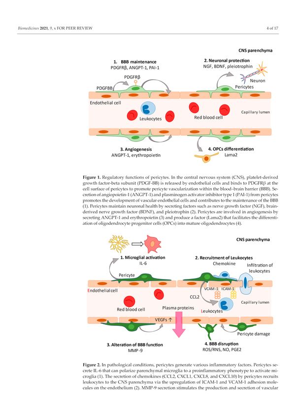
Biomedicines 2021, 9, 759                                                                                                  3 of 16

                            be used with caution [44]. Although there is no scientific consensus on what constitutes
                            true pericytes [45], the current review focuses on studies using definitive pericyte-related
                            markers and anatomy.

                            Figure 1. Regulatory functions of pericytes. In the central nervous system (CNS), platelet-derived
                            growth factor-beta subunit (PDGF-BB) is released by endothelial cells and binds to PDGFRβ at the
                            cell surface of pericytes to promote pericyte vascularization within the blood–brain barrier (BBB).
                            Secretion of angiopoietin-1 (ANGPT-1) and plasminogen activator inhibitor type 1 (PAI-1) from
                            pericytes promotes the development of vascular endothelial cells and contributes to the maintenance
                            of the BBB (1). Pericytes maintain neuronal health by secreting factors such as nerve growth factor
                            (NGF), brain-derived nerve growth factor (BDNF), and pleiotrophin (2). Pericytes are involved in
                            angiogenesis by secreting ANGPT-1 and erythropoietin (3) and produce a factor (Lama2) that facilitates
                            the differentiation of oligodendrocyte progenitor cells (OPCs) into mature oligodendrocytes (4).

                            Figure 2. In pathological conditions, pericytes generate various inflammatory factors. Pericytes
                            secrete IL-6 that can polarize parenchymal microglia to a proinflammatory phenotype to activate
                            microglia (1). The secretion of chemokines (CCL2, CXCL1, CXCL8, and CXCL10) by pericytes recruits
                            leukocytes to the CNS parenchyma via the upregulation of ICAM-1 and VCAM-1 adhesion molecules
                            on the endothelium (2). MMP-9 secretion stimulates the production and secretion of vascular
                            endothelial growth factor (VEGF), resulting in endothelial dysfunction (3). Secretion of reactive
                            oxygen species/reactive nitrogen species (ROS/RNS), nitric oxide (NO), and prostaglandins (PGE2)
                            by pericytes lead to vasodilation and breaching of the blood–brain barrier. Pericytes themselves are
                            morphologically altered by inflammatory mediators (4).
Biomedicines 2021, 9, 759 4 of 16
Table 1. Common markers used to identify pericytes in the central nervous system of mice that also label other cell types.
Marker Cells Labeled Main Function Reference(s)
PDGFRβ
(platelet-derived growth factor receptor beta) Fibroblasts, SMCs, pericytes Tyrosine kinase receptor [14,41]
NG2 OPCs, NSCs, SMCs, pericytes
(CSPG4; chondroitin sulfate proteoglycan 4) Cell-membrane proteoglycan [46]
CD13
(aminopeptidase N) Fibroblasts, SMCs, pericytes Cell-membrane aminopeptidase [14]
αSMA
(actin, aortic smooth muscle) SMCs, myofibroblasts, pericytes Cytoskeletal protein [14]
Desmin SMCs, pericytes Intermediate filament [14]
Rgs5
SMCs, pericytes Regulator of G protein [47]
(regulator of G protein signaling 5)
CD146 SMCs, pericytes
(cell surface glycoprotein MUC18) Membrane proteins [48]
SUR2 SMCs, pericytes
(sulfonylurea receptor 2) Potassium-channel [47,49]
Kir6.1
(K+ channel pore-forming subunit) SMCs, fibroblasts, pericytes Potassium-channel [47,49]
NeuroTrace 500/525 Pericytes - [50]
(fluorescent Nissl dye/FluoroNissl Green)
Vitronectin SMCs, Pericytes Complement-binding protein [49,51]
Note: NSCs, neural stem cells; OPCs, oligodendrocyte progenitor cells; SMCs, smooth muscle cells.
3. Pericytes and Neuroinflammation
Evidence accumulated from experimental models and human samples implicates im-
munological processes in the pathogenesis of epilepsy [1,4]. The involvement of pericytes
in the CNS immune responses has attracted significant attention. Pericytes present hetero-
geneous signals to the surrounding cells and actively modulate inflammatory responses in
a tissue- and context-dependent manner. The expression of various pattern-recognition
receptors (PRRs), including toll-like receptors (TLRs) and nucleotide-binding and oligomer-
ization domain (NOD)-like receptor families, has been detected in brain pericytes [52].
Given the abundance of surface receptors, pericytes can respond to inflammatory medi-
ators, such as monocyte chemoattractant protein-1 (MCP-1/CCL2) and tumor necrosis
factor (TNF)-α, which in turn induce the secretion of CCL2, nitric oxide (NO), and several
cytokines [7–9,53]. Pericytes act as promoters of both the innate and adaptive immune
system [43]. In the CNS, microglia are a hallmark of the immune response, which produce
cytokines such as interleukin (IL)-1β, TNF-α, IL-6, and various other chemokines [54],
and related effector pathways, including cyclooxygenase-2 (COX-2)/prostaglandin (PGE2)
and complement factors [55]. The rapid activation of microglia impairs neuronal function
by inducing inflammatory mediators, such as NO, reactive oxygen species (ROS), and
proinflammatory cytokines [56,57].
Pericytes have been shown to be more sensitive to proinflammatory cytokines com-
pared to other cells in the NVU [9,11–13]. Specifically, cytokine and chemokine release
profiles from brain pericytes in response to TNF-α are distinct to those of other cell types
comprising the NVU, and TNF-α-stimulated pericytes release macrophage inflammatory
protein (MIP)-1α and IL-6. Among BBB cells, pericytes stimulated with TNF-α induced
the highest levels of iNOS and IL-1β mRNA expression, which indicates the activation
of BV-2 microglia [9]. The mechanism underlying TNF-α-induced IL-6 release involves
the inhibitor kappa B (IκB)-nuclear factor kappa-light-chain-enhancer of activated B cells
(NFκB) and the Janus family of tyrosine kinase (JAK)-signal transducer and activator of
transcription (STAT) 3 pathways [13]. NFκB plays a key role in inflammation, immune,
and stress-related responses, as well as in the regulation of cell survival and in the growth
of neural processes in developing peripheral and central neurons [58]. These findings
indicate that the activated brain pericytes trigger the development of uncoordinated NVU
function, including glial activation, and may act as sensors at the BBB in TNF-α-mediated
brain inflammation.Biomedicines 2021, 9, 759 5 of 16
Pericytes also release anti-inflammatory factors, highlighting their involvement in
regeneration and protection [7,59,60]. Pericytes respond to lipopolysaccharide (LPS), se-
crete anti-inflammatory cytokines such as IL-10 and IL-13 [61], and produce neurotrophins
such as nerve growth factor (NGF) and brain-derived neurotrophic factor (BDNF), which
regulate neuronal development [42,62]. Pericytes upregulate neurotrophin-3 production
in response to hypoxia, resulting in increased NGF production in astrocytes, thereby
protecting neurons from hypoxia-induced apoptosis [62]. These actions highlight the
neuroprotective functions of pericytes under pathological conditions.
4. Pericytes and Epilepsy
Table 2 summarizes the research on pericytes and epilepsy.
Table 2. Research and key findings on pericytes and epilepsy.
No. Patients/Model Species Key Findings Reference
• Degeneration of pericytes (aggregates of
cellular debris within the basement
membrane) with the morphological changes
1 Intractable complex partial seizures Humans in pericyte-basement membrane unit [63]
thickness and pericyte cytoplasmic density
were observed in the spiking area of
microvessels in an electron microscopy study
of brain tissue
• PDGFRβ+ cells are distributed around the
TLE with HS Humans cerebrovasculature and are present in the
brain parenchyma of human TLE specimens
• Constitutive cerebrovascular NG2DsRed
2 pericyte coverage is impaired in response to [2]
SE in vivo or seizure-like events in vitro
NG2DsRed or C57BL/6J mice • Redistribution of parenchymal and vascular
Mice PDGFRβ+ cells occurs in vitro and in vivo
(intraperitoneal KA injections)
• Vascular and parenchymal PDGFRβ+ cells
partially co-localize with NG2DsRed and
NG2, but not with IBA-1 (indicators
of microglia)
• FCD and TLE-HS display the highest
PDGFRβ immunoreactivity at the
microvasculature identifying pericytes
• Cryptogenic epilepsy patients also showed a
FCD, TLE without HS, Humans similar immune response pattern, although to
cryptogenic epilepsy a lesser extent than that in FCD
• The amount of perivascular PDGFRβ
immunoreactivity was found to be associated
3 with increased hippocampal angiogenesis in [25]
tissues from patients with TLE-HS
• Pericyte-vascular dysplasia was detected in
Neurovascular dysplasia rat model hippocampi corresponding to
(Sprague-Dawley rats with pre-natal neuronal heterotopias
Mice
exposure to methyl-axozy methanoic • Severe SE was associated with a
acid), pilocarpine region-specific increase in
PDGFRβ immunoreactivity
• Chronic IFN-γ treatment blocks signaling
4 TLE Humans through PDGFRβ by enhancing agonist [26]
PDGF-BB
• TGFβ1 decreased pericyte proliferation and
decreased phagocytosis
Drug-resistant TLE • TGFβ1 also upregulates the expression of
5 human IL-6, MMP-2, and NOX4, which disrupt the [27]
(microarray analysis)
function of the BBB, and these responses to
TGFβ1 may not be therapeutic for the
neurovascular systemBiomedicines 2021, 9, 759 6 of 16
Table 2. Cont.
No. Patients/Model Species Key Findings Reference
• NG2 mural cells are added and removed from
veins, arterioles, and capillaries after
status epilepticus
Dynamics of NG2 mural cells under SE • Loss of NG2 mural cells is proportional to
6 Mice seizure severity and vascular pathology (e.g., [28]
with systemic KA injection in mice
rigidity, perfusion, and permeability)
• Treatment with PDGF-BB reduced NG2 mural
cell loss, vascular pathology, and epileptiform
electroencephalogram activity
• Pericyte-microglia assemblies with
IBA1/HLA microglial cells outlining the
capillary wall were observed in TLE-HS and
TLE with or without HS, FCD Humans FCD-IIb specimens
• Proinflammatory cytokines such as IL-1β
7 cause morphological changes and IL-6 causes [29]
cell damage in human-derived pericytes
NG2DsRed/C57BL6 (unilateral
• IL-1β elicited pericyte morphological changes
Mice and pericyte-microglia clustering in
intra-hippocampal KA injections)
NG2DsRed hippocampal slices
• Multicellular scarring occurs at the outer
capillary wall in the hippocampus during
seizure progression
• PDGFRβ stromal cells and collagens III and
8 NG2DsRed/C57BL6 (unilateral Mice [30]
intra-hippocampal KA injections) IV participate in the localized pericyte-glial
scarring and capillary pathology in
hippocampal subregions
• PDGFRβ is a proposed anti-inflammatory
entry point for chronic disease stages in vivo
• Pericytes regulate changes in vascular
diameter in response to neuronal activity
• Recurrent seizures are associated with
Transgenic mice (4-aminopyridine or impaired neurovascular coupling and
9 Mice [31]
low-Mg2+ conditions) increased BBB permeability in capillaries
• Recurrent seizures lead to depolarization of
pericytic mitochondria and subsequent
vasoconstriction
• PDGFRβ levels were increased from 1 h to 4
days after CCI in the injured ipsilateral
hippocampus prior to increased expression of
Traumatic brain injury model markers of microglia and astrocytes; this
10 (C57BL/6J mice with CCI and Mice supports the postulated role of pericytes as [32]
pilocarpine injections) initiators of the CNS immune response
• Treatment with imatinib on postoperative
days 0–4 reduced seizure susceptibility,
demonstrating the usefulness of imatinib
in vitro
CCI, controlled cortical impact; FCD, focal cortical dysplasia, HS, hippocampal sclerosis; IP, intraperitoneal; KA, kainic acid; PDGF-BB,
platelet-derived growth factor-beta subunit; SE, status epilepticus; TBI, traumatic brain injury; TLE, temporal lobe epilepsy.
5. Blood-Brain Barrier Disruption in the Pathogenesis of Epilepsy
Experimental evidence of BBB impairment in the pathogenesis of epilepsy has been
demonstrated in patients and animal models [64–67], which is a hallmark of epilepsy. BBB
disruption can also directly induce seizure activity and exacerbate epileptogenesis; the
relationship between epilepsy and BBB breakdown is bidirectional [64,65].
BBB dysfunction and subsequent infiltration of serum albumin into the brain leads to
changes in epileptogenesis, including astrocyte changes, neuroinflammation, excitatory
synapse formation, and pathological plasticity [68,69]. These BBB alterations are not only
due to leakage, as demonstrated by Evans Blue staining [65]. There is involvement of vari-
ous inflammatory mediators as nondisruptive changes at the molecular level of pericytes
are also involved in the changes of the BBB; specifically, they secrete various mediators asBiomedicines 2021, 9, 759 7 of 16
follows: IL-1β, TNF-α, IFN-γ, matrix metalloproteinases (MMPs), ROS/reactive nitrogen
species (RNS), (NO), and prostaglandin E2 (PGE2). Pericyte-derived MMP-9 upregulation
in the cerebral microvasculature can cause endothelial dysfunction through degradation of
tight junctions and extracellular matrices, resulting in subsequent pericyte loss from the
microvasculature and BBB disruption [11,43]. Moreover, the secretion of ROS/RNS, NO,
and PGE2 lead to vasodilation and breaching of the BBB [9]. Epileptic seizures can cause
pericytes surrounding the blood vessels to rearrange [2] and morphologically alter, which
is facilitated by the inflammatory mediators [29,30]. These series of alterations are thought
to be linked to the pathogenesis of epilepsy, although further details are warranted.
6. Leukocyte Recruitment and Peripheral-to-Central Infiltration
Pericytes regulate the migration of leukocytes across the BMVEC barrier and secrete
key molecules that support the BBB [17,18]. Chemokines (CCL2, CXCL1, CXCL8, and
CXCL10) secreted by pericytes in both basal and inflammatory states recruit peripheral
immune cells, including monocytes, B and T cells, and neutrophils, to the CNS parenchyma
via upregulation of intercellular adhesion molecule-1 (ICAM-1) and vascular cell adhe-
sion molecule-1 (VCAM-1) on the endothelium [7–9,70]. Although the human brain is
considered an immune-privileged area [68,71], this is not preserved during inflammatory
conditions. Analysis of brain parenchyma in patients with epilepsy showed that there
have been both positive [72,73] and negative [74] reports on the occurrence of infiltration of
peripheral leukocytes into the brain tissue. Recent experimental research demonstrated that
peripheral-to-CNS cell infiltration, particularly monocytes, occurs in the status epilepticus
(SE) model, without evidence of infections or immune disorders [20,75,76]. The possibility
of classifying peripheral monocytes and indigenous microglia, which have been considered
difficult to differentiate, has been increased using genetic engineering [75,77,78].
In chemokine receptor 2 (CCR2)-knockout mice, the CCL2 receptor, which blocks
peripheral monocyte invasion into the brain tissue, attenuated neuronal damage in SE mod-
els [75]. Analysis of the brain tissue from pediatric patients with drug-resistant epilepsy
(DRE) revealed that seizure frequency was correlated with the number of infiltrating pe-
ripherally activated CD3+ T cells and monocytes, but not microglia [19]. Current analysis
of pediatric patients with DRE also demonstrated a correlation between the number of
seizures and intracellular IL-1β levels in monocytes [79], while experimental data and
human research attributed seizure-induced neuronal death to the activation of resident mi-
croglia [78,80]. Whether the peripheral monocytes or the resident microglia are the primary
triggers of epilepsy, as well as the extent to which the infiltrated cells are significant, remains
to be determined; nevertheless, the combination of the roles of the pericytes in maintaining
the BBB integrity, producing inflammatory mediators, and recruiting leukocytes indicate
that the pericytes could be intimately involved in the pathogenesis of epilepsy.
7. Clinical Evidence Links Pericytes to Epilepsy
The disarray of the pericyte-basal lamina interface in patients with epilepsy was
first described in 1990 [63]. Evidence of pericyte degeneration with basement membrane
unit thickness and cytoplasmic density has also been reported in most of the spiking
area microvessels in human brain tissues of intractable complex partial seizures using an
electron microscope [63].
With the advent of PDGFRβ, though a nonspecific CNS pericyte marker, the immunos-
taining reports of the presence of PDGFRβ+ cells have emerged in the brain specimens
of patients with intractable epilepsy in focal cortical dysplasia (FCD) and temporal lobe
seizures (TLE) [2,25,29]. In tissues from patients with refractory TLE and hippocampal scle-
rosis (HS), the presence of PDGFRβ+ cells associated with blood vessels and parenchyma
was observed, although findings were heterogenous [2]. Indeed, the highest perivascular
PDGFRβ immunoreactivity was detected in patients with TLE-HS, specifically in the mi-
crovasculature [2]. Tissue from patients with cryptogenic epilepsy has exhibited a similar
immune response pattern, although to a lesser extent than that of FCD. Increased perivascu-Biomedicines 2021, 9, 759 8 of 16
lar PDGFRβ immunoreactivity was associated with increased hippocampal vascularization
in the cells of patients with TLE-HS [25].
Another study of TLE and FCD specimens revealed robust PDGFRβ-positive cell
pericyte immunoreactivity surrounding the blood vessels, particularly in TLE with HS
specimens, with aggregation of IBA1/HLA microglial cells and pericyte-microglia out-
lining the capillary wall [29]. The morphological changes in pericytes were induced by
proinflammatory cytokines, including IL-1β, TNFα, and IL-6; in particular, IL-6 exposure
was drastically associated with apoptosis, suggesting pericyte damage [29].
Collectively, the accumulation of pericytes (PDGFRβ-positive cells) in the cerebral
vascular regions was consistently observed in patients with refractory epilepsy [2,25,29].
The degree of accumulation correlates to some extent with the clinical picture [25,29], and
morphological changes of the pericytes might be due to proinflammatory cytokines [29].
In addition, the amount of angiogenesis, which is associated with epileptogenesis, was
related to the number of PDGFRβ-positive cells [25], suggesting a relationship between
PDGFRβ-positive cells and the pathogenesis of epilepsy.
8. Experimental Evidence Links Pericytes to Epilepsy
An in vivo study of NG2DsRed mice, which enabled the visualization of cerebrovas-
cular pericytes, revealed heterogeneous perivascular prominence of NG2DsRed cells with
PDGFRβ expression in an SE model induced by intraperitoneal kainic acid (KA) [2]. These
heterogeneous perivascular patterns of PDGFRβ+ cells are inconsistent with the afore-
mentioned human tissue findings [2,25,29], which have also been observed in a rat model
of neurovascular dysplasia SE, particularly in the hippocampus with a neurovascular
dysplasia SE rat model [25].
An in vitro and in vivo study by Milesi et al. demonstrated that the parenchymal and
vascular PDGFRβ+ cells were redistributed, alongside partial colocalization of vascular
and parenchymal PDGFRβ+ cells with NG2DsRed and NG2, but not with IBA-1 [2]. These
findings, suggesting that the accumulation of pericytes and microglia is associated with
epileptic seizure events, have been documented in recent studies [29,30].
Klement et al. employed a model of TLE (associated with HS) in NG2DsRed mice
to assess the impact of seizure progression on capillary pericytes and surrounding glial
cells [29]. In vivo, SE mice presenting with spontaneous recurrent seizures (SRS) exhib-
ited disorganized NG2DsRed-positive pericyte somata in the hippocampus at 72 h and
1 week after SE (epileptogenesis) in the hippocampus. Pericyte modifications clustered
with IBA1-positive microglia, surrounding capillaries, and overlapped topographically
with pericytes lodged within microglial cells [29]. Residual microglial clustering was also
observed surrounding NG2DsRed pericytes in SRS, proinflammatory mediators, such
as IL-1β, IL-6, TNF-α, and particularly IL-1β; however, the in vitro study in humans re-
vealed that IL-6 induced these morphological changes of pericyte-microglia clustering in
NG2DsRed hippocampal slices [29]. In addition, Klement et al. also reported a pericyte-glia
perivascular scar with capillary leaks in the hippocampus during seizure activity. These
scars in the cornu ammonis region developed an abnormal distribution or accumulation
of extracellular matrix collagen III/IV as the seizure progressed [30]. In vitro experiments
induced by 4-aminopyridine and low-Mg2+ conditions repeated seizures that cause vaso-
constriction associated with the depolarization of mitochondria in pericytes and gradual
neurovascular disconnection, suggesting that the pericyte damage causes vascular dys-
function in epilepsy [31]. The gradual progression of neurovascular decoupling during
recurrent seizures suggests that pericyte damage induces vascular dysfunction in epilepsy
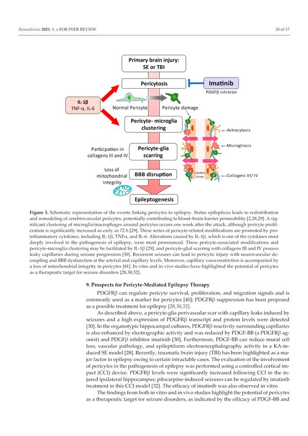
(Figure 3) [31].Biomedicines 2021, 9, 759 9 of 16
Figure 3. Schematic representation of the events linking pericytes to epilepsy. Status epilepticus leads
to redistribution and remodeling of cerebrovascular pericytes, potentially contributing to blood–brain
barrier permeability [2,28,29]. A significant clustering of microglia/macrophages around pericytes
occurs one week after the attack, although pericyte proliferation is significantly increased as early as
72 h [29]. These series of pericyte-related modifications are promoted by proinflammatory cytokines,
including IL-1β, TNFα, and IL-6. Alterations caused by IL-1β, which is one of the cytokines most
deeply involved in the pathogenesis of epilepsy, were most pronounced. These pericyte-associated
modifications and pericyte-microglia clustering may be facilitated by IL-1β [29], and pericyte-glial
scarring with collagens III and IV process leaky capillaries during seizure progression [30]. Recurrent
seizures can lead to pericytic injury with neurovascular decoupling and BBB dysfunction at the
arterial and capillary levels. Moreover, capillary vasoconstriction is accompanied by a loss of
mitochondrial integrity in pericytes [81]. In vitro and in vivo studies have highlighted the potential
of pericytes as a therapeutic target for seizure disorders [28,30,32].
9. Prospects for Pericyte-Mediated Epilepsy Therapy
PDGFRβ can regulate pericyte survival, proliferation, and migration signals and is
commonly used as a marker for pericytes [40]; PDGFRβ suppression has been proposed as
a possible treatment for epilepsy [28,30,32].
As described above, a pericyte-glia perivascular scar with capillary leaks induced by
seizures and a high expression of PDGFRβ transcript and protein levels were detected [30].
In the organotypic hippocampal cultures, PDGFRβ reactivity surrounding capillaries is
also enhanced by electrographic activity and was reduced by PDGF-BB (a PDGFRβ agonist)
and PDGFβ inhibitor imatinib [30]. Furthermore, PDGF-BB can reduce mural cell loss,
vascular pathology, and epileptiform electroencephalography activity in a KA-induced SE
model [28]. Recently, traumatic brain injury (TBI) has been highlighted as a major factor in
epilepsy owing to certain intractable cases. The evaluation of the involvement of pericytes
in the pathogenesis of epilepsy was performed using a controlled cortical impact (CCI)
device. PDGFRβ levels were significantly increased following CCI in the injured ipsilateral
hippocampus; pilocarpine-induced seizures can be regulated by imatinib treatment in this
CCI model [32]. The efficacy of imatinib was also observed in vitro.
The findings from both in vitro and in vivo studies highlight the potential of pericytes
as a therapeutic target for seizure disorders, as indicated by the efficacy of PDGF-BB and
imatinib in blocking PDGFRβ. However, both PDGFRβ and PDGF-BB are required for the
pericyte coating of the BBB in the developing CNS [38,41]. Under pathological conditions,Biomedicines 2021, 9, 759 10 of 16
mural cells in the immediate postacute phase (SE, ischemic stroke, and head trauma)
require support from the PDGFRβ activation [28]; hence, the inflammatory involvement of
PDGFRβ may be relevant in long-term progression as well as in chronic stages.
When considering the pharmacological modulation of pericyte signaling pathways as
a means of attenuating disease progression and capillary pathology, the impact of pericyte
modulation in the epileptic brain must consider the activation state of the glial cells and
the disease stage (e.g., acute vs. chronic) [29]. Further, considering the distinct functions of
PDGFRβ at different developmental stages, the timing of PDGFRβ inhibition needs to be
carefully studied; moreover, avoiding imatinib in the acute phase of the disease may be
considered. It remains debatable whether the changes in pericytes and accumulation of
microglia associated with PDGFR expression in this series of studies should be suppressed.
Transforming growth factor-beta 1 (TGFβ1) is a multifaceted cytokine in the brain
that plays a role in regulating cell proliferation, differentiation, survival, and scar for-
mation [82,83]. Since 1989, the possibility of PDGF-induced TGF-β signaling has been
suggested [84]; PDGFR-β and TGF-β with PDGFR-β might mediate the endothelial
cell/pericyte interaction to protect the BBB integrity [33]. The potential involvement
of TGF-β in epileptogenesis has been recognized from an experimental model showing
TGF-β upregulation as part of the inflammatory response [85]. Microarray analysis of
TGFβ1-stimulated human brain pericytes isolated from intractable TLE demonstrated
inhibition of pericyte proliferation and phagocytosis by TGFβ1 [27]. However, TGFβ1 also
enhanced the expression of IL-6, MMP-2, and NOX4, which can disrupt BBB functioning;
thus, these reactions caused by TGFβ1 might not lead to the treatment of the neurovascular
system [27].
Although the brain pericyte-derived TGF-β contributes to the upregulation of BBB
functions [86], suppression of TGFβ1 indicates improvement in epilepsy [87]. Losartan, an
angiotensin-type 1 receptor (AT1) antagonist, prevents phosphorylation of Smad proteins
of TGF-β signaling [88,89], which has demonstrated both neuroprotective and antineuroin-
flammatory effects [90–92].
These in vitro studies also suggest that human-derived pericytes are morphologically
altered by proinflammatory cytokines that induce apoptosis [29], indicating the potential of
targeting IFN-γ for pericyte-mediated epilepsy treatment [26]. IFN-γ is a central component
of the CNS inflammatory response and is secreted by microglia, astrocytes, endothelial
cells, and circulating immune cells [93–95]. This classical inflammatory mediator has
been implicated in CNS diseases, including epilepsy [96,97]. Altering the proportion of
microglial phenotypes via IFN-γ treatment improved the prognosis in a mouse model of
epilepsy [98].
Notably, in epileptiform conditions, IL-1β, a neurotoxic cytokine and one of the
cytokines chiefly involved in the pathogenesis of epilepsy, prominently contributes to the
morphological changes in the pericytes [29]. There is evidence that the IL-1/IL-1R1 axis
plays an important role in the inflammatory response in epilepsy, as presented by Vezzani
et al. in an excellent review [4,99]. IL-1β agonist, the IL-1 receptor antagonist (IL-1RA),
has already been tested for clinical application for epileptic syndromes using anakinra,
and has shown favorable clinical outcomes [100–103]. The use of anakinra on pericytes in
status epilepticus has not yet been investigated. To ensure the involvement of pericytes in
epilepsy, it is worthwhile to confirm that anakinra suppresses the morphological changes
in pericytes and reduces seizures.
Previous reports have demonstrated that inhibition of pericytes could have positive
effects of neuroprotection [26,28,30,32]; however, there is also a concern that the suppres-
sion of pericytes by TGFβ1 may not necessarily have a positive effect on the CNS [27].
Since TGFβ1 suppresses pericyte phagocytosis and reduces the expression of central leuko-
cyte trafficking chemokines and adhesion molecules while increasing the expression of
proinflammatory cytokines and enzymes that promote BBB disruption, a paradoxical
reaction has been reported [27]. The TGFβ1 response of pericytes may differ from theBiomedicines 2021, 9, 759 11 of 16
anti-inflammatory response of microglia [104–107]; therefore, further studies are required
to obtain any effect on this nonuniform response.
In the pathogenesis of epilepsy, pericytes adopt a phenotype that is neither solely
pro- nor anti-inflammatory [27]. Merely suppressing pericytes may not be sufficient to
improve the treatment of epilepsy, and it may be necessary to seek a treatment tailored
to the affected child in combination with various therapies that have been introduced in
recent reviews [108].
10. Conclusions
In this review, we present evidence for the substantive role of pericytes in the patho-
genesis of epilepsy. The roles of pericytes in maintaining BBB integrity, producing inflam-
matory secretions, and recruiting leukocytes highlights the potential role of pericytes in the
pathogenesis of epilepsy. Pericytes may also act as sensors of inflammatory processes in
the CNS and regulating them may lead to the development of novel therapies for epilepsy.
However, as there remains a lack of absolute molecular markers for pericytes, and since
pericytes originate from multiple cellular sources and vary in morphology, localization as
well as function in different tissues leaves several issues to be addressed. In addition, we
are unable to determine whether brain inflammation is an initiator or a consequence of a
systemic inflammatory process.
Several reports have suggested entry points that may also act as a basis for various
neurovascular therapies, including anakinra [100,101] and losartan [87], though the level
of evidence for both drugs is limited for the establishment of treatment for epilepsy. These
drugs provide an avenue for novel therapeutic, anti-inflammatory, or cerebrovascular repair
to mitigate epileptic pathophysiology. Unfortunately, definitive treatments for epilepsy
are currently lacking. BBB integrity and systemic peripheral inflammation may contribute
to epilepsy and hold potential for molecular biomarkers and targets in the treatment of
epilepsy. Moreover, human pluripotent stem cell-derived brain pericyte-like cells induced
BBB properties in BMECs, resulting in strengthening of the barrier and a reduction in
transcytosis [109]. These stem cell techniques could be applied to examine the possibility
of new strategies to selectively target pericytes and the role of pericytes in epilepsy more
specifically. Novel tools to control pericytes should be developed to target inflammatory
vascular-related processes during seizure progression or activity.
Author Contributions: Conceptualization, G.Y.; investigation, K.K. and S.M.; writing—original draft
preparation, G.Y.; writing—review and editing, F.T.; visualization, S.D.; supervision, Y.K. and H.K.
All authors have read and agreed to the published version of the manuscript.
Funding: This study was funded by the Kawano Masanori Memorial Foundation for Promotion of
Pediatrics in Japan under grant number 30-7 and the Japan Epilepsy Research Foundation under
grant number 20012. APC funded by the Japan Epilepsy Research Foundation.
Institutional Review Board Statement: We confirm that we have read the journal’s position on the
issues associated with ethical publication and affirm that this report is consistent with these guidelines.
Informed Consent Statement: Not applicable.
Data Availability Statement: The datasets generated and/or analyzed during the current study are
available at the PubMed database repository (https://pubmed.ncbi.nlm.nih.gov/, accessed on 31
May 2021).
Conflicts of Interest: The authors declare no conflict of interest.
References
1. Vezzani, A.; Balosso, S.; Ravizza, T. The role of cytokines in the pathophysiology of epilepsy. Brain. Behav. Immun. 2008, 22,
797–803. [CrossRef] [PubMed]
2. Milesi, S.; Boussadia, B.; Plaud, C.; Catteau, M.; Rousset, M.C.; De Bock, F.; Schaeffer, M.; Lerner-Natoli, M.; Rigau, V.; Marchi, N.
Redistribution of PDGFRβ cells and NG2DsRed pericytes at the cerebrovasculature after status epilepticus. Neurobiol. Dis. 2014,
71, 151–158. [CrossRef]Biomedicines 2021, 9, 759 12 of 16
3. Marchi, N.; Banjara, M.; Janigro, D. Blood-brain barrier, bulk flow, and interstitial clearance in epilepsy. J. Neurosci. Methods 2016,
260, 118–124. [CrossRef]
4. Vezzani, A.; Balosso, S.; Ravizza, T. Neuroinflammatory pathways as treatment targets and biomarkers in epilepsy. Nat. Rev.
Neurol. 2019, 15, 459–472. [CrossRef] [PubMed]
5. Löscher, W.; Friedman, A. Structural, Molecular, and Functional Alterations of the Blood-Brain Barrier during Epileptogenesis
and Epilepsy: A Cause, Consequence, or Both? Int. J. Mol. Sci. 2020, 21, 591. [CrossRef]
6. Nishibori, M.; Wang, D.; Ousaka, D.; Wake, H. High Mobility Group Box-1 and Blood-Brain Barrier Disruption. Cells 2020, 9, 2650.
[CrossRef] [PubMed]
7. Kovac, A.; Erickson, M.A.; Banks, W.A. Brain microvascular pericytes are immunoactive in culture: Cytokine, chemokine, nitric
oxide, and LRP-1 expression in response to lipopolysaccharide. J. Neuroinflamm. 2011, 8, 139. [CrossRef] [PubMed]
8. Jansson, D.; Rustenhoven, J.; Feng, S.; Hurley, D.; Oldfield, R.L.; Bergin, P.S.; Mee, E.W.; Faull, R.L.; Dragunow, M. A role for
human brain pericytes in neuroinflammation. J. Neuroinflamm. 2014, 11, 104. [CrossRef] [PubMed]
9. Matsumoto, J.; Takata, F.; Machida, T.; Takahashi, H.; Soejima, Y.; Funakoshi, M.; Futagami, K.; Yamauchi, A.; Dohgu, S.; Kataoka, Y.
Tumor necrosis factor-α-stimulated brain pericytes possess a unique cytokine and chemokine release profile and enhance
microglial activation. Neurosci. Lett. 2014, 578, 133–138. [CrossRef] [PubMed]
10. Rustenhoven, J.; Jansson, D.; Smyth, L.C.; Dragunow, M. Brain Pericytes as Mediators of Neuroinflammation. Trends Pharmacol.
Sci. 2017, 38, 291–304. [CrossRef]
11. Takata, F.; Dohgu, S.; Matsumoto, J.; Takahashi, H.; Machida, T.; Wakigawa, T.; Harada, E.; Miyaji, H.; Koga, M.; Nishioku, T.; et al.
Brain pericytes among cells constituting the blood-brain barrier are highly sensitive to tumor necrosis factor-α, releasing matrix
metalloproteinase-9 and migrating in vitro. J. Neuroinflamm. 2011, 8, 106. [CrossRef]
12. Machida, T.; Takata, F.; Matsumoto, J.; Takenoshita, H.; Kimura, I.; Yamauchi, A.; Dohgu, S.; Kataoka, Y. Brain pericytes are the
most thrombin-sensitive matrix metalloproteinase-9-releasing cell type constituting the blood-brain barrier in vitro. Neurosci. Lett.
2015, 599, 109–114. [CrossRef]
13. Matsumoto, J.; Dohgu, S.; Takata, F.; Machida, T.; Bölükbaşi Hatip, F.F.; Hatip-Al-Khatib, I.; Yamauchi, A.; Kataoka, Y. TNF-α-
sensitive brain pericytes activate microglia by releasing IL-6 through cooperation between IκB-NFκB and JAK-STAT3 pathways.
Brain Res. 2018, 1692, 34–44. [CrossRef] [PubMed]
14. Armulik, A.; Genové, G.; Mäe, M.; Nisancioglu, M.H.; Wallgard, E.; Niaudet, C.; He, L.; Norlin, J.; Lindblom, P.; Strittmatter, K.; et al.
Pericytes regulate the blood-brain barrier. Nature 2010, 468, 557–561. [CrossRef] [PubMed]
15. Armulik, A.; Genové, G.; Betsholtz, C. Pericytes: Developmental, physiological, and pathological perspectives, problems, and
promises. Dev. Cell 2011, 21, 193–215. [CrossRef] [PubMed]
16. Sweeney, M.D.; Zhao, Z.; Montagne, A.; Nelson, A.R.; Zlokovic, B.V. Blood-Brain Barrier: From Physiology to Disease and Back.
Physiol. Rev. 2019, 99, 21–78. [CrossRef]
17. Winkler, E.A.; Bell, R.D.; Zlokovic, B.V. Central nervous system pericytes in health and disease. Nat. Neurosci. 2011, 14, 1398–1405.
[CrossRef]
18. Stark, K.; Eckart, A.; Haidari, S.; Tirniceriu, A.; Lorenz, M.; von Brühl, M.L.; Gärtner, F.; Khandoga, A.G.; Legate, K.R.;
Pless, R.; et al. Capillary and arteriolar pericytes attract innate leukocytes exiting through venules and ‘instruct’ them with
pattern-recognition and motility programs. Nat. Immunol. 2013, 14, 41–51. [CrossRef]
19. Xu, D.; Robinson, A.P.; Ishii, T.; Duncan, D.S.; Alden, T.D.; Goings, G.E.; Ifergan, I.; Podojil, J.R.; Penaloza-MacMaster, P.;
Kearney, J.A.; et al. Peripherally derived T regulatory and gammadelta T cells have opposing roles in the pathogenesis of
intractable pediatric epilepsy. J. Exp. Med. 2018, 215, 1169–1186. [CrossRef] [PubMed]
20. Yamanaka, G.; Morichi, S.; Takamatsu, T.; Watanabe, Y.; Suzuki, S.; Ishida, Y.; Oana, S.; Yamazaki, T.; Takata, F.; Kawashima, H.
Links between Immune Cells from the Periphery and the Brain in the Pathogenesis of Epilepsy: A Narrative Review. Int. J. Mol.
Sci. 2021, 22, 4395. [CrossRef]
21. Fernández-Klett, F.; Potas, J.R.; Hilpert, D.; Blazej, K.; Radke, J.; Huck, J.; Engel, O.; Stenzel, W.; Genové, G.; Priller, J. Early
loss of pericytes and perivascular stromal cell-induced scar formation after stroke. J. Cereb. Blood Flow Metab. 2013, 33, 428–439.
[CrossRef]
22. Göritz, C.; Dias, D.O.; Tomilin, N.; Barbacid, M.; Shupliakov, O.; Frisén, J. A pericyte origin of spinal cord scar tissue. Science 2011,
333, 238–242. [CrossRef]
23. Reeves, C.; Pradim-Jardim, A.; Sisodiya, S.M.; Thom, M.; Liu, J.Y.W. Spatiotemporal dynamics of PDGFRβ expression in pericytes
and glial scar formation in penetrating brain injuries in adults. Neuropathol. Appl. Neurobiol. 2019, 45, 609–627. [CrossRef]
24. Rivera, F.J.; Hinrichsen, B.; Silva, M.E. Pericytes in Multiple Sclerosis. Adv. Exp. Med. Biol. 2019, 1147, 167–187. [CrossRef]
[PubMed]
25. Garbelli, R.; de Bock, F.; Medici, V.; Rousset, M.C.; Villani, F.; Boussadia, B.; Arango-Lievano, M.; Jeanneteau, F.; Daneman, R.;
Bartolomei, F.; et al. PDGFRβ(+) cells in human and experimental neuro-vascular dysplasia and seizures. Neuroscience 2015, 306,
18–27. [CrossRef]
26. Jansson, D.; Scotter, E.L.; Rustenhoven, J.; Coppieters, N.; Smyth, L.C.; Oldfield, R.L.; Bergin, P.S.; Mee, E.W.; Graham, E.S.;
Faull, R.L.; et al. Interferon-γ blocks signalling through PDGFRβ in human brain pericytes. J. Neuroinflamm. 2016, 13, 249.
[CrossRef] [PubMed]Biomedicines 2021, 9, 759 13 of 16
27. Rustenhoven, J.; Aalderink, M.; Scotter, E.L.; Oldfield, R.L.; Bergin, P.S.; Mee, E.W.; Graham, E.S.; Faull, R.L.; Curtis, M.A.;
Park, T.I.; et al. TGF-beta1 regulates human brain pericyte inflammatory processes involved in neurovasculature function. J.
Neuroinflamm. 2016, 13, 37. [CrossRef]
28. Arango-Lievano, M.; Boussadia, B.; De Terdonck, L.D.T.; Gault, C.; Fontanaud, P.; Lafont, C.; Mollard, P.; Marchi, N.; Jeanneteau, F.
Topographic Reorganization of Cerebrovascular Mural Cells under Seizure Conditions. Cell Rep. 2018, 23, 1045–1059. [CrossRef]
29. Klement, W.; Garbelli, R.; Zub, E.; Rossini, L.; Tassi, L.; Girard, B.; Blaquiere, M.; Bertaso, F.; Perroy, J.; de Bock, F.; et al.
Seizure progression and inflammatory mediators promote pericytosis and pericyte-microglia clustering at the cerebrovasculature.
Neurobiol. Dis. 2018, 113, 70–81. [CrossRef]
30. Klement, W.; Blaquiere, M.; Zub, E.; deBock, F.; Boux, F.; Barbier, E.; Audinat, E.; Lerner-Natoli, M.; Marchi, N. A pericyte-glia
scarring develops at the leaky capillaries in the hippocampus during seizure activity. Epilepsia 2019, 60, 1399–1411. [CrossRef]
[PubMed]
31. Prager, O.; Kamintsky, L.; Hasam-Henderson, L.A.; Schoknecht, K.; Wuntke, V.; Papageorgiou, I.; Swolinsky, J.; Muoio, V.;
Bar-Klein, G.; Vazana, U.; et al. Seizure-induced microvascular injury is associated with impaired neurovascular coupling and
blood-brain barrier dysfunction. Epilepsia 2019, 60, 322–336. [CrossRef] [PubMed]
32. Sakai, K.; Takata, F.; Yamanaka, G.; Yasunaga, M.; Hashiguchi, K.; Tominaga, K.; Itoh, K.; Kataoka, Y.; Yamauchi, A.; Dohgu, S.
Reactive pericytes in early phase are involved in glial activation and late-onset hypersusceptibility to pilocarpine-induced seizures
in traumatic brain injury model mice. J. Pharmacol. Sci. 2021, 145, 155–165. [CrossRef]
33. Sweeney, M.D.; Ayyadurai, S.; Zlokovic, B.V. Pericytes of the neurovascular unit: Key functions and signaling pathways. Nat.
Neurosci. 2016, 19, 771–783. [CrossRef]
34. Cheng, J.; Korte, N.; Nortley, R.; Sethi, H.; Tang, Y.; Attwell, D. Targeting pericytes for therapeutic approaches to neurological
disorders. Acta Neuropathol. 2018, 136, 507–523. [CrossRef]
35. Rouget, C. Note sur le developpement de la tunique contractile des vaisseaux. C. R. L’académie Sci. 1874, 59, 559–562.
36. Zimmermann, K.W. Der feinere bau der blutcapillares. Z. Anat. Entwicklungsgesch. 1923, 68, 3–109. [CrossRef]
37. Ando, K.; Fukuhara, S.; Izumi, N.; Nakajima, H.; Fukui, H.; Kelsh, R.N.; Mochizuki, N. Clarification of mural cell coverage of
vascular endothelial cells by live imaging of zebrafish. Development 2016, 143, 1328–1339. [CrossRef] [PubMed]
38. Winkler, E.A.; Bell, R.D.; Zlokovic, B.V. Pericyte-specific expression of PDGF beta receptor in mouse models with normal and
deficient PDGF beta receptor signaling. Mol. Neurodegener. 2010, 5, 32. [CrossRef] [PubMed]
39. Thomsen, M.S.; Routhe, L.J.; Moos, T. The vascular basement membrane in the healthy and pathological brain. J. Cereb. Blood
Flow Metab. 2017, 37, 3300–3317. [CrossRef] [PubMed]
40. Hellström, M.; Kalén, M.; Lindahl, P.; Abramsson, A.; Betsholtz, C. Role of PDGF-B and PDGFR-beta in recruitment of vascular
smooth muscle cells and pericytes during embryonic blood vessel formation in the mouse. Development 1999, 126, 3047–3055.
[CrossRef]
41. Lindahl, P.; Johansson, B.R.; Levéen, P.; Betsholtz, C. Pericyte loss and microaneurysm formation in PDGF-B-deficient mice.
Science 1997, 277, 242–245. [CrossRef]
42. Nikolakopoulou, A.M.; Montagne, A.; Kisler, K.; Dai, Z.; Wang, Y.; Huuskonen, M.T.; Sagare, A.P.; Lazic, D.; Sweeney, M.D.;
Kong, P.; et al. Pericyte loss leads to circulatory failure and pleiotrophin depletion causing neuron loss. Nat. Neurosci. 2019, 22,
1089–1098. [CrossRef]
43. Bhattacharya, A.; Kaushik, D.K.; Lozinski, B.M.; Yong, V.W. Beyond barrier functions: Roles of pericytes in homeostasis and
regulation of neuroinflammation. J. Neurosci. Res. 2020, 98, 2390–2405. [CrossRef]
44. Vanlandewijck, M.; He, L.; Mäe, M.A.; Andrae, J.; Ando, K.; Del Gaudio, F.; Nahar, K.; Lebouvier, T.; Laviña, B.; Gouveia, L.; et al.
A molecular atlas of cell types and zonation in the brain vasculature. Nature 2018, 554, 475–480. [CrossRef]
45. Attwell, D.; Mishra, A.; Hall, C.N.; O’Farrell, F.M.; Dalkara, T. What is a pericyte? J. Cereb. Blood Flow Metab. 2016, 36, 451–455.
[CrossRef] [PubMed]
46. Marques, S.; van Bruggen, D.; Vanichkina, D.P.; Floriddia, E.M.; Munguba, H.; Väremo, L.; Giacomello, S.; Falcão, A.M.; Meijer, M.;
Björklund, Å.K.; et al. Transcriptional Convergence of Oligodendrocyte Lineage Progenitors during Development. Dev. Cell 2018,
46, 504–517.e7. [CrossRef]
47. Bondjers, C.; He, L.; Takemoto, M.; Norlin, J.; Asker, N.; Hellström, M.; Lindahl, P.; Betsholtz, C. Microarray analysis of blood
microvessels from PDGF-B and PDGF-Rbeta mutant mice identifies novel markers for brain pericytes. FASEB J. 2006, 20,
1703–1705. [CrossRef]
48. Iacobaeus, E.; Sugars, R.V.; Törnqvist Andrén, A.; Alm, J.J.; Qian, H.; Frantzen, J.; Newcombe, J.; Alkass, K.; Druid, H.;
Bottai, M.; et al. Dynamic Changes in Brain Mesenchymal Perivascular Cells Associate with Multiple Sclerosis Disease Duration,
Active Inflammation, and Demyelination. Stem. Cells Transl. Med. 2017, 6, 1840–1851. [CrossRef] [PubMed]
49. Zeisel, A.; Hochgerner, H.; Lönnerberg, P.; Johnsson, A.; Memic, F.; van der Zwan, J.; Häring, M.; Braun, E.; Borm, L.E.;
La Manno, G.; et al. Molecular Architecture of the Mouse Nervous System. Cell 2018, 174, 999–1014.e22. [CrossRef]
50. Damisah, E.C.; Hill, R.A.; Tong, L.; Murray, K.N.; Grutzendler, J. A fluoro-Nissl dye identifies pericytes as distinct vascular mural
cells during in vivo brain imaging. Nat. Neurosci. 2017, 20, 1023–1032. [CrossRef] [PubMed]
51. Sweeney, M.D.; Sagare, A.P.; Zlokovic, B.V. Blood-brain barrier breakdown in Alzheimer disease and other neurodegenerative
disorders. Nat. Rev. Neurol. 2018, 14, 133–150. [CrossRef]Biomedicines 2021, 9, 759 14 of 16
52. Navarro, R.; Compte, M.; Álvarez-Vallina, L.; Sanz, L. Immune Regulation by Pericytes: Modulating Innate and Adaptive
Immunity. Front. Immunol. 2016, 7, 480. [CrossRef]
53. Nehmé, A.; Edelman, J. Dexamethasone inhibits high glucose-, TNF-alpha-, and IL-1beta-induced secretion of inflammatory and
angiogenic mediators from retinal microvascular pericytes. Investig. Ophthalmol. Vis. Sci. 2008, 49, 2030–2038. [CrossRef]
54. Fabene, P.F.; Bramanti, P.; Constantin, G. The emerging role for chemokines in epilepsy. J. Neuroimmunol. 2010, 224, 22–27.
[CrossRef] [PubMed]
55. Vezzani, A.; Aronica, E.; Mazarati, A.; Pittman, Q.J. Epilepsy and brain inflammation. Exp. Neurol. 2013, 244, 11–21. [CrossRef]
[PubMed]
56. Glass, C.K.; Saijo, K.; Winner, B.; Marchetto, M.C.; Gage, F.H. Mechanisms underlying inflammation in neurodegeneration. Cell
2010, 140, 918–934. [CrossRef] [PubMed]
57. Kim, J.Y.; Kim, N.; Yenari, M.A. Mechanisms and potential therapeutic applications of microglial activation after brain injury.
CNS Neurosci. Ther. 2015, 21, 309–319. [CrossRef]
58. Gutierrez, H.; Hale, V.A.; Dolcet, X.; Davies, A. NF-kappaB signalling regulates the growth of neural processes in the developing
PNS and CNS. Development 2005, 132, 1713–1726. [CrossRef] [PubMed]
59. Bodnar, R.J.; Yang, T.; Rigatti, L.H.; Liu, F.; Evdokiou, A.; Kathju, S.; Satish, L. Pericytes reduce inflammation and collagen
deposition in acute wounds. Cytotherapy 2018, 20, 1046–1060. [CrossRef]
60. Minutti, C.M.; Modak, R.V.; Macdonald, F.; Li, F.; Smyth, D.J.; Dorward, D.A.; Blair, N.; Husovsky, C.; Muir, A.;
Giampazolias, E.; et al. A Macrophage-Pericyte Axis Directs Tissue Restoration via Amphiregulin-Induced Transforming Growth
Factor Beta Activation. Immunity 2019, 50, 645–654.e6. [CrossRef]
61. Gaceb, A.; Özen, I.; Padel, T.; Barbariga, M.; Paul, G. Pericytes secrete pro-regenerative molecules in response to platelet-derived
growth factor-BB. J. Cereb. Blood Flow Metab. 2018, 38, 45–57. [CrossRef]
62. Ishitsuka, K.; Ago, T.; Arimura, K.; Nakamura, K.; Tokami, H.; Makihara, N.; Kuroda, J.; Kamouchi, M.; Kitazono, T. Neurotrophin
production in brain pericytes during hypoxia: A role of pericytes for neuroprotection. Microvasc. Res. 2012, 83, 352–359. [CrossRef]
[PubMed]
63. Liwnicz, B.H.; Leach, J.L.; Yeh, H.S.; Privitera, M. Pericyte degeneration and thickening of basement membranes of cerebral
microvessels in complex partial seizures: Electron microscopic study of surgically removed tissue. Neurosurgery 1990, 26, 409–420.
[CrossRef]
64. Van Vliet, E.A.; da Costa Araújo, S.; Redeker, S.; van Schaik, R.; Aronica, E.; Gorter, J.A. Blood-brain barrier leakage may lead to
progression of temporal lobe epilepsy. Brain 2007, 130, 521–534. [CrossRef]
65. Marchi, N.; Angelov, L.; Masaryk, T.; Fazio, V.; Granata, T.; Hernandez, N.; Hallene, K.; Diglaw, T.; Franic, L.; Najm, I.; et al.
Seizure-promoting effect of blood-brain barrier disruption. Epilepsia 2007, 48, 732–742. [CrossRef]
66. Marchi, N.; Granata, T.; Ghosh, C.; Janigro, D. Blood-brain barrier dysfunction and epilepsy: Pathophysiologic role and
therapeutic approaches. Epilepsia 2012, 53, 1877–1886. [CrossRef]
67. Uprety, A.; Kang, Y.; Kim, S.Y. Blood-brain barrier dysfunction as a potential therapeutic target for neurodegenerative disorders.
Arch. Pharm. Res. 2021, 44, 487–498. [CrossRef] [PubMed]
68. Ivens, S.; Kaufer, D.; Flores, L.P.; Bechmann, I.; Zumsteg, D.; Tomkins, O.; Seiffert, E.; Heinemann, U.; Friedman, A. TGF-beta
receptor-mediated albumin uptake into astrocytes is involved in neocortical epileptogenesis. Brain 2007, 130, 535–547. [CrossRef]
69. Weissberg, I.; Wood, L.; Kamintsky, L.; Vazquez, O.; Milikovsky, D.Z.; Alexander, A.; Oppenheim, H.; Ardizzone, C.; Becker, A.;
Frigerio, F.; et al. Albumin induces excitatory synaptogenesis through astrocytic TGF-β/ALK5 signaling in a model of acquired
epilepsy following blood-brain barrier dysfunction. Neurobiol. Dis. 2015, 78, 115–125. [CrossRef]
70. Pieper, C.; Marek, J.J.; Unterberg, M.; Schwerdtle, T.; Galla, H.J. Brain capillary pericytes contribute to the immune defense in
response to cytokines or LPS in vitro. Brain Res. 2014, 1550, 1–8. [CrossRef] [PubMed]
71. Galea, I.; Bernardes-Silva, M.; Forse, P.A.; van Rooijen, N.; Liblau, R.S.; Perry, V.H. An antigen-specific pathway for CD8 T cells
across the blood-brain barrier. J. Exp. Med. 2007, 204, 2023–2030. [CrossRef]
72. Fabene, P.F.; Mora, G.N.; Martinello, M.; Rossi, B.; Merigo, F.; Ottoboni, L.; Bach, S.; Angiari, S.; Benati, D.; Chakir, A.; et al. A role
for leukocyte-endothelial adhesion mechanisms in epilepsy. Nat. Med. 2008, 14, 1377–1383. [CrossRef] [PubMed]
73. Ravizza, T.; Gagliardi, B.; Noe, F.; Boer, K.; Aronica, E.; Vezzani, A. Innate and adaptive immunity during epileptogenesis and
spontaneous seizures: Evidence from experimental models and human temporal lobe epilepsy. Neurobiol. Dis. 2008, 29, 142–160.
[CrossRef] [PubMed]
74. Marchi, N.; Teng, Q.; Ghosh, C.; Fan, Q.; Nguyen, M.T.; Desai, N.K.; Bawa, H.; Rasmussen, P.; Masaryk, T.K.; Janigro, D.
Blood-brain barrier damage, but not parenchymal white blood cells, is a hallmark of seizure activity. Brain Res. 2010, 1353,
176–186. [CrossRef]
75. Varvel, N.H.; Neher, J.J.; Bosch, A.; Wang, W.; Ransohoff, R.M.; Miller, R.J.; Dingledine, R. Infiltrating monocytes promote brain
inflammation and exacerbate neuronal damage after status epilepticus. Proc. Natl. Acad. Sci. USA 2016, 113, E5665–E5674.
[CrossRef]
76. Broekaart, D.W.M.; Anink, J.J.; Baayen, J.C.; Idema, S.; de Vries, H.E.; Aronica, E.; Gorter, J.A.; van Vliet, E.A. Activation of the
innate immune system is evident throughout epileptogenesis and is associated with blood-brain barrier dysfunction and seizure
progression. Epilepsia 2018, 59, 1931–1944. [CrossRef] [PubMed]You can also read