UPDATE ON THE BETHESDA SYSTEM FOR REPORTING THYROID CYTOLOGY - ESTHER ROSSI MD PHD MIAC DIVISION OF ANATOMIC PATHOLOGY AND CYTOLOGY CATHOLIC ...
←
→
Page content transcription
If your browser does not render page correctly, please read the page content below
UPDATE ON THE
Bethesda system for
 reporting thyroid
     cytology

    Esther Rossi MD PhD MIAC
 Division of Anatomic Pathology
           and Cytology
Catholic University of Sacred Heart
             Rome, Italy
Present
< Current State and Global Impact of TBSRTC >

USA BETHESDA              AUSTRALASIA            JAPAN THYROID
                                                                                    CLASSIFICATION         ASSOCIATION 2013
     UK RCPath 2015                   ITALY 2014          2008                      2014
Diagnostic category               Diagnostic category     Terminology               Categories             Terminology

Thy1/Thy1c
Non-diagnostic for cytological    TIR 1: Non-diagnostic   I. Non-diagnostic                                Inadequate (non
diagnosis                         TIR 1C: Cystic                                        Non-diagnostic     diagnostic)
Unsatisfactory, consistent with
cyst

Thy2/Thy2c                        TIR 2: Non-                                               Benign         Normal or benign
                                                          II. Benign
Non-neoplastic, benign cystic     malignant/benign

Thy 3a                                                    III. Atypia of                                   Indeterminate
Neoplasm possible – atypia        TIR 3A: Low-risk        undetermined              Indeterminate or       A. Follicular
present                           indeterminate lesion    significance (AUS) or     Follicular lesion of        Neoplasm
                                  (LRIL)                  follicular lesion u.s.    undetermined           •    A1 favor benign
                                                          (FLUS)                    significance           •    A2 border-line
                                                                                                           •    A3 favor malignant
                                                                                                           B. Others (atypia in
                                                                                                                non-follicular
Thy3f                          TIR 3B: High-risk          IV. Follicular neoplasm
                                                                                                                patterned lesions)
Neoplasm possible - suggesting indeterminate lesion       or suspicious for a       Suggestive of a
follicular neoplasm            (HRIL)                     follicular neoplasm       follicular neoplasm

                                                                                    Suspicious of          Malignancy suspected
Thy 4                             TIR 4: Suspicious of    V. Suspicious of          malignancy
Suspicious of malignancy          malignancy              malignancy

Thy5 Malignant                    TIR 5: Malignant        VI. Malignant
                                                                                        Malignant                     3
                                                                                                             Malignancy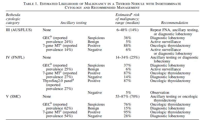
TBSRTC
Used world over
2011 CAP Study – 77% of Labs in the US*
540,000 links on Google
115,000 citations on Google Scholar
1,751 articles on PubMed (unique)
*Auger M, et al, Arch Pathol Lab Med 2013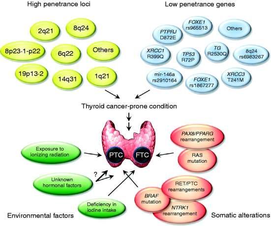
TBSRTC – Diagnostic Categories Nondiagnostic/Unsatisfactory Benign AUS/FLUS * FN/SFN * Suspicious for Malignancy Malignant * Why two names?
Definitions, criteria, explanatory notes Over 40 contributing authors Digital Image Atlas – 170 pages http://www.papsociety.org 200 color images $40 Chinese, Spanish, Japanese, Turkish

TBSRTC - PROBABILISTIC APPROACH AND
     RELATIONSHIP TO CLINICAL ALGORITHMS

                             ROM (%)         Management

Nondiagnostic                   1-4         Repeat FNA with US

Benign                         0.5-5.5          Follow-up
AUS/FLUS                    ~5-10 (15-25)      Repeat FNA
FN/Suspicious for a FN         15-30            Lobectomy
Suspicious for Malignancy      60-77        Lobectomy or total
                                              thyroidectomy
Malignant                      96-99        Total thyroidectomy
FUTURE
TBSRTC II
March 2018
200 pages, 200 figures
Issues and Recommendations for possible
modifications in TBSRTC 2 (Chapter based)
Recent advances with potential impact on TBSRTC
TBSRTC PANEL Co-Leaders Bill Faquin (USA) Marc Pusztaszeri (Switzerland) Esther Diana Rossi (Italy) Members Manon Auger (Canada) Zubair Baloch (USA) Justin Bishop (USA) Massimo Bongiovanni (Switzerland) Ashish Chandra (UK) Guido Fadda (Italy) M. Hirokawa (Japan) Soonwon Hong (Korea) Kennichi Kakudo (Japan) Jeffrey Krane (USA) Ritu Nayar (USA) Sareh Parangi (USA) Beatrix Cochand-Priollet (France) Fernando Schmitt (Luxembourg)
Tasks of TBSRTC Panel:

•Pubmed literature review of thyroid
cytology from 2010 to present

•Divided efforts into subgroups
corresponding to each of the 6
TBSRTC diagnostic categories
•2-6 panel members per subgroup

•Email discussions among subgroup
members, and face to face meeting at
USCAP in Seattle

•IAC Symposium presentation –
Yokohama, Japan
•Publication of manuscript detailing
the panel’s consensus
recommendations for TBSRTC II
THE BETHESDA SYSTEM FOR REPORTING THYROID
CYTOPATHOLOGY: PROPOSED MODIFICATIONS AND
  UPDATES FOR THE SECOND EDITION FROM AN
           INTERNATIONAL PANEL
What are the prospects for the second edition of TBSRTC Atlas?
•IAC Symposium organized by Drs. Syed Ali
and Philippe Vielh to address past, present,
& future of TBSRTC
•Many advances, large amount of published
literature, and new questions for TBSRTC:
•Reporting of selected uncommon
entities (e.g. parathyroid adenoma)
•Refinements to the ROM for each
corresponding diagnostic category
•NIFTP and its impact on the
indeterminate categories of TBSRTC
•2015 ATA Guidelines – impact on
clinical management algorithms
•Diagnostic category names – continue
with multiple options or reduce to one?
•Quality control: laboratory metrics for
monitoring
•Many more diagnostic category-
specific issues…THE NONDIAGNOSTIC THYROID FNA:
CRITERIA AND FOLLOW-UP
Rationale
Adequacy criteria
Frequency of Nondiagnostic
Management
Possible Future ScenarioFACTORS CONTRIBUTORY TO
INADEQUATETHYROID FNA
• Inadequate history
• Inadequate specimen
Quantity and quality of representative cells
• Suboptimal preparation
• Interpretative and diagnostic errorsRATIONALE FOR A NON-DIAGNOSTIC CATEGORY:
WHY IS ADEQUACY A PROBLEM IN THYROID FNA?
1) TO REDUCE “FALSE NEGATIVE” DIAGNOSES ARISING FROM
INSUFFICIENT SAMPLING
If adequacy criteria work
Malignancy rate in
Malignancy rate in
“Adequate” / Benign
“Inadequate” cases Greater than
cases
2) SAMPLES WITH LOW CELLULARITY
cystic lesions are common
Poor quality sampling by inexperienced aspirators
Vascular components
Colloid-rich nodulesADEQUACY
CRITERIA
• Criteria are not evidence-based
• Similar in all the current classification systems:
Bethesda
British
Italian
JapaneseNONDIAGNOSTIC: BETHESDA CRITERIA
DEFINITION: A specimen is Nondiagnostic if it fails to meet the adequacy
criterion
ADEQUACY CRITERION: At least 6 groups, each with at least 10 benign-
appearing, well-visualized follicular cells
Same criteria irrespective of cytological preparation ( smears, LBC, Cell-block)
“Cyst fluid ( macrophages) only” cases included as a subset
EXCEPTION Thyroiditis = BENIGN
Abundant Colloid= BENIGN
Any atypiaSome attempts of different epithelial
quantification
• Frost et al. Cancer 1998;84:17-25
– ThinPrep - 6 clusters of 10 epithelial cells
• 5% inadequate rate
• Renshaw. Am J Clin Pathol 2002;118:518-521
– At least 10 follicular cells lacking atypia with no Hürthle cells
• Michael et. al. Diagn Cytopathol 2007; 35:792-797
– ThinPrep cases
• At least 200 cells
• Renshaw. Diagn Cytopathol 2010
• 30 epithelial cells lacking atypia with no Hürthle cellsNONDIAGNOSTIC* RATES
Study authors Number of Nondiagnostic Rate
nodules biopsied (%)
Grant et al, 1988 8219 21
Yoder, et al, 2006 1043 5
Yassa et al, 2007 3589 13
Yang et al, 2007 4703 10
Theoharis et al, 2009 3037 12
Nayar et al, 2009 5194 5
Marchevsky et al, 2010 879 13
Hryhorczuk et al, 2011 1344 22
Renshaw, 2011 7089 24
Al Maqbali et al, 2012 1657 16
Coorough et al, 2013 4286 6
Ferreira et al, 2014 15,292 7
Range 5-24%
* defined using Bethesda System criteriaBTSRTC:MANAGEMENT OF ND FNA
Repeat FNA with ultrasound
guidance no sooner than 3 month
later
Partially cystic nodules that are
repeatedly ND need close
observation or surgical excision[A12] Nondiagnostic cytology ■ RECOMMENDATION 10 A) For a nodule with an initial nondiagnostic cytology result, FNA should be repeated with US guidance and, if available, on-site cytologic evaluation (Strong recommendation, Moderate-quality evidence) B) Repeatedly nondiagnostic nodules without a high suspicion sonographic pattern require close observation or surgical excision for histopathologic diagnosis (Weak recommendation, Low-quality evidence) C) Surgery should be considered for histopathologic diagnosis if the cytologically nondiagnostic nodule has a high suspicion sonographic pattern, growth of the nodule (greater than 20% in two dimensions) is detected during ultrasound surveillance, or clinical risk factors for malignancy are present (Weak recommendation, Low-quality evidence)
THE FUTURE OF
ADEQUACY
FROM THE COMPOSITE OUTLINE OF THE
KEY ISSUES IDENTIFIED BY OUR TBSRTC
(MAILS AND USCAP16)
Cystic lesions:
Should still be reported as Non-diagnostic with an explanatory note. Update management
according to revised ATA guidelines. The sample reports and the explanatory notes in TBSRTC
regarding cystic lesions are adequate.
Repeat FNA after ND result:
The wait time for repeating an FNA after a ND result can be less than 3 months (as suggested by
the revised ATA guidelines). However, it should be explained that reactive atypia and cellular
changes may be present if the delay to repeat FNA is shortened.
Adequacy criteria and preparation method:
Clarification is needed pertaining to the specific adequacy criteria for smears vs. liquid based
preparations: ThinPrep and Surepath alone or in combination with smears.BENIGN NODULES
CATEGORY
                             « BENIGN »

•   The most important category in terms of percentage of
    nodules
•   percentage should be around 90-92% of all nodules
•   Mostly concerns 60-70% of all nodules

•   With some variations depending on:
                  1.  the sampler performance
               2.    the cytopathologist training
            3.     The local epidemiological data
Literature Results
Authors Cases Non Benign FLUS or FN/ SM Malignant
Number diagn AUS FNHC
Cochand-Priollet B 2210 14.3% 65.5% 11% 4.9% 2.3% 2%
et al 2012 (23.6%) (15.2%) (58.7%) (100%)
Mastorakis et al 500 NA 49% 9.4% 1.2% 10.6% 26.8%
Cytopathology (23.4%) (96%) (100%)
2012
500 NA 72.2% 5% 2.2% 3.2% 12.2%
(8%) (87.5%) (100%)
Lacoste-Collin L 1317 31.6% 48% 7.8% 7% 3% 2.6%
et al 2012 (18.5%) (22.2%) (55.6%) (100%)
Bongiovanni 7686 2% 54.7% 6.3% 25.3% 6.3% 4%
et al 2012
(14.4%) 32.1% (74.9%) 99.4%
Park JH et al 2014 1730 13.3% 40.6% 9.1% 0.4% 19.3% 17.3%
(35.3%) (5.6%) (69%) (50%) (98.7%) (98.9%)
BethesdaBTSRTC:MANAGEMENT OF BN FNA
• Follow-up for 6-18 month
intervals and for at least 3-5
years
• Repeat FNA for nodules with
significant growth or US
abnormalitiesRECOMMENDATION 11
If the nodule is benign on cytology, further immediate diagnostic studies or treatment are not required (Strong
recommendation, High-quality evidence)
RECOMMENDATION 23
Given the low false negative rate of US-guided FNA cytology and the higher yield of missed malignancies based upon
nodule sonographic pattern rather than growth, the follow up of thyroid nodules with benign cytology diagnoses should be
determined by risk stratification based upon ultrasound pattern.
A)Nodules with high suspicion US pattern: repeat US and US-guided FNA within 12 months (Strong recommendation,
Moderate-quality evidence)
B) Nodules with low to intermediate suspicion US pattern: repeat US at 12-24 months.
If sonographic evidence of growth (20% increase in at least two nodule dimensions with a minimal increase of 2 mm or
more than a 50% change in volume) or development of new suspicious sonographic features, the FNA could be
repeated or observation continued with repeat US, with repeat FNA in case of continued growth (Weak
recommendation, Low-quality evidence).
C) Nodules with very low suspicion US pattern (including spongiform nodules): the utility of surveillance US and
assessment of nodule growth as an indicator for repeat FNA to detect a missed malignancy is limited. If US is repeated, it
should be done at > 24 months (No Weak recommendation, Insufficient Low-quality evidence).CONCLUSION • No major changes were suggested for this category • ROM: Several recent studies have confirmed that the ROM is very low for this category (≤ 3%) • Diagnoses: More LBC imagines IgG4 thyroïditis should be included in the “thyroïditis” chapter • Follow-up: Risk stratification based upon ultrasound pattern (ATA 2015 revised guidelines) can be used to guide follow-up of thyroid nodules with benign cytology
AUS/FLUS
USA BETHESDA             AUSTRALASIA            JAPAN THYROID
                                                                                     CLASSIFICATION         ASSOCIATION 2013
     UK RCPath 2015                     ITALY 2014          2008                     2014
DIAGNOSTIC CATEGORY DIAGNOSTIC                              TERMINOLOGY              CATEGORIES             TERMINOLOGY
                    CATEGORY

Thy1/Thy1c
Non-diagnostic for cytological      TIR 1: Non-diagnostic   I. Non-diagnostic                               Inadequate (non
diagnosis                           TIR 1C: Cystic                                       Non-diagnostic     diagnostic)
Unsatisfactory, consistent with
cyst

Thy2/Thy2c                          TIR 2: Non-                                              Benign         Normal or benign
                                                            II. Benign
Non-neoplastic, benign cystic       malignant/benign

Thy 3a                                                      III. Atypia of                                  Indeterminate
Neoplasm possible – atypia          TIR 3A: Low-risk        undetermined             Indeterminate or       A. Follicular
present                             indeterminate lesion    significance (AUS) or    Follicular lesion of        Neoplasm
                                    (LRIL)                  follicular lesion u.s.   undetermined           •    A1 favor benign
                                                            (FLUS)                   significance           •    A2 border-line
                                                                                                            •    A3 favor malignant
                                                                                                            B. Others (atypia in
Thy3f                             Indeterminate lesion
                               TIR 3B: High-risk            or neoplasm
                                                  IV. Follicular indeterminate                                   non-follicular
                                                                                                                 patterned lesions)
                                             pathologist??
Neoplasm possible - suggesting indeterminate lesion
                                                  or suspicious for a   Suggestive of a
follicular neoplasm            (HRIL)                       follicular neoplasm      follicular neoplasm

                                                                                     Suspicious of          Malignancy suspected
Thy 4                               TIR 4: Suspicious of    V. Suspicious of         malignancy
Suspicious of malignancy            malignancy              malignancy

Thy5 Malignant                      TIR 5: Malignant        VI. Malignant
                                                                                         Malignant                    34
                                                                                                              Malignancy
Thyroid and
atypia in TBSRTC
Reporting AUS/FLUS
AUS/FLUS both options to report
this category
“Architectural” vs “Cytologic” atypia
Criteria (Describe 9 scenarios)
“Indeterminate” or Grey Zone in Recommended clarification of
Thyroid Cytopathology category
A narrative comment/ differential
Morphology and outcome differ diagnosis
from SFN/FNs and SMs
Avoid “buzz words” overlapping
with SM or PM categories
Not all Atypical Thyroid FNA’s
require surgical excision Recommended TBSRTC Rate
◦ Approx. 7% of all thyroid FNAREPORTING RATES FOR TBSRTC CATEGORIES Overall ~ 8-10% seems to be the experience in labs with high volume experience
Which are the
criteria used to
diagnose an FNA as
“AUS/FLUS?
AUS/FLUS

  1. CYTOLOGIC ATYPIA

        And/Or

2. ARCHITECTURAL ATYPIA
NUCLEAR ATYPIA IN THYROID
Nuclear pleomorphism
Nuclear enlargement
Hyperchromasia
Prominent nucleoli
Nuclear Anaplasia
Changes in Nuclear Chromatin
Reactive
Neoplastic
Nuclear atypia in benign thyroid lesionsARCHITECTURAL ATYPIA Papillary Formations Microfollicles
Am J Clin Pathol 2011; 136: 572-577

Architectural atypia: 24% risk of malignancy

Cytologic atypia: 50% risk of malignancy
How to manage AUS/FLUS lesion?

What to do with AUS/FLUS nodule?
ATA guidelines 2015
MANAGEMENT -AUS/FLUS
1.    Repeat FNA
     ◦ is a suitable follow-up option in ATA 2015
     ◦ limited cellularity contributes to the initial AUS/FLUS interpretation
     ◦ Need clinical correlation (US findings, TSH/antibody titer correlation, etc.)

2.    Surgery
     ◦ Generally discouraged for initial AUS/FLUS
     ◦ Reasonable option for second AUS

3.    Molecular testing
     ◦ Acceptable consideration for AUS/FLUS
     ◦ Reflexive molecular testing is not mandated for all AUS/FLUS

ATA 2015- All clinical, radiologic, pathologic, and molecular findings must
    be integrated for the most informed, accurate, and individualized
                                 assessment
WHAT’S AGAIN?????
Changes in
                     Histopathology
                     Nomenclature

Changes in the Implied Risk of Malignancy for TBSRTC
                         Categories
                         AUS/FLUS
           Suspicious for Follicular Neoplasm
       Suspicious for Malignancy – 50% decrease
   (Strickland et al. Thyroid 2015 & Faquin et al. Cancer
                     Cytopathology 2015)
Non-Invasive Follicular Thyroid Neoplasm
with Papillary-Like Nuclear Features (NIFTP)
Cancer Cytopathology,
2015.THE BETHESDA SYSTEM FOR REPORTING
THYROID CYTOPATHOLOGY: PROPOSED
MODIFICATIONS AND UPDATES FOR THE SECOND
EDITION FROM AN INTERNATIONAL PANEL
The panel endorses AUS/FLUS
Widely accepted and included in ATA 2015
Only one term would be selected by a laboratory
Recommendations for subclassifying AUS/FLUS
( importance of nuclear atypia)THE BETHESDA SYSTEM FOR REPORTING THYROID
CYTOPATHOLOGY: PROPOSED MODIFICATIONS AND
UPDATES FOR THE SECOND EDITION FROM AN
INTERNATIONAL PANEL
Common patterns include: Architectural Atypia
Nuclear atypia
Oncocytic features
Compromised samples lacking any Atypia should be
classified as NDFOLLICULAR NEOPLASMS/SUSPICIOUS
FOR FOLLICULAR NEOPLASM
The panel favors the use of one term
Widely accepted and included in ATA 2015
Only one term would be selected by a laboratory
Current diagnostic criteria could be further definedTHE BETHESDA SYSTEM FOR REPORTING THYROID
CYTOPATHOLOGY: PROPOSED MODIFICATIONS AND
UPDATES FOR THE SECOND EDITION FROM AN
INTERNATIONAL PANEL
Due to NIFTPs, a follicular patterned lesion with nuclear
atypia can be classified as FN rather than SM
Long-established standard of care is diagnostic surgical
excision
ATA 2015 guidelines provide the option of molecular testingFOLLICULAR NEOPLASM, HURTHLE CELL TYPE/
SUSPICIOUS FOR A FOLLICULAR NEOPLASM,
HURTHLE CELL TYPE
Panel Recommendations:
Two names not ideal but accepted due to current use
Use of term “oncocytic” rather than “Hurthle cell” is
preferred to coordinate with WHO terminologyFOLLICULAR NEOPLASM, HURTHLE CELL TYPE/ SUSPICIOUS FOR A
FOLLICULAR NEOPLASM, HURTHLE CELL TYPE
Panel Recommendations:
Rare cases reported containing abundant colloid
Can abundant colloid exclude oncocytic (Hürthle
cell) carcinoma in thyroid fine needle aspiration?
Cytohistological correlation of 127 oncocytic
(Hürthle cell) lesions.
Yang GC1, Schreiner AM, Sun W. Cytopathology, 2013
Jun;24(3):185-93FOLLICULAR NEOPLASM, HURTHLE CELL TYPE/
SUSPICIOUS FOR A FOLLICULAR NEOPLASM,
HURTHLE CELL TYPE
Panel Recommendations:
Molecular testing using Afirma may overestimate the
ROM in aspirates of Hurthle cell neoplasms and lead
to overtreatment
Performance of the Afirma Gene Expression Classifier
in Hurthle Cell Thyroid Nodules Differs
from Other Indeterminate Thyroid Nodules
Eran Brauner,1,* Brittany J. Holmes,2,* Jeffrey F. Krane,3
Michiya Nishino,4 David Zurakowski,5James V. Hennessey,6
William C. Faquin,2,* and Sareh Parangi1,*SUSPICIOUS FOR
MALIGNANCY
The group did not have major issues with this category, however, the following
are suggested as footnote explanations and comments
A major proportion of cases (>50%) classified as “SFM” are found to be
follicular variant of PTC
The change in terminology of the encapsulated PTC to “Non-invasive Follicular
Tumor with Papillary-like Nuclei (NIFT-P)” will cause a change in the
absolute ROM
Several differential diagnosesSUSPICIOUS FOR
MALIGNANCY:
Molecular testing:
Utility of molecular testing using panels with high positive
predictive value have future relevance for NIFTP (e.g. RAS vs
BRAFV600E), and may be useful for management
Liquid based preparations:
Differences in cytological features of PTC between conventional
versus liquid-based preparations should be addressedUPDATE ON PTC
In general, essential diagnostic criteria for all types of
PTC, conventional and variants, remain the same
only minor fine-tuning and wordingUPDATE ON PTC
Key proposed modifications
relate to
Liquid-based cytology (LBC)
PTC variants
Follicular variant PTC (FVPTC)
Hyalinizing trabecular tumorPTC VARIANTS
Since the implementation of TBSRTC
in 2008, several PTC variants have
been better characterized
histologically,
cytologically, and
molecularly
Deserve more descriptions and
illustrationsPDC
KEY ISSUES FOR TBSRTC
An oncocytic variant of PDTC has also been described
and mentioned in the new WHO classification of thyroid
tumors
The presence of Hürthle cells does not exclude a
diagnosis of PDTC
miR-150 and miR-23b differently expressed in WDTC vs
PDTC
TERT promoter mutations are highly prevalent in
advanced cancers.UNDIFFERENTIATED CARCINOMA
KEY ISSUES FOR TBSRTC
No major issues were suggested for this category.
Molecular:
High rates of MAPK mutations, p53 mutations, and TERT mutations.
Immunohistochemistry:
PAX8 is usually retained in UTC while TTF-1 and thyroglobulin are
usually negative.MTC
No major changes
2015 ATA guidelines defined that calcitonin levels can be
helpful (blood and FNA washout fluid)
Rare morphological variants
Overlaps with OFN, PDTC, neuroendocrine tumors
Update from the 2015 ATA guidelines for managementMETASTATIC TUMORS
BACKGROUND
0.16% prevalence of all aspirated nodules
1.4 to 3% of all patients with surgical removed nodules
20-40% of cases with synchronous primary tumor
Most common malignancies : RCC ( 48.1%)
Colon-rectal Ca (10.4%)
Lung Ca (8.3%)
Breast Ca (7.8%)
Sarcoma ( 4%)DIAGNOSIS OF METASTATIC
TUMORS
TBSRTC defines the criteria and
explanatory notes for some
metastatic tumors including:
RCC
Melanoma
Breast Ca
Lung Ca
Other malignancies highlighted in the
ATLAS picturesDIAGNOSTIC CRITERIA FOR METS Moderately-highly cellular samples Single cell, small clusters, fragmented papillae, sheets Variable cellular size and shape Mostly depending on the primary tumor
HOW MUCH RELIABLE IS FNA
FOR METS?
Chung et al found 73.7% correct metastatic diagnoses
Pusztaszeri M et al 87% correct secondary neoplasms
Hegerova et al misdagnosed 6% as FNs or PTCs
Rossi et al reported 100% correct diagnoses with the support
of ancillary techniquesFEW SUGGESTIONS FOR
CHAPTER OF METASTATIC
LESIONS
Extend to few other neoplasms ( i.e metastatic neuroendocrine tumors )
Some explanatory notes for other cytological preparations (LBC)
Role of ancillary techniques (expanded array of markers: e.g. PAX-8 and
GATA-3 and others)
Distinction Between Primary Thyroid neoplasms (UTC) and Metastases
Management sectionLYMPHOMA
Primary (less frequent) or secondary malignancy
(more frequent)
Mostly non-Hodgkin lymphomas (NHL) of B-cell
phenotype (98%)
Two thirds preceded by HT
Most of them: Diffuse large B cell Lymphoma
MALT
Three Different pattern of lymphomas on FNACTBSRTC FOR LYMPHOMA
CURRENT TBSRTC VERSION POSSIBLE TBSRTC REVISION
Definition of Lymphoma No major issues for this
category
Explanatory notes
Few additional explanatory
notes for Ancillary
Sample Reports techniques
Section on managementMOLECULAR MODELS
ROLE OF MOLECULAR ANALYSIS
                        DIAGNOSIS OF CARCINOMA
                             Risk stratification
                             Personalized management

                      DX: BENIGN/INDETERMINATE

Molecular Analysis
                     DX: MALIGNANT NEOPLASM

                       PROGNOSTIC
                       PARAMETERS
WHICH MOLECULAR TESTING??
rapidly evolving
area and that no
specific molecular
test is preferred at
presentHIGH PPV
Asuragen Panel (ThyGenX)
•Done on FNA specimens
•Panel includes BRAF, N/H/K-RAS, RET/PTC, &
PAX8/PPARg
•Nikiforov et al, JCEM 2011, reported on 1,056 nodules
with 87 positive mutations, risk of cancer was 87% to
95%; sensitivity was 60% (high PPV, low NPV)HIGH PPV 7 GENES 12 GENES 55 GENES Asuragen ThyroSeq v1 ThyroSeq v2
HIGH NPV

    Veracyte Afirma Gene Expression
                Classifier
•Gene expression (167 genes) on FNA
•Alexander et al, NEJM 2012, showed that for
265 nodules, NPV was 95%
•McIver et al, JCEM 2014, did not confirm
completely those results
MOLECULAR TESTINGS AND THYROID
MALIGNANCIES PTC and FVPTC:the BRAFV600E
mutation is 99.5% specific for PTC
Encapsulated FVPTC: 80% risk of
associating BRAFK601E mutation
FTC: RAS mutations
MTC: 1) all MEN2A, MEN2B,, FMTC
and 50% of sporadic have RET
germline mutations
2)18%–80% of sporadic MTCs
lacking somatic RET mutations have
somatic mutations of HRAS, KRAS,
or rarely NRAS
PDTC: TERT mutations and
microRNA
UDTC : High rates of MAPK
mutations, p53 mutations, and TERT
mutations.WHAT’S
AGAIN?
??
The evaluation of miRNAs on
thyroid FNAC: the promising
role of miR-375 in follicular
neoplasms
Rossi ED et al, Endocrine 2016FUTURE DIRECTIONS
Our working group has provided several conservative
recommendations based on the available literature for
potential changes and improvements of TBSRTC.
The data from thyroid FNA studies based on changes
in surgical pathology diagnoses were important for
recommending additional changes in TBSRTC.
The role of molecular tests still needs to be defined.
They are not going to replace thyroid FNA cytology but
they certainly play A ROLE in the current management.THANK U FOR YOUR ATTENTION
You can also read