A Multiplex Thyroid-Specific Assay for Quantification of Circulating Thyroid Cell-Free RNA in Plasma of Thyroid Cancer Patients
←
→
Page content transcription
If your browser does not render page correctly, please read the page content below
ORIGINAL RESEARCH
                                                                                                                                             published: 25 August 2021
                                                                                                                                       doi: 10.3389/fgene.2021.721832

                                              A Multiplex Thyroid-Specific Assay
                                              for Quantification of Circulating
                                              Thyroid Cell-Free RNA in Plasma of
                                              Thyroid Cancer Patients
                                              Samantha Peiling Yang 1,2* † , Lian Chye Winston Koh 3* † , Kiat Whye Kong 3 ,
                                              Rajeev Parameswaran 4 , Kelvin Siu Hoong Loke 5 , Kee Yuan Ngiam 4 , Wee Boon Tan 4 ,
                                              Thomas Loh 6 , David Chee Eng Ng 5 , Boon Cher Goh 7 , Joanne Ngeow 8,9 and
                                              E. Shyong Tai 1,2
                                              1
                                                Endocrinology Division, Department of Medicine, National University Hospital, Singapore, Singapore, 2 Endocrinology
                                              Division, Department of Medicine, Yong Loo Lin School of Medicine, National University of Singapore, Singapore, Singapore,
                           Edited by:
                                              3
                                                Molecular Engineering Lab, Institute of Molecular and Cell Biology (IMCB), A∗ STAR, Singapore, Singapore, 4 Department
                       Ryan Spengler,         of Endocrine Surgery, National University Hospital, Singapore, Singapore, 5 Department of Nuclear Medicine and Molecular
     University of Wisconsin-Madison,         Imaging, Singapore General Hospital, Singapore, Singapore, 6 Department of Otolaryngology Surgery, National University
                        United States         Hospital, Singapore, Singapore, 7 Department of Medical Oncology, National University Hospital, Singapore, Singapore,
                                              8
                                                Cancer Genetics Service, Division of Medical Oncology, National Cancer Centre, Singapore, Singapore, 9 Lee Kong Chian
                        Reviewed by:
                                              School of Medicine, Nanyang Technological University, Singapore, Singapore
               Udayan Bhattacharya,
              Technion Israel Institute
                  of Technology, Israel       Background: The standard of care for thyroid cancer management is thyroidectomy
                       Anna Lewinska,
        University of Rzeszow, Poland         and adjuvant radioactive iodine (RAI). There is a paucity of clinical tool that quantifies
                *Correspondence:              residual thyroid volume reliably for precise adjuvant RAI dosing. Serum thyroglobulin
             Samantha Peiling Yang            (TG), tumour marker for thyroid cancer, takes 4 weeks for complete clearance due
                 mdcyp@nus.edu.sg
                                              to its long half-life, and might be undetectable in 12% of structural disease patients.
             Lian Chye Winston Koh
    Winston_Koh@imcb.a-star.edu.sg            It detects recurrence with a sensitivity of 19–40%, mainly attributed to issue of TG
     † These   authors have contributed       antibody interference with TG immunometric assay. We hypothesise that the quantity
                    equally to this work      of thyroid-specific cell-free RNA (cfRNA) is indicative of amount of thyroid tissues, and
                   Specialty section:
                                              that during thyroid surgery, cfRNA levels decrease accordingly.
         This article was submitted to
                                              Methods: We identified 11 biologically significant and highly expressed thyroid-specific
                                  RNA,
               a section of the journal       targets from Human Protein Atlas and literature. To assess for a fall in thyroid-
                  Frontiers in Genetics       specific cfRNA level, we recruited 16 patients undergoing thyroid surgery or RAI for
           Received: 07 June 2021             malignant or benign thyroid disease, and tracked longitudinal trend of cfRNA. To assess
         Accepted: 04 August 2021
         Published: 25 August 2021            the utility of cfRNA in detecting metastatic thyroid cancer, cfRNA of 11 patients at
                              Citation:       intermediate to high risk of recurrence was measured during surveillance and at time
        Yang SP, Koh LCW, Kong KW,            of clinical recurrence.
         Parameswaran R, Loke KSH,
  Ngiam KY, Tan WB, Loh T, Ng DCE,            Results: The multiplex assay was capable of amplifying and quantifying multiple thyroid-
 Goh BC, Ngeow J and Tai ES (2021)
                                              specific genes in a single reaction. The selected targets were amplified successfully
   A Multiplex Thyroid-Specific Assay
       for Quantification of Circulating      from RNA extracted directly from the thyroid (positive control), indicating that they
     Thyroid Cell-Free RNA in Plasma          were highly expressed within thyroid tissue. These cfRNAs were present in plasma, in
           of Thyroid Cancer Patients.
             Front. Genet. 12:721832.
                                              amounts quantifiable using qRT-PCR. Four cfRNA transcripts (TPO, GFRA2, IVD, TG)
    doi: 10.3389/fgene.2021.721832            fell post-treatment in more than 50% of cohort. The thyroid peroxidase (TPO) cfRNA fell

Frontiers in Genetics | www.frontiersin.org                                         1                                         August 2021 | Volume 12 | Article 721832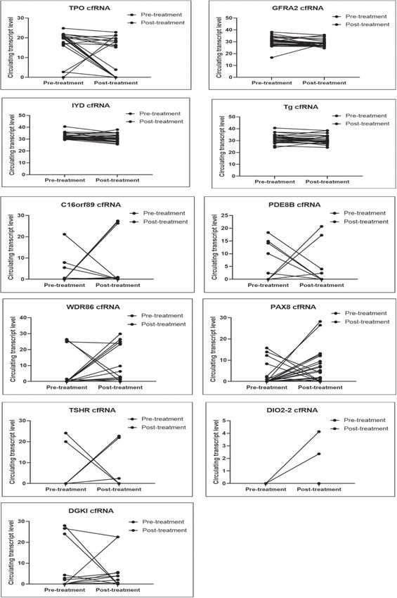
Yang et al.                                                                                                                      Cell-Free RNA in Thyroid Cancer

                                              post-therapy in 63% of cohort by 80%, as early as 1 day post-treatment, supporting
                                              the potential role as early indicator of remnant thyroid tissue volume. We demonstrated
                                              the clinical relevance of circulating TPO cfRNA by tracking temporal changes in setting
                                              of peri-treatment, recurrence, and TG Ab positive state.
                                              Conclusion: Using a multiplex pre-amplification approach, the TPO cfRNA was a
                                              potential biomarker that can track residual thyroid mass. It can be further optimised for
                                              quantification of thyroid volume to guide RAI doses and for detection of thyroid cancer
                                              recurrence.
                                              Keywords: thyroid-specific transcripts, thyroglobulin antibody positivity, thyroid cancer surveillance follow-up,
                                              thyroglobulin assay, thyroid cancer recurrence

INTRODUCTION                                                                         of plasma and the need for higher sensitivity for detection of
                                                                                     thyroid tissues.
The current standard of care for management of thyroid cancer                            Unlike other solid organ malignancies where the primary
has been total thyroidectomy followed by post-operative adjuvant                     organ is still in situ after excision of tumour, thyroid cancer
radioactive iodine (RAI) for majority of patients. The decision                      treatment involves removal of the entire thyroid gland as part
for RAI and the I–131 dosage, ranging from 30 to 250 mCi per                         of the treatment strategy. This would have led to the fall of
dose, is determined by the predicted tumour burden and risk of                       thyroid-specific cell-free RNA (cfRNA) transcripts. Therefore any
recurrence. In patients with advanced distant metastatic disease,                    elevation in these circulating tissue-specific RNA levels could
higher doses of 100–250 mCi of RAI is usually recommended                            potentially indicate significant residual thyroid tissue or tumour
(Haugen et al., 2016). There is a lack of precision for estimation                   burden since these transcripts reflect the presence of the cells
of adjuvant RAI treatment dosages due to the absence of a clinical                   originating from thyroid tissue. We had previously shown that
tool that quantifies residual thyroid tissue volume reliably. The                    cfRNA can change over 6–8 h (Koh et al., 2014). As such, cfRNA
current tumour marker, serum thyroglobulin that has a half-life                      could be a potential real-time indicator of the residual thyroid
of 65 h, may take at least 7–10 half-lives (4 weeks) for complete                    tissue volume as opposed to tumour marker, serum thyroglobulin
thyroglobulin clearance in the absence of metastases (Hocevar                        that may take at least 4 weeks for complete thyroglobulin
et al., 1997). One study had shown that undetectable baseline                        clearance in the absence of metastases.
thyroglobulin at 1 month post-surgery was associated with                                In our study, we aim to demonstrate the feasibility of cfRNA in
absence of structural disease on post-operative scans (Giovanella                    tracking thyroid tissue volume as a proof-of-concept to allow for
et al., 2008), whereas another study showed that undetectable                        further clinical study design to (1) examine the clinical utility in
thyroglobulin levels 2–6 months post-surgery was associated                          thyroid cancer risk stratification for guidance on RAI treatment,
with radioactive iodine avid metastatic disease in 12% of cases                      and (2) to assess its performance in detecting thyroid cancer
(Robenshtok et al., 2013).                                                           recurrence.
   The role of post-surgical pre-ablative diagnostic radioiodine                         Thyroid cancer surveillance for recurrence is performed
scan to estimate the remnant amount of thyroid tissue had been                       using ultrasound scan of neck, cross-sectional imaging in some
explored. However, there had been studies reporting that low                         patients, along with serum thyroglobulin (TG) to monitor disease
doses of RAI given for these diagnosis was associated with higher                    burden in response to treatment (Robbins et al., 2004). It detects
risk of remnant ablation failure, hypothesised to be due to the                      recurrence in thyroid cancer with a sensitivity of 19–40% and
stunning effect of the RAI dose for diagnostic scan rendering                        specificity of 92–97% (Schlumberger et al., 2007). The rather
lower RAI uptake with the subsequent treatment dose of RAI (Hu                       low sensitivity of thyroglobulin for detecting recurrence leaves
et al., 2004; Verburg et al., 2009). As such, this is not routinely                  room for the development of molecular tools. In addition,
done in clinical practice (Haugen et al., 2016).                                     cfRNA will address the issue of anti-thyroglobulin antibodies
   Using circulating tumour DNA (ctDNA) to estimate residual                         (present in 25% of thyroid cancer patients), that affects the
thyroid cancer tissue can be challenging. In a study evaluating the                  reliability of thyroglobulin immunoassay (Giovanella et al.,
performance of ctDNA using digital PCR, the authors pointed                          2011; Spencer, 2011). Similarly, in a review of papillary thyroid
out that ctDNA was detected mainly in patients with solid                            carcinoma cohort in our institution, 24% of the patients (20/83)
tumours outside the brain (112 of 136; 82%). In contrast, less than                  had anti-thyroglobulin antibodies (TG Ab) (Sek et al., 2021).
50% of patients with medulloblastomas or metastatic cancers of                       TG Ab interferes with the serum thyroglobulin immunometric
the kidney, prostate, and thyroid harboured detectable ctDNA                         assay (IMA) measurement causing TG underestimation with
(Bettegowda et al., 2014). Compared to ctDNA with only two                           the risk of missing detection of persistent or recurrent
copies per cell, multiple copies of tissue-specific RNA are present,                 thyroid cancer. In this clinical setting, alternative methods of
providing a higher chance of detection. As such, cfRNA can                           assessing serum TG using radioimmunoassay (RIA) or liquid
potentially overcome the challenges of using small input amount                      chromatography/tandem mass spectrometry (LC-MS/MS) had

Frontiers in Genetics | www.frontiersin.org                                      2                                      August 2021 | Volume 12 | Article 721832
Yang et al.                                                                                                         Cell-Free RNA in Thyroid Cancer

been studied (Netzel et al., 2015). These were reported to have            TG, thyroid uptake on radioiodine scan (if available), and
no interference from TG Ab. However, the functional sensitivity            neck ultrasound.
of RIA (0.5–1.0 ug/L) and LC-MS/MS (1–2 ug/L) are lower                       Treatment response was classified as excellent, indeterminate,
than IMA method (0.05–1.0 ug/L), and they are not readily                  biochemical incomplete or structural incomplete based on
available at most institutions. In this clinical scenario, TG Ab           the American Thyroid Association (ATA) thyroid cancer
had been used as a surrogate tumour marker for thyroid cancer.             management guidelines (Haugen et al., 2016). Patients were
However, TG Ab reduction in level post-treatment is usually                considered to have an excellent treatment response if they
delayed (half-life 10 weeks). The TG Ab titre falls in 75% of              had non-stimulated TG < 0.2 ug/L or stimulated serum
patients following complete treatment, but only 50% of these               TG < 1.0 ug/L after RAI ablation, with no detectable TG Ab
patients have undetectable TG Ab after 4 years of follow-up. It            and no structural disease on neck ultrasound scan and/or cross-
is uncertain if this TG Ab persistence is due to continued TG              sectional or radioiodine imaging. Patients were considered to
production by persistent thyroid tissues not detected by imaging           have an indeterminate response if they had a non-stimulated
or a stigma of continued activity of plasma cells (Spencer, 2013).         serum TG between 0.2 and 1.0 ug/L, stimulated serum TG ≥ 1–
Lastly, patients with poorly differentiated thyroid cancers lose           10 ug/L after RAI ablation, stable or declining TG Ab or
the ability to produce thyroglobulin, making the measurement of            non-specific changes on neck ultrasound scan and/or cross-
thyroglobulin an unreliable reflection of tumour burden in these           sectional or radioiodine imaging. Patients were considered to
patients (Robbins et al., 2004).                                           have biochemical incomplete treatment response if they had
   Targeting circulating RNA of thyroid origin in thyroid                  a non-stimulated TG > 1 ug/L after RAI ablation, simulated
cancer patients is a promising biomarker to monitor disease                serum TG > 10 ug/L or rising TG/TG Ab values without
status. Utility of thyroid-specific mRNA transcripts such as               structural evidence of disease on imaging. Patients with structural
thyroid peroxidase (TPO), sodium-iodide symporter (NIS),                   evidence of disease on imaging were considered to have structural
thyroglobulin (TG), and thyroid stimulating hormone receptor               incomplete response.
(TSHR) in predicting thyroid cancer recurrences and metastases                The study was conducted from May 2018 to Oct 2020, and the
had been assessed previously using whole blood (Barzon et al.,             study protocol was approved by the local ethics board committee
2004). TPO and TSHR mRNA extracted from whole blood                        (NHG DSRB Study Reference Number: 2017/00632). Written
showed significant correlation with disease status; they showed            informed consent was obtained from all participants.
higher specificity (65–81%) but lower sensitivity (40–53%), while
TG and NIS mRNA showed high sensitivity (60–73%) but low                   Clinical Laboratory Measurement
specificity (29–48%). Thyroid-specific cell free RNA (cfRNA)               The serum TG was measured using immunometric assay,
from the plasma fraction instead of whole blood can provide                e411 (Roche) or E170 (Roche) with a functional sensitivity of
better sensitivity without the cellular background and will have to        0.5 ug/L before 2015. Subsequently, the TG assay was changed
be studied to ascertain if it could offer better sensitivity than TG       in 2015 to Kryptor (Brahms) with a functional sensitivity of
for use in detection of cancer recurrence. cfRNA had been used             0.15 ug/L, and to e411 (Roche) in 2017 with a functional
for tumour surveillance in other tumours; for example, cfRNA               sensitivity of 0.1 ug/L. Anti-TG antibodies were measured using
showing PD-L1 expression had been monitored in metastatic                  radioimmunoassay Immulite 2000 (Siemens) assay before 2017,
gastric cancer patients (Shen et al., 2016).                               or e411 (Roche) from 2017.
                                                                              The analytical functional sensitivity of TG Ab was 20
                                                                           IU/mL for Immulite 2000 (Siemens) assay and Roche assay.
MATERIALS AND METHODS                                                      We considered a detectable level of TG Ab according to the
                                                                           functional sensitivity (limit of quantification) as TG Ab positive
Clinical Sample Collection                                                 state. Of note, reference values of TG Ab are reported to
In order to demonstrate a fall of thyroid-specific cfRNA                   distinguish individuals with and without thyroid autoimmune
after thyroid surgery, 16 patients undergoing total or hemi-               disease. Whereas detectable concentrations of TG Ab (i.e., above
thyroidectomy for thyroid cancer or benign thyroid disease were            the functional sensitivity) even below the normal reference range
recruited. Peripheral blood pre- and post-surgery was assessed for         cut-off can interfere with serum TG (Spencer et al., 2014).
a fall in levels of circulating thyroid-specific cfRNA. To establish
the temporal trend of the cfRNA titre with thyroidectomy,
post-surgical cfRNA was evaluated at multiple time points                  Selection of Thyroid-Specific Targets for
at the following time intervals 24 h, 1 week, 1 month, and                 Measurements in Plasma
6 months post-surgery. There were two healthy volunteers                   A data-driven approach leveraging on thyroid-specific targets
included as control group.                                                 derived from the Human Protein Atlas to measure tissue-specific
   To assess the utility of thyroid-specific cfRNA in detecting            RNA within the plasma was adopted. Genes that fall into the
recurrent or persistent metastatic thyroid cancer, peripheral              category of “Tissue-enriched genes” in thyroid tissues were
blood of 11 patients at American Thyroid Association (ATA)-                selected (Uhlén et al., 2015; Atlas, 2021). Genes that are highly
intermediate to high of recurrence (Haugen et al., 2016) was               expressed and have more than four times fold-change when
collected during surveillance visits and at time of clinical               compared to other tissues were selected for analysis. Selected
recurrence. The cfRNA titres was correlated with serum                     targets were also verified in literature to be biologically relevant

Frontiers in Genetics | www.frontiersin.org                            3                                    August 2021 | Volume 12 | Article 721832
Yang et al.                                                                                                            Cell-Free RNA in Thyroid Cancer

before including into the panel (Lindahl et al., 2001; Afink et al.,          primers mix and 26 mU of RNase H2 enzyme. Emulsion was
2010; Li et al., 2012; Fernández et al., 2015; Soto-Pedre et al., 2017;       generated by adding three parts of 10% 008-FluoroSurfactant
Targovnik et al., 2017). Eventually, 11 thyroid-specific genes were           (RAN Biotechnologies) in 3M FluorinertTM Engineered Fluid
targeted: TPO, TG, GFRA2, IYD, PDE8B, WDR86, C16orf89,                        (3M, Cat no. FC-40) to 1 part of PCR reaction mixture. The
DGKI, DIO2, TSHR, and PAX8.                                                   mixture was vortexed until it became cloudy and uniform.
                                                                              Thermocycling started with enzyme activation at 94◦ C for 2 min,
Circulating RNA Extraction                                                    followed by 20 cycles of denaturation (94◦ C, 15 s), annealing
Consistent and reliable blood collection protocols are critical               (55◦ C, 30 s), and extension (68◦ C, 1 min). Reaction recovered
to maintain the integrity of circulating nucleic acid assays. We              from emulsion PCR was top up with the same amount of
collected blood in Streck cfRNA tubes containing a stabilising                polymerase and RNase H2 used in emulsion PCR. Thermocycling
reagent to prevent cell lysis and reduce degradation of RNA                   started with enzyme activation at 94◦ C for 2 min, followed by 20
(Fernando et al., 2012).                                                      cycles of denaturation (94◦ C, 15 s), hybridization (78◦ C, 10 min),
   cfRNA was extracted from 1 mL of plasma using                              annealing (55◦ C, 30 s), and extension (68◦ C, 1 min).
Plasma/Serum Circulating and Exosomal RNA Purification
Kit (Norgen, Cat no. 42800). The residual DNA in the cfRNA                    Quantification of Pre-amplified Gene
was digested using RNase-Free DNase I Kit (Norgen, Cat no.                    Targets Using qPCR
25720). Extracted cfRNA was purified using RNA Clean and                      To monitor the expression of the targeted cfRNA across the
ConcentratorTM -5 (Zymo, Cat no. ZYR.R1016), yielding 24 µL                   different time point, qPCR was performed for 60 cycles with
of cfRNA per sample.                                                          Maxima SYBR Green/ROX qPCR Master Mix (ThermoFisher
                                                                              Scientific, Cat no. K0221).
Reverse Transcription and Emulsion
Based Targeted Pre-amplification                                              Spike-In Controls and Quality Controls
The multiplex approach is based on RNase H-dependent PCR                      Assays for Ensuring Consistent
(rhPCR), a method that provides increased target specificity
over traditional PCR (Figure 1). Compared to traditional PCR,
                                                                              Comparisons Across Patient Samples
                                                                              There were two types of quality control. One for technical
rhPCR requires an enzyme, RNase H2, and uses blocked primers
                                                                              amplification efficiency and another for extraction efficiency.
(rhPrimers obtainable from IDT) in place of conventional PCR
                                                                                  Each sample is spiked with the same amount of commercially
primers. rhPrimers contain a single RNA base and a 30 blocking
                                                                              obtained Luciferase RNA that is not normally found in normal
group. The blocking group must be removed by RNase H2 before
                                                                              plasma (L4561, Promega). 10,000 copies of Luciferase Control
extension by DNA polymerase. This additional condition results
                                                                              RNA was spiked in with 10 µL of the extracted cfRNA
in less off target amplification and less primer-dimers are formed,
                                                                              at the reverse transcription step. The spiked-in served as a
resulting in increased specificity and efficient amplification of
                                                                              technical control for multiplex amplification efficiency control
the circulating RNA that we are targeting. In this work, we
                                                                              to normalise for any unintended variation in the experiment.
specifically designed a panel of rhPrimers to amplify thyroid
                                                                              Median Ct value of luciferase RNA across all samples is set as
and housekeeping genes (Supplementary Table 1). In addition,
                                                                              the benchmark. In each sample, all gene targets Ct values are
we performed rhPCR in an emulsion that physically separated
                                                                              offset till the Luciferase control Ct in the sample matches up
the reaction into multiple reaction-in-oil droplets, which aids in
                                                                              to the benchmark.
avoiding the formation of unproductive artefacts.
                                                                                  Subsequently to normalise for possible variations during
    Sequencing is used primarily as quality control of the qPCR
                                                                              RNA extraction, geometric mean of the previous luciferase-
assay. This is to ensure that the amplicons match up to the
                                                                              adjusted Ct values for housekeeping genes (ACTB, GAPDH, and
intended primers design. Considerations were given to replace
                                                                              RPS18) are calculated and similarly offset in each sample till the
the qPCR assay with sequencing, however, due to the cost
                                                                              housekeeping geometric mean matches up to the cohort average.
of sequencing which scale with each additional samples, we
                                                                              In Supplementary Figure 1, we demonstrate that normalising
deployed it only once primarily to ensure that the amplicons
                                                                              using the luciferase and geometric mean of housekeeping genes
generated are correct.
                                                                              result in tighter distribution of amplification curves.
    10 µL of extracted cfRNA was annealed with a final
concentration of 0.4 µM of reverse primers mix in the presence
of 10,000 copies of luciferase control RNA (Promega, Cat no.                  Data Analysis
L4561) and a final concentration 2 mM of dNTPs at 65◦ C for                   Analysis of the data used normalised Ct values with respect to
5 min. Reverse transcription of cfRNA was performed using                     the housekeeping genes and corrected for amplification efficiency
SuperscriptTM III Reverse Transcriptase (Invitrogen, Cat no.                  using spiked in luciferase RNA amount. All sequencing analysis
18080044) at 25◦ C for 5 min, 50◦ C for 50 min, and enzyme                    was performed and plotted in R using ggplot. We started with
inactivation at 95◦ C for 3 min.                                              normalising the raw Ct values with Luc and Housekeeping genes.
    cDNA from reverse transcription was added to the PCR                      We verified the amplified product by referring to the melt curve.
mixture of PlatinumTM Taq DNA Polymerase (Invitrogen, Cat                     For an undetermined Ct/incorrect product, Ct value of 60 was
no. 10966) with a final concentration of 0.5 µM of rh PCR                     assigned. This was because 60 cycles were ran on qPCR. The final

Frontiers in Genetics | www.frontiersin.org                               4                                    August 2021 | Volume 12 | Article 721832
Yang et al. Cell-Free RNA in Thyroid Cancer
FIGURE 1 | Workflow of reverse transcription and amplification of cfRNA. cfRNA, spiked-in with luciferase RNA control, was reverse transcript with SuperscriptTM III
Reverse Transcriptase (Invitrogen, Cat no. 18080044). The product was amplified with rhPCR primers and PlatinumTM Taq DNA Polymerase (Invitrogen, Cat no.
10966) using emulsion and CoT PCR. The residual primers were removed with Exonuclease I (New England Biolabs, Cat no. M0293). Amplified products were used
for qPCR quantification and sequencing.
Ct value was then deducted from 60 to allow easy visualisation of and TSHR RNA were not detectable in healthy controls
the changes in the RNA expression. (Supplementary Figure 2A and Table 2). Similarly, in thyroid
Level of each circulating cfRNA was represented as median patients, only low level circulating DIO2-2 RNA was detected
with interquartile range. Statistical significance was determined in 2 patients with Ct values of 56 and 58 (data not shown).
according to p-values generated by Wilcoxon Signed Rank t test. The circulating TSHR RNA was only detected in a single time
Stata software (Version15.1; StataCorp, TX, United States) was point of 7 patients (7/16, 44%) with Ct values ranging 36–58
used for statistical analysis. The cfRNA level graphs were plotted (data not shown).
using GraphPad Prism v9 (GraphPad Software).
Thyroid-Specific cfRNA Levels Fall
Following Surgical or Pharmacological
RESULTS Ablation of the Thyroid Tissue
We evaluated the 11 circulating thyroid-specific cfRNA levels
Multiplex Assay Is Capable of Amplifying in patients undergoing thyroid surgery for (i) benign thyroid
and Quantifying Multiple conditions (e.g., benign thyroid nodules, hyperthyroidism from
Thyroid-Specific Genes in a Single Graves’ disease; n = 3), (ii) or malignant thyroid nodules
Reaction (n = 14), (iii) and patients with recurrent/persistent thyroid
RNA extracted directly from the thyroid (commercially obtained cancer undergoing repeat thyroid surgery or radioactive iodine
from Takara Bioscience) was ran as a positive control using the adjuvant therapy (n = 10). All the thyroid cancer patients had
assay. Successful amplification of these targets collaborates the papillary thyroid cancer (PTC) except for a patient with both PTC
notion that the selected genes were highly expressed within the and poorly differentiated thyroid cancer, and another patient
thyroid tissue (Figure 2). with follicular thyroid cancer. Their baseline characteristics is
summarised in Table 1. Four of the circulating plasma cfRNA
Thyroid-Specific RNA Transcripts Are transcripts, namely TPO (thyroid peroxidase), IYD (iodotyrosine
deiodinase), GFRA2 (glial cell line-derived neurotrophic factor
Present in Plasma and in Amounts family receptor alpha-2), and TG cfRNA, fell post-treatment
Quantifiable Using Our Approach of in more than 50% of the study cohort, reaching statistical
qRT-PCR significance (Table 2 and Figure 3). The TPO cfRNA fell post-
The 11 selected thyroid-specific RNA transcripts (TPO, therapy in 63% of the study cohort with a magnitude of fall of
TG, GFRA2, IYD, PDE8B, WDR86, C16orf89, DGKI, 80%. Of note, in some patients, TPO cfRNA started to fall as early
DIO2, TSHR, and PAX8) were amplified from healthy as 1 day post-treatment. The magnitude of fall in cfRNA level
volunteers (Supplementary Figure 2A) and thyroid patients post-treatment in IYD, TG, and GFRA2 cfRNA were smaller at
(Supplementary Figure 2B). Both circulating DIO2-2 RNA 5 to 8%. Similar to TPO cfRNA, in some patients, IYD, TG, and
Frontiers in Genetics | www.frontiersin.org 5 August 2021 | Volume 12 | Article 721832Yang et al. Cell-Free RNA in Thyroid Cancer
FIGURE 2 | Positive amplification curves and melt curves of all thyroid-specific RNA transcripts using directly extracted thyroid RNA.
GFRA2 cfRNA started to fall as early as 1 day post-treatment This case illustrates that TPO cfRNA falls after thyroid surgery,
supporting the potential for cfRNA to be early indicators of and TPO transcription is increased by TSH stimulation as
remnant thyroid tissue volume post-treatment. indicated by increased circulating cfRNA levels.
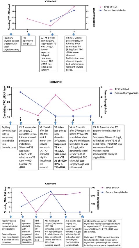
Next, we reviewed the utility of cfRNA measurement in a
thyroid cancer patient with innate production of thyroglobulin
Thyroid-Specific cfRNA Transcripts antibodies (TG Ab) due to underlying autoimmune lymphocytic
Capture Temporal Trends in Clinical thyroiditis. In this clinical setting, TG Ab usually interferes with
Course of Thyroid Cancer Patients the serum thyroglobulin immunometric assay measurement.
Temporal trends were showcased in three different clinical This patient (CBN019) had Stage I papillary thyroid carcinoma
scenarios (peri-operative period, TG Ab positive state, with cervical lymph node metastases [T2N1bM0], treated with
recurrent/persistent thyroid cancer), reflecting different aspects total thyroidectomy and adjuvant RAI 7 weeks later (Figure 4B).
where cfRNA measurements can make an impact. At time point 1, evaluated 2 days after RAI under TSH stimulated
We demonstrated the expected variability of the observed state, serum TG was elevated at 2.5 ug/L, TG Ab was high
TPO cfRNA level peri-operatively and under thyroid stimulating at >4000 IU/ml (normal reference rangeYang et al. Cell-Free RNA in Thyroid Cancer
TABLE 1 | Baseline characteristics of patients. RAI (time point 3), in the TSH stimulated state, both her serum
TG (256 ug/L) and TPO cfRNA were elevated. Her radioiodine
Characteristics n = 27
scan showed thyroid bed RAI uptake indicating residual thyroid
Age at diagnosis in years (median ± interquartile range) 54 (40–69) tissue. At time point 4, during surveillance at 6 months after neck
Gender, n (%) dissection, both serum TG (2 ug/L) and TPO cfRNA were still
Female 16 (59.3%) elevated, even though these levels were lower than levels detected
Ethnicity, n (%) at time point 1 (before neck dissection and RAI). Ultrasound scan
Chinese 14 (51.9%) of neck showed left thyroid bed nodule. This was later biopsied
Malay 2 (7.4%) and proven to be persistent papillary thyroid cancer (time
Indian 1 (3.7%) point 5). The biopsy needle washing TG level was elevated at
Others 10 (37.0%) 5180 ug/L, without detectable TG Ab. Concurrently, both serum
Thyroglobulin Antibody Status TG (1.3 ug/L) and TPO cfRNA were elevated. Subsequently, she
Present 17 (63.0%) underwent another course RAI treatment. Her radioiodine scan
Absent 7 (25.9%) showed the same thyroid bed RAI uptake though less intense,
Not known 3 (11.1%) indicating that the previous RAI had partially ablated the tumour.
Thyroid histology This case illustrates that TPO cfRNA can be a potential tool to
Papillary thyroid cancer 22 (81.5%) detect recurrent or persistent thyroid cancer, and its level can fall
Follicular thyroid cancer 1 (3.7%) within a day after treatment, indicating that measurements are
Poorly differentiated thyroid cancer 1 (3.7%) instant snapshots of the molecular physiology in time.
Benign thyroid nodule 3 (11.1%)
Including a patient with Grave’s disease
And another with autoimmune thyroiditis DISCUSSION
Thyroid cancer stage (AJCC eighth edition/TNM stage)
I 15 (55.6%) Key Study Findings
II 5 (18.5%) In this study, we aimed to assess the performance of circulating
III 0 thyroid-specific cfRNA in quantifying remnant thyroid gland
IVA 1 (3.7%) tissue or malignancy. Circulating tissue-specific RNA had also
IVB 3 (11.1%) been previously reported to be present at low levels. To increase
Not applicable for benign nodules 3 (11.1%) sensitivity of our assay, we measured multiple thyroid-specific
Risk of recurrence targets using a multiplex pre-amplification approach. In addition,
Low 7 (25.9%) a data-driven bioinformatics approach was adopted to design a
Intermediate 7 (25.9%) multiplex primer design assay specific to thyroid targets with
High 10 (37.1) minimal cross-interaction. Integrating multiple measurements
Not applicable for benign nodules 3 (11.1%) targeting the thyroid, we found the TPO cfRNA to be a potential
Response to therapy circulating biomarker that can track the residual thyroid mass
Excellent 10 (37.0%) in patients, with levels falling dynamically as early as 1 day
Indeterminate 6 (22.3%) after treatment. We have demonstrated the clinical relevance
Biochemical incomplete 3 (11.1%) of circulating TPO cfRNA by tracking the changes in levels
Structural incomplete 5 (18.5%) throughout the patient clinical course in setting of the peri-
Not applicable for benign nodules 3 (11.1%) treatment, recurrence, and TG Ab positive state.
Follow-up duration (in months) (median ± interquartile range) 33 (21–46) The identification of a circulating cfRNA that changes in real-
time and reflects molecular physiology in time, allows for further
clinical study design to (1) examine the clinical utility in remnant
was on the upwards trend with similar undetectable serum TG thyroid mass quantification for precise guidance on RAI doses,
and elevated TG Ab. Her neck ultrasound showed indeterminate and (2) to assess its performance in detecting thyroid cancer
finding of atypical lymph nodes that needed to be followed- recurrence, especially in patients with TG Ab that interferes with
up. This case illustrates the application of cfRNA technique for existing serum TG immunometric assays.
detection of persistent thyroid cancer in the setting of TG Ab
positivity that interferes with TG immunometric assay. Harnessing Thyroid-Specific Targets
We next analysed cfRNA time course for a patient (CBN001) Beyond Thyroglobulin
who had persistent papillary thyroid cancer despite initial total The current standard of care relies on quantification of
thyroidectomy (Figure 4C). She had elevated serum TG post- circulating thyroid-specific protein TG to reflect thyroid tissue
surgery, in the absence of TG Ab, and was found to have cervical mass (Haugen et al., 2016). However, the most widely used
lymph node metastases. Just prior to neck dissection surgery, at TG assay, the immunometric assay underestimates serum TG
time point 1, she had elevated serum TG (72 ug/L) and TPO in the presence of TG Ab due to the formation of TG-TG Ab
cfRNA levels. After neck dissection surgery, her TPO cfRNA complex that leads to a reduction of free TG, which is measured
level fell within a day (time point 2). On day of her adjuvant by immunometric assay (Rosario et al., 2021). Several studies
Frontiers in Genetics | www.frontiersin.org 7 August 2021 | Volume 12 | Article 721832Yang et al. Cell-Free RNA in Thyroid Cancer
TABLE 2 | Circulating levels of thyroid-specific cfRNA transcript candidates pre- and post-treatment in patients undergoing thyroidectomy or adjuvant
radioactive iodine therapy.
Total n = 27 Fall with Proportion of Mean level in Median level Median level % fall in median p value
treatment cohort with healthy pre-treatment of patients post-treatment of cfRNA level between
(surgery or RAI) fall post- volunteers [60 (interquartile range) [60 patients (interquartile pre- and
(n) treatment - Ct value] - Ct value] range) [60 - Ct value] post-treatment levels
TPO RNA 17 63% 27.4 19.8 3.9 −80% 0.0079
(2.7–21.3) (0–18.6)
TG RNA 20 74% 18.9 32.1 29.6 −8% 0.0264
(29.4–33.6) (28.3 – 33.6)
IYD RNA 20 74% 20.1 31.8 30.3 −5% 0.0058
(30.7–32.9) (28.3 – 32.9)
GFRA2 RNA 19 70% 25.0 30.4 28.2 −7% 0.0110
(28.1–32.4) (26.5 – 29.1)
WDR86 RNA 4 15% 4.6 0 0 –
(0–0) (0–6.3)
PAX8 RNA 5 19% 10.3 0 1.9 –
(0–0.9) (0–8.6)
DGKI RNA 4 15% 8.0 0 0 –
(0–0) (0–2.0)
PDE8B RNA 5 19% 8.9 0 0 –
(0–0) (0–0)
C16orf89 RNA 4 15% 0 0 0 –
(0–0) (0–0)
TSHR RNA 2 7% 0 0 0 –
(0–0) (0–0)
DIO2-2 RNA 0 0% 0 0 0 –
(0–0) (0–0)
Rows in bold represent cfRNA transcripts that fall post-treatment.
had assessed the utility of TG measurement by LC-MS/MS. as iodotyrosine dehalogenase 1, DEHAL1) to recycle iodide,
They had shown feasibility in detecting circulating TG via preventing iodide from being leaked out of thyroid cells
mass spectrometry technique. However, this needs to be further (Targovnik et al., 2017; Citterio et al., 2019). As such, circulating
optimised to improve the current sensitivity of 40–60% as there levels of TPO, IYD, and TG cfRNA whose function are specific
were patients with structural disease and TG Ab with negative TG to the thyroid gland could plausibly reflect thyroid mass. GFRA2
measured on LC-MS/MS (Spencer et al., 2014; Netzel et al., 2015; was found to be highly expressed in brain and thyroid tissue
Azmat et al., 2016; Guastapaglia et al., 2020). TG measurement (Atlas, 2021), and it was enriched in papillary thyroid cancer from
by radioimmunoassay (RIA) is an alternative technique for TG the TCGA dataset (Cancer Genome Atlas Research Network,
assessment in the presence of TG Ab though it had been reported 2014). It mediates activation of the RET tyrosine kinase receptor
to be associated with false-positive TG measurement in patients and is a candidate gene for RET-associated diseases. It had been
with TG Ab (Netzel et al., 2015). The assessment of circulating TG shown to be present on immunohistochemistry stain in case
mRNA was shown to correctly identify 93% (13/14) of patients series of papillary, follicular, and medullary thyroid cancer, as well
with structural disease, negative serum TG, in the presence of as follicular adenoma (Lindahl et al., 2001).
elevated TG Ab (Boldarine et al., 2010). This remains to be further The current tumour marker serum TG takes at least 4 weeks
validated. for complete clearance whereas levels of TPO cfRNA changes as
We found that other circulating thyroid-specific targets early as 1 day after treatment. With further optimisation, it could
such as TPO, IYD, TG, and GFRA2 cfRNA fell accordingly potentially allowing for early quantification of residual thyroid
post-treatment for thyroid cancer, and could be potential mass and precise, timely planning of adjuvant RAI dosage after
biomarkers to reflect thyroid mass. Thyroglobulin (TG), thyroid thyroid surgery.
peroxidase (TPO), and iodotyrosine deiodinase (IYD) are
involved in thyroid hormone biosynthesis. Thyroid peroxidase Limitations
oxidises iodide ions to iodine atoms for addition onto tyrosine The quantification provided in the form of 60-ct values for
residues on thyroglobulin, forming mono-iodotyrosine (MIT) circulating cfRNA does not vary proportionally to the expected
and di-iodotyrosine (DIT). Thyroid hormones, thyroxine (T4) amount of thyroid mass. For example, the level of TPO cfRNA
formation involves oxidative coupling of 2 DIT, whereas in patients with previous hemi-thyroidectomy is not half of
triiodothyronine (T3) de novo formation involves coupling of the level in patients with an entire thyroid gland in situ. The
MIT and DIT. Some free MIT and DIT released by thyroid cfRNA RT-qPCR technique requires further optimisation for
cells are scavenged by iodotyrosine deiodinase (also known better precision. Some patients with thyroid cancer did not shed
Frontiers in Genetics | www.frontiersin.org 8 August 2021 | Volume 12 | Article 721832Yang et al. Cell-Free RNA in Thyroid Cancer FIGURE 3 | Plots comparing pre- and post- treatment levels of thyroid-specific cfRNA transcripts candidates. The circulating cfRNA levels plotted along Y-axis were derived from 60- Ct values. Frontiers in Genetics | www.frontiersin.org 9 August 2021 | Volume 12 | Article 721832
Yang et al.                                                                                                                               Cell-Free RNA in Thyroid Cancer

  FIGURE 4 | Longitudinal trend of thyroid-specific cfRNA in clinical course of thyroid cancer patients. The circulating cfRNA levels plotted along Y-axis were derived
  from 60- Ct values. (A) Peri-operative and pre-RAI TPO cfRNA level and serum thyroglobulin level at three time points in patient with papillary thyroid cancer. (B) TPO
  cfRNA level and serum thyroglobulin (TG) level in the setting of positive thyroglobulin antibody (TG Ab) in patient with papillary thyroid cancer and lymph node (LN)
  metastases. (C) TPO cfRNA level and serum thyroglobulin (TG) level in a patient with papillary thyroid cancer and persistent lymph node (LN) metastases undergoing
  further ablative therapy.

Frontiers in Genetics | www.frontiersin.org                                         10                                          August 2021 | Volume 12 | Article 721832
Yang et al.                                                                                                                                     Cell-Free RNA in Thyroid Cancer

TPO cfRNA into the circulation and had measurable circulating                                 The patients/participants provided their written informed
TG, GFRA2, IYD cfRNA levels instead. It is possible that different                            consent to participate in this study. Written informed consent
clonal thyroid cancer cells could preferentially produce different                            was obtained from the individual(s) for the publication of
thyroid proteins due to genetic aberrations, and the use of                                   any potentially identifiable images or data included in this
a multiplex cfRNA panel that detects several thyroid-specific                                 article.
targets might be needed. The patient and healthy volunteer
sample size is small. The authors have an ongoing study to recruit
a larger cohort for validation of the findings in this study.                                 AUTHOR CONTRIBUTIONS
Future Directions                                                                             SY, LK, and ET contributed to conception and design of the study.
Building on the foundation laid by the current study, future work                             SY organized the clinical database. LK designed the primers. LK
could employ the digital emulsion PCR technique to quantify the                               and KK performed the nucleic acid extraction and sequencing.
thyroid-specific targets identified and interrogate if the sensitivity                        SY and LK performed the statistical analysis, analysed the data,
for detection of these low-abundance targets with small fold                                  and wrote the first draft of the manuscript. SY, RP, KL, KN, WT,
changes could be improved.                                                                    and TL contributed clinical samples. SY, LK, JN, RP, KN, and
   Conceptually, harnessing the presence of multiple circulating                              DN contributed to manuscript revision. All authors read and
thyroid-specific cfRNA in the form of a multiplex thyroid                                     approved the submitted version.
transcript kit for the estimation of remnant thyroid tissue can
potentially improve the sensitivity of detecting and quantifying
thyroid mass; when complemented with the pre-existing tumour                                  FUNDING
marker, serum TG that has high specificity, it has the potential
of improving the accuracy of quantification of thyroid mass to                                SY was funded by NUHS Joint Grant Call (MOE Tier 1),
guide RAI dosing decision and detect tumour recurrence even in                                NUHS Clinician Scientist Program (NCSP), and NMRC Research
the presence of TG Ab.                                                                        Fellowship. LK was funded by A∗ STAR (IBN/MEL) core funding
                                                                                              support.

DATA AVAILABILITY STATEMENT
                                                                                              ACKNOWLEDGMENTS
The original contributions presented in the study are included
in the article/Supplementary Material, further inquiries can be                               We would like to acknowledge our funding agencies.
directed to the corresponding authors.

                                                                                              SUPPLEMENTARY MATERIAL
ETHICS STATEMENT
                                                                                              The Supplementary Material for this article can be found
The studies involving human participants were reviewed and                                    online at: https://www.frontiersin.org/articles/10.3389/fgene.
approved by NHG DSRB Study Reference Number: 2017/00632.                                      2021.721832/full#supplementary-material

REFERENCES                                                                                       differentiated thyroid carcinoma. J. Clin. Endocrinol. Metab. 95, 1726–1733.
                                                                                                 doi: 10.1210/jc.2009-1354
Afink, G. B., Veenboer, G., de Randamie, J., Keijser, R., Meischl, C., Niessen, H.,           Cancer Genome Atlas Research Network (2014). Integrated genomic
   et al. (2010). Initial characterization of C16orf89, a novel thyroid-specific gene.           characterization of papillary thyroid carcinoma. Cell 159, 676–690.
   Thyroid 20, 811–821. doi: 10.1089/thy.2009.0366                                            Citterio, C. E., Targovnik, H. M., and Arvan, P. (2019). The role of thyroglobulin
Atlas, T. H. P. (2021). The Thyroid Gland-Specific Proteome. Protein Atlas version               in thyroid hormonogenesis. Nat. Rev. Endocrinol. 15, 323–338. doi: 10.1038/
   18.0. Available online at: https://www.proteinatlas.org/humanproteome/tissue/                 s41574-019-0184-8
   thyroid+gland (accessed December 1, 2017).                                                 Fernández, L. P., López-Márquez, A., and Santisteban, P. (2015). Thyroid
Azmat, U., Porter, K., Senter, L., Ringel, M. D., and Nabhan, F. (2016).                         transcription factors in development, differentiation and disease. Nat. Rev.
   Thyroglobulin liquid chromatography–tandem mass spectrometry has a low                        Endocrinol. 11, 29–42. doi: 10.1038/nrendo.2014.186
   sensitivity for detecting structural disease in patients with antithyroglobulin            Fernando, M. R., Norton, S. E., Luna, K. K., Lechner, J. M., and Qin, J.
   antibodies. Thyroid 27, 74–80. doi: 10.1089/thy.2016.0210                                     (2012). Stabilization of cell-free RNA in blood samples using a new collection
Barzon, L., Boscaro, M., Pacenti, M., Taccaliti, A., and Palù, G. (2004). Evaluation             device. Clin. Biochem. 45, 1497–1502. doi: 10.1016/j.clinbiochem.2012.
   of circulating thyroid-specific transcripts as markers of thyroid cancer relapse.             07.090
   Int. J. Cancer 110, 914–920. doi: 10.1002/ijc.20182                                        Giovanella, L., Ceriani, L., Suriano, S., Ghelfo, A., and Maffioli, M. (2008).
Bettegowda, C., Sausen, M., Leary, R. J., Kinde, I., Wang, Y., Agrawal, N., et al.               Thyroglobulin measurement before rhTSH-aided 131I ablation in detecting
   (2014). Detection of circulating tumor DNA in early- and late-stage human                     metastases from differentiated thyroid carcinoma. Clin. Endocrinol. 69, 659–
   malignancies. Sci. Transl. Med. 6:224ra24.                                                    663. doi: 10.1111/j.1365-2265.2008.03244.x
Boldarine, V. T., Maciel, R. M. B., Guimarães, G. S., Nakabashi, C. C. D., Camacho,           Giovanella, L., Suriano, S., Ceriani, L., and Anton Verburg, F. (2011). Undetectable
   C. P., Andreoni, D. M., et al. (2010). Development of a sensitive and specific                thyroglobulin in patients with differentiated thyroid carcinoma and residual
   quantitative reverse transcription-polymerase chain reaction assay for blood                  radioiodine uptake on a postablation whole-body scan. Clin. Nucl. Med. 36,
   thyroglobulin messenger ribonucleic acid in the follow-up of patients with                    109–112. doi: 10.1097/rlu.0b013e318203bb84

Frontiers in Genetics | www.frontiersin.org                                              11                                            August 2021 | Volume 12 | Article 721832
Yang et al. Cell-Free RNA in Thyroid Cancer
Guastapaglia, L., Kasamatsu, T. S., Nakabashi, C. C. D., Camacho, C. P., Maciel, Sek, K. S., Tsang, I., Yong Lee, X., Albaqmi, O. H., Morosan Allo, Y. J., Rosmarin,
R. M. B., Vieira, J. G. H., et al. (2020). The role of a new polyclonal M. C., et al. (2021). Frequent neck US in papillary thyroid cancer likely detects
competitive thyroglobulin assay in the follow-up of patients with differentiated non-actionable findings. Clin. Endocrinol. 94, 504–512. doi: 10.1111/cen.
thyroid cancer with structural disease but low levels of serum thyroglobulin by 14325
immunometric and LC-MS/MS methods. Endocrine 72, 784–790. doi: 10.1007/ Shen, J. Y. C., Usher, J., Samberg, D., Ishiba, T., Danenberg, K., Lenz, H., et al.
s12020-020-02526-8 (2016). PD-L1 and HER2 expression in gastric cancer (GC) patients (pts) using
Haugen, B. R., Alexander, E. K., Bible, K. C., Doherty, G. M., Mandel, S. J., cell-free RNA (cfRNA). J. Clin. Oncol. 34(15 Suppl), e15539–e15539.
Nikiforov, Y. E., et al. (2016). 2015 american thyroid association management Soto-Pedre, E., Siddiqui, M. K., Doney, A. S., Palmer, C. N. A., Pearson, E. R.,
guidelines for adult patients with thyroid nodules and differentiated thyroid and Leese, G. P. (2017). Replication confirms the association of loci in FOXE1,
cancer: the american thyroid association guidelines task force on thyroid PDE8B, CAPZB and PDE10A with thyroid traits: a genetics of diabetes audit
nodules and differentiated thyroid cancer. Thyroid 26, 1–133. and research tayside study (GoDARTS). Pharmacogenet. Genomics 27, 356–362.
Hocevar, M., Auersperg, M., and Stanovnik, L. (1997). The dynamics of serum doi: 10.1097/fpc.0000000000000299
thyroglobulin elimination from the body after thyroid surgery. Eur. J. Surg. Spencer, C. (2013). Commentary on: implications of thyroglobulin antibody
Oncol. 23, 208–210. doi: 10.1016/s0748-7983(97)92292-7 positivity in patients with differentiated thyroid cancer: a clinical position
Hu, Y. H., Wang, P.-W., Wang, S.-T., Lee, C.-H., Chen, H.-Y., Chou, F.-F., et al. statement. Thyroid 23, 1190–1192. doi: 10.1089/thy.2013.0496
(2004). . Influence of 131I diagnostic dose on subsequent ablation in patients Spencer, C., Petrovic, I., Fatemi, S., and LoPresti, J. (2014). Serum thyroglobulin
with differentiated thyroid carcinoma: discrepancy between the presence of (Tg) monitoring of patients with differentiated thyroid cancer using sensitive
visually apparent stunning and the impairment of successful ablation. Nucl. (second-generation) immunometric assays can be disrupted by false-negative
Med. Commun. 25, 793–797. doi: 10.1097/01.mnm.0000126626.17166.ed and false-positive serum thyroglobulin autoantibody misclassifications. J. Clin.
Koh, W., Pan, W., Gawad, C., Christina Fan, H., Kerchner, G. A., Wyss-Coray, Endocrinol. Metab. 99, 4589–4599. doi: 10.1210/jc.2014-1203
T., et al. (2014). Noninvasive in vivo monitoring of tissue-specific global gene Spencer, C. A. (2011). Clinical utility of thyroglobulin antibody (TgAb)
expression in humans. Proc. Natl. Acad. Sci. U.S.A. 111, 7361–7366. doi: 10. measurements for patients with differentiated thyroid cancers (DTC). J. Clin.
1073/pnas.1405528111 Endocrinol. Metab. 96, 3615–3627. doi: 10.1210/jc.2011-1740
Li, Q. L., Chen, F.-J., Lai, R., Guo, Z.-M., Luo, R., and Yang, A.-K. (2012). Targovnik, H. M., Citterio, C. E., and Rivolta, C. M. (2017). Iodide handling
ZCCHC12, a potential molecular marker of papillary thyroid carcinoma: a disorders (NIS, TPO, TG, IYD). Best Pract. Res. Clin. Endocrinol. Metab. 31,
preliminary study. Med. Oncol. 29, 1409–1417. doi: 10.1007/s12032-011- 195–212. doi: 10.1016/j.beem.2017.03.006
0018-6 Uhlén, M., Fagerberg, L., Hallström, B. M., Lindskog, C., Oksvold, P., Mardinoglu,
Lindahl, M., Poteryaev, D., Yu, L., Arumae, U., Timmusk, T., Bongarzone, I., A., et al. (2015). Tissue-based map of the human proteome. Sci. 347:1260419.
et al. (2001). Human glial cell line-derived neurotrophic factor receptor alpha doi: 10.1126/science.1260419
4 is the receptor for persephin and is predominantly expressed in normal and Verburg, F. A., Verkooijen, R. B. T., Stokkel, M. P. M., and van Isselt, J. W.
malignant thyroid medullary cells. J. Biol. Chem. 276, 9344–9351. doi: 10.1074/ (2009). The success of 131I ablation in thyroid cancer patients is significantly
jbc.m008279200 reduced after a diagnostic activity of 40 MBq 131I. Nuklearmedizin 48, 138–142.
Netzel, B. C., Grebe, S. K. G., Carranza Leon, B. G., Castro, M. R., Clark, doi: 10.3413/nukmed-0225
P. M., Hoofnagle, A. N., et al. (2015). Thyroglobulin (Tg) testing revisited: TG
assays, TGAB assays, and correlation of results with clinical outcomes. J. Clin. Conflict of Interest: The authors declare that the research was conducted in the
Endocrinol. Metab. 100, E1074–E1083. absence of any commercial or financial relationships that could be construed as a
Robbins, R. J., Srivastava, S., Shaha, A., Ghossein, R., Larson, S. M., Fleisher, M., potential conflict of interest.
et al. (2004). Factors influencing the basal and recombinant human thyrotropin-
stimulated serum thyroglobulin in patients with metastatic thyroid carcinoma. Publisher’s Note: All claims expressed in this article are solely those of the authors
J. Clin. Endocrinol. Metab. 89, 6010–6016. doi: 10.1210/jc.2003-031573 and do not necessarily represent those of their affiliated organizations, or those of
Robenshtok, E., Grewal, R. K., Fish, S., Sabra, M., and Michael Tuttle, R. (2013). A the publisher, the editors and the reviewers. Any product that may be evaluated in
low postoperative nonstimulated serum thyroglobulin level does not exclude this article, or claim that may be made by its manufacturer, is not guaranteed or
the presence of radioactive iodine avid metastatic foci in intermediate-risk endorsed by the publisher.
differentiated thyroid cancer patients. Thyroid 23, 436–442. doi: 10.1089/thy.
2012.0352 Copyright © 2021 Yang, Koh, Kong, Parameswaran, Loke, Ngiam, Tan, Loh,
Rosario, P. W., Côrtes, M. C. S., and Franco Mourão, G. (2021). Follow-up of Ng, Goh, Ngeow and Tai. This is an open-access article distributed under
patients with thyroid cancer and antithyroglobulin antibodies: a review for the terms of the Creative Commons Attribution License (CC BY). The use,
clinicians. Endocr. Relat. Cancer 28, R111–R119. distribution or reproduction in other forums is permitted, provided the original
Schlumberger, M., Hitzel, A., Toubert, M. E., Corone, C., Troalen, F., Schlageter, author(s) and the copyright owner(s) are credited and that the original publication
M. H., et al. (2007). Comparison of seven serum thyroglobulin assays in the in this journal is cited, in accordance with accepted academic practice. No
follow-up of papillary and follicular thyroid cancer patients. J. Clin. Endocrinol. use, distribution or reproduction is permitted which does not comply with
Metab. 92, 2487–2495. these terms.
Frontiers in Genetics | www.frontiersin.org 12 August 2021 | Volume 12 | Article 721832You can also read