A multipronged approach for the detection of leptospirosis in captive sloth bears (Melursus ursinus) in Agra and Bannerghatta sloth bear rescue ...
←
→
Page content transcription
If your browser does not render page correctly, please read the page content below
FULL PAPER
                                 Wildlife Science

                                 A multipronged approach for the detection
                                 of leptospirosis in captive sloth bears
                                 (Melursus ursinus) in Agra and Bannerghatta
                                 sloth bear rescue centers in India
                                 Karikalan MATHESH1)#, Sabarinath THANKAPPAN2)#*, Yosef DENEKE3),
                                 Beena VAMADEVAN4), Chandra Mohan SIDDAPPA1), Anil Kumar SHARMA1),
                                 Ilayaraja SELVARAJ5), Arun SHA6) and Ashok KUMAR7)
                                 1)Center  for Wildlife, ICAR-IVRI, Izzatnagar, Uttar Pradesh 243122, India
                                 2)Clinical Bacteriological Laboratory, ICAR-IVRI, Mukteshwar, Uttarakhand 263138, India
                                 3)School of Veterinary Medicine, Jimma University, P.O. Box 378, Ethiopia
                                 4)Veterinary Pathology Division, ICAR-IVRI, Izzatnagar, Uttar Pradesh 243122, India
                                 5)Agra Bear Rescue Center, Wildlife SOS, Keetham, Agra, Uttar Pradesh 283101, India
                                 6)Bannerghatta Bear Rescue Center, Wildlife SOS, Bengaluru 560083, India
                                 7)Krishi Bhawan, ICAR, New Delhi 110001, India

                                    ABSTRACT. Leptospirosis is an exacerbating factor responsible for the drastic decline of sloth
                                    bear population in India. In this study, a multipronged approach based on antigen detection
                                    using Polymerase Chain Reaction (PCR) employing G1/G2 and LigBF/LigBR primers, antibody
                                    detection using Microscopic Agglutination Test (MAT) and recombinant LigBCon1-5 antigen
                                    based Latex Agglutination Test (rLigBCon1-5 LAT), serum biochemistry using hepatic (serum
                                    glutamate oxalo acetic transaminase (SGOT) and serum glutamate pyruvic transaminase (SGPT)
                                    and renal biomarkers (blood urea nitrogen (BUN) and Creatinine) and gross/histopathological
                                    evidence in liver and kidneys were employed to investigate leptospirosis in captive sloth bears.
                                    A total of 133 serum samples collected from Agra (n=113) and Bannerghatta (n=20) sloth bear
                                    rescue centers were screened using MAT and rLigBCon1-5 LAT. A total of 87 and 78 sera tested
                                    positive by MAT and LAT respectively. Pyrogenes was the leading serovar obtained using MAT
                                    followed by Icterohaemorrhagiae, Javanica, Grippotyphosa, Canicola and Tarassovi. The relative
J. Vet. Med. Sci.                   sensitivity, specificity and accuracy of rLigBCon1-5 LAT in comparison to MAT were 89.66%,
83(7): 1059–1067, 2021              100% and 93.23% respectively. PCR performed on hepatic and renal tissues showed amplicon of
                                    285 and 219 base pairs for G1/G2 and LigBF/LigBR primers respectively. Gross evidence (icteric
doi: 10.1292/jvms.21-0082           liver, severely engorged hepatic sinusoids, congested kidneys with necrotic white spots on sub
                                    capsular surface), histopathology (severe hepatic degeneration and tubulointerstitial nephritis)
                                    and elevated hepatic/renal biomarkers were suggestive of leptospirosis. This study suggests that
Received: 19 February 2021
                                    rLigBCon1-5 LAT can be employed as a pen-side test for detecting leptospirosis in sloth bears.
Accepted: 20 April 2021
                                    KEY WORDS: histopathology, latex agglutination test, leptospira, microscopic agglutination test,
Advanced Epub:
                                    serum biochemistry
          17 May 2021

   Sloth bears are classified as vulnerable in the International Union for the Conservation of Nature and Natural Resources (IUCN)
Red List of Threatened Species in 2020 [14]. Sloth bear population in India has declined alarmingly due to habitat loss, poaching,
the use of bear gall bladder in traditional medicine [33] and predation by tigers [6]. Infectious diseases such as leptospirosis and
tuberculosis have also contributed to the decline of sloth bear population [38, 39]. However, reporting of leptospirosis in sloth
bears has been rare due to unavailability of penside diagnostics. In India, the only instance where leptospirosis was previously
reported in sloth bear was in Nehru Zoological Park, Hyderabad [38].
   Microscopic agglutination test (MAT) is the gold standard serological test for detecting leptospirosis [3]. MAT provides
information about Leptospira serogroups circulating in various animal species in a geographical area [11]. The effectiveness of the

*Correspondence to: Thankappan, S.: drsabari143ivri@gmail.com
#These authors contributed equally to this work.

(Supplementary material: refer to PMC https://www.ncbi.nlm.nih.gov/pmc/journals/2350/)
©2021 The Japanese Society of Veterinary Science
                  This is an open-access article distributed under the terms of the Creative Commons Attribution Non-Commercial No Derivatives (by-nc-nd)
                  License. (CC-BY-NC-ND 4.0: https://creativecommons.org/licenses/by-nc-nd/4.0/)

                                                                                                                                                  1059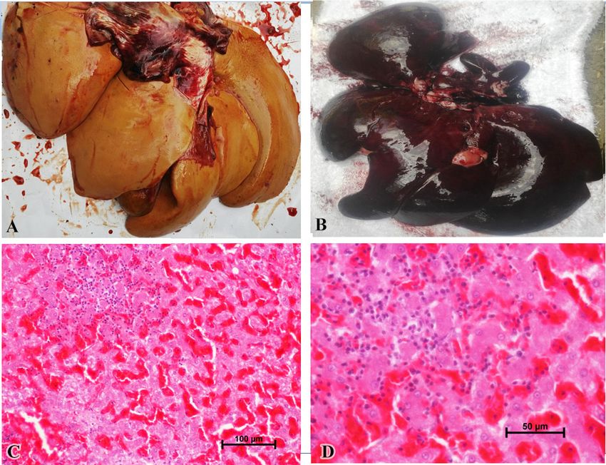
K. MATHESH ET AL.

killed whole cell leptospiral vaccine is serovar-specific and this serovar-specific protection of killed leptospiral vaccine has been
well documented [20].Vaccination in sloth bears against leptospirosis in India is performed using imported canine vaccines. Hence,
ample opportunity exists for developing indigenous leptospira vaccines targeting sloth bears utilizing the information generated by
performing MAT in this study.
   The inherent pitfalls of MAT have forced disease investigators to search for alternative field oriented tests [8]. A noteworthy
example is Latex Agglutination Test (LAT) which is a highly economical test suited for large-scale screening of sera
samples without using sophisticated equipments [32]. The advent of recombinant DNA technology enabled the use of outer
membrane proteins (OMPs) which can circumvent shortcomings of MAT when employed in diagnostic assays [22]. Leptospira
Immunoglobulin-Like Proteins (Lig proteins) are the serodiagnostic markers for detecting acute leptospirosis and Lig proteins
based immunodiagnostic assays would address the under-reporting of leptospirosis [12]. Hence, latex beads coated with first to fifth
tandem repeat domains of the conserved region of LigB protein (rLigBCon1-5) has been employed in this study for detection of
leptospirosis in sloth bears.
   The abnormalities in hepatorenal function serves as an indicator for suspicious case of leptospirosis and based on elevation in
the liver function tests such as serum glutamate oxalo acetic transaminase (SGOT) and serum glutamate pyruvic transaminase
(SGPT) as well as renal function tests such as blood urea nitrogen (BUN) and creatinine, clinicians can develop their own clinical
algorithm towards the confirmation of leptospirosis [26, 27]. Hence, serum biochemistry profile of terminally ill sloth bears has
been performed to study patterns which will augment disease diagnosis.
   A diagnostic PCR assay employing Lig gene was developed for the detection of pathogenic Leptospira interrogans in biological
samples with high sensitivity with a detection limit of six leptospires [29]. Several researchers have successfully employed PCR
as a diagnostic tool to maximize disease investigation credibility since positive PCR test results in tissue samples in the absence
of isolation of the organism by culture attempts have been reported [16, 36]. Hence, in this study diagnostic PCR employing LigB
gene reported previously [2] has been used to detect leptospirosis in biological samples of sloth bear whose results have been
compared with well established PCR assay [17].
   Therefore, the aim of the present study is to conduct a multi-pronged approach for the detection of leptospirosis in sloth bears
comprising of antigen and antibody detection which will be supported by serum biochemistry and gross/histopathology findings in
hepatic and renal tissues of sloth bears.

MATERIALS AND METHODS
Collection of sloth bear serum samples
   A total of 133 sera samples (113 and 20 sloth bear sera samples from Bear Rescue Centers located in Agra and Bannerghatta,
respectively) were submitted to Center for Wildlife, ICAR-IVRI by sloth bear rescue center authorities. Base Map of Sloth bear
rescue centers located in Agra and Bannerghatta are depicted in Fig. 1. The rescue center authorities restrained the animals using
standard protocols [33]. Briefly, the chemical immobilization of sloth bears was done using a combination of ketamine (5 mg/kg body
weight; Ketamil®, Troy Laboratories, Smitfield, NSW, Australia) and xylazine (2 mg/kg body weight; Xylazil®, Troy Laboratories)
delivered using a blow pipe on unsuspecting sloth bears thereby causing minimal stress to the animals. Blood was drawn from the
jugular vein 10–15 min after immobilization using a 20-guage sterile hypodermic needle in vacutainer (Becton Dickinson, Franklin
Lakes, NJ, USA). Serum was separated by centrifugation at 2,000 rpm for 20 min and stored at −20°C until used.

Histopathology
   Tissues such as liver and kidneys of all the dead sloth bears were fixed in 10% NBF (Neutral Buffered Formalin) and processed
for histopathology following standard procedures [23]. Briefly, the tissues were kept under running tap water overnight and
transferred to an automatic tissue processor (Leica Biosystems, Wetzler, Hesse, Germany) for further processing. Tissues were
dehydrated in ascending grades of ethanol, cleared in xylene and embedded with melted paraffin wax (Merck, Germany) (melting
point 60°C to 62°C), and paraffin blocks of the tissues were made. These tissue blocks were trimmed and sectioned at 5 µm
thickness using a microtome (Thermo Shandon, Woonsocket, RI, USA) placed on glass slides and were stained with hematoxylin
and eosin.

Serum biochemistry
   Serum samples of eleven sloth bears that succumbed to leptospirosis were subjected for serum biochemistry evaluation to
determine the elevation of hepatic and renal markers. The hepatic biomarkers such as SGOT and SGPT and renal biomarkers such
as creatinine and serum urea level for determining BUN were estimated using Modified IFCC method, Reitman and Frankel’s
method, Alkaline picrate method and modified Berthelot method, respectively, according to the instructions provided by the
manufacturer (Coral Clinical Systems, Tulip Diagnostics Private Limited, Verna, Goa, India).

PCR amplification of liver and kidney tissue samples
   Nucleic acids were extracted from sloth bear liver and kidney samples (25 mg each) using the DNeasy Blood &
Tissue Kit (Qiagen, Hilden, Germany) according to the manufacturer’s instructions which would yield 10–30 µg DNA
from liver and 15–30 µg DNA from kidney. PCR was performed using two sets of primers i.e. LigBF/LigBR (F3/B3)
(5′-AACCGGTCTGGTAGGATT-3′ and 5′-GAATCGGGGACGATGGAT-3′) and G1/G2 (5′-CTGAATCGCTGTATAAAAGT-3′

J. Vet. Med. Sci. 83(7): 1059–1067, 2021                                                                                       1060
DIAGNOSIS OF LEPTOSPIROSIS IN SLOTH BEARS
and 5′-GGAAAACAAATGGTCGGAAG-3′). The reaction conditions for LigBF/LigBR (F3/B3) and G1/G2 were followed as
described by [2] and [17] respectively. Briefly, both the PCR amplifications were carried out using 1.5 mM MgCl2 buffer, 200
µM of each dNTP, 20 pM of each primer, 1 U of recombinant Taq DNA Polymerase (Genetix Biotech Asia Pvt. Ltd., New Delhi,
India), and 10 ng of genomic DNA. PCR Amplification using G1/G2 and LigBF/LigBR primers was done using initial cycle of
95°C for 5 min followed by 30 amplification cycles with denaturation at 94°C for 1 min, annealing at 55°C and 54°C for 1 min
for G1/G2 and LigBF/LigBR (F3/B3) respectively and extension at 72°C for 1 min with a final extension of 72°C for 7 min and 5
min for G1/G2 and LigBF/LigBR (F3/B3) respectively. In both the PCR tests, saprophytic leptospires (Leptospira biflexa serovar
Patoc) was used as negative control and Leptospira interrogans serovar Icterohaemorrhagiae was used as positive control. The PCR
product was visualized using UV trans-illuminator (Bio-Rad, Hercules, CA, USA) after electrophoresis in 1.5% agarose gel stained
with ethidium bromide (0.1 µg/ml).
MAT
MAT was performed for screening of anti-leptospiral antibodies in sloth bears as described previously [37]. Briefly, serum
samples were diluted 1:50 in phosphate buffered saline (PBS) and a volume of leptospiral antigen, equal to the diluted serum
volume, was added to each well, making the final serum dilution 1/100 in the screening test. Four–eight day old live leptospiral
antigens (approx. 2 × 10⁸ leptospires/ml) of 16 leptospiral serovars (Leptospira interrogans serovar Australis strain Ballico, L.
interrogans serovar Autumnalis strain Akiyami A, L. interrogans serovar Ballum strain S102, L. interrogans serovar Bataviae strain
van Tienen, L. interrogans serovar Canicola strain Hond Utrecht IV, L. kirschneri serovar Cynopteri strain 3522C, L. interrogans
serovar Djasiman strain Djasiman, L. kirschneri serovar Grippotyphosa strain Moskva V, L. borgpetersenii serovar Hardjo strain
Hardjoprajitno, L. interrogans serovar Hebdomadis strain Hebdomadis, L. interrogans serovar Icterohaemorrhagiae strain RGA,
L. borgpetersenii serovar Javanica strain Veldrat Batavia 46, L. noguchii serovar Louisiana strain LSU 1945, L. interrogans
serovar Pomona strain Pomona, L. interrogans serovar Pyrogenes strain Salinem and L. borgpetersenii serovar Tarassovi strain
Perepelitsin) were used for performing MAT. The microtiter plates were incubated for 2 hr at 29°C and the serum-antigen mixtures
were examined using dark field microscopy. A positive outcome in MAT suggestive of exposure/seropositivity was defined as any
single serum sample found to have >50% reduction in the number of free non-agglutinable leptospires in the test when compared to
the control at 1 in 100 serum dilution for at least one leptospiral serovar.
Induction of expression and purification of recombinant LigBCon1-5 antigen
Escherichia coli (E. coli) M15 strain harboring recombinant plasmid pQE30-LigBCon1-5 was used for rLigBCon1-5
antigen production as described previously [13]. E. coli M15 strain was grown in Luria Bertani broth (Difco, Sparks, MD,
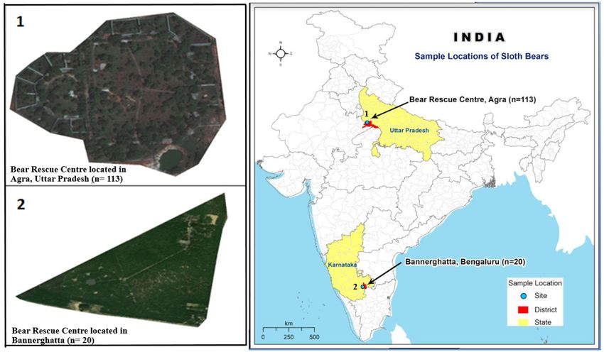
Fig. 1. Base map of Sloth bear rescue centers located in Agra and Bannerghatta from where sera samples were collected (Map was generated
using ArcGIS 10.5 software).
J. Vet. Med. Sci. 83(7): 1059–1067, 2021 1061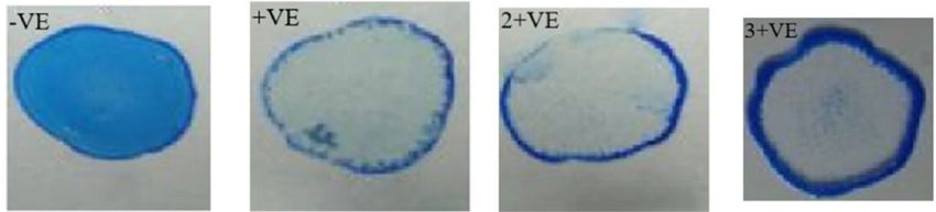
K. MATHESH ET AL.

USA) till the spectrometric reading at OD600 nm reached 0.5–0.7. The cells were then induced with 1 mM Isopropyl β-D-1-
thiogalactopyranoside (IPTG) (Sigma-Aldrich, St. Louis, MO, USA) and allowed to grow further for 6 hr at 37°C. Cells were
harvested and the proteins analyzed by Sodium dodecyl sulfate–polyacrylamide gel electrophoresis (SDS-PAGE). Purification
of recombinant protein was done using nickel–nitrilotriacetic acid (Ni-NTA) agarose affinity chromatography (Qiagen) as per
instructions of the manufacturer and analysis of purified recombinant protein was done on SDS-PAGE.

rLigBCon1-5 based LAT
   Latex beads were sensitized with rLigBCon1-5 antigen as described previously [13] with slight modifications. A 10% suspension
of latex particles (0.8 µm diameter, Sigma-Aldrich,) was washed thrice with glycine buffered saline (Glycine 0.1 M, NaCl 0.17
M; pH 8.2). Finally, the latex beads were made into a 2% suspension with glycine buffered saline which was later mixed with
an equal volume of rLigBCon1-5 antigen (1 mg/ml) diluted in the same buffer. The mixture was incubated at 37°C for 6 hr in a
shaking platform to ensure constant mixing. The sensitized latex beads were further blocked with Bovine Serum Albumin (Difco)
(5 mg/ml) and incubated overnight. Latex beads were centrifuged and the pellet was finally resuspended in glycine buffered saline
as a 2% suspension containing 0.02% sodium azide. The sensitized latex beads were stored at 4°C until use. LAT was performed
on glass slides by mixing equal volume of serum (20 µl) and sensitized beads (20 µl). The result was read within 2 min. Samples
were considered positive when there is formation of agglutination. A score of 3+ve, 2+ve and 1+ve were designated to sera which
showed agglutination within 30 sec, 30 sec to 1 min and 2 min respectively as described previously [34]. Samples were considered
negative if no agglutination was observed.

Statistical analysis
  The evaluation of rLigBCon1-5 LAT for detection of anti-leptospira antibodies in sloth bear as compared with MAT was
determined using Kappa statistics. The relative sensitivity, specificity and accuracy (in percentage) of rLigBCon1-5 LAT in
comparison to MAT has been calculated as described previously [13]. The calculation of predictive values (in percent) for positive
and negative test results was done as per standard method [18].

RESULTS
MAT
  The most frequently encountered leptospiral serovar in sloth bears was Pyrogenes (n=64) followed by Icterohaemorrhagiae
(n=59), Grippotyphosa (n=29), Javanica (n=25), Canicola (n=9) and Tarassovi (n=2). The leptospiral serovar distribution in Agra
and Bannerghatta sloth bear rescue centers are shown in Table 1. Out of eleven dead sloth bears which succumbed to leptospirosis,
seven sloth bears had a sera titer of 1in 800 while four showed a sera titer of 1in 400.

Serum biochemistry of dead sloth bears
   SGOT (IU/l) levels suggestive of liver function were found to be elevated in all the dead sloth bears while SGPT (IU/l) levels
showed marginal increase in most dead sloth bears as shown in Table 2. Moreover, Creatinine (mg/dl) and BUN (mg/dl) levels
were also found to be elevated in all the dead sloth bears as shown in Table 2. One noteworthy finding was the disproportional
exaggerated elevation in SGOT levels and the significantly higher SGOT/SGPT ratio during the terminal stages of the infection in
all the dead sloth bears. A positive correlation was also observed between MAT titer and SGOT/SGPT ratio since dead sloth bears
which had MAT titer of 1 in 800 had SGOT/SGPT ratio in the range of 3.42–7.19 while dead sloth bears which had MAT titer of
1in 400 had SGOT/SGPT ratio in the range of 2.59–3.58.

Gross and histopathology findings of liver and kidneys
  All the dead sloth bears had mucous membranes which were icteric in nature and post mortem revealed serosanguinous fluid

 Table 1. Leptospira serovars and their antibody titer found in bear rescue centers located in Agra and Bannerghatta
  Leptospira serovars whose                   Sloth bears seropositive by MAT                                  Sloth bears seropositive by MAT
  agglutinins are present in                  in Agra sloth bear rescue center                             in Bannerghatta sloth bear rescue center
   sloth bear sera samples        Total positivea 1 in 100 1 in 200 1 in 400 1 in 800            Total positiveb 1 in 100 1 in 200 1 in 400 1 in 800
  Pyrogenes                             64            32         20          11          1             0              0          0          0          0
  Icterohaemorrhagiae                   56            17         22          10          7             3              1          1          0          1
  Javanica                              25            22          2           1          0             0              0          0          0          0
  Grippotyphosa                         23            22          1           0          0             6              4          2          0          0
  Canicola                               9             7          2           0          0             0              0          0          0          0
  Tarassovi                              2             2          0           0          0             0              0          0          0          0
 a) Cumulative figure of sera positive for various serovars (179) exceed 79 (total Microscopic Agglutination Test (MAT) positive sera in Agra bear rescue
 center) as several sera (n=55) reacted with multiple leptospiral serovars. b) Cumulative figure of sera positive for various serovars (9) exceed 8 (total MAT
 positive sera in Bannerghatta bear rescue center) as one sera (n=1) reacted with multiple leptospiral serovars.

J. Vet. Med. Sci. 83(7): 1059–1067, 2021                                                                                                                   1062
DIAGNOSIS OF LEPTOSPIROSIS IN SLOTH BEARS

present inside the abdominal cavity (Supplementary Fig. 1). The most significant postmortem finding in the dead sloth bears was
the gross appearance of liver which was yellowish in color due to jaundice in six animals (Fig. 2A) while liver of five sloth bears
were highly congested and haemorrhagic with soft and friable consistency (Fig. 2B). The gall bladders of all the dead sloth bears
were highly distended (Supplementary Fig. 2). The histopathology of liver sections revealed severely engorged sinusoids (Fig.
2C and 2D) with severe hepatic degeneration (Supplementary Fig. 3) and multifocal areas of hepatic necrosis surrounded by
inflammatory cells in liver parenchyma of dead sloth bears (Supplementary Fig. 4). On gross examination, the kidneys appeared
soft, swollen and congested with tiny pale white spots (necrotic foci) on sub capsular surface in four dead sloth bears (Fig. 3A
and 3B). The major histopathological findings of renal tissue in sub-acute cases include moderate to severe lymphoplasmacytic
and neutrophilic tubulointerstitial nephritis (Supplementary Fig. 5) with tubular degeneration, necrosis, mineralization and mild

Table 2. Serum biochemistry profile of dead sloth bears

Serial No of                      Normal SGOT        Normal SGPT           Normal SGOT/         Normal BUN                Normal
                MAT       SGOTb       level   SGPTc      level   SGOT/SGPT               BUNd               Creatinine Creatinine level
 dead sloth                                                                 SGPT Ratio          (mean + SD)
                titer      (IU/l) (mean + SD) (IU/l) (mean + SD)   Ratio                (mg/dl)              (mg/dl)    (mean + SD)
bear (DSB)a                          (IU/l)             (IU/l)              (mean + SD)           (mg/dl)                  (mg/dl)
DSB No 1       1 in 800    276   56.37 ± 27.02    43.0   36.78 ± 7.52       6.42        1.53 ± 0.35      31.9    6.24 ± 1.49      1.8       1.08 ± 0.24
DSB No 2       1 in 800    196                    39.0                      5.03                         26.0                     1.6
DSB No 3       1 in 800    345                    48.0                      7.19                         26.5                     2.1
DSB No 4       1 in 800    208                    42.0                      4.95                         40.5                     2.0
DSB No 5       1 in 800    165                    40.5                      4.07                         32.0                     1.8
DSB No 6       1 in 800    174                    45.0                      3.87                         36.0                     1.9
DSB No 7       1 in 800    135                    39.5                      3.42                         36.0                     1.7
DSB No 8       1 in 400    129                    36.0                      3.58                         28.1                     1.6
DSB No 9       1 in 400    114                    44.0                      2.59                         29.5                     1.6
DSB No 10      1 in 400    112                    38.5                      2.91                         35.5                     1.6
DSB No 11      1 in 400    125                    39.5                      3.16                         26.0                     1.9
a) The names of dead sloth bears are kept confidential following request from bear rescue park authorities. b) SGOT stands for serum glutamate oxalo acetic
transaminase. c) SGPT stands for serum glutamate pyruvic transaminase. d) BUN stands for blood urea nitrogen.

   Fig. 2. Gross and histopathology lesions of liver of dead sloth bears. A) Soft and friable icteric liver showing yellowish color due to jaundice
      and B) highly congested and haemorrhagic liver which were soft and friable. C, D) Liver showing severely engorged sinusoids with
      degeneration of hepatocytes and infiltration of mononuclear cells in the hepatic parenchyma hematoxylin-eosin (HE) ×200 & HE ×400.

J. Vet. Med. Sci. 83(7): 1059–1067, 2021                                                                                                            1063
K. MATHESH ET AL.
interstitial fibrosis (Fig. 3C). However, in chronic cases, inflammation is less severe and is accompanied by pronounced interstitial
fibrosis and tubular atrophy (Fig. 3D).
Polymerase chain reaction of tissue samples
PCR amplification of kidney tissue samples using G1/G2 and LigBF/LigBR (F3/B3) primers revealed positive test results as
indicated by amplicons of 285 bp and 219 bp respectively. The detection of Leptospira sp. DNA in the renal tissue of all dead sloth
bears by PCR employing both G1/G2 and LigBF/LigBR (F3/B3) primers provided diagnostic confirmation that leptospirosis was
responsible for the mortality of sloth bears in both sloth bear rescue centers due to renal failure. Further, the results obtained using
PCR helped to corroborate the gross and histopathological findings observed in renal tissue which were suggestive for leptospirosis.
Recombinant LigBCon1-5 antigen expression and utilization in LAT
We have obtained a high level expression of rLigBCon1-5 protein estimated at approximately 20 mg of protein per liter of IPTG
induced culture. LAT developed using rLigBCon1-5 antigen showed a clear-cut agglutination with positive sera which can be
visualized easily and can clearly differentiate negative sera showing homogeneous suspension (Fig. 4).
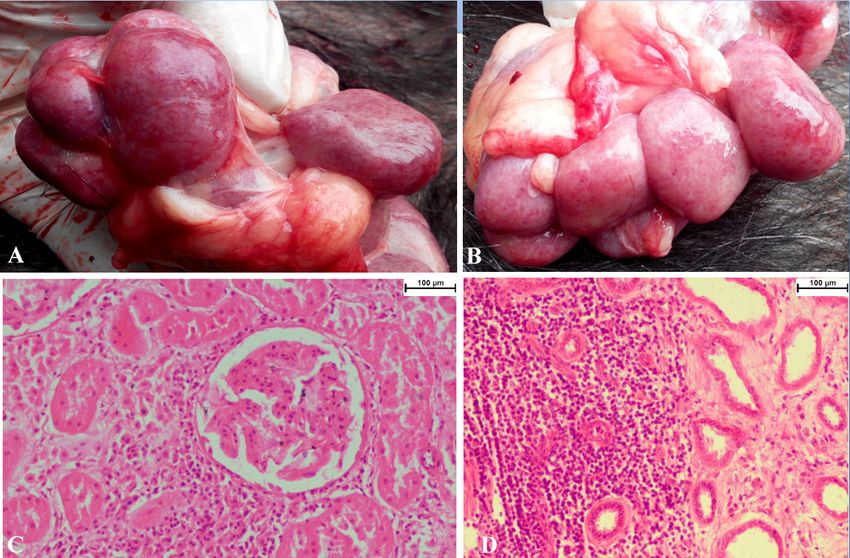
Fig. 3. Gross and histopathology lesions of kidneys of dead sloth bears. A, B) Soft, swollen and congested kidneys with tiny pale white
spots (necrotic foci) on subcapsular surface. C) Histopathology of kidneys showing tubular degeneration, necrosis, mineralization and mild
interstitial fibrosis hematoxylin-eosin (HE) ×200. D) Histopathology of kidneys in chronic case showing pronounced interstitial fibrosis
and tubular atrophy with less severe inflammation HE ×200.
Fig. 4. Latex agglutination test results of sloth bear sera samples using recombinant LigBCon1-5 protein.
J. Vet. Med. Sci. 83(7): 1059–1067, 2021 1064DIAGNOSIS OF LEPTOSPIROSIS IN SLOTH BEARS Correlation between MAT titer and LAT score A positive correlation exists between MAT titer and LAT score. Sloth bear sera (n=23) showing MAT titer ≥1 in 400 gave 3+ve LAT score and the agglutination intensity was high with the agglutinins forming a thick halo at the periphery while the center was virtually empty (Fig. 4). Sera samples (n=27) showing 1 in 200 MAT titer gave 2+ve LAT score with moderate agglutination intensity. Sloth bear sera (n=28) showing 1 in 100 MAT titer gave 1+ve LAT score (low intensity of agglutination). Nine sera with 1in 100 MAT titer showed no agglutination with rLigBCon1-5 based LAT (Fig. 4). Out of 133 sera sample tested, 87 (65.41%) and 78 (58.65%) sera samples were found positive by MAT and rLigBCon1-5 based LAT respectively. The relative sensitivity, specificity, accuracy, positive and negative predictive values of rLigBCon1-5 based LAT in comparison to MAT were 89.66%, 100%, 93.23%, 100% and 83.64% respectively. Further, Kappa value of 0.86 (95% C.I. 0.77–0.95) for rLigBCon1-5 based LAT indicates high agreement with MAT. DISCUSSION Sloth bears are enlisted in Appendix I of CITES (the Convention on International Trade in Endangered Species of Wild Fauna and Flora) and these endangered animals are protected under Schedule I of the Indian Wildlife Protection Act of 1972 [14]. The five predominant leptospiral serovars (Pyrogenes, Icterohaemorrhagiae, Grippotyphosa, Javanica and Canicola) observed in the present study were also among the five leading serovars reported in seroprevalence studies conducted in four rat species in India [7]. Globally, free living rodents are implicated as the only mammals capable of maintaining close contact with captive wildlife in zoos [19]. This clearly suggests the role of rodents as the main agent for transmission of leptospirosis in both sloth bear rescue centers. There was clear evidence of rodent infestation in granaries as well as sloth bear enclosures. The rodents were attracted to sloth bear enclosures due to the food leftover by sloth bears and the rodent urination on leftover food might be one probable source of infection. Hence, a concerted effort for rodent control was launched which included setting up rodent traps and rodenticides at strategic locations especially at entry/exit points within the rescue park premises. The leftover food was immediately disposed off from sloth bear enclosures following meal time. In order to counteract the post deluge spike in leptospirosis, prophylactic use of doxycycline @5 mg/kg body weight was given prior to monsoon season. Moreover, both the rescue centers started the practice of biannual vaccination with Nobivac DHPPi / RL in captive sloth bears with encouraging results. The dedicated effort of sloth bear rescue center staff ensured that there was no mortality in both rescue centers for the past five years. Further, the MAT titers of recuperating sloth bears have tapered to tolerable levels (MAT titer ≤1 in 100). In this study, mucous membranes of all the dead sloth bears were icteric in nature. Moreover, the livers of six sloth bears were yellowish in color due to jaundice. The reason is due to invasion of the intercellular junctions of host hepatocytes by pathogenic leptospires which contributed to the disruption of the junction and the subsequent leakage of bile from bile canaliculi contributing to jaundice and elevation in SGOT levels in serum [25]. In this study, pale white spots (necrotic foci) were observed on sub capsular surface of kidneys of four dead sloth bears. The gross findings observed in kidneys in the present study are in concordance with the observations made by [4] who suggested that Leptospira spp. were associated with white-spotted bovine kidneys based on PCR results using LipL32 primers. The kidneys of sloth bears which died due to sub-acute leptospirosis showed tubulointerstitial nephritis characterized by severe degeneration of proximal convoluted tubules with interstitial and periglomerular infiltration of mononuclear cells and mild fibrosis. Our observations are in concordance with findings of [35] and [30] who observed acute tubulointerstitial nephritis as well as acute tubular necrosis in patients with acute leptospirosis. The typical pathological findings observed in sloth bears which died due to chronic leptospirosis was interstitial and periglomerular fibrosis and infiltration of plasma cells, lymphocytes and few macrophages. Chronic Leptospira interrogans infection was studied in a mouse model by [15] and had suggested that the leptospiral colonization of the kidneys triggers renal fibrosis which is characterized by the pathological accumulation of extra-cellular matrix components that can compromise the kidney functions of patients with leptospirosis. The serum biochemistry of all the dead sloth bears revealed that both SGOT and SGPT levels were elevated. The mean ± standard deviation for SGOT and SGPT in apparently healthy bears (both sex combined) is 56.37 ± 27.02 IU/l and 36.78 ± 7.52 respectively [9]. However, all the dead sloth bears showed SGOT levels on the higher range of 112–345 IU/l while SGPT levels ranged from 36–48 IU/l. The average SGOT/SGPT ratio in normal sloth bears is approximately 1.53 ± 0.35 [9]. One hallmark feature observed in serum biochemistry in all the eleven sloth bears which succumbed to leptospirosis was the significantly higher SGOT/SGPT ratio during the terminal stages of the infection. The SGOT levels elevated progressively without a concomitant change of SGPT during the acute disease course in all dead sloth bears. Our experience with regard to SGOT/SGPT levels in terminal stages of leptospirosis suggests that SGOT/SGPT ratio can serve as a valuable prognostic parameter for leptospirosis with a ratio of ≥4.0 indicative of a grave prognosis for the disease in sloth bears. We have observed SGOT/SGPT ratio ranging from 2.59–7.19 in sloth bears which succumbed to leptospirosis. The disproportionate and exaggerated elevation in SGOT levels and high SGOT/SGPT ratio in human patients who died due to leptospirosis was also observed by [10]. All the dead sloth bears showed BUN levels on higher range of 26–40.5 mg/dl whereas the normal range of BUN in sloth bear is 6.24 ± 1.49 (95% confidence Interval 5.7–6.7) [9]. BUN is used to evaluate kidney function and elevations in BUN level are often a result of a decrease in Glomerular Filtration Rate (GFR) which clearly suggests kidney damage resulting in decreased excretion of urea in urine [24]. All the dead sloth bears showed creatinine levels on slightly higher range of 1.6–2.1 mg/dl whereas the normal range of creatinine in sloth bear is 1.08 ± 0.24 (95% confidence Interval 1.0–1.15) [9]. An elevation in the serum creatinine concentration usually reflects J. Vet. Med. Sci. 83(7): 1059–1067, 2021 1065
K. MATHESH ET AL.

a reduction in the glomerular filtration rate (GFR) suggestive of kidney damage [31].
   Our study clearly suggests that ‘Point of care testing’ for leptospirosis in both sloth bear rescue centers can be achieved utilizing
rLigBCon1-5 based LAT. Our results are in agreement with findings of [5] and [13] where LAT using recombinant LigBCon1-5
protein have yielded high sensitivity and specificity while investigating canine and bovine leptospirosis respectively. In future,
we are planning to employ rLigBCon1-5 antigen based LAT to identify sloth bears that contract leptospirosis despite vaccination
with Nobivac® DHPPi/RL. Diagnostic assays based on recombinant LigB antigen would be a valuable tool to identify animals
that contract leptospirosis despite vaccination [28]. Vaccination of sloth bears with Nobivac® DHPPi/RL (Intervet, Kenilworth, NJ,
USA) will generate agglutinins against serogroups Icterohaemorrhagiae and Canicola but fail to confer cross protection against
natural infection with serovars belonging to unrelated serogroups. This main drawback of leptospira bacterin based vaccines of
limited efficacy spectrum cannot be solved by increasing the amount of protective antigen [1]. Hence, it is of paramount importance
to conduct regular epidemiological surveillance in sloth bear rescue centers to unravel temporal variation due to the emergence of
new serovars which can become causative agent for leptospirosis in sloth bears despite vaccination. Therefore, the authors of this
manuscript are in favor of ‘tailored leptospiral vaccines’ which was also proposed by [21] which contain serovars representative
of those present in the population to be immunized. The MAT results of this study have convinced the authors that the tailored
leptospiral vaccine should include serovars Pyrogenes (most frequently encountered serovar), Grippotyphosa and Javanica along
with the existing leptospira vaccine serovars such as Icterohaemorrhagiae and Canicola.
   In conclusion, the multipronged approach adopted in the present study to diagnose leptospirosis in sloth bears based on antigen
detection (using PCR employing G1/G2 primers and LigBF/LigBR (F3/B3) primers), antibody detection (MAT and rLigBCon1-5
based LAT), serum biochemistry profile (SGOT, SGPT, Creatinine and BUN) and gross/histopathological findings of liver and
kidney would permit the implementation of intervention strategies in sloth bears based on early case detection and timely initiation
of antimicrobial therapy.

CONFLICT OF INTEREST. All authors have no potential conflicts of interest.

ACKNOWLEDGMENTS. We sincerely acknowledge the logistical support provided by Director, ICAR-IVRI and Head, Center
for Wildlife (Dr. Abhijit M Pawde). We also acknowledge the funds received from Project ‘National Referral Center on Wildlife
Healthcare’ provided by Central Zoo Authority of India (CZA). Further, we acknowledge the funding received from Indian Council of
Agricultural Research (ICAR) funded project titled “Outreach Program on Zoonotic diseases” [ICAR Grant number 3021].

REFERENCES

 1. Adler, B. 2015. Vaccines against leptospirosis. Curr. Top. Microbiol. Immunol. 387: 251–272. [Medline]
 2. Ali, S. A., Kaur, G., Boby, N., Sabarinath, T., Solanki, K., Pal, D. and Chaudhuri, P. 2017. Rapid and visual detection of Leptospira in urine by
    LigB-LAMP assay with pre-addition of dye. Mol. Cell. Probes 36: 29–35. [Medline] [CrossRef]
 3. André-Fontaine, G. 2016. Leptospirosis in domestic animals in France: serological results from 1988 to 2007. Rev. Sci. Tech. 35: 913–923.
    [Medline] [CrossRef]
 4. Azizi, S., Tajbakhsh, E., Hajimirzaei, M. R., Gholami Varnamkhast, M., Sadeghian, H. and Oryan, A. 2012. Evaluation of ‘white-spotted kidneys’
    associated with leptospirosis by polymerase chain reaction based LipL32 gene in slaughtered cows. J. S. Afr. Vet. Assoc. 83: 69. [Medline]
    [CrossRef]
 5. Behera, S. K., Sabarinath, T., Kumar, A., Ali, S. A. and Chaudhuri, P. 2016. Evaluation of recombinant LigB based in-house latex agglutination
    assay for sero-surveillance of canine leptospirosis. J. Vet. Public Health 14: 19–23.
 6. Biswas, S. and Sankar, K. 2002. Prey abundance and food habits of tigers (Panthera tigris tigris) in Pench National Park, Madhya Pradesh, India. J.
    Zool. (Lond.) 256: 411–420. [CrossRef]
 7. Boey, K., Shiokawa, K. and Rajeev, S. 2019. Leptospira infection in rats: a literature review of global prevalence and distribution. PLoS Negl. Trop.
    Dis. 13: e0007499. [Medline] [CrossRef]
 8. Budihal, S. V. and Perwez, K. 2014. Leptospirosis diagnosis: competancy of various laboratory tests. J. Clin. Diagn. Res. 8: 199–202. [Medline]
 9. Chandra Mohan, S., Nair, S. V., Gupta, R., Karikalan, M., Ilayaraja, S., Shanmugam, A. A. and Sharma, A. K. 2018. Biochemical reference intervals
    for semi-captive sloth bears (Melursus ursinus ursinus) in India. Int. J. Livest. Res. 8: 311–315.
10. Chang, M. L., Yang, C. W., Chen, J. C., Ho, Y. P., Pan, M. J., Lin, C. H. and Lin, D. Y. 2005. Disproportional exaggerated aspartate transaminase is
    a useful prognostic parameter in late leptospirosis. World J. Gastroenterol. 11: 5553–5556. [Medline] [CrossRef]
11. Collantes, T. M. A., David, J. M. F., Vergara, E. J. S., Armea, S. R. D. and Flores, M. L. S. 2016. Detection of pathogenic leptospires and analysis of
    factors and clinical signs associated with canine leptospirosis. Philipp. J. Vet. Anim. Sci. 42: 41–48.
12. Croda, J., Ramos, J. G., Matsunaga, J., Queiroz, A., Homma, A., Riley, L. W., Haake, D. A., Reis, M. G. and Ko, A. I. 2007. Leptospira
    immunoglobulin-like proteins as a serodiagnostic marker for acute leptospirosis. J. Clin. Microbiol. 45: 1528–1534. [Medline] [CrossRef]
13. Deneke, Y., Sabarinath, T., Gogia, N., Lalsiamthara, J., Viswas, K. N. and Chaudhuri, P. 2014. Evaluation of recombinant LigB antigen-based
    indirect ELISA and latex agglutination test for the serodiagnosis of bovine leptospirosis in India. Mol. Cell. Probes 28: 141–146. [Medline]
    [CrossRef]
14. Dharaiya N., Bargali H.S., Sharp T. 2020. Melursus ursinus (amended version of 2016 assessment). The IUCN Red List of Threatened Species
    2020: e.T13143A166519315.
15. Fanton d’Andon, M., Quellard, N., Fernandez, B., Ratet, G., Lacroix-Lamandé, S., Vandewalle, A., Boneca, I. G., Goujon, J. M. and Werts, C. 2014.
    Leptospira Interrogans induces fibrosis in the mouse kidney through Inos-dependent, TLR- and NLR-independent signaling pathways. PLoS Negl.
    Trop. Dis. 8: e2664. [Medline] [CrossRef]
16. Fearnley, C., Wakeley, P. R., Gallego-Beltran, J., Dalley, C., Williamson, S., Gaudie, C. and Woodward, M. J. 2008. The development of a real-time
    PCR to detect pathogenic Leptospira species in kidney tissue. Res. Vet. Sci. 85: 8–16. [Medline] [CrossRef]

J. Vet. Med. Sci. 83(7): 1059–1067, 2021                                                                                                            1066
DIAGNOSIS OF LEPTOSPIROSIS IN SLOTH BEARS
17. Gravekamp, C., Van de Kemp, H., Franzen, M., Carrington, D., Schoone, G. J., Van Eys, G. J. J. M., Everard, C. O. R., Hartskeerl, R. A. and
Terpstra, W. J. 1993. Detection of seven species of pathogenic leptospires by PCR using two sets of primers. J. Gen. Microbiol. 139: 1691–1700.
[Medline] [CrossRef]
18. Jacobson, R. H. 1998. Validation of serological assays for diagnosis of infectious diseases. Rev. Sci. Tech. 17: 469–526. [Medline] [CrossRef]
19. Jung, B. Y., Choi, J. S., Kim, K. T., Song, Y. K., Lee, S. H., Lee, K. W., Kim, J. Y. and Moon, O. K. 2007. Seroprevalence of leptospirosis in Korean
municipal zoo animals. J. Vet. Med. Sci. 69: 861–863. [Medline] [CrossRef]
20. Koizumi, N. and Watanabe, H. 2005. Leptospirosis vaccines: past, present, and future. J. Postgrad. Med. 51: 210–214. [Medline]
21. Levett, P. N. 2001. Leptospirosis. Clin. Microbiol. Rev. 14: 296–326. [Medline] [CrossRef]
22. Lin, X., Chen, Y. and Yan, J. 2008. Recombinant multiepitope protein for diagnosis of leptospirosis. Clin. Vaccine Immunol. 15: 1711–1714.
[Medline] [CrossRef]
23. Luna, L. G. 1968. Manual of Histologic Staining Methods of the Armed forces Institute of Pathology, 3rd ed., McGraw-Hill Book Co., Washington,
D.C.
24. Macedo, E. and Mehta, R. L. 2013. National kidney foundation primer on kidney diseases. pp. 294–303. In: National Kidney Foundation Primer on
Kidney Diseases, 6th ed. (Gilbert S. and Weiner D. eds.), Saunders, Philadelphia.
25. Miyahara, S., Saito, M., Kanemaru, T., Villanueva, S. Y. A. M., Gloriani, N. G. and Yoshida, S. 2014. Destruction of the hepatocyte junction by
intercellular invasion of Leptospira causes jaundice in a hamster model of Weil’s disease. Int. J. Exp. Pathol. 95: 271–281. [Medline] [CrossRef]
26. Mutoh, Y., Koizumi, N., Morino, E., Hayakawa, K., Kato, Y. and Ohmagari, N. 2017. Leptospirosis cases in the Tokyo metropolitan area, Japan.
Jpn. J. Infect. Dis. 70: 669–671. [Medline] [CrossRef]
27. Nagarajan, P., Sundhararajan, A., Jeyaseelan Senthinath, T., Robert, A. A., Natarajaseenivasan, K. and Danialas, J. P. I. 2015. Analysis of
biochemical parameters of laboratory confirmed leptospirosis samples. J. Pharm. Biol. Sci. 3: 240–244.
28. Palaniappan, R. U. M., Chang, Y. F., Hassan, F., McDonough, S. P., Pough, M., Barr, S. C., Simpson, K. W., Mohammed, H. O., Shin, S.,
McDonough, P., Zuerner, R. L., Qu, J. and Roe, B. 2004. Expression of leptospiral immunoglobulin-like protein by Leptospira interrogans and
evaluation of its diagnostic potential in a kinetic ELISA. J. Med. Microbiol. 53: 975–984. [Medline] [CrossRef]
29. Palaniappan, R. U., Chang, Y. F., Chang, C. F., Pan, M. J., Yang, C. W., Harpending, P., McDonough, S. P., Dubovi, E., Divers, T., Qu, J. and
Roe, B. 2005. Evaluation of lig-based conventional and real time PCR for the detection of pathogenic leptospires. Mol. Cell. Probes 19: 111–117.
[Medline] [CrossRef]
30. Salkade, H. P., Divate, S., Deshpande, J. R., Kawishwar, V., Chaturvedi, R., Kandalkar, B. M. and Vaideeswar, P. 2005. A study of sutopsy findings
in 62 cases of leptospirosis in a metropolitan city in India. J. Postgrad. Med. 51: 169–173. [Medline]
31. Samra, M. and Abcar, A. C. 2012. False estimates of elevated creatinine. Perm. J. 16: 51–52. [Medline] [CrossRef]
32. Senthilkumar, T., Subathra, M., Phil, M., Ramadass, P. and Ramaswamy, V. 2008. Rapid serodiagnosis of leptospirosis by latex agglutination test
and flow-through assay. Indian J. Med. Microbiol. 26: 45–49. [Medline] [CrossRef]
33. Shanmugam, A. A., Kumar, J. K., Selvaraj, I. and Selvaraj, V. 2008. Hematology of sloth bears (Melursus ursinus ursinus) from two locations in
India. J. Wildl. Dis. 44: 509–518. [Medline] [CrossRef]
34. Smits, H. L., van der Hoorn, M. A., Goris, M. G., Gussenhoven, G. C., Yersin, C., Sasaki, D. M., Terpstra, W. J. and Hartskeerl, R. A. 2000. Simple
latex agglutination assay for rapid serodiagnosis of human leptospirosis. J. Clin. Microbiol. 38: 1272–1275. [Medline] [CrossRef]
35. Tanaka, K., Tanabe, K., Nishii, N., Takiue, K., Sugiyama, H. and Wada, J. 2017. Sustained Tubulointerstitial Inflammation in Kidney with Severe
Leptospirosis. Intern. Med. 56: 1179–1184. [Medline] [CrossRef]
36. Tulsiani, S. M., Graham, G. C., Dohnt, M. F., Burns, M. A. and Craig, S. B. 2011. Maximizing the chances of detecting pathogenic leptospires
in mammals: the evaluation of field samples and a multi-sample-per-mammal, multi-test approach. Ann. Trop. Med. Parasitol. 105: 145–162.
[Medline] [CrossRef]
37. USDA (United States Department of Agriculture) National Veterinary Services Laboratories. 1987. Microtiter technique for detection of Leptospira
antibodies. Proc. Annu. Meet. U. S. Anim. Health Assoc. 91: 65–73.
38. Veeraselvam, M., Sridhar, R., Senthikumar, T. M. A., Jayathangaraj, M. G. and Rajesh, N. V. 2013. Seroprevalence of leptospirosis in captive sloth
bears (Melursus ursinus). Indian Vet. J. 90: 113–114.
39. Veeraselvam, M., Sridhar, R., Senthilkumar, T. M. A., Perumal, P. and Jayathangaraj, M. G. 2015. Diagnosis of Mycobacterium bovis in captive
sloth bears (Melursus ursinus) by polymerase chain reaction. Proc. Zool. Soc. 68: 109–111. [CrossRef]
J. Vet. Med. Sci. 83(7): 1059–1067, 2021 1067You can also read