A Transcriptomic Study Reveals That Fish Vibriosis Due to the Zoonotic Pathogen Vibrio vulnificus Is an Acute Inflammatory Disease in Which ...
←
→
Page content transcription
If your browser does not render page correctly, please read the page content below
ORIGINAL RESEARCH
                                                                                                                                                  published: 01 April 2022
                                                                                                                                        doi: 10.3389/fmicb.2022.852677

                                              A Transcriptomic Study Reveals That
                                              Fish Vibriosis Due to the Zoonotic
                                              Pathogen Vibrio vulnificus Is an
                                              Acute Inflammatory Disease in Which
                                              Erythrocytes May Play an Important
                                              Role
                                              Carla Hernández-Cabanyero1, Eva Sanjuán1, Felipe E. Reyes-López2,3, Eva Vallejos-Vidal2,4,
                                              Lluis Tort3 and Carmen Amaro1*
                                              1
                                               Instituto Universitario de Biotecnología y Biomedicina (BIOTECMED), Universitat de València, Valencia, Spain, 2 Centro de
                          Edited by:          Biotecnología Acuícola, Facultad de Química y Biología, Universidad de Santiago de Chile, Santiago, Chile, 3 Department of
                    David Kornspan,           Cell Biology, Physiology, and Immunology, Universitat Autònoma de Barcelona, Bellaterra, Spain, 4 Facultad de Medicina
     Kimron Veterinary Institute, Israel      Veterinaria y Agronomía, Universidad de Las Américas, Santiago, Chile
                       Reviewed by:
                       Keun Hwa Lee,
                                              Vibrio vulnificus is a marine zoonotic pathogen associated with fish farms that is considered
    Hanyang University, South Korea
      Maria Del Mar Ortega-Villaizan,         a biomarker of climate change. Zoonotic strains trigger a rapid death of their susceptible
Miguel Hernández University of Elche,         hosts (fish or humans) by septicemia that has been linked to a cytokine storm in mice.
                                  Spain
                        Panpan Zhao,          Therefore, we hypothesize that V. vulnificus also causes fish death by triggering a cytokine
                Jilin University, China       storm in which red blood cells (RBCs), as nucleated cells in fish, could play an active role.
                  *Correspondence:            To do it, we used the eel immersion infection model and then analyzed the transcriptome
                      Carmen Amaro
                                              in RBCs, white BCs, and whole blood using an eel-specific microarray platform. Our
                 carmen.amaro@uv.es
                                              results demonstrate that V. vulnificus triggers an acute but atypical inflammatory response
                   Specialty section:         that occurs in two main phases. The early phase (3 h post-infection [hpi]) is characterized
         This article was submitted to
                                              by the upregulation of several genes for proinflammatory cytokines related to the mucosal
               Systems Microbiology,
               a section of the journal       immune response (il17a/f1 and il20) along with genes for antiviral cytokines (il12β) and
             Frontiers in Microbiology        antiviral factors (ifna and ifnc). In contrast, the late phase (12 hpi) is based on the
         Received: 11 January 2022            upregulation of genes for typical inflammatory cytokines (il1β), endothelial destruction
          Accepted: 07 March 2022
           Published: 01 April 2022           (mmp9 and hyal2), and, interestingly, genes related to an RNA-based immune response
                            Citation:         (sidt1). Functional assays revealed significant proteolytic and hemolytic activity in serum
Hernández-Cabanyero C, Sanjuán E,             at 12 hpi that would explain the hemorrhages characteristic of this septicemia in fish. As
   Reyes-López FE, Vallejos-Vidal E,
                                              expected, we found evidence that RBCs are transcriptionally active and contribute to this
        Tort L and Amaro C (2022) A
  Transcriptomic Study Reveals That           atypical immune response, especially in the short term. Based on a selected set of marker
   Fish Vibriosis Due to the Zoonotic         genes, we propose here an in vivo RT-qPCR assay that allows detection of early sepsis
      Pathogen Vibrio vulnificus Is an
       Acute Inflammatory Disease in          caused by V. vulnificus. Finally, we develop a model of sepsis that could serve as a basis
    Which Erythrocytes May Play an            for understanding sepsis caused by V. vulnificus not only in fish but also in humans.
                      Important Role.
        Front. Microbiol. 13:852677.          Keywords: Vibrio vulnificus, zoonotic pathogen, blood, erythrocytes, European eel, host-pathogen relationship,
   doi: 10.3389/fmicb.2022.852677             immune response

Frontiers in Microbiology | www.frontiersin.org                                      1                                              April 2022 | Volume 13 | Article 852677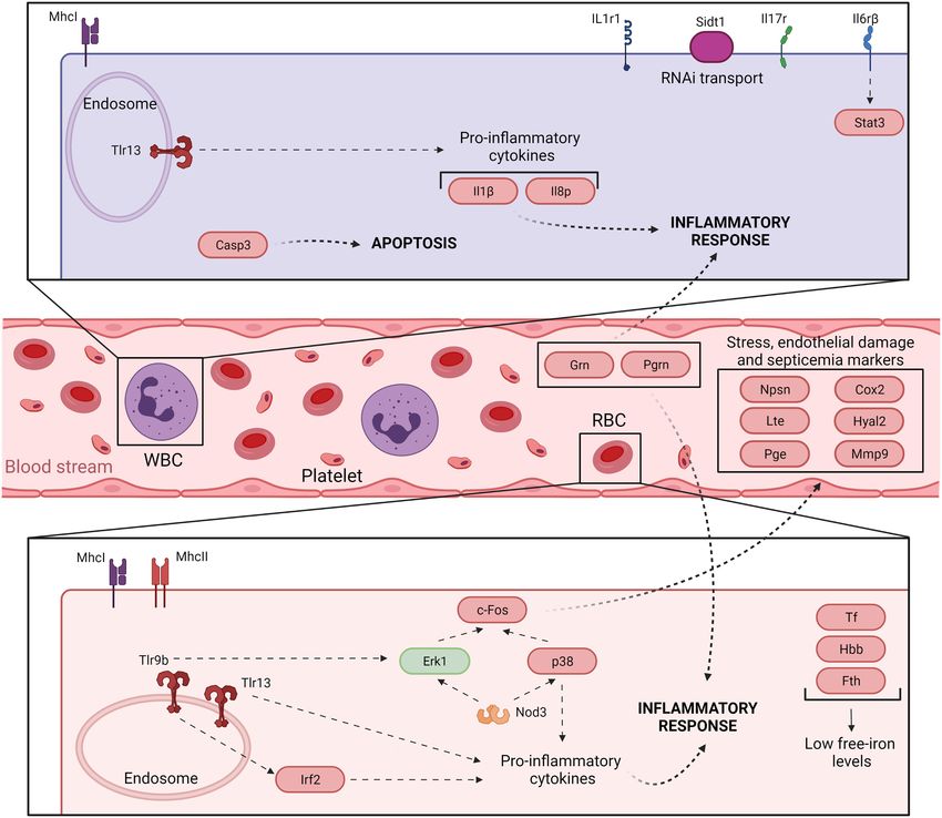
Hernández-Cabanyero et al.                                                                                           Eel Vibriosis Due to V. vulnificus

INTRODUCTION                                                               microarray platform containing probes for thousands of immune-
                                                                           related genes (Callol et al., 2015b). Since fish red blood cells
Fish vibriosis encompasses a group of diseases with common                 (RBCs) are nucleated cells involved in defense against viruses
clinical signs caused by different genus Vibrio species (Amaro             (Workenhe et al., 2008; Morera et al., 2011; Dahle et al., 2015;
et al., 2020). Among these species, Vibrio vulnificus stands out           Nombela et al., 2017; Nombela and Ortega-Villaizan, 2018),
as the only one linked to zoonotic cases acquired through                  we also considered analyzing the RBCs-associated transcriptome.
contact with diseased fish, mainly farmed fish (Amaro et al.,              Accordingly, we studied and compared the transcriptome
2015; Oliver, 2015). Moreover, it is the only one that can                 associated with RBCs, white BCs (WBCs), and whole blood
cause rapid death by septicemia in both humans and fish                    (B) at 0-, 3- and 12-h post-infection (hpi). We then validated
(Ceccarelli et al., 2019; Amaro et al., 2020).                             the results obtained by RT-qPCR and performed a series of
    The severity of outbreaks caused by V. vulnificus in fish              functional confirmatory assays. Our results suggest that
farms is highly dependent on water temperature since the                   V. vulnificus triggers an acute but atypical inflammatory response
highest mortality rates occur at temperatures above 25°C (Amaro            in two main phases. The early phase (detectable at 3 hpi) is
et al., 1995). This temperature dependence explains why this               characterized by the upregulation of important genes for
vibriosis (hereafter Vv-vibriosis) mainly affects fish reared above        proinflammatory cytokines related to the mucosal immune
22°C, as well as why clinical cases in humans and animals                  response (il17a/f1 [in RBCs] and il20 [in RBCs and WBCs])
are increasing with global warming (Ceccarelli et al., 2019;               along with antiviral cytokine genes (il12β [in both cell types])
Amaro et al., 2020). Part of the reason for this dependence                and antiviral factors (ifna and ifnc [in WBCs only]). The late
is that an increase in temperature above 22°C significantly                phase (detectable at 12 hpi) is characterized by the upregulation
increases the transcription of several pathogen genes related              of genes for typical inflammatory cytokines (il1β), endothelial
to colonization and resistance to innate immunity in fish, thus            destruction (mmp9 and hyal2), and genes related to an RNA-based
favoring disease transmission and unbalancing the host-pathogen            immune response (sidt1), all of them detected in B samples.
relationship toward the pathogen (Hernández-Cabanyero et al.,              Functional assays revealed significant proteolytic and hemolytic
2020). These data correlate with field data and underline the              activity in serum at 12 hpi that would explain the hemorrhages
relevance of V. vulnificus as a biological barometer of climate            characteristic of this septicemia. As expected, we found evidence
change (Baker-Austin et al., 2012, 2018).                                  that RBCs are transcriptionally active and may contribute to
    Vv-vibriosis differs from other vibriosis in that death from           this atypical immune response, especially in the short term.
septicemia occurs very quickly and without the pathogen                    We also selected a series of marker genes and validated in
reaching as high numbers in the blood or tissues as it does                vivo an RT-qPCR assay for early detection of Vv-vibriosis.
in other vibriosis (Valiente and Amaro, 2006; Valiente et al.,             Finally, we developed a model of septicemia that could serve
2008a). Previous studies in eels infected by immersion showed              as a basis for understanding sepsis caused by V. vulnificus not
that the pathogen infects animals through water, colonize the              only in fish but also in humans.
gill and intestinal epithelium and cause local inflammation
that favors its entry into the blood (Marco-Noales et al., 2001;
Callol et al., 2015a). Furthermore, a series of additional studies         MATERIALS AND METHODS
demonstrated that once in the bloodstream, the pathogen
produces a series of iron-regulated proteins that allow it to              Animal Maintenance
resist innate immunity and survive (Pajuelo et al., 2016;                  Adult European eels (Anguilla anguilla) of around 20 g (50%
Hernández-Cabanyero et al., 2019). However, although toxins                lethal dose [LD50] determination) or 100 g (sample collection
and exoenzymes that could cause cell death and/or tissue injury            for transcriptomic experiments) of body weight were purchased
are known (Jeong and Satchell, 2012; Lee et al., 2013), is less            from a local eel farm (Valenciana de Acuicultura SA, Spain)
clear the mechanism by which such a small number of bacteria               that does not vaccinate against V. vulnificus. Eel maintenance
triggers rapid death by sepsis. Murciano et al. (2017) shed                and all the experiments described above were performed at
light on how this bacterium could cause rapid death by sepsis.             28°C in 180-liter tanks containing either 60 (infection
The authors infected mice by intraperitoneal injection with a              experiments) or 120 L (the rest of experiments and animal
zoonotic strain. They showed that the animal’s death was related           maintenance) of saline water (SW, 1.5% NaCl, pH 7) with a
to an early cytokine storm triggered by the pathogen. However,             system of aeration and filtration in the facilities of the Central
although the mouse is the animal model used to study human                 Service for Experimental Research (SCSIE) of the University
vibriosis, it is neither a natural host for V. vulnificus nor is           of Valencia (Spain).
injection a natural route of infection. Therefore, it would have
to be shown that the pathogen triggers a cytokine storm by                 Bacterial Strain, Growth Media, and
using one of its natural animal hosts infected by the natural route.       Conditions
    Given the above, the main objective of this study was to               The V. vulnificus strain CECT 4999 (Spanish Type Culture
demonstrate that V. vulnificus causes an early cytokine storm              Collection; hereafter R99), which was isolated from a diseased
in fish. To do so, we infected eels by immersion (Amaro et al.,            eel in Spain (Lee et al., 2008), was used in this study. It was
1995) with the same strain used by Murciano et al. (2017).                 routinely grown in Tryptone Soy Agar or Luria-Bertani broth,
We analyzed the transcriptome in blood using an eel-specific               both supplemented with 1% NaCl (TSA-1 and LB-1, respectively)

Frontiers in Microbiology | www.frontiersin.org                        2                                      April 2022 | Volume 13 | Article 852677
Hernández-Cabanyero et al.                                                                                            Eel Vibriosis Due to V. vulnificus

with gentle agitation (100 rpm), at 28°C for 18 h to reach a               RNA Extraction, Microarray Hybridization,
concentration of 109 colony forming units (CFU)/ml for bath                and Data Analysis
infection. Bacterial concentration was checked before and after            Total RNA (from eels B, RBCs, and WBCs obtained at 0, 3,
bath infection by drop-plate counting in TSA-1 plates (Hoben               and 12 hpi) was extracted with NucleoZOL (Macherey-Nagel)
and Somasegaran, 1982). The bacterial strain was stored in                 following the manufacturer’s instructions. Possible contaminating
LB-1 plus glycerol (20%) at −80°C.                                         DNA was eliminated using TURBO™ DNase (Ambion) and
                                                                           then, RNA was cleaned with RNA Cleanup and Concentration
In vivo Bacterial Challenge, Sample                                        Micro Kit RNA (Thermo Scientific) according to the
Collection, and Preparation                                                manufacturer’s instructions. RNA integrity and quality were
Before the transcriptomic experiments, the LD50 of R99 to the              verified with a 2,100 Bioanalyzer (Agilent), and only high-
eel stock was determined by immersion challenge, according                 quality samples (RNA Integrity Number [RIN] ≥ 7.5) were
to Amaro et al. (1995). Briefly, groups of 6 eels of 20 g were             selected and used for hybridization with the microarray.
immersed in tanks containing either seawater (SW; control                      For hybridization, it was used a custom eel-specific microarray
group) or serial decimal concentrations of R99 strain in SW                platform that contains 42,403 probes (3 per target) of
(from 1 × 10⁸ to 1 × 10⁵ CFU/ml) for 1 h. Eels were then transferred       60-oligonucleotide in length (accession number GPL16775)
to new tanks containing fresh SW and monitored for 1 week.                 corresponding to each one of the ORFs identified in the eel
Moribund animals were microbiologically analyzed to confirm                immune-transcriptome determined by Callol et al. (2015b).
that they were infected with V. vulnificus (liver sampling in              Since the eel genome was not available at the moment the
TSA-1 and TCBS, followed by serological confirmation), and                 microarray was designed, the eel immune-transcriptome was
LD50 was calculated according to Reed and Muench (1938).                   annotated by similarity with other genomes, searching sequence
For transcriptomic experiments, eels of 100 g were distributed             homologies against NCBI’s non-redundant protein, and NCBI’s
into two groups, the tested (n = 24 individuals) and the control           redundant nucleotide database by bestBLAST iterative
group (n = 6 individuals). Individuals were then immersed either           methodology (Callol et al., 2015b). Therefore, the microarray
in an infective bath containing 2 × 10⁶ CFU of the strain R99              genes in this work refer to those annotated genes. General
(the previously estimated LD50; tested group) or in sterilized             procedures to obtain labeled cDNA were performed as previously
SW (control group). After 1 h of immersion, fish were transferred          described by Callol et al. (2015b).
separately into new tanks and kept under constant conditions                   Microarray data were extracted from raw images with Feature
until sampling. We selected as sampling points, time zero (0               Extraction software (Agilent technologies). Quality reports were
hpi; used as another control for the analysis), 3 hpi [as the              generated and checked for each array. Extracted raw data were
early time at which most V. vulnificus virulence factors are               imported and analyzed with Genespring GX 14.5 software
expressed in vivo (Lee et al., 2013; Callol et al., 2015b; Murciano        (Agilent technologies). The 75% percentile normalization was
et al., 2017; Hernández-Cabanyero et al., 2019)], and 12 hpi               used to standardize arrays for comparisons. All samples were
[the average time at which eels start to die (Amaro et al.,                analyzed at gene level using a relative analysis, comparing
2015)]. Six live eels were randomly sampled at the selected                each sample against a reference sample (0 hpi sample of each
times. Prior sampling, eels were anesthetized with MS222                   cell type). Supplementary Figure S1 summarizes the
(50 mg/l), and around 2.5 ml of blood per individual was                   experimental design and all the comparisons performed. Statistical
extracted from the caudal vein with heparinized syringes. Bled             analysis available in Genespring software was run. One-way
eels were then sacrificed using an overdose of MS222 (150 mg/l).           analysis of variance (ANOVA; p < 0.05) followed by Tukey’s
Next, a volume of 0.5 ml of the sampled blood was used for                 pairwise comparisons were performed to describe transcriptomic
bacterial drop-plate counting on TSA-1 and blood cell counts               profile differences along the time for each cell type in response
(RBCs and WBCs), a volume of 1 ml was used as a whole                      to V. vulnificus infection.
blood sample (B), and the rest was processed to get RBCs                       Transcriptomic data are available at Gene Expression Omnibus
and WBCs samples. To this end, blood was centrifuged at                    (GEO) database with accession number GSE196944.
800 × g for 5 min. Serum was removed from cells and stored
at −80°C until use (for fuctional assays, see below). The pelleted
cells were washed with 1 ml of Phosphate Buffered Saline (PBS,             Validation of Microarray Results by
pH 7), centrifuged again at 800 × g for 5 min, and the final               RT-qPCR
pellet was resuspended in the same volume of PBS. Then, a                  RT-qPCR was performed in parallel to hybridization to validate
density gradient separation was carried out by mixing the                  the microarray results. Table 1 lists the genes, the conditions
suspension with Ficoll®-Paque Premium (Sigma-Aldrich; vol:vol)             in which the samples were taken, and the control sample used
and centrifugation at 720 × g for 30 min. RBCs and WBCs layers             in each case to calculate the fold induction.
were collected and washed in PBS. We assured that the samples              Supplementary Table S1 lists the primers used. cDNA samples
were not contaminated with other cellular populations by                   were obtained from RNA using Maxima H Minus Reverse
observation under the microscope. Finally, the different samples           Transcriptase (Thermo Scientific). Then, qPCR was performed
were treated with 1 ml of NucleoZOL (Macherey-Nagel) and                   on cDNA using Power SYBR® green PCR Mastermix on a
stored at −80°C until use. All the in vivo experiments were                StepOnePlus™ Real-Time PCR System. The CT values were
performed in triplicate.                                                   determined with StepOne Software v2.0 to establish the relative

Frontiers in Microbiology | www.frontiersin.org                        3                                       April 2022 | Volume 13 | Article 852677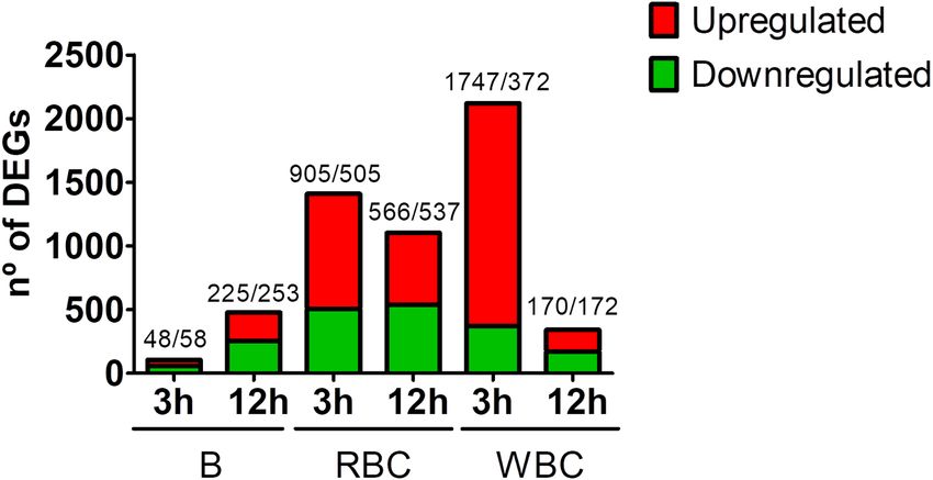
Hernández-Cabanyero et al.                                                                                                                Eel Vibriosis Due to V. vulnificus

TABLE 1 | Microarray validation by RT-qPCR.                                                   positive bacteriolytic activity measured as inhibition halo of
                                                                                              bacterial growth was determined. Three independent technical
Gene name             Gene           Sample                             FC1
                      acronym
                                                                                              replicates of bacteriolytic activity were performed for each
                                                             Array        RT-qPCR             biological sample of serum.

Beta-catenin-like     bcl2           B 3 vs. 0 hpi          1.52 (=)          1.87 (=)
protein 1
                                                                                              Design and Validation of a New RT-qPCR
Interleukin 1beta     il1β           B 12 vs. 0 hpi        17.16 (++)     23.44 (++)          Assay to the Early Detection of
Interleukin 10        il10r          RBCs 3 vs. 0 hpi       4.94 (+)       5.24 (+)           Vv-Vibriosis
receptor subunit
                                                                                              A selection of genes (npsn, cox2, mmp9 and sidt1) have been
beta
Beta-catenin-like     bcl2           RBCs 12 vs. 0 hpi      −1.90 (=)         −1.03 (=)
                                                                                              used to develop a new RT-qPCR assay to the early detection
protein 1                                                                                     of Vv-vibriosis. The list of primers in Supplementary Table S1.
p53                   p53            WBCs 3 vs. 0 hpi       6.76 (+)          7.77 (+)        Eel infection, blood sampling, sample processing, and RT-qPCR
Interleukin 6         il6r           WBCs 12 vs. 0 hpi      6.83 (=)          4.87 (+)        procedure were performed as described on the previous sections.
receptor subunit
                                                                                              Statistical analysis was performed using GraphPad Prism 7.
beta precursor
                                                                                              Data were analyzed by ANOVA analysis followed by the post-hoc
Comparison of fold change (FC) values obtained by microarray and RT-qPCR. In case             multiple comparison by Bonferroni’s method that was run for
of RT-qPCR, results were obtained using act as the reference gene and the fold                each gene to determine differences between groups (p < 0.05).
induction (2-ΔΔCt) for each gene was calculated. Primers used are listed in
Supplementary Table S1. FC value represents the mean obtained from 3 independent
biological samples.
1
 FC: fold change values qualitative classification: =, −2 < X < 2; +, 2 ≤ X < 10; ++,
10 ≤ X < 25; ND, non-detected as differentially expressed.
                                                                                              RESULTS
                                                                                              Cell Analysis
mRNA levels of the tested genes, using eel actin (act) as the                                 First, we monitored the presence of the pathogen in blood
gold standard (Paria et al., 2016) and the fold induction (2-ΔΔCt)                            and found bacterial counts (0 hpi;
Hernández-Cabanyero et al. Eel Vibriosis Due to V. vulnificus
intracellular ones (tlr3, tlr7, and tlr9b). Consistent with this,
genes encoding major histocompatibility complex (Mhc) class
I (mhcI) and class II (mhcII) were also upregulated, mhcI by
RBCs and WBCs, and mhcII only by RBCs. Thus, although
V. vulnificus is an extracellular pathogen, our results point to
activation of intracellular pathogen recognition and processing
mechanisms frequently associated with viral infection (Lund
et al., 2004).
In all samples (B, RBCs and WBCs), we also found upregulated
genes for cathepsins B, L, and S, a group of lysosomal proteases
that play a key role in cellular protein turnover. Cathepsins
are associated with Tlr signaling pathways in blood cells to
FIGURE 1 | Magnitude of the eel immune response against V. vulnificus the extent that their inhibition blocks Tlr3-, Tlr7,- and Tlr9-
represented as the number of differentially expressed genes (DEGs) in blood mediated responses (Matsumoto et al., 2008).
(B), red blood cells (RBCs) and white blood cells (WBCs) samples. Bars
represent total DEGs (sum of upregulated [red] and downregulated [green]
DEGs) of each sampling point (3 hpi; 12 hpi) against time zero (0 hpi) of each Pathogen Control and Destruction
type of sample. The numbers of up/downregulated DEGs are indicated on the We found evidence of an antibacterial response in blood of infected
top of each bar.
eels, suggesting that the host immune response tried to eliminate
the pathogen after the infection. Our data showed the upregulation
were less transcriptionally active at 12 hpi, which is compatible of multiple genes related to pathogen growth inhibition (transferrin
with a loss of functionality caused directly or indirectly by [RBCs] and hepcidin [WBCs], the hormone that controls iron
the pathogen. Interestingly, the number of DEGs detected in sequestration, pathogen tagging (i.e., complement factor C3 and
the B samples was much lower than that found in the RBCs lectins), and pathogen destruction (i.e., complement factors C5-C9,
and WBCs samples (Figure 1). This apparent anomaly could Lbp/Bpi protein and genes related to activation of phagocytosis;
be explained by the cellular heterogeneity of the blood, which Table 2; Supplementary Table S2). Complement genes were
could negatively affect the normalization of the data and be the upregulated by RBCs and WBCs, especially at 3 hpi, although
cause of high outlier removal. the strongest and most varied response was associated with WBCs
Overall, the transcriptomic results were reliable thanks to at 3 hpi. Similarly, both cell types’ upregulated genes encoding
the similar fold change values obtained by microarray lectins at 3 hpi, especially the galectin and intelectin. Complement/
hybridization and RT-qPCR for a set of genes (Table 1). Venn lectin-tagged bacteria can be recognized and phagocytosed more
diagrams showing common DEGs in RBCs and WBCs throughout easily by host phagocytes. In accordance, we found several
infection revealed a cell-specific response, as most transcripts upregulated genes that could be related to phagocytosis and
that change their transcription level were not shared between bacterial killing, such as those involved in cytoskeleton
both cell types (Figure 2). rearrangements and nitric oxide synthesis (i.e., inos that was only
The DEGs by the different blood fractions are shown in upregulated by WBCs at 3 hpi), as well as genes related to signal
Supplementary Table S2, and a selection of them by putative transduction in common with other cellular processes that will
function is listed in Table 2. Based on this information, be discussed in the following sections (Table 2;
we highlight the following genes and processes that could Supplementary Table S2).
be related to a harmful defensive response (which could cause Regarding antibacterial activity, our results showed the gene
self-damage in host tissues and favor its death) of eel against encoding Lbp/Bpi, an antibacterial protein produced by different
V. vulnificus: cell types (Inagawa et al., 2002), to be highly upregulated, but
only in B samples. Surprisingly, the gene coding for nephrosin
(npsn), which has recently been linked to the antibacterial
Pathogen Detection and Antigen Presentation
activity of the immune system in fish (Di et al., 2017), was
Systems
the most strongly upregulated gene in the B samples at both
RBCs and WBCs upregulated multiple pattern recognition
3 and 12 hpi (Table 2; Supplementary Table S2).
receptor (Prr) genes as well as major histocompatibility complex
(Mhc) genes, the former mainly at 3 hpi and the latter at
both 3 and 12 hpi (Table 2; Supplementary Table S2). This Cell Death
result suggested that not only WBCs but also RBCs could act We detected upregulation of multiple genes related to the
as antigen-presenting cells. Among the Prr genes, we found activation of cell death by apoptosis and/or autophagy, but
upregulated tlr genes (Toll-like receptors [Tlrs]), some of which interestingly only in the WBCs fraction with the sole exception
were specifically associated with cell type: i.e., tlr7, tlr6, and of p53 (RBCs, 3 hpi). Thus, autophagy could be related to
tlr5s (encoding the soluble form of Tlr5) with WBCs and the upregulation of autophagy-related proteins (atg9 and atg2a);
tlr9b with RBCs (Figure 2; Table 2). Some of these tlr genes and apoptosis to p53, caspase-8 (casp8), and genes encoding
are related to the detection of mainly extracellular antigens apoptosis-inducing factors, all upregulated at 3 hpi (Table 2;
(tlr20a, tlr21, tlr6 and tlr5s) and others to the detection of Supplementary Table S2).
Frontiers in Microbiology | www.frontiersin.org 5 April 2022 | Volume 13 | Article 852677Hernández-Cabanyero et al. Eel Vibriosis Due to V. vulnificus
TABLE 2 | Eel blood transcriptome after V. vulnificus infection.
Gene1 FC2
B RBCs WBCs
3 hpi 12 hpi 3 hpi 12 hpi 3 hpi 12 hpi
Pathogen detection and antigen presentation systems
PRR
tlr13 – 4.3 – – – –
tlr9a – −2.6 – – – –
tlr20a – – 4.2 – 9.6 –
tlr3 – – 3.4 – 3.7 –
tlr21 – – 3.1 – 4.9 –
tlr9b – – 2.2 1.8 – –
tlr7 – – – – 4.1 –
tlr6 – – – – 3.0 –
tlrs5 – – – – 2.2 –
tlr20f – – – – −5.5 –
Antigen presentation
mhcII – 2.7 3–1.7 1.5 – –
mhcI – – 4.8–2 84.4 5.3–3.2 30.6
AP-1 complex subunit sigma 3 – – – −2.0 3.8 –
AP-1 complex subunit gamma 1 – – – – 2.1 –
Cathepsins
Cathepsin L – 3.6 – – 5.6 –
Cathepsin S precursor – 2.9 1.8 – – –
Cathepsin B – – 2.5 5.3 – –
Pathogen control and destruction
Pathogen growth inhibition
Transferrin – 2.7 2.5–2.1 1.8 – –
Transferrin receptor (tfr1) – 2.4 – – – –
Aminolevulinic acid – 2.0 – – – 2.1
Hemoglobin subunit – – – 3.7 – –
Ferritin – – – 2.8–2.1 – –
Hepcidin – – – – 10.2 –
Complement system
C5a receptor – 14.1 – – – –
Complement factor Bf-2 – 6.5–5.3 – – – –
Complement factor B/C2 – 5.9 – – 2.4 –
Complement factor B – 4.6 – – – –
C3a receptor 1 – – 6.5 – 7.5 –
C3c – – 4.2 – 4.2 –
C7-1 – – 4.1 – 3.3 –
C4BPB – – 4.1 3.0 7.7 –
C3 – – 3.9 – 7.4 –
C3-H2 – – 3.6 – 4.2 –
C3-H1 – – 3.5 – 2.9 –
C3-S – – 3.1 1.9 5.6–2.6 1.9
factor D precursor – – 2.8 – – –
C4-2 – – 2.2 – 2.1 2.7
C5 – – – – 12.8 –
C5-2 – – – – 6.1 –
C4 – – – – 4.1 –
C4b – – – – 4.0 –
C3-3 – – – – 3.6 –
C1R/C1S subunit of Ca2 + − – – – – 3.4–2.9 –
dependent
C7 – – – – 3.0 –
Complement factor I – – – – 2.3 –
C3 precursor – – – – 2.2 –
C1q, B chain – – – – – 7.7
Antibacterial effectors
Nephrosin (npsn) 10.1–6.3 150.6–99 – – – –
Lpb/Bpi 4.5–3.3 41.7–25 – – – –
(Continued)
Frontiers in Microbiology | www.frontiersin.org 6 April 2022 | Volume 13 | Article 852677Hernández-Cabanyero et al. Eel Vibriosis Due to V. vulnificus
TABLE 2 | Continued
Gene1 FC2
B RBCs WBCs
3 hpi 12 hpi 3 hpi 12 hpi 3 hpi 12 hpi
Nitric oxide synthase – – – – 3.4 –
Lectins
Mannose-6-phosphate receptor- – 14.0 – – – –
binding
C-type lectin receptor – – 3.8 – 11.5-(−6) –
gal3 – – 3.2 – 4.3 1.7
gal4 – – 2.9 1.7 16.1 –
Intelectin – – 2.5 – 12.6 –
Mannose binding lectin 2 – – – 3.0 3.1–2.1 –
Fucolectin 2 – – – – 2.7 –
Fucolectin 4 – – – – 2.1 1.9
Cytoskeleton rearrangements
Tubulin-related genes – 27.2 – – 2.0 –
Myosin-related genes – 5.9 7.6–2.6 6–1.6 8.2–2.2 –
itpr1, itpr3 – – 4.9–2.7 – – –
Actin-related genes – 3.1 3.4 7–1.5 4.7–2 1.8
Coronin-1a – – 2.9 – – –
Cell death
Apoptosis
atg9 – – – – 3.3 –
atg2a – – – – 3.3 –
Autophagy
p53 – – 6.8 – 9.9 –
Calpain – – – −3.9 −4.7 –
p53 apoptosis effector related to – – – – 33.5 –
PMP-22
Apoptosis-inducing factor 3 – – – – 6.8 –
casp8 – – – – 3.0 –
casp3 – 4.8*
Inflammatory response
Signal transducers and transcriptional factors
klf6 2.2 8.5–5.2 – – – –
src-family tyrosine kinase SCK – 17.2 – – – –
traf3 – 5.4 – – – –
socs3 – 5.1 – – – –
c-fos – 3.7 – 16.9 – –
jun-b – 3.5 – 11.1 – –
map2k6 – – 8.6 – 25.3 –
nod1 – – 4.9 – 4.3 –
p38 – – 4.5 4.2 4.0 –
map3k2 – – 4.5 – 4.2 –
pak1 – – 3.3 – 6.9 –
map3k5 – – 3.1 – 6.9 –
mapk7 – – −2.0 −2.0 – –
irak4 – – −2.4 – – –
jak1 – – −2.4 – – –
erk1 – – −1.9 −2.0 – –
NF-Κβ inhibitor alpha – – – 3.3 −4.8 –
nod3 – – – 2.8 3.6 –
klf13 – – – 2.2 – –
map3k4 – – – −2.3 – –
c-myc binding protein – – – −5.5 – –
mapk6 – – – – 24.3 –
NF-Κβ p105 subunit – – – – 4.8 –
Kdel receptor 3 – – – – 4.3 –
mapk14 – – – – 4.0 –
stat3 – – – – 3.5 3.1
(Continued)
Frontiers in Microbiology | www.frontiersin.org 7 April 2022 | Volume 13 | Article 852677Hernández-Cabanyero et al. Eel Vibriosis Due to V. vulnificus
TABLE 2 | Continued
Gene1 FC2
B RBCs WBCs
3 hpi 12 hpi 3 hpi 12 hpi 3 hpi 12 hpi
nlrc3 receptor – – – – 2.6 –
cop9 – – – – 2.1 –
Inflammatory cytokines and related proteins
Interferon induced protein 2 – 20.1 – – – –
il1β – 17.2 – – 3.5* 27.8*
Granulin – 9.3 – – – –
IL8 precursor – 7.5 – – – –
IL1β receptor type 1 soluble – 5.5 – – −4.2 –
Progranulin type 1 – 5.5 – – – –
IL1 receptor type 1 – 4.4 – – −6.2 –
il12β – – 8.5 – 14.8 –
IL10 receptor β – – 4.9 – 22.2 –
il17a/f1 – – 4.4 – – –
il20 – – 2.7 – 6.0 –
irf3 – – 2.4 – – –
nuclear factor interleukin 3-regulated – – – 10.1 3.5 –
protein
IL6 receptor subunit β precursor – – – 6.8 4.3 2.4
irf2A – – – 2.7 – –
irf2B – – – 2.2 – –
IRF2, promoter region – – – 2.2 – –
Tumor necrosis factor receptor – – – – 15.9 –
(tnfrsf12a)
TNF receptor member 27 – – – – 6.0 –
irf1 – – – – 3.3–2.8 –
IL1 receptor-like – – – – 3.1 –
ifnc1 – – – – 2.6 –
Allograft inflammatory factor-1 – – – – 2.6 –
TNF receptor associated factor 2 – – – – 2.4 –
ifna2 – – – – 2.1 –
il17r – – – – – 2.2
Chemokines and receptors
CC CK3 – 3.6 – – – –
C-C receptor type 4 – – 6.2 – – –
CK 21 precursor – – 4.1 – – –
CCL4 – – – – 8.6 13.4
CK 4 precursor – – – – 8.2 –
CK 19 precursor – – – – 5.2 –
CK 10 precursor – – – – 2.2 –
Septicemia markers
Ciclooxigenase-2 (cox2) 5.5 32.1 – – – –
Hyaluronidase-2 (hyal2) 2.5 26.6 – – – –
mmp9 or gelatinase B – 61.5–40 – – – –
Leukotriene – 5.2 – – – –
Prostaglandin – 11.0 – – – –
Coagulation factors
Coagulation factor VIII – 22.4–9.1 – – – –
Platelet receptor Gi24 – 2.0 – – – –
Antithrombin protein – – 3.8 – 4.1 –
Thrombin protein – – 3.4 – – –
Thrombospondin – – – – 7.1–3 –
Coagulation factor V – – – – 4.6 –
Fibrinogen – – – – 4.0 –
Angiotensinogen – – – – 3.3 –
Plasminogen – – – – 2.0 1.9
Multiple coagulation factor – – – – – 3.1
Angiogenesis and hematopoiesis
angpt2 – 5.9 – – −3.7 –
(Continued)
Frontiers in Microbiology | www.frontiersin.org 8 April 2022 | Volume 13 | Article 852677Hernández-Cabanyero et al. Eel Vibriosis Due to V. vulnificus
TABLE 2 | Continued
Gene1 FC2
B RBCs WBCs
3 hpi 12 hpi 3 hpi 12 hpi 3 hpi 12 hpi
angpt1 – – 5.6 – 7.4 –
cldn19 – – 3.2 – 11.6 –
cldn1 – – 3.2 – 7.2 –
cldn18 – – – – 15.4 –
cldn4 – – – – 4.1 –
cldn29a – – – – 3.0 –
Epigenetic response
Histone H2B – −2.3 – – – –
Histone H2AFX – – 3.2 2.2 – –
Anti-silencing protein – – 2.3 – 3.1 –
Histone acetyltransferase type B – – 2.2 – 2.6 –
catalytic subunit
Histone deacetylase 3 – – 2.2 – – –
Histone gene cluster XlH3-A (h1a, – – −1.4 – – –
h2b, h3, h4)
Histone acetyltransferase MYST2 – – −1.4 – – –
Histone H1x – – −3.5 – – –
Histone H3.3 – – – 1.3 – –
euchromatic histone-lysine – – – −1.7 – –
N-methyltransferase 1b (ehmt1b)
Histone acetyltransferase MYST4 – – – −2.7 – –
Histone deacetylase 1 – – – −2.7 – –
Histone H2A.Z – – – −4.0 – –
Histone deacetylase 2 – – – −4.3 – –
Histone H1 – – – – 7.4–5.9 –
Histone H2AV – – – – −1.8 −1.6
Relationship between systemic and mucosal immunity
muc2A – – 2.5 – 4.4 –
RNA-based response
Systemic RNA deficient-1 (sidt1) – 10.5 – – 5.4* 2.2*
Stress-related response
Hypoxia-inducible factor 1 alpha – 3.5 – – – –
Glutathione peroxidase – −2.1 −1.5 −1.7 −2.0 –
hsp90 (cochaperone activator of Tlr9) – – 3.6 5–1.8 36.9–8.2 –
Inositol hexakisphosphate (insP6) – – 2.0 2.9 – –
hsp70 – – – 31.7–2.2 25.2 9.1
Osmotic stress gene – – – 6.7 – –
List of selected differentially expressed genes (DEGs) from eels infected with V. vulnificus R99 strain. DEGs are grouped according to their putative biological function. The fold change
(FC) values are based on the comparison between the time indicated on top of each column (3 hpi; 12 hpi) compared to time zero (0 hpi) for the same type of sample [B (blood),
RBCs (red blood cells), or WBCs (white blood cells)]. FC value represents the mean obtained from 3 independent biological samples.
–Not detected as differentially expressed.
1
Identified DEGs are indicated.
2
FC: fold change value for each individual gene. See Supplementary Table S2 for specific gene and fold-change value.
*
Relevant mRNAs detected by RT-qPCR.
Inflammatory Response the Nf-Κβ and Mapk signaling pathways, enhancing the
We also found evidence of early regulation of different pathways transcription of proinflammatory cytokines (Kim et al., 2016).
that trigger a proinflammatory response. At the signal The rest of the genes mentioned above are part of the Jak/
transduction level, this response consisted of upregulation by Stat signaling pathway, which is also involved in activating
RBCs and WBCs of nucleotide-binding oligomerization domain the inflammatory response (Rawlings et al., 2004). Accordingly,
containing proteins (nod1 and nod3), as well as the genes for we also found upregulated by WBCs at 3 hpi, a gene for an
Mapk kinases map2K6, map3K2, and map4K5, whereas mapK6, Nlrc3 receptor that acts as a negative regulator of all these
NF-Κβ (p105 subunit), and signal transducer and activator of processes (Schneider et al., 2013), suggesting an attempt by
transcription 3 (stat3) were only upregulated by WBCs (Table 2; the immune system to counteract the activation of the
Supplementary Table S2). Indeed, Nod1 and Nod3 activate inflammatory response against V. vulnificus.
Frontiers in Microbiology | www.frontiersin.org 9 April 2022 | Volume 13 | Article 852677Hernández-Cabanyero et al. Eel Vibriosis Due to V. vulnificus
intravascular coagulation, such as those encoding coagulation
A
factor VIII, and genes encoding leukotrienes, prostaglandins,
and cyclooxygenase (i.e., cox2), all of which are considered
markers of the acute phase of the disease (Peters-Golden
et al., 2005; Gómez-Abellán and Sepulcre, 2016; Wang et al.,
2016). Leukotrienes increase leukocyte accumulation, phagocytic
capacity for microbial ingestion and elimination, and the
generation of other proinflammatory mediators (Peters-Golden
et al., 2005). Cyclooxygenases enhance prostaglandin production
(Smith et al., 2000), leading to the induction of the immune
B response (Gómez-Abellán and Sepulcre, 2016). More
importantly, genes for matrix metalloproteinases that are
implicated in endothelial damage (i.e., mmp9; Pedersen et al.,
2015) were among the most overexpressed genes in B samples
at 12 hpi (Table 2; Supplementary Table S2). Related to
this damage, genes related to endothelial regeneration
(angiogenesis) were also upregulated (angiopoietins angpt2
and angpt1 in B and RBCs samples, respectively; Table 2;
Supplementary Table S2).
Epigenetic Response
FIGURE 2 | Red blood cells (RBCs) and white blood cells (WBCs) elicit a Several histone-related genes (acetylases, deacetylases, and
different immune response against V. vulnificus. Venn diagram depicting the methyltransferases, among others) were found to be DEGs
overlap of the differentially expressed genes (DEGs) between RBCs and
(both up- and downregulated) mainly by RBCs (Table 2;
WBCs at 3 hpi (A) and 12 hpi (B).
Supplementary Table S2). This effect could be associated with
an epigenetic response probably related to modulation of the
The activated proinflammatory response was also evidenced immune response by gene silencing through methylation
by the early (3 hpi) upregulation of genes for several tumor (Medzhitov and Horng, 2009; Shakespear et al., 2011).
necrosis alpha (Tnfα) receptors, interleukins (Ils), and their receptors In parallel, we also detected a strongly upregulated gene for
and interferon (Ifn) and related proteins by RBCs and WBCs an anti-silencing protein in RBCs and WBCs samples at 3
with a common (il12β, il10 receptor β, and il20) and cell-type- hpi (Table 3; Supplementary Table S2). Anti-silencing proteins
specific pattern (RBCs: il17a/f1; WBCs: several Tnf receptors, Il1 are evolutionarily conserved proteins that act as histone
receptor-like, ifnc1, ifna2, and irf1) followed by a strong upregulation chaperones and are required for various chromatin-mediated
of genes for Il1β, two receptors for Il1β, Il8 precursor, progranulin cellular processes. Recently, it has been demonstrated that all
and granulin in B samples at 12 hpi (Table 2). Granulins are these proteins are involved in antiviral mechanisms promoting
multifunctional proteins produced after proteolytic processing of Ifnb production (Liu et al., 2016).
progranulin (Bateman et al., 1990) that enhance the production
of proinflammatory cytokines such as Tnfα and Il8 (Park et al.,
Relationship Between Systemic and Mucosal
2011). Related to these results, we highlight that Il1β is the main
Immunity
proinflammatory cytokine in both humans and fish (Zou and
It was not surprising to find DEGs that evidenced a link
Secombes, 2016) and that Ifna has been related to the immune
between systemic and mucosal immunity that our group had
response against virus (Zou and Secombes, 2016). Multiple genes
previously shown to occur in eels vaccinated against V. vulnificus
for Tnfα- and interferon-induced proteins were also detected in
(Esteve-Gassent et al., 2003). Among them, it should
all the samples (Table 2; Supplementary Table S2). In parallel,
be highlighted muc2A, a gene for a mucolipin secreted by
a few genes encoding for anti-inflammatory cytokine receptors
mucosal cells that is involved in binding to bacteria for killing
(e.g., il10r) were found early upregulated by RBCs and WBCs,
(McGuckin et al., 2011; Brinchmann, 2016) and that was
which could be interpreted as an attempt by the organism to
upregulated at 3 hpi by both RBCs and WBCs (Table 2;
control the cytokine storm and restore homeostasis.
Supplementary Table S2).
Since we detected upregulation of the inflammatory gene
markers il1β and caspase-3 (casp3) in B samples and not in
WBCs samples, we performed RT-qPCR with the same samples RNA-Based Response
and found both to be upregulated in WBCs (il1β at 3 and One of the most striking results of the present study was the
12 hpi; caps3 only at 12 hpi; Table 2). strong upregulation by B samples of a gene for a specific
transporter of a systemic interference RNA, sidt1 (systemic
Sepsis Markers RNAi deficient-1; Li et al., 2015), which was detected at 12
Cells present in B samples upregulated multiple markers of hpi (Table 2). This result strongly suggested that a systemic
sepsis at 12 hpi. For example, marker genes for disseminated RNAi may be acting during the immune response against
Frontiers in Microbiology | www.frontiersin.org 10 April 2022 | Volume 13 | Article 852677Hernández-Cabanyero et al. Eel Vibriosis Due to V. vulnificus
TABLE 3 | Proteolytic, hemolytic and bacteriolytic activity of eel serum before infected by immersion with V. vulnificus. To do so, we selected
and after V. vulnificus infection.
the most upregulated genes related with antibacterial activity
Serum sample Proteolytic Hemolytic Bacteriolytic
(npsn), endothelial damage and acute phase of infection
activity1 activity2 activity3 (cox2 and mmp9) and the transporter of a systemic interference
RNA (sidt1; Table 2). We infected eels with V. vulnificus
Non-infected – 1:2 1:2 and analyzed the expression of the selected genes in blood
0 hpi – 1:8 1:4
of the infected animals at 3 and 12 hpi compared to
3 hpi 1:8 1:8 1:4
12 hpi 1:4 1:4 1:8
non-infected eels. All the selected genes were easily detected
upregulated in blood of the infected animals, especially cox2
Eels were infected by immersion and the lytic activities were determined in serum from and sidt1 at 3 hpi (Table 4). Therefore, we propose that
non-infected eels (control), and eels infected at different hours post infection (hpi).
Results are presented as the titter (maximal dilution of serum with a positive result in 3
this easy and fast methodology could be used to the early
independent biological samples) of the corresponding activity. diagnose of Vv-vibriosis.
–Without activity.
1
Proteolytic activity: evaluated by plating 5 μl of the serum samples and dilutions (serial
dilution 1:2 to 1:64 on PBS) on 1% agarose plates supplemented with 5% casein. The
maximal serum dilution that produced a transparent halo was considered as the titter of DISCUSSION
this activity.
2
Hemolytic activity: evaluated by plating 5 μl of the serum samples and dilutions (serial V. vulnificus is an emerging zoonotic pathogen associated
dilution 1:2 to 1:64 on PBS) on 1% agarose plates supplemented with 1% erythrocytes
with fish farms as all clonal groups defined in the species
(bovine erythrocytes from Sigma). The maximal serum dilution that produced a
transparent halo was considered as the titter of this activity. have emerged from outbreaks of fish vibriosis in farms and
3
Bacteriolytic activity: evaluated by plating 5 μl of the serum samples and dilutions (serial contain clinical isolates from fish and humans (Roig et al.,
dilution 1:2 to 1:64 on PBS) on LB-1 plates inoculated with a V. vulnificus lawn. The 2018; Carmona-Salido et al., 2021). Interestingly, this species
maximal serum dilution that inhibited bacterial growth was considered as the titter of
this activity.
uses both generalist and host-specific virulence mechanisms,
the former mainly related to its toxins and exoenzymes,
and the latter to resistance to innate immunity (Hernández-
TABLE 4 | Early diagnosis of fish vibriosis due to V. vulnificus by RT-qPCR. Cabanyero et al., 2019). Using both, V. vulnificus can survive
and cause rapid death by septicemia in hosts as evolutionarily
Gene name Gene acronym 3 hpi 12 hpi
distant as humans and eels. Previous studies using mice as
Nephrosin npsn 4.2 (+) 1.5 (=)
an animal model suggested that sepsis death of their original
Cyclooxygenase 2 cox2 21.9 (++) 10.5 (++) hosts may be due to an early cytokine storm triggered by
Matrix metalloproteinase-9 mmp9 3.1 (+) 2.9 (+) the pathogen during its interaction with the immune system
Systemic RNAi deficient-1 sidt1 11.31 (++) 2.5 (+) (Murciano et al., 2017). In this work, we set out to demonstrate
Blood samples taken at 3 and 12 hpi from infected and control animals were used
this hypothesis using one of the natural hosts of the disease,
to determine the expression of genes selected as septicemic markers by RT-qPCR. the eel, and reproducing the natural conditions of infection
Results were obtained using act as the reference gene and the fold induction (2-ΔΔCt) for with a representative strain of the most studied zoonotic
each gene was calculated. Primers used are listed in Supplementary Table S1. Fold
group. For the study, a microarray platform was used that
induction values qualitative classification: =, −2 < X < 2; +, 2 ≤ X < 10; ++, 10 ≤ X < 25.
was designed from the transcriptome of the hematopoietic
organs of eels stimulated with viral/bacterial PAMPs and
V. vulnificus. It is well known that systemic RNAi, common
was consequently enriched in immune genes (Callol
to all vertebrates, is involved in ancestral innate defense
et al., 2015b).
mechanisms against viral infections (Li et al., 2015). Since
First, we highlight the critical role that eel RBCs appear
we detected upregulation of this gene in the B samples and
to play in the defense against V. vulnificus and, probably,
not in WBCs samples, we performed RT-qPCR on the same
against bacterial pathogens in general. We suspected that
samples and found the gene to be upregulated in WBCs at
RBCs were immunologically active cells because we had
3 and 12 hpi (Table 2).
observed that bacteria agglutinated in the presence of eel
erythrocytes in vitro (Lee et al., 2013). In this work, we found
Functional Assays that RBCs do indeed activate multiple lectin genes in response
The transcriptomic results were confirmed by evaluating different to V. vulnificus infection that could exert this antibacterial
enzymatic and lytic activities in eel serum samples. We detected function. We also found that RBCs are genetically primed
proteolytic, hemolytic, and bacteriolytic activities in serum that to act as antigen-presenting cells, as they also activate the
were significantly increased at 3 hpi and 12 hpi compared to transcription of extracellular and intracellular Prrs (Tlrs and
those found in serum samples at 0 hpi and those found in Nods), as well as Mhc classes I and II. Similar results were
serum samples from non infected animals (Table 3). previously found in rainbow trout RBCs which express mhcII
in response to virus (Nombela et al., 2019). In addition,
Early Diagnosis of Fish Septicemia by they are genetically prepared to produce proinflammatory
RT-qPCR cytokines such as Il17 and Il20 as well as Il12β, whose
The use of selected gene markers for the early detection hypothetic function will be commented on later (Rutz et al.,
of fish septicemia was evaluated by RT-qPCR from eels 2014; Zou and Secombes, 2016). Although demonstrating
Frontiers in Microbiology | www.frontiersin.org 11 April 2022 | Volume 13 | Article 852677Hernández-Cabanyero et al. Eel Vibriosis Due to V. vulnificus that RBCs act as antigen-presenting cells or produce these fraction of WBCs. Related to this, we also observed a drastic cytokines are beyond the scope of this work, these results reduction in the transcription of most of the genes that are consistent with what we know about eel vibriosis, and had been upregulated at 3 hpi. In contrast, RBCs changed with the results we have obtained when analyzing the other their transcriptional pattern by stopping to transcribe genes blood fractions. for proinflammatory cytokines and chemokines and Thus, eel WBCs also appear to be very active during transcribing genes for MhcI, c-Fos, JunB, Irf2A, Irf2B, and the first hours of infection, upregulating the transcription cathepsin B. An overproduction of c-Fos, JunB, and cathepsin of Prr genes for extracellular and intracellular antigens and, B has been linked in fish to tissue repair and the over- interestingly, only MhcI, the form of Mhc associated with activation of irf2A and irf2B with inhibition of interferons intracellular antigen presentation. In this regard, our results alpha and beta (Sato et al., 2009; Botwright et al., 2021), suggest that both RBCs and WBCs may overexpress Tlrs both processes probably related with an attempt to that in fish detect double-stranded RNA (Tlr3 and Tlr13) control the strong immune response that was activated and DNA (Tlr9 and Tlr21) both extracellularly (Tlr21) and at 3 hpi. intracellularly (Tlr3, Tlr9, and Tlr13), again suggesting that Thus, the pathogen would activate an atypical cytokine V. vulnificus could be recognized and processed as if it was storm at 3 hpi, followed by a strong inflammation at 12 an intracellular pathogen. We also observed that eel WBCs hpi and blood cell stress and death. This strong inflammation could produce an orthologue of mammalian Tlr6 whose could also lead to endothelial destruction, evidenced by a function is unknown, as it has not been previously described significant strong activation of sepsis markers related to in any fish species. As expected, we also found considerable this destruction; a result compatible with natural disease evidence that eel WBCs could produce numerous antibacterial given that this disease is known as hemorrhagic septicemia compounds and act as phagocytic cells, especially in the (Ince et al., 2016). Beneath this inflammatory response, short term after infection. we found evidence for the activation of a systemic RNAi. Our transcriptomic results also suggest that signaling Systemic RNAi are part of the conserved biological response pathways would converge in RBCs and WBCs on Iraq4 and mechanisms to double-stranded RNA and are involved in Traf3, consistent with activation at 3 hpi of an atypical resistance to endogenous and exogenous pathogenic nucleic proinflammatory response typically antiviral (Table 2; acids (Abubaker et al., 2014). Its function in fish innate Supplementary Table S2). Thus, RBCs and WBCs activated immunity is entirely unknown. Taking all the above mentioned the transcription of genes for Il12β, Il17, and Il20 and several results into account, we hypothesized that V. vulnificus could genes for type 1 interferons. These interleukins have been activate a response against endogenous RNA that, in turn, linked to mucosal inflammation in mice and humans, especially would trigger the cytokine storm. In fact, it has been recently in inflammatory bowel diseases (Rutz et al., 2014; Zou and published the activation of this kind of response in patients Secombes, 2016; Moschen et al., 2019), while Il12β has been with sepsis (Chousterman et al., 2017). Further, linked to antiviral response in both fish and humans (Sakai we hypothesized that the toxin RtxA1 would be one of the et al., 2021). This result is very interesting. Firstly, because responsible virulence factors. links systemic and mucosal immunity, which correlates with Previous studies demonstrated that mutants deficient in previous results showing that eels vaccinated via mucosal this toxin kept the ability to infect and invade the bloodstream route, produce both mucosal and systemic antibodies against but were unable to cause death by sepsis in fish while were V. vulnificus that protect them against Vv-vibriosis (Fouz attenuated in virulence and unable to activate the early et al., 2001; Esteve-Gassent et al., 2003). Secondly, the cytokine storm in mice (Lee et al., 2013; Murciano et al., production of Il12β, interferon type 1, and their regulators 2017). Similarly, in human immune cells, the RtxA1 toxin together with the activation of genes for intracellular antigen enhances inflammatory pathways (Kim et al., 2020). In recognition and processing mentioned above strongly evidence addition, this toxin has an intracellular existence as it is that this pathogen could be recognized as if it was an secreted after contact with the eukaryotic cell, associates intracellular pathogen. Finally, we also found strong evidence with the cell membrane for its terminal ends, and forms a that this atypical early immune response leads to a typical pore that allows the central module to enter, self-process, inflammatory response at 12 hpi, with upregulation of and release the functional domains that attack the cell il1β, il8, and the il1βr that were detected in B and WBCs (Satchell, 2015). Studies are in progress to demonstrate samples. this hypothesis. In parallel to all these processes, cell death mechanisms Our concluding remarks are summarized in Figures 3 and by autophagy and apoptosis are probably activated, especially 4, which present a model of the immune response against in WBCs. At the same time, RBCs mainly would suffer a V. vulnificus that sheds light on the comprehension of the stressful situation, as indicated by the strong upregulation disease caused by this zoonotic pathogen in its hosts. It of stress markers (Table 2; Supplementary Table S2). should be noticed that although mammalian RBCs are not Although an increase in the number of WBCs occurs as nucleated and thus considered not active during the immune a natural response in bacterial infections, we did not observe response, a recent study has demonstrated that human and this proliferation in response to V. vulnificus, which would murine RBCs are involved in the innate immune response be compatible with death by apoptosis or autophagy of a to virus (Lam et al., 2021). Therefore, the proposed model Frontiers in Microbiology | www.frontiersin.org 12 April 2022 | Volume 13 | Article 852677
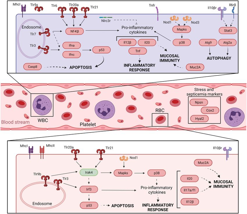
Hernández-Cabanyero et al.                                                                                                                   Eel Vibriosis Due to V. vulnificus

 FIGURE 3 | Model of the immune response in eel blood against V. vulnificus infection: early phase of vibriosis (3 hpi). The model shows the resultant proteins
 produced by the main transcripts differentially expressed by eels’ blood cells during the early phase of Vv-vibriosis (at 3 hpi with V. vulnificus R99 strain) infection.
 The putative translated proteins from the major immune-related pathways are represented in a code color depending on the gene modulation: upregulated (red) and
 downregulated (green).

could potentially be extended to all V. vulnificus hosts,                                (sidt1), could be used for the early detection of septicemia
including humans. According to our model, V. vulnificus                                  caused by V. vulnificus infection, as we could diagnose it
indeed triggers an acute but atypical inflammatory response                              from blood samples from artificially infected eels by using
that occurs in two main phases. In the early phase (3 hpi;                               an RT-qPCR targeting this gene. However, this proposal
Figure 3), the pathogen triggers the upregulation of a series                            should be validated with other fish species and by reproducing
of proinflammatory cytokine genes related to the mucosal                                 the vibriosis caused by other Vibrios to determine whether
immune response (il17a/f1 and il20) along with antiviral                                 this gene marker is exclusive of Vv-vibriosis. Functional
cytokine genes (il12β) and antiviral factors (ifna and ifnc),                            assays also highlighted that the serum from infected animals
while in the late phase (12 hpi; Figure 4) the upregulation                              is proteolytic, hemolytic, and bacteriolytic, partially confirming
of genes for typical inflammatory cytokines (il1β), endothelial                          the transcriptomic results. Finally, we found considerable
destruction (mmp9 and hyal2) and, interestingly, genes related                           evidence that RBCs are transcriptionally active and that
to an RNA-based immune response. Remarkably, some of                                     they may contribute significantly to this atypical immune
these genes, especially the gene for systemic RNAi transporter                           response, especially in the short term.

Frontiers in Microbiology | www.frontiersin.org                                     13                                                April 2022 | Volume 13 | Article 852677
You can also read