Acitretin mitigates uroporphyrin induced bone defects in congenital erythropoietic porphyria models - Nature
←
→
Page content transcription
If your browser does not render page correctly, please read the page content below
www.nature.com/scientificreports
OPEN Acitretin mitigates
uroporphyrin‑induced bone defects
in congenital erythropoietic
porphyria models
Juliana Bragazzi Cunha1,6*, Jared S. Elenbaas2,6, Dhiman Maitra1,6, Ning Kuo1,
Rodrigo Azuero‑Dajud1, Allison C. Ferguson3, Megan S. Griffin3, Stephen I. Lentz4,
Jordan A. Shavit3 & M. Bishr Omary1,5*
Congenital erythropoietic porphyria (CEP) is a rare genetic disorder leading to accumulation of uro/
coproporphyrin-I in tissues due to inhibition of uroporphyrinogen-III synthase. Clinical manifestations
of CEP include bone fragility, severe photosensitivity and photomutilation. Currently there is no
specific treatment for CEP, except bone marrow transplantation, and there is an unmet need for
treating this orphan disease. Fluorescent porphyrins cause protein aggregation, which led us to
hypothesize that uroporphyrin-I accumulation leads to protein aggregation and CEP-related bone
phenotype. We developed a zebrafish model that phenocopies features of CEP. As in human patients,
uroporphyrin-I accumulated in the bones of zebrafish, leading to impaired bone development.
Furthermore, in an osteoblast-like cell line, uroporphyrin-I decreased mineralization, aggregated
bone matrix proteins, activated endoplasmic reticulum stress and disrupted autophagy. Using high-
throughput drug screening, we identified acitretin, a second-generation retinoid, and showed that it
reduced uroporphyrin-I accumulation and its deleterious effects on bones. Our findings provide a new
CEP experimental model and a potential repurposed therapeutic.
Porphyrias are a group of inherited disorders due to defects in the heme biosynthetic pathway1,2. One such
example is congenital erythropoietic porphyria (CEP), most commonly caused by loss of function mutation in
uroporphyrinogen III synthase (UROS; Fig.S1), the enzyme that catalyzes the third step of the heme biosyn-
thetic pathway1,3. CEP is rare, with ~ 250 cases reported to d
ate4,5. It is autosomal recessive, and associated with
reduced UROS activity (5% of normal) and consequent accumulation of uro/coproporphyrin-I (uro/copro-I)
in bone marrow, erythrocytes, plasma, and increased uro/copro-I excretion in urine and stool1,3,5,6. CEP is char-
acterized by severe photosensitivity, with skin fragility and blistering of sun-exposed areas1,5,6. Scaring due to
secondary skin infections and bone resorption contribute to disfigurement of light-exposed areas3. Other clinical
manifestations of this multisystem disease include chronic ulcerative keratitis, hemolysis, which may require
repeated blood transfusions in severe cases, nonimmune hydrops fetalis, red urine since birth, erythrodontia
and osteodystrophy1,3,6,7. Currently, there is no specific pharmacological treatment for CEP, with interventions
being life-style-related (e.g. avoidance of sun) or complex procedures, including bone marrow t ransplantation3,8,9.
Fluorescent porphyrin accumulation in porphyria causes organelle specific protein oxidation and aggrega-
tion through mechanisms that involve type-II photosensitive reactions and secondary oxidative s tress10–17. We
posit that porphyrin-mediated protein aggregation in CEP plays a major mechanistic role in tissue damage that
involves accumulation of fluorescent uro/copro-I.
We demonstrated that uro-I injection of zebrafish larvae mimics features of CEP, including uro-I accumula-
tion in bones and bone deformation, as judged by decreased vertebra and operculum volume. Uro-I treatment
of an osteoblastic human osteosarcoma cell line, Saos-2, caused significant decrease in mineral matrix synthesis
1
Center for Advanced Biotechnology and Medicine, Rutgers University, Piscataway 08854, USA. 2Medical Scientist
Training Program, Washington University, Saint Louis 63110, USA. 3Department of Pediatrics, Division of Pediatric
Hematology/Oncology, University of Michigan, Ann Arbor 48109, USA. 4Department of Internal Medicine, Division
of Metabolism, Endocrinology and Diabetes, University of Michigan, Ann Arbor 48109, USA. 5Department of
Molecular and Integrative Physiology, University of Michigan Medical School, Ann Arbor 48109, USA. 6These
authors contributed equally: Juliana Bragazzi Cunha, Jared S. Elenbaas, and Dhiman Maitra *email: bragazzi@
umich.edu; bo163@cabm.rutgers.edu
Scientific Reports | (2021) 11:9601 | https://doi.org/10.1038/s41598-021-88668-9 1
Vol.:(0123456789)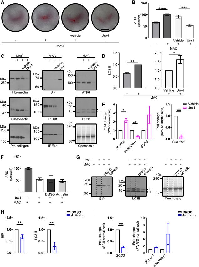
www.nature.com/scientificreports/
Figure 1. Zebrafish model of CEP develops bone phenotype resembling human disease. (A) 6dpf zebrafish
larvae were injected with uro-I or vehicle and imaged by confocal microscopy at 7dpf. Porphyrin was detected
only in the bones of uro-I-injected group. Arrowhead-operculum; box-vertebrae. (B) Larvae were treated as in
(A) and injected with calcein prior to imaging. Arrowhead-operculum; arrow-4th vertebra. (C) Quantification
of bone volume in larvae from (B); bone volume was normalized to vehicle-injected larvae set to 100%. Symbols
represent individual larvae (14–18/group) from 4–5 independent experiments. (D) Larvae were treated as
in (A). At 7dpf bones were harvested and imaged by epifluorescence microscopy pre and post HCl bone
demineralization, arrow-notochord. (E) Hydroxyapatite was incubated with calcein/uro-I/copro-I and imaged
by epifluorescence microscopy. Scale bars: 200 µm (A-D); 50 µm (E). Three-dimensional image reconstruction
(A, B) was performed using Imaris 3D software v7.7 (http://imaris.oxinst.com/). **p < 0.01.
and proteotoxicity. Using high-throughput drug screening, we identified acitretin, a 2 nd generation retinoid, as
an effective drug that mitigates some of the harmful effects of uro-I in zebrafish and Saos-2 cells.
Results
An inducible zebrafish model mimics bone defects of human CEP. Uro-I injected zebrafish larvae
showed porphyrin fluorescence in bone tissue (Fig. 1A). To confirm that uro-I binds specifically to bone, lar-
vae were co-injected with calcein (bone-specific dye) and imaged. Calcein and uro-I fluorescence co-localized
(Fig. 1B), confirming uro-I bound to bone. Additionally, uro-I-injected larvae exhibited severe photosensitiv-
ity and had to be shielded from light to prevent their death (not shown). Next, we assessed uro-I-mediated
bone defect by measuring the volume of the operculum and 4th vertebra. Notably, uro-I injection significantly
decreased operculum and 4th vertebra volume (Fig. 1C).
Bone matrix is composed of protein/organic (including collagen/fibronectin/osteonectin) and inorganic com-
ponents (minerals, mostly hydroxyapatite)18,19. We tested whether uro-I binds to the protein/organic or inorganic
parts of bone matrix by demineralizing bones of uro-I-injected larvae. Demineralization caused loss of uro-I
fluorescence, indicating that uro-I is extractable from the mineral matrix (Fig. 1D). We validated this finding
Scientific Reports | (2021) 11:9601 | https://doi.org/10.1038/s41598-021-88668-9 2
Vol:.(1234567890)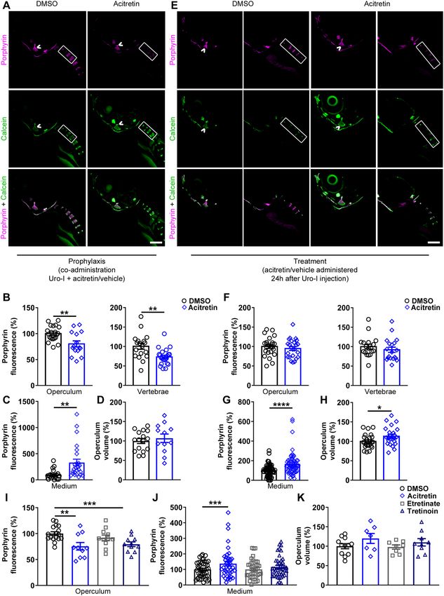
www.nature.com/scientificreports/
in vitro using hydroxyapatite crystals. Uro-I, but not copro-I, bound to hydroxyapatite, with calcein binding
used as a positive control (Fig. 1E). Therefore, uro-I binds to the inorganic bone matrix and its administration to
zebrafish phenocopies three major features of CEP: osteal accumulation, bone defects, and severe photosensitivity.
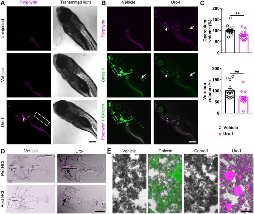
Acitretin mitigates uro‑I effects in bones of zebrafish larvae. To identify potential drugs to treat
CEP, we used our zebrafish CEP model and performed high-throughput screening of 1,280 small molecules by
co-administering drug and uro-I injection (Fig.S2). Acitretin, a second-generation retinoid commonly used
to treat psoriasis, decreased uro-I accumulation in bones (not shown). We validated the screening results and
further characterized acitretin as a potential treatment for CEP by testing whether it had a prophylactic effect.
Uro-I injected larvae were immediately transferred to either acitretin- or vehicle-containing medium. After 24 h,
acitretin-treated larvae had significantly reduced porphyrin fluorescence in their bones (operculum and verte-
brae, Fig. 2A, B) and increased uro-I excretion into the medium (Fig. 2C), but no effect on operculum volume
(Fig. 2D). To assess the therapeutic potential of acitretin, larvae were injected with uro-I then transferred after
24 h to medium containing either acitretin or vehicle and incubated for further 24 h then imaged. Acitretin
did not decrease bone porphyrin fluorescence (Fig. 2E, F), but it increased uro-I excretion to the medium and
operculum volume (Fig. 2G, H).
Since acitretin is a retinoid, we tested whether the protective effects are specific to acitretin or shared by
other retinoids. Etretinate (second-generation retinoid and precursor of acitretin), tretinoin (all trans-retinoic
acid) or acitretin were co-administered to zebrafish with uro-I. In addition to acitretin, tretinoin significantly
reduced bone porphyrin (Fig. 2I). Unlike acitretin, etretinate and tretinoin did not increase uro-I excretion into
the medium (Fig. 2J), and retinoids did not prevent loss in bone volume (Fig. 2K). Thus, both retinoids, acitretin
and tretinoin, prevent uro-I accumulation in zebrafish larvae. Hence, using our novel zebrafish CEP model, we
demonstrated that acitretin attenuated uro-I-mediated bone damage by modulating the dynamics of uro-I bone
binding and excretion.
Uro‑I impairs osteoblastic mineralization by aggregating matrix proteins, promoting ER
stress and inhibiting autophagy. To elucidate the molecular mechanism of uro-I-mediated bone dam-
age, we used Saos-2 cells, a human osteosarcoma cell line with osteoblastic features20. Mineralization was stimu-
lated by treating cells with a mineralization activation cocktail (MAC) and assayed by alizarin red S (ARS)
staining. As expected, Saos-2 cells manifested a mineralization phenotype when cultured for 3 days in MAC-
supplemented medium, while in uro-I + MAC supplemented medium, mineralization decreased significantly
(Fig. 3A, B). Uro-I also caused marked photosensitivity, leading to cell death when cells were not shielded from
light (not shown).
Since fluorescent porphyrins cause protein aggregation or loss of antibody reactivity when tested by immuno-
blotting, we tested whether uro-I-mediated inhibition of Saos-2 mineralization led to aggregation of bone matrix
proteins. Blotting Saos-2 cell lysates prepared from uro-I or vehicle treated cells using antibodies to fibronectin,
osteonectin and type 1 pro-collagen showed a distinct loss of monomer for these proteins after uro-I treatment
(Fig. 3C). We attribute the loss of antibody reactivity to epitope masking after uro-I binding and subsequent
oxidation and aggregation, as shown previously for PP-IX13. The loss of matrix protein monomers and aggrega-
tion was verified by mass spectrometry (Fig.S3).
Given the effect of uro-I on protein aggregation, we tested whether uro-I treatment initiates unfolded pro-
tein response (UPR) and endoplasmic reticulum (ER) stress. We observed upregulation of BiP, consistent with
UPR and ER s tress21 (Fig. 3C). Other ER stress markers, including PERK, IRE1α and ATF6, were likely oxidized
and aggregated, as judged by monomer loss (Fig. 3C). Our findings suggest a non-canonical form of ER stress,
which we have observed upon PP-IX a ccumulation13, that involves aggregation and possibly inactivation of ER
resident proteins and chaperones.
Autophagy modulates exocytosis of hydroxyapatite crystals and thus plays an important role in bone miner-
alization by o steoblasts22,23. Since uro-I inhibited mineralization, we tested whether it also disrupted autophagy.
As expected, MAC-treated Saos-2 cells showed increased LC3-II (Fig. 3D, left panel). Uro-I treatment also
increased LC3-II levels (Fig. 3D, right panel). The likely explanation for the increased LC3-II is not increased
autophagy but a slowing of autophagic flux, which could lead to stalling of exocytosis of mineral-loaded vesicles
and decreased mineralization of bone m atrix24.
To further characterize uro-I-mediated impairment of mineralization in Saos-2 cells, we performed gene
expression analysis to probe for alterations in the stress response pathway. Genes that were differentially regu-
lated two-fold or more after uro-I treatment were assessed further. Uro-I treatment increased HSPA5 (2.1x)
and SOD3 (2.8x), while SERPINH1 decreased (3.3x) (Fig. 3E, left panel). Since HSPA5 encodes BiP, HSPA5
upregulation supports the BiP upregulation observed biochemically (Fig. 3C). SERPINH1, a collagen-specific
chaperone, downregulation may account for collagen misfolding and aggregation. Because type 1 collagen is
the most abundant protein in bone matrix25, we assessed COL1A1 expression and observed a 90% reduction in
COL1A1 after uro-I treatment (Fig. 3E, right panel). This finding supports uro-I-induced loss of mineralization,
since collagen serves as a matrix for mineral deposition.
We next asked whether acitretin can protect from the effects of uro-I, by treating Saos-2 cells with uro-I in
the presence of acitretin. Although acitretin did not prevent uro-I-mediated loss of mineralization (Fig. 3F), it
blunted the ER stress response by reducing BiP level and normalized the autophagic flux by reducing LC3-II
(Fig. 3G, H). Acitretin also downregulated SOD3 3.8-fold, thereby suggesting that acitretin mitigates the oxida-
tive stress caused by uro-I (Fig. 3I). Upregulation of COL1A1 (1.7x) and SERPINH1 (5.4x) was also observed.
Taken together, our data demonstrate that acitretin mitigates uro-I-mediated proteotoxicity and oxidative
stress. However, under the conditions tested, acitretin did not rescue the impairment of mineralization caused
Scientific Reports | (2021) 11:9601 | https://doi.org/10.1038/s41598-021-88668-9 3
Vol.:(0123456789)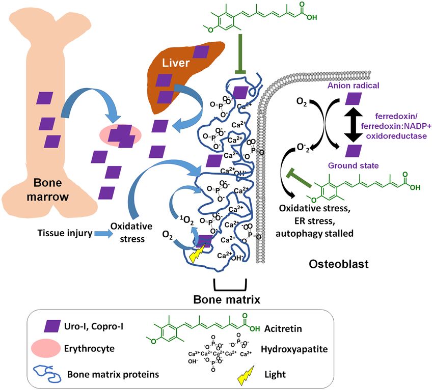
www.nature.com/scientificreports/
Figure 2. Acitretin mitigates CEP bone phenotype in zebrafish. (A) 6dpf larvae were injected with uro-I and transferred to medium
containing acitretin or DMSO. At 7dpf larvae were injected with calcein and imaged by confocal microscopy. Quantification of
porphyrin fluorescence (B), porphyrin excretion (C) and operculum volume (D) from experiment in (A). Symbols represent
individual larvae (12–25/group) from 3–4 independent experiments. (E) 6dpf larvae were injected with uro-I. At 7dpf they were
transferred to medium containing acitretin or DMSO. At 8dpf larvae were injected with calcein and imaged by confocal microscopy.
Quantification of porphyrin fluorescence (F), porphyrin excretion (G) and operculum volume (H) from experiment in (E).
Arrowhead-operculum; box-vertebrae (A, E). Symbols represent individual larvae (18–64/group) from 3–4 independent experiments.
(I-K) Larvae were treated as in (A) with the indicated retinoid or DMSO and porphyrin fluorescence (I), porphyrin excretion (J)
and operculum volume (K) were assayed. Bone volume was normalized to DMSO-treated larvae set to 100%, (D, H, K). Porphyrin
excretion was normalized to DMSO-treated larvae set to 100%, (C, G, J). Symbols represent individual larvae (7–44/group) from
2–4 independent experiments. Scale bars: 200 µm. Three-dimensional image reconstruction (A, E) was performed using Imaris 3D
software v7.7 (http://imaris.oxinst.com/). *p < 0.05, **p < 0.01, ***p < 0.001, ****p < 0.0001.
Scientific Reports | (2021) 11:9601 | https://doi.org/10.1038/s41598-021-88668-9 4
Vol:.(1234567890)www.nature.com/scientificreports/
Figure 3. Saos-2 cells mimic CEP zebrafish model. (A, B) Mineralization in Saos-2 cells treated with MAC ± Uro-I
was assayed using ARS staining (photograph, A; quantification, B). Staining was normalized to MAC only-treated cells
(set to 100%). (C) Cell lysates from experiment in (A) were blotted with the indicated antibodies. (D) Quantification
of LC3-II shown in (C). LC3-II level was normalized to MAC only (left panel) or vehicle-treated (right panel), set
to 100%. (E) RT2 Profiler PCR Array (left panel) and qPCR (right panel). Relative gene expression is represented
as fold change normalized to housekeeping gene. Data are from 2 independent experiments. (F) Acitretin does not
rescue reduced mineral matrix phenotype in uro-I-treated cells. ARS staining quantification as in (B). (G) Acitretin
normalizes ER stress (BiP) and autophagy (LC3-II) markers. Dashed lines represent non-adjacent lanes in the gel.
Coomassie-stained gel (C,G) shows equal protein loading. Full-length blots and gels (C,G) are presented in Fig.S4.
(H) Quantification of LC3-II. LC3-II level was normalized to DMSO-treated cells set to 100%. (I) Gene expression
profiling as in (E). *p < 0.05, **p < 0.01, ***p < 0.001, ****p < 0.0001.
Scientific Reports | (2021) 11:9601 | https://doi.org/10.1038/s41598-021-88668-9 5
Vol.:(0123456789)www.nature.com/scientificreports/
Figure 4. Proposed model of CEP pathogenesis. UROS inhibition leads to production of uro/copro-I mostly in
erythrocytes and liver, which is transported through blood to the bones. Uro-I causes bone damage by binding
to hydroxyapatite, causing oxidative and ER stress, protein aggregation and stalled autophagy. Acitretin partially
rescues uro-I-induced bone damage by reducing oxidative and ER stress and restoring autophagic flux.
by uro-I treatment of Saos-2 cells. A possible explanation for why mineralization was not normalized by acitretin
is that ER stress and autophagy pathways need to be normalized in order for cells to have their mineralization
ability restored. Alternatively, acitretin may act differently on various cell types which is one major advantage
offered by the in vivo zebrafish system.
Discussion
Uro-I is a fluorescent porphyrin capable of types I/II-photosensitized r eactions26–28, which explains the observed
photosensitivity in CEP, damage to digits and facial features3. However, light is unlikely to reach deep internal
tissues, which are also affected in CEP. Of note, we observed uro-I-mediated protein aggregation and decreased
mineralization in the dark. Previous studies had also reported dark effects of porphyrins. For example, uro-I
increased collagen biosynthesis in human skin fi broblasts29, and inhibited erythrocytic uroporphyrinogen decar-
boxylase activity30. A 2-hit model could explain light-independent porphyrin-mediated protein aggregation
and proteotoxicity whereby, in absence of light, a secondary oxidant source (eg. inflammatory cells) causes
protein oxidation followed by porphyrin binding to oxidized protein, yielding protein aggregates11,12 (Fig. 4).
CEP is frequently associated with superinfections and osteolysis3,31. Hence, infiltrating immune cell-generated
oxidants might serve as a secondary source of oxidant, leading to uro-I mediated protein aggregation in internal
organs such as bones. Additionally, uro-I might generate oxidants by acting as a substrate for the ferredoxin/
ferredoxin:NADP + oxidoreductase system32. Although ferredoxin/ferredoxin:NADP + oxidoreductase are com-
monly associated with hepatic microsomes, they are also expressed in osteoblasts33 and could metabolize uro-I
to generate oxidants in the absence of light.
The differences in charge and polarity of uro-I and PP-IX might explain the striking difference in their tissue
localization. Retro-orbitally injected PP-IX accumulated in zebrafish liver10, while uro-I accumulated preferen-
tially in bone (Fig. 1). Of note, liver cancer cell lines do not uptake uroporphyrin (unpublished data), possibly
Scientific Reports | (2021) 11:9601 | https://doi.org/10.1038/s41598-021-88668-9 6
Vol:.(1234567890)www.nature.com/scientificreports/
due to its high negative charge that prevents traversing the cell m embrane34. Based on our data, we propose that
negatively charged uro-I binds to Ca2+ in hydroxyapatite (Fig. 4) and thus bone and Saos-2 cells are affected by
uro-I. This association with bone matrix causes uro-I to have a different protein aggregation signature compared
to PP-IX, which is primarily internalized. PP-IX aggregated intracellular proteins such as keratins and glycer-
aldehyde 3-phosphate d ehydrogenase13,15,35, whereas uro-I affected extracellular bone matrix proteins (Fig. 3).
Oxidants such as singlet oxygen, a major oxidant produced by photosensitive r eactions26, have extremely small
intracellular diffusion distance (10-20 nm) and lifetime (10–40 ns)36–38. Binding of uro-I to bone matrix causes
a ‘sensitizer-acceptor’ coupling, as observed for other diffusible oxidants39,40, and greatly increases the oxidation
efficiency and specificity. Of note, oxidized fibronectin reduces mineralization of rat calvarial osteoblasts in
vitro41. The high selectivity of uro-I localization to bone matrix might provide a pathway to develop photody-
namic therapeutic agents for bone cancers such as osteosarcoma.
The management of CEP is challenging, with current therapeutic options focusing on bone marrow/hemat-
opoietic stem cell transplantation3, and by avoidance of sun and light exposure, including the use of protective
clothing42–45. There are also potential experimental therapeutic approaches including gene t herapy46, proteasomal
inhibitors47,48, iron c helation49,50 and p
hlebotomy51. Most recently, the repurposed use of ciclopirox, an approved
broad-spectrum antifungal agent, showed promising results in the treatment of CEP using a mouse m odel52.
However, there are limitations to these approaches, such as complications from transplantation and neurotoxic
side effects of proteasome inhibitors53. Currently there are no known pharmaceuticals that act by clearance of
uro-I, and in this regard acitretin provides a novel approach. Acitretin might also act as an antioxidant (Fig. 4)
due to its hyperconjugated nucleophilic double bonds. Thus, through a combination of destabilizing uro-I-bone
matrix interaction and antioxidant activity, acitretin could ameliorate CEP manifestations (Fig. 4). Acitretin also
offers a drug repurposing advantage since it is already approved for psoriasis t reatment54.
Materials and methods
Zebrafish experiments and cell culture. Zebrafish (Danio rerio) experiments were conducted using
ABxTL hybrid and NHGRI-1 wild type zebrafish lines. All animal procedures were approved by the Rutgers
University Institutional Animal Care and Use Committee (protocol number PROTO201900147) and performed
in compliance with federal guidelines and the standards of the NIH Guide for the Care and Use of Laboratory
Animals55, the Rutgers University IACUC Policy Handbook and the Animal Research: Reporting of In Vivo
Experiments (ARRIVE) guidelines.
Saos-2 cells were purchased from ATCC. Cells were maintained in McCoy 5A medium supplemented with
15% FBS, penicillin/streptomycin, non-essential amino acids, Hepes and L-glutamine. To induce mineralization,
cells were treated with mineralization activation cocktail (MAC), consisting of 5 mM β-glycerophosphate, 50
µM ascorbic acid and 10 nM d examethasone56.
Uro‑I solution preparation and treatment of zebrafish larvae and Saos‑2 cells. Uro-I (uropor-
phyrin-I dihydrochloride; Frontier Scientific, Catalog#:U830-1) was initially resuspended in 0.1 M NaOH and
the pH was adjusted to neutral using 0.2 M N a2HPO4. Six days post fertilization (dpf), ABTL zebrafish larvae
were injected via the retro-orbital route with approximately 3nL of 7.2 mM uro-I solution and control larvae were
injected with vehicle (0.1 M NaOH in 0.2 M N a2HPO4). After injection, larvae were immediately transferred to
Petri dishes wrapped with heavy duty aluminum foil and kept in a dark incubator, at 28.5 °C for 24 h. Where
indicated, 7 dpf larvae were injected with approximately 2 nL of 0.2% w/v calcein (Sigma, Catalog#:C0875) 2 h
prior to imaging.
Saos-2 cells were plated in 12-well plates (1.5 × 10⁵ cells/well) and allowed to attach overnight. Cells were then
treated with uro-I (144 µM final concentration) or vehicle in medium containing MAC for 3 days. Experiments
were conducted in a dark room and cells were kept shielded from light in a tissue culture incubator.
Confocal microscopy imaging and quantification. Seven dpf ABxTL zebrafish larvae were anesthe-
tized with tricaine-S (Syndel) and immobilized in 0.5% low melt agarose. Fluorescent z-series were captured
using an Olympus FV500 confocal microscope (10X objective, confocal aperture of 300 µµ) with an optical
thickness of 10 µm and z-step size of 10 µm. Calcein was excited with a 488 nm argon laser and emission was
captured between 505 and 525 nm. Porphyrin was excited with a 405 nm laser diode and emission was captured
above 560 nm. Three-dimensional image reconstruction and quantification of fluorescent signal and bone vol-
ume were performed using Imaris 3D visualization and analysis software v7.7 (Bitplane).
In vivo and in vitro binding of uro‑I. Six dpf ABxTL zebrafish larvae were injected with uro-I or vehicle
(as described above) and 24 h later were euthanized by tricaine-S overdose on ice bath. Bones were harvested as
previously described57. Briefly, soft tissue was removed by incubating larvae with Accumax solution (Millipore-
Sigma, Catalog#:A7089) under vigorous shaking. Bones were collected using a 70 µm cell strainer, followed by
demineralization with 1.2 M HCl. Fluorescent images were captured prior to and after the demineralization step
using a Zeiss Axio Imager M2 fluorescence microscope. Porphyrin signal was captured using the red fluorescent
channel.
10 mg hydroxyapatite (Acros Organics, Catalog#:1306–06-5) was incubated with 1 mM uro-I, 1 mM copro-I
(coproporphyrin-I dihydrochloride, Frontier Scientific, Catalog#:C654-1), vehicle or 0.2% calcein for 30 min
in the dark and vortexed every five minutes. Samples were washed and imaged by epifluorescence microscopy
as described above.
Scientific Reports | (2021) 11:9601 | https://doi.org/10.1038/s41598-021-88668-9 7
Vol.:(0123456789)www.nature.com/scientificreports/
High throughput drug screening. Unbiased high throughput drug screening was performed using the
Prestwick library (Prestwick Chemical), which consists of 1,280 small molecules chosen by the manufacturer
for their bioavailability and safety. A pooled approach, where four compounds were tested together, was used in
order to optimize animal use and investigation of drugs with potential for CEP treatment. Zebrafish E3 medium
(100 µL/well) was transferred to a 96-well half area imaging plate (Corning, cat. n. 3880) using a Multidrop
dispenser (ThermoFisher Scientific). Compounds (0.4 µL of 2 mM stock) were added to the wells using a multi-
channel plate handling robot (Biomek FX, Beckman Coulter Life Sciences). This step was performed four times
in order to pool four compounds into one well: one 384-well stock plate yielded one 96-well test plate. Control
wells contained 1.6 µL of DMSO.
Six dpf NHGRI-1 zebrafish larvae were injected retro-orbitally with approximately 2nL of a solution of uro-I
(10 mM) and calcein (0.2% w/v). Immediately after injection, larvae were transferred to a 96-well test plate (two
larvae in 50 µL of E3 medium/well), including the DMSO control wells. Larvae were kept in the dark, at 28.5 °C.
After 24 h, they were anesthetized with tricaine-S, centrifuged at 500xg for two minutes and imaged using the
ImageXpress Micro Cellular Imaging and Analysis System (Molecular Devices). Positive hits were selected based
on visual identification of calcein signal increase and porphyrin signal decrease compared to DMSO-treated
larvae. Compounds in test wells that met the inclusion criterion were tested individually in the same manner as
described above (Figure S2).
Acitretin validation and treatment. A dose-response curve with acitretin (Selleck Chemicals, Houston,
TX) was conducted (0.5–12.5 µM) and 10 µM was observed to yield consistent results, without being toxic to
zebrafish larvae. Validation and characterization of acitretin as a potential treatment for CEP was performed. Six
dpf ABxTL zebrafish larvae injected with uro-I were immediately transferred to 10 cm plastic dishes containing
10 µM acitretin or DMSO in E3 medium (prophylaxis protocol, Fig. 2A), and incubated for 24 h in the dark at
28.5 °C. Porphyrin binding to bones and bone volume were analyzed by confocal microscopy as described above.
Porphyrin excretion into the medium was quantified. Uro-I-injected larvae were transferred to 96-well plates,
one larva/well, 100µL of 10 µM acitretin or DMSO/well. Medium was collected after 24 h and porphyrin was
quantified as described previously13. Etretinate (Selleck Chemicals, Houston, TX) and tretinoin (Selleck Chemi-
cals, Houston, TX) treatment was performed as described for acitretin.
In addition to being used as prophylaxis, we evaluated whether acitretin had a therapeutic effect. Six dpf
ABxTL zebrafish larvae were injected with uro-I and 24 h later they were transferred to E3 medium containing
10 µM acitretin or DMSO. Porphyrin binding, excretion and bone volume were analyzed as described above. For
Saos-2 cells, they were treated with 10 µM acitretin or DMSO in medium containing MAC and uro-I.
Alizarin Red S (ARS) staining and quantification. Cell mineralization was quantified by ARS (Sigma
reviously58 with minor modifications. Briefly, cells were fixed with
Aldrich St. Louis, MO) staining as described p
100% ethanol at 37 °C for 1 h, stained with 40 mM (pH4.2) ARS solution for 20 min in an orbital shaker. Cells
were washed and ARS was extracted by incubation of fixed cells with 10% (v/v) acetic acid, followed by scrap-
ing, incubation of suspension (85 °C, 10 min), centrifugation and neutralization of supernatant with 10% (v/v)
ammonium hydroxide. ARS standard curve (from 2–0.02 mM) and samples were transferred in triplicate to a
96-well plate and absorbance was measured at 405 nm.
Cell harvest, immunoblotting and mass spectrometry. Saos-2 cells were lysed in ice cold RIPA
buffer (Sigma Aldrich, St. Louis, MO) with protease inhibitor cocktail (Thermo Scientific, Waltham, MA) and
scraped. Whole cell lysate was kept in the dark until reducing SDS-PAGE sample buffer was added. Immunob-
lotting, band densitometry and mass spectrometry were conducted as described previously12,13. The antibodies
used and their vendors are as listed. Antibodies to the indicated antigens (and sources) are: ATF6, BiP, LC3B
(Cell Signaling Technology, Danvers, MA); fibronectin HFN 7.1, pro-collagen SP1.D8, osteonectin AON-1
(Developmental Studies Hybridoma Bank; Iowa City, Iowa); IRE1α, PERK (Invitrogen, Carlsbad, CA); lamin
A/C (Santa Cruz Biotechnology, Dallas, TX).
Gene expression profiling. Saos-2 cells RNA was extracted using RNeasy mini kit (Qiagen, Cata-
log#:74,104). and gene expression was carried using the R T2 Profiler PCR Array for human cellular stress
responses (Qiagen, Catalog#:PAHS-019ZA) following manufacturer’s instructions. A previously described
qPCR59 was performed for COL1A1 and SERPINH1 (IDT Integrated DNA Technologies, PrimeTime assay ID
Hs.PT.58.15517795 and Hs.PT.56a.26865778, respectively).
Statistical analysis. Statistical analysis was performed using GraphPad Prism v8 (GraphPad Software).
Unpaired two-tailed Student’s t-test was used to determine statistical significance. Error bars represent standard
error of the mean. *p < 0.05, **p < 0.01, ***p < 0.001, ****p < 0.0001.
Received: 28 November 2020; Accepted: 15 April 2021
Referenceseferences
1. Puy, H., Gouya, L. & Deybach, J.-C. Porphyrias. The Lancet 375, 924–937. https://doi.org/10.1016/S0140-6736(09)61925-5 (2010).
Scientific Reports | (2021) 11:9601 | https://doi.org/10.1038/s41598-021-88668-9 8
Vol:.(1234567890)www.nature.com/scientificreports/
2. Ajioka, R. S., Phillips, J. D. & Kushner, J. P. Biosynthesis of heme in mammals. Biochimica et Biophysica Acta BBA Mol. Cell Res.
1763, 723–736. https://doi.org/10.1016/j.bbamcr.2006.05.005 (2006).
3. Erwin, A. L. & Desnick, R. J. Congenital erythropoietic porphyria: recent advances. Mol. Genet. Metab. 128, 288–297 (2019).
4. Di Pierro, E. et al. Congenital erythropoietic porphyria linked to GATA1-R216W mutation: challenges for diagnosis. Eur. J. Hae-
matol. 94, 491–497. https://doi.org/10.1111/ejh.12452 (2015).
5. Yasuda, M., Chen, B. & Desnick, R. J. Recent advances on porphyria genetics: Inheritance, penetrance & molecular heterogeneity,
including new modifying/causative genes. Mol. Genet. Metab 128, 320–331. https://doi.org/10.1016/j.ymgme.2018.11.012 (2019).
6. Balwani, M. & Desnick, R. J. The porphyrias: advances in diagnosis and treatment. Blood 120, 4496–4504. https://doi.org/10.1182/
blood-2012-05-423186 (2012).
7. Di Pierro, E., Brancaleoni, V. & Granata, F. Advances in understanding the pathogenesis of congenital erythropoietic porphyria.
Br. J. Haematol. 173, 365–379. https://doi.org/10.1111/bjh.13978 (2016).
8. Bonkovsky, H. L. et al. Porphyrin and heme metabolism and the porphyrias. Compr. Physiol. 3, 365–401. https://doi.org/10.1002/
cphy.c120006 (2013).
9. Katugampola, R. P. et al. Congenital erythropoietic porphyria: a single-observer clinical study of 29 cases. Br. J. Dermatol. 167,
901–913. https://doi.org/10.1111/j.1365-2133.2012.11160.x (2012).
10. Elenbaas, J. S. et al. A precursor-inducible zebrafish model of acute protoporphyria with hepatic protein aggregation and multio-
rganelle stress. FASEB J. 30, 1798–1810. https://doi.org/10.1096/fj.201500111R (2016).
11. Maitra, D. et al. Porphyrin-induced protein oxidation and aggregation as a mechanism of porphyria-associated cell injury. Cell
Mol. Gastroenterol. Hepatol. 8, 535–548. https://doi.org/10.1016/j.jcmgh.2019.06.006 (2019).
12. Maitra, D. et al. Oxygen and conformation dependent protein oxidation and aggregation by porphyrins in hepatocytes and light-
exposed cells. Cell Mol. Gastroenterol. Hepatol. 8, 659–682. https://doi.org/10.1016/j.jcmgh.2019.05.010 (2019).
13. Maitra, D. et al. Ambient light promotes selective subcellular proteotoxicity after endogenous and exogenous porphyrinogenic
stress. J. Biol. Chem. 290, 23711–23724. https://doi.org/10.1074/jbc.M114.636001 (2015).
14. Saggi, H. et al. Loss of hepatocyte beta-catenin protects mice from experimental porphyria-associated liver injury. J. Hepatol. 70,
108–117. https://doi.org/10.1016/j.jhep.2018.09.023 (2019).
15. Singla, A. et al. Lamin aggregation is an early sensor of porphyria-induced liver injury. J. Cell Sci. 126, 3105–3112. https://doi.org/
10.1242/jcs.123026 (2013).
16. Zhang, H. et al. Tumor-selective proteotoxicity of verteporfin inhibits colon cancer progression independently of YAP1. Sci. Signal
8, ra 98. https://doi.org/10.1126/scisignal.aac5418 (2015).
17. Maitra, D. et al. Protoporphyrin-IX nanostructures modulate their protein aggregation ability via differential oxidation and protein
binding. bioRxiv, 2021.2001.2011.426224, doi:https://doi.org/10.1101/2021.01.11.426224 (2021).
18. Crockett, J. C., Rogers, M. J., Coxon, F. P., Hocking, L. J. & Helfrich, M. H. Bone remodelling at a glance. J. Cell Sci. 124, 991–998.
https://doi.org/10.1242/jcs.063032 (2011).
19. Raggatt, L. J. & Partridge, N. C. Cellular and molecular mechanisms of bone remodeling. J. Biol. Chem. 285, 25103–25108. https://
doi.org/10.1074/jbc.R109.041087 (2010).
20. Rodan, S. B. et al. Characterization of a human osteosarcoma cell line (Saos-2) with osteoblastic properties. Can. Res. 47, 4961–4966
(1987).
21. Pavelka, K., Vojtisek, O., Bremova, A., Dostal, C. & Kralova, M. A new nonsteroid antirheumatic drug diclofenac in the treatment
of rheumatoid arthritis (preliminary report. Fysiatr. Revmatol. Vestn. 55, 355–359 (1977).
22. Nollet, M. et al. Autophagy in osteoblasts is involved in mineralization and bone homeostasis. Autophagy 10, 1965–1977. https://
doi.org/10.4161/auto.36182 (2014).
23. Yin, X. et al. Autophagy in bone homeostasis and the onset of osteoporosis. Bone Res. 7, 28. https://doi.org/10.1038/s41413-019-
0058-7 (2019).
24. Bottini, M. et al. Matrix vesicles from chondrocytes and osteoblasts: their biogenesis, properties, functions and biomimetic models.
Biochim Biophys Acta Gen Subj 532–546, 2018. https://doi.org/10.1016/j.bbagen.2017.11.005 (1862).
25. N. Cooper, L. & Maas, M. C. in Encyclopedia of marine mammals (Third Edition) (eds Bernd Würsig, J. G. M. Thewissen, & Kit M.
Kovacs) 114–118 (Academic Press, 2018).
26. Baptista, M. S. et al. Type I and Type II photosensitized oxidation reactions: guidelines and mechanistic pathways. Photochem.
Photobiol. 93, 912–919. https://doi.org/10.1111/php.12716 (2017).
27. Takeshita, K., Olea-Azar, C. A., Mizuno, M. & Ozawa, T. Singlet oxygen-dependent hydroxyl radical formation during uropor-
phyrin-mediated photosensitization in the presence of NADPH. Antioxid. Redox Signal. 2, 355–362 (2000).
28. Herrmann, G., Bolsen, K., Prenzel, K., Goerz, G. & Scharffetter-Kochanek, K. Photosensitization of uroporphyrin augments the
ultraviolet A-induced synthesis of matrix metalloproteinases in human dermal fibroblasts. J. Investig. Dermatol. 107, 398–403
(1996).
29. Varigos, G., Schiltz, J. R. & Bickers, D. R. Uroporphyrin I stimulation of collagen biosynthesis in human skin fibroblasts. A unique
dark effect of porphyrin. J. Clin. Invest. 69, 129–135. https://doi.org/10.1172/jci110423 (1982).
30. Afonso, S. G., Chinarro, S., De Salamanca, R. E. & Batlle, A. M. D. C. Further Evidence on the Photodynamic and the Novel Non-
Photodynamic Inactivation of Uroporphyrinogen Decarboxylase by Uroporphyrin I. J. Enzym. Inhib. 5, 225–233. https://doi.org/
10.3109/14756369109080061 (1991).
31. Horner, M. E. et al. Cutaneous porphyrias part I: epidemiology, pathogenesis, presentation, diagnosis, and histopathology. Int. J.
Dermatol. 52, 1464–1480. https://doi.org/10.1111/ijd.12305 (2013).
32. Morehouse, K. M. & Mason, R. P. The enzymatic one-electron reduction of porphyrins to their anion free radicals. Arch. Biochem.
Biophys. 283, 306–310 (1990).
33. Teplyuk, N. M. et al. The osteogenic transcription factor runx2 controls genes involved in sterol/steroid metabolism, including
CYP11A1 in osteoblasts. Mol. Endocrinol. (Baltimore, Md.) 23, 849–861. https://doi.org/10.1210/me.2008-0270 (2009).
34. Krishnamurthy, P., Xie, T. & Schuetz, J. D. The role of transporters in cellular heme and porphyrin homeostasis. Pharmacol. Ther.
114, 345–358. https://doi.org/10.1016/j.pharmthera.2007.02.001 (2007).
35. Snider, N. T. et al. Energy determinants GAPDH and NDPK act as genetic modifiers for hepatocyte inclusion formation. J. Cell
Biol. 195, 217–229. https://doi.org/10.1083/jcb.201102142 (2011).
36. Davies, M. J. Reactive species formed on proteins exposed to singlet oxygen. Photochem. Photobiol. Sci. 3, 17–25. https://doi.org/
10.1039/b307576c (2004).
37. Moan, J. & Berg, K. The photodegradation of porphyrins in cells can be used to estimate the lifetime of singlet oxygen. Photochem.
Photobiol. 53, 549–553. https://doi.org/10.1111/j.1751-1097.1991.tb03669.x (1991).
38. Pattison, D. I., Rahmanto, A. S. & Davies, M. J. Photo-oxidation of proteins. Photochem. Photobiol. Sci. 11, 38–53. https://doi.org/
10.1039/c1pp05164d (2012).
39. Klaper, M., Fudickar, W. & Linker, T. Role of distance in singlet oxygen applications: a model system. J. Am. Chem. Soc. 138,
7024–7029. https://doi.org/10.1021/jacs.6b01555 (2016).
40. Zheng, L. et al. Apolipoprotein A-I is a selective target for myeloperoxidase-catalyzed oxidation and function impairment in
subjects with cardiovascular disease. J. Clin. Investig. 114, 529–541. https://doi.org/10.1172/JCI200421109 (2004).
41. Suzuki, H., Hayakawa, M., Kobayashi, K., Takiguchi, H. & Abiko, Y. H2O2-derived free radicals treated fibronectin substratum
reduces the bone nodule formation of rat calvarial osteoblast. Mech. Ageing Dev. 98(2), 113–125 (1997).
Scientific Reports | (2021) 11:9601 | https://doi.org/10.1038/s41598-021-88668-9 9
Vol.:(0123456789)www.nature.com/scientificreports/
42. Matthew, H., Anthony, H. & Donald, R. Congenital erythropoietic porphyria–long-term follow up of a case and review. (2017).
43. Badminton, M. N. & Elder, G. H. Management of acute and cutaneous porphyrias. Int. J. Clin. Pract. 56, 272–278 (2002).
44. Balwani, M., Bloomer, J., Desnick, R., of the NIH-Sponsored, P. C. & Network, R. D. C. R. in GeneReviews®[Internet] (University
of Washington, Seattle, 2017).
45. Erwin, A. L., Balwani, M. & Desnick, R. in GeneReviews®[Internet] (University of Washington, 2013).
46. Robert-Richard, E. et al. Effective gene therapy of mice with congenital erythropoietic porphyria is facilitated by a survival advan-
tage of corrected erythroid cells. Am. J. Hum. Genet. 82, 113–124 (2008).
47. Ben Bdira, F. et al. Tuning intracellular homeostasis of human uroporphyrinogen III synthase by enzyme engineering at a single
hotspot of congenital erythropoietic porphyria. Hum. Mol. Genet. 23, 5805–5813 (2014).
48. Blouin, J.-M. et al. Missense UROS mutations causing congenital erythropoietic porphyria reduce UROS homeostasis that can be
rescued by proteasome inhibition. Hum. Mol. Genet. 26, 1565–1576 (2017).
49. Blouin, J. M. et al. Iron chelation rescues hemolytic anemia and skin photosensitivity in congenital erythropoietic porphyria. Blood
https://doi.org/10.1182/blood.2020006037 (2020).
50. Egan, D. N., Yang, Z., Phillips, J. & Abkowitz, J. L. Inducing iron deficiency improves erythropoiesis and photosensitivity in con-
genital erythropoietic porphyria. Blood 126, 257–261. https://doi.org/10.1182/blood-2014-07-584664 (2015).
51. Mirmiran, A. et al. Phlebotomy as an efficient long-term treatment of congenital erythropoietic porphyria. Haematologica Online
ahead of print, doi:https://doi.org/10.3324/haematol.2019.228270 (2020).
52. Urquiza, P. et al. Repurposing ciclopirox as a pharmacological chaperone in a model of congenital erythropoietic porphyria. Sci.
Transl. Med. https://doi.org/10.1126/scitranslmed.aat7467 (2018).
53. Blouin, J.-M. et al. Therapeutic potential of proteasome inhibitors in congenital erythropoietic porphyria. Proc. Natl. Acad. Sci.
110, 18238–18243 (2013).
54. Dunn, L. K., Gaar, L. R., Yentzer, B. A., O’Neill, J. L. & Feldman, S. R. Acitretin in dermatology: a review. J. Drugs Dermatol. 10,
772–782 (2011).
55. National Research Council Committee for the Update of the Guide for the, C. & Use of Laboratory, A. in Guide for the Care and
Use of Laboratory Animals (National Academies Press (US) Copyright © 2011, National Academy of Sciences., 2011).
56. Wiens, M. et al. Osteogenic potential of biosilica on human osteoblast-like (SaOS-2) cells. Calcif. Tissue Int. 87, 513–524. https://
doi.org/10.1007/s00223-010-9408-6 (2010).
57. Kessels, M. Y. et al. Proteomics analysis of the zebrafish skeletal extracellular matrix. PLoS ONE 9, e90568. https://d
oi.o
rg/1 0.1 371/
journal.pone.0090568 (2014).
58. Harper, E. et al. TRAIL attenuates RANKL-mediated osteoblastic signalling in vascular cell mono-culture and co-culture models.
PLoS ONE 12, e0188192. https://doi.org/10.1371/journal.pone.0188192 (2017).
59. Elenbaas, J. S. et al. Lamin A/C maintains exocrine pancreas homeostasis by regulating stability of RB and activity of E2F. Gastro-
enterology 154, 1625–1629. https://doi.org/10.1053/j.gastro.2018.01.024 (2018).
Acknowledgements
We thank the following research cores at the University of Michigan: Center for Chemical Genomics; Microscopy,
Imaging and Cellular Physiology Core (MICPC) Imaging Laboratory; and the Proteomics Research Facility.
Author contributions
Conceptualization: J.B.C., J.S.E., D.M., M.B.O.; Methodology: J.B.C., J.S.E., J.A.S.; Investigation: J.B.C., J.S.E.,
N.K., R.A.D., A.C.F., M.S.G., S.I.L.; Writing original draft: J.B.C., D.M.; Review and editing of the manuscript:
J.B.C., D.M., M.B.O.; Review of final version prior to submission: all authors; Overall Project Supervision: M.B.O.;
Funding acquisition: M.B.O., J.A.S.
Funding
This work is supported by NIH R01 DK116548 (M.B.O.), R35 HL150784 (J.A.S.), and P30 DK020572 (MICPC
Imaging Lab).
Competing interests
The authors declare no competing interests. A provisional patent application for the use of retinoids as a possible
therapy for CEP has been filed.
Additional information
Supplementary Information The online version contains supplementary material available at https://doi.org/
10.1038/s41598-021-88668-9.
Correspondence and requests for materials should be addressed to J.B.C. or M.B.O.
Reprints and permissions information is available at www.nature.com/reprints.
Publisher’s note Springer Nature remains neutral with regard to jurisdictional claims in published maps and
institutional affiliations.
Open Access This article is licensed under a Creative Commons Attribution 4.0 International
License, which permits use, sharing, adaptation, distribution and reproduction in any medium or
format, as long as you give appropriate credit to the original author(s) and the source, provide a link to the
Creative Commons licence, and indicate if changes were made. The images or other third party material in this
article are included in the article’s Creative Commons licence, unless indicated otherwise in a credit line to the
material. If material is not included in the article’s Creative Commons licence and your intended use is not
permitted by statutory regulation or exceeds the permitted use, you will need to obtain permission directly from
the copyright holder. To view a copy of this licence, visit http://creativecommons.org/licenses/by/4.0/.
© The Author(s) 2021
Scientific Reports | (2021) 11:9601 | https://doi.org/10.1038/s41598-021-88668-9 10
Vol:.(1234567890)You can also read