Avian Liver: The Forgotten Organ - Review - MDPI
←
→
Page content transcription
If your browser does not render page correctly, please read the page content below
Review
Avian Liver: The Forgotten Organ
Faegheh Zaefarian 1,*, Mohammad Reza Abdollahi 1, Aaron Cowieson 2 and Velmurugu
Ravindran 1
 1 Monogastric Research Centre, School of Agriculture and Environment, Massey University, Private Bag 11
   222, Palmerston North 4442, New Zealand; M.Abdollahi@massey.ac.nz (M.R.A.);
   V.Ravindran@massey.ac.nz (V.R.)
 2 DSM Nutritional Products, Wurmisweg 576, CH-4303 Kaiseraugst, Switzerland; aaron.cowieson@dsm.com

 * Correspondence: F.Zaefarian@massey.ac.nz; Tel.: +64-6-9518282

 Received: 10 January 2019; Accepted: 11 February 2019; Published: 15 February 2019

 Simple Summary: The liver is a multi-purpose organ, with involvement in bile secretion, and lipid,
 carbohydrate and protein metabolism, as well as a number of other metabolic functions. This organ
 can adapt easily to changes in feed and the environment. Being at the centre of a number of
 digestive, metabolic and productive activities, it is essential to have a better understanding of this
 organ and the factors affecting liver functionality.

 Abstract: Despite having huge responsibilities in avian species, published reports on the influence
 of dietary factors and other possible constraints on the size, development and function of liver are
 limited. Consideration of the factors that could influence and alter liver function is therefore of
 critical relevance. In the current review, aspects of liver structure and function, and the influence of
 feed restriction, anti-nutritional factors, structural components and feed additives on liver are
 discussed. Effects of feed technology techniques such as thermal treatment and pelleting, feed
 particle size and whole grain feeding on the liver are also reviewed. A discussion of lipogenesis and
 lipid storage in poultry is presented to provide a better understanding and to differentiate the
 normal pathways of lipid metabolism from abnormal (i.e., disordered) pathways. The liver is the
 main site of fat synthesis in poultry, but under certain conditions, excessive fat can accumulate in
 the liver and cause problems. Factors contributing to the fatty liver syndrome are also examined.

 Keywords: liver functions; liver metabolism; feed restriction; anti-nutritional factors; feed
 processing techniques; poultry

1. Introduction
     Similar to mammals, the liver in birds is involved in an array of metabolic and homeostatic
functions and considered as a biochemical factory responsible for most of the synthesis, metabolism,
excretion, and detoxification processes. It plays an important role in digestion and metabolism,
regulating the production, storage, and release of lipids, carbohydrates and proteins [1]. The liver
produces a variety of proteins, including blood proteins, enzymes, hormones, clotting and immune
factors. It functions as both an endocrine and exocrine gland [1]. In order to maintain a healthy bird,
this organ should be kept in an excellent condition. Better understanding of the metabolic functions
and the factors that can cause disruptions in the liver is important for the production of heathy birds.
     It is the intention of this paper to review the available literature on the constituents, structure
and metabolic functions of the liver and to provide an overview of dietary factors, which can
influence the liver function in poultry. The focus will be on the influence of (i) feed restriction, (ii)
anti-nutritional factors, (iii) structural components (iv) feed additives, and (v) feed processing
techniques. Moreover, metabolic disorders related to the liver dysfunction will also be considered.

Animals 2019, 9, 63; doi:10.3390/ani9020063                                    www.mdpi.com/journal/animals
Animals 2019, 9, 63 2 of 23
2. Structure and Functional Units in Avian Species
The liver is an accessory organ of the digestive system and the largest gland of the body. The
liver, located in the anterior end of the body cavity, has its conformation modified to fit the contour
of the internal surfaces of the body wall, as well as the adjacent and enclosed structures, such as the
heart and pericardial cavity, the underlying gizzard, spleen, gall bladder, the loop of the intestine
and the lungs [2]. It is divided into two lobes, namely right and left, which are joined cranially at the
midline. The right lobe is larger and separated from the left lobe by a deep fissure. In the domestic
fowl and turkey, the left lobe is subdivided into the dorsal and ventral parts [1,2]. Visceral peritoneum
covers the liver and closely adheres to its surface. There are several attachments and ligaments, such
as triangular ligaments and lesser omentum holding this organ in place. Lobules, the functional unit
of liver, are hexagonal in shape. Each lobe of the liver has approximately 100,000 lobules. Lobules are
separated from each other by interlobular septum. The liver lobule is formed by parenchymal cells
(hepatocytes) and non-parenchymal cells. Hepatocytes occupy almost 80% of the total liver volume
and perform numerous liver functions. Non-parenchymal liver cells are localised in the sinusoidal
wall. Two types of the cells compose the sinusoids, namely endothelia cells that coat the sinusoids
and macrophages. The latter are referred to as Kupffer and are star-shaped and confined to the liver.
Kupffer cells phagocytise pathogens, cell debris and, damaged red and white blood cells [3].
Sinusoids allow large plasma proteins from the bloodstream into the spaces surrounding the
hepatocytes.
The hepatocyte is a complex cell with a large nucleus and many mitochondria which contain
granules. Lysosomes, rough and smooth endoplasmic reticulum, Golgi apparatus, and other
organelles are also found in the hepatocyte [4]. Hepatocytes are arranged in plates that radiate
longitudinally outward from central vein. At each corner of the hexagonal lobule, there is a portal
triad consisting of a branch of the hepatic artery, a branch of portal vein and the bile duct. The liver
receives blood from the intestine and the general circulation [5]. The liver receives oxygenated blood
from the hepatic artery and deoxygenated blood from the hepatic portal vein, which contains
nutrients, drugs and toxins from the digestive tract. Branches from both arteries enter the liver
sinusoids where the hepatocytes remove some nutrients and toxins. As the blood passes through
sinusoids, metabolites from hepatocytes are secreted into the blood. This blood then passes to the
central vein and then into the hepatic vein (Figures 1 and 2).
Hepatic portal vein
Hepatic artery
(contains drugs and toxins absorbed in digestive tract)
(supplies oxygenated blood)
Liver sinusoids Hepatocyte
(absorbs nutrients, detoxifies some drugs
and produces bile)
Central vein
Hepatic vein
Inferior vena cava
Figure 1. Blood flow through the liver lobule (Adapted from Akers and Denbow [5]).Animals 2019, 9, 63 3 of 23
.
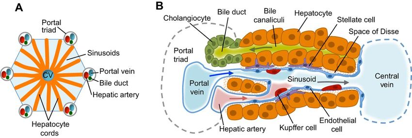
Figure 2. Liver structure and cell types (A) Functional unit of the liver lobules; Each lobule is
composed of a central vein (CV), the portal triad consists of a portal vein, biliary duct and hepatic
artery. Hepatocyte cords are separated by sinusoids that carry blood from the portal triads to the
central vein. (B) Hepatocytes secrete bile salts into the bile canaliculi that lead to the bile duct. Stellate
cells are in the space of Disse between the hepatocyte cords and sinusoids. Kupffer cells, which are
the specialised macrophages of the liver, are also located in sinusoids. Epithelial cells lining the bile
ducts are called Cholangiocytes (Adapted from Gordillo et al. [6]).
3. Composition and Size
The avian liver has much less connective tissue than the mammalian liver and it lacks a true
lobular structure [4]. In avian species, the liver is larger than in mammals when compared to body
size. The size and colour of the liver depends on age and body weight [1,7]. In 21-day old broiler
chickens fed maize-soy diets, the liver comprises approximately 27 g/kg body weight (BW) and
protein content of the liver was 690 g/kg liver mass. There were no differences in the contents of
protein, fat and ash between soybean oil- and tallow-supplemented diets (Table 1).
Table 1. Liver weight and proximate analysis of liver in broiler chickens fed maize-based diets (d 21) 1.
Weight DM Protein Ash Fat
Fat Source
(g/kg BW) (g/kg) (g/kg) (g/kg) (g/kg)
Soybean oil 26.9 241 690 59 125
Tallow 26.3 234 688 60 130
1 Each value represents the mean of 12 birds.
Bowes and Julian [8] compared the liver weights of broilers at 9 and 42 days of age and observed
that over this period the relative weight of liver decreased by 35.3 %, from 37.7 to 24.4 g/kg BW,
respectively. Nir et al. [9] found an increase in the relative liver weight from 25 g/kg BW at hatch to
46 and 48 g/kg BW on day 8 in egg-type and meat-type chickens, respectively. The relative weights
declined thereafter, but the reductions were greater in egg-type than in meat-type chicks. By day 14,
the liver weight of meat-type chicks exceeded that of the egg-type chicks. Ravindran et al. [10]
reported that the relative weights of the liver in birds fed wheat-based diets increased to day 14 and
decreased thereafter. In their study, the relative weights were 35, 38, 43, 33, 30 and 26 g/kg BW at 1,
7, 14, 21, 28 and 35 days post-hatch, respectively. The heavier relative liver size in early age
presumably enable birds to metabolised nutrient more efficiently, due to the lower feed intake and
endogenous enzyme secretions.Animals 2019, 9, 63 4 of 23
4. Liver Functions
The liver is a vital organ that is involved in a wide range of functions including the metabolism
of fat, carbohydrate, protein, vitamins and minerals, removal of waste products and detoxification.
The liver is the main storage site of fat-soluble vitamins (A, D, K and E) as well as vitamin B12,
glycogen, some minerals (Fe and Cu) and is also involved in the activation of vitamin D. The liver is
the main site of phagocytosis by Kupffer cells, which destroy aged blood cells and pathogens that
may enter via the hepatic portal blood [5].
4.1. Fat Metabolism
The liver plays the main role in lipogenesis, providing lipids destined to be used by all tissues
and the liver itself [11]. In contrast to mammals, the synthesis of fat in birds is greater in the hepatic
tissue and very limited in the adipose tissue [11]. In laying hens, the liver plays a major role in the
synthesis and metabolism of fat. Fats that are metabolised in the liver are derived from three main
sources: dietary fat, depot fat and fat from de novo fatty acid synthesis (from feed carbohydrates).
Similar to mammals, the digestion and absorption of dietary fats in avian species occur in the small
intestine [12]. However, due to a poorly developed intestinal lymphatic system in birds, dietary fatty
acids are drained directly into the portal blood system (instead of the lymphatic system) as very low-
density lipoproteins (VLDL) which are termed portomicrons [13]. Portomicrons are chylomicrons
and contain 90% triglycerides which combined with free and esterified cholesterol, lipoprotein and
phospholipids [11,14]. From the portal blood system, most of the portomicrons pass through the liver
before they reach the rest of the circulation. This unique feature predisposes the birds to fat
accumulation in the liver [15]. Liver hepatocytes can store triglycerides from portomicrons as well as
metabolise fatty acids to ATP, synthesise lipoproteins and phospholipids or store the released energy
in tissues as fat deposits [16]. The transport of triglycerides from the liver into adipose tissues or the
oviduct is facilitated by different classes of lipoproteins.
4.1.1. Gall Bladder, Bile Formation and Secretion
The gall bladder, which stores the bile, is a thin-walled muscular green sac found on the ventral
surface of liver. The liver is the source of the bile which plays an important role in the emulsification
of dietary fats, an essential step in fat digestion and absorption. Bile is produced in the hepatocytes
of the liver and secreted into the bile canaliculi. Canaliculi are the intracellular canals between the
hepatocytes and transport bile for storage in the gall bladder [17]. In birds, in contrast to mammals,
two bile ducts enter the duodenum just below the gizzard exit. The right and left hepatic ducts
combine to form a common hepato-enteric duct, which then goes to the duodenum. However, a
hepato-cystic duct branches from the right hepatic duct and connects to the gall bladder which, in
turn, is drained by the cystico-enteric duct into the duodenum [18]. The bile ducts generally drain
into the duodenum at a site very near the pancreatic ducts and occur on the ascending loop of the
duodenum. In some avian species (e.g., ostrich and pigeons), the ducts empty into the descending
loop of the duodenum [18]. Bile is a major source of fat secretion into the duodenum and account for
the observed negative fat digestibility in this segment [12,19–21]. Moreover, digesta and bile are
shuttled between the gastric region and duodenum via anti-peristaltic refluxes to enhance the
enzymatic and mechanical action of digestion. This action may also increase the net concentration of
fat in the duodenum [12].
Relatively little is known about biliary secretion in birds due to the complex anatomy in which
bile enters the intestine via both hepato-enteric and the cystico-enteric ducts. Bile flow is stimulated
by feed [22], presence of bile salts in the blood [23] and cholecystokinin (CCK) secretion [24]. In
ruminants, pigs and poultry, there is relatively continuous secretion of bile into the intestine. The
sphincter of hepato-pancreatic ampulla (sphincter of oddi) in these species is less well defined. In
dogs and cats, a continuous secretion into intestine is unnecessary because these species eat only once
or twice a day [25]. Parasympathetic signals traveling along the vagus nerve can stimulate bile
production by the liver. Neural and hormonal stimuli can also stimulate bile secretion. The sphincterAnimals 2019, 9, 63 5 of 23
of oddi controls the entry of bile into the duodenum. The presence of fatty acids, particularly medium
and long chain fatty acids in the chyme stimulates duodenal entero-endocrine cells to secrete CCK
and cause the contraction of smooth muscle of the gall bladder and relax the sphincter of oddi. Bile
is then squeezed into the cystic duct and through the common bile duct [5]. The biliary secretion rate
has been reported to be 24.2 uL/min in fasted birds [26].
4.1.2. Bile Composition and Functions
Bile consists of water, electrolytes, bile acids, bile salts, and neutral fats such as cholesterol,
glycerides, phospholipids (e.g., lecithin), bile pigments and some proteins [27,28]. Bile is the main
source of endogenous fat and fatty acids and plays an important role as an emulsifier in the digestion
and absorption of fat by reducing the tension at the oil-water interface [28]. Bile is slightly acidic in
chickens (5.9), ducks (6.1) and turkeys (6.0) [1,5]. Bile activates pancreatic lipase as well as prevents
the denaturation of this enzyme when it leaves the surface of emulsified fat droplets [29]. Electrolytes
and water in the bile are reabsorbed by the gall bladder epithelium and there is some passive
absorption of fat-soluble compounds such as cholesterol. Bile salts become concentrated 10-20-fold
in the gall bladder [25]. Cholesterol, the precursor of bile, is first hydrolysed with 7-α-hydroxylase to
form cholic and chenodeoxycholic acids to form taurocholic and glycocholic acids, which make them
water soluble at the pH of bile. Bile acids are then conjugated with taurine or glycine and secreted as
bile salts. During fat digestion, bile salts along with free fatty acids and lecithin combine to form
micelles within the intestinal lumen. The polar ends of molecules are arranged outside of the micelle
and the non-polar ends face the inside of the micelle where fat-soluble vitamins are found [28].
Bile acids which are synthesised from cholesterol are termed primary bile acids. The conjugation
lowers the isoelectric point and forms charged complexes in the bile and in the intestinal lumen,
where the pH is about 6.0. This ensures that the bile salts are not passively absorbed in the upper
intestine but are available throughout the small intestine for fat digestion and absorption [25]. The
bile salts, glycocholate and taurocholate, are readily absorbed throughout the small intestine, with
the rate of absorption being higher towards the distal end [30]. This presumably aids the recycling of
bile salts to the liver for reuse [24]. Hurwitz et al. [19] estimated that 90% of bile salts are reabsorbed
in the jejunum and ileum. Bile salts escaping intestinal absorption enter the hindgut, where they are
deconjugated and dehydroxylated by bacteria. The resulting products are known as secondary bile
salts and may cause colonic epithelial damage and diarrhoea [25].
Endogenous secretion rates of the bile pigments, biliverdin and bilirubin, in chickens have been
reported to be 14.7 and 0.9 µg/kg/min, respectively [31]. Excretion rates for total endogenous bile
pigments were found to be greater in chickens than in non-avian species. Biliverdin concentrations
are higher than those of bilirubin because the chicken has very low concentrations of glucuronyl
transferase and biliverdin reductase [24]. The green colour observed in avian excreta is possibly
because of biliverdin and the brown colour is likely due to bacterial reduction of biliverdin to
bilirubin and subsequent dehydrogenation [32].
In germ-free chickens, only cholic, allocholic, and chenodeoxycholic acids are observed [24]. In
conventionally raised chickens, however, other bile acids originating from the intestinal microbiome
are also detected. Elkin et al. [33] found that chenodeoxycholyltaurine and cholyltaurine are the
predominant bile acids in chickens and turkeys, whereas chenodeoxycholyltaurine and
phocaecholyltaurine predominate in ducks. Yeh and Hwang [34] reported that bile from ducks
contained a high amount of taurochenodeoxycholic acid, followed by cholic, ursodeoxycholic,
chenodeoxycholic, lithochilic, deoxycholic and taurocholic acids. In chickens, a high concentration of
glycolithocholic acid was observed, followed by taurocholic, lithocholic, taurolithocholic,
chenodeoxycholic and cholic acids (Table 2). Among wild birds, chenodeoxycholic acid was the
primary bile acid, with cholic and allocholic acids dominating in carnivorous species [24].Animals 2019, 9, 63 6 of 23
Table 2. Concentration of bile acids in the bile of the chicken and duck (mg/g).
Bile Acid Chicken Duck
Cholic acid 9.6 ± 0.5 45.2 ± 2.3
Chenodeoxycholic acid 25.2 ± 2.2 28.2 ± 1.6
Ursodeoxycholic acid n.d. 43.5 ± 2.1
Deoxycholic acid n.d. 31.6 ± 1.9
Lithocholic acid 68.7 ± 2.1 37.5 ± 2.1
Taurocholic acid 152.6 ± 3.1 16.8 ± 1.5
Taurochenodeoxycholic acid n.d. 97.5 ± 3.4
Taurolithocholic acid 35.9 ± 0.6 n.d.
Glycolithocholic acid 228.4 ± 1.6 n.d.
n.d. = not detected; Source: Yeh and Hwang [34].
Secretion of bile is thought to be limited in young birds, especially during the first week of life,
resulting in reduced fat digestion and absorption [28]. In addition, young birds are unable to recycle
bile salts efficiently as older birds and this decreases the pool of bile salts, which may also contribute
to the poor digestion of fat [35].
4.1.3. Lipogenesis and Lipoprotein Formation
Because poultry feed formulations contain low concentrations of fat (usually less than 50 g/kg),
the liver plays an important role in lipogenesis and the conversion of glucose to triglycerides, which
can be used by all tissues, including the liver itself [11]. Klasing [36] stated that avian hepatic cells
can synthesise saturated fatty acids from non-lipid substrates (i.e., de novo synthesis) and oxidise
them to mono- and di-unsaturated fatty acids. Lipogenesis occurs via a series of linked-reactions
including glycolysis, citric acid cycle and fatty acid synthesis [37]. Hermier [11] found that lipogenesis
in the liver of chickens is high and particularly active in laying hens producing eggs, because of high
oestrogen secretion. De novo hepatic fatty acid synthesis depends on the availability of dietary
carbohydrate to provide acetyl coenzyme A [11].
While the main products of de novo hepatic lipogenesis are triglycerides, the liver is also the
major site of phospholipid and cholesterol synthesis. These lipids, along with proteins, are the
components of lipoproteins. In birds, apart from portomicrons, which transport lipids from the
gastrointestinal tract (GIT) to the liver via the portal circulation, very lowdensity lipoprotein (VLDL),
intermediate density lipoproteins (IDLs), low-density lipoprotein (LDL), and high density
lipoproteins (HDL) are the four classes of lipoprotein particles that are synthesised and secreted by
the liver [38,39]. The classification of lipoproteins is based on density, which varies depending on the
proportion of lipids and proteins that they contain [39]. As mentioned above, VLDL are synthesised
in the liver and released into the bloodstream, transporting lipids to other tissues. IDL are the residues
of VLDL following their metabolism by other tissues, while LDL originate from the metabolism of
VLDL and IDL after lipolysis by both lipoprotein lipase and hepatic lipase. HDL, which begin as disc-
shaped particles, originate primarily in the liver and presumably also in the intestine. Another type
of lipoprotein is vitellogenin, which is synthesised in the liver of laying birds, under the influence of
oestrogen during the production phase and exported directly to the ovaries, where it participates in
the formation of the egg yolk [39]. Speake et al. [40] reported that approximately 24% dry mass of egg
yolk comes from vitellogenin lipoproteins. The VLDL is the major lipoprotein responsible for the
transport of lipids from the liver to the oocyte and accounts for 60% of the dry yolk mass. Griffin and
Hermier [41] stated that VLDL concentrations rise at the onset of lay, while HDL concentrations are
approximately halved [41]. The specific proteins of the lipoprotein are also synthesised in the liver
[42]. Triglycerides are preferentially associated with apolipoprotein B into VLDL particles, whereas
most of the phospholipids and cholesterol are associated with apolipoprotein A-1 in HDL. During
egg production, the synthesis of yolk lipoproteins by the liver is faster than their mobilisation from
the hepatocytes, which may increase the liver size and lipid content. Additionally, the rate of theAnimals 2019, 9, 63 7 of 23
VLDL absorption by ovarian follicles is not as rapid as hepatic release, resulting in 2–10-fold increase
in circulating triglycerides [36].
Literature regarding the regulation of lipoprotein synthesis and secretion, and its hormonal
regulation in the liver of birds is scant. However, Tarlow et al. [43] reported that insulin increased de
novo lipogenesis and VLDL synthesis, whereas thyroxine and glucagon have the opposite effects.
4.2. Carbohydrate Metabolism
In the developing embryo, liver carbohydrates become available by the action of glycogenic or
gluconeogenic enzymes [24]. The concentration of glycogen in the liver (mostly) and muscle (to a
lesser extent) fluctuates throughout embryonic life. A day after hatching, both the heart and liver
glycogen concentrations decrease dramatically to 40 and 16% of pre-hatching values, respectively
[44]. Liver glycogen concentrations remain relatively low for several weeks post-hatch and increase
to adult levels by 4 months. The plasma glucose concentration in chickens approaches 150–160 mg/dL
at hatching and gradually increases in 4–6 weeks to 190–220 mg/dL [24]. Goodridge [45] reported that
the activity of Krebs cycle increases throughout the incubation period, especially in the liver, and
continues to rise during embryogenesis and early post-hatch growth.
In general, the liver, with the help of the pancreas, maintains a constant concentration of blood
glucose. When blood glucose concentrations are high, the liver converts glucose to glycogen
(glycogenesis) and triglycerides so that the energy can be stored until needed. When blood glucose
concentrations drop, the liver can break down glycogen to glucose (glycogenolysis) and releases the
glucose into the bloodstream. In response to immediate glucose demand, the liver can convert certain
amino acids and fats, as well as lactic acid to glucose [5].
4.3. Protein Metabolism
The liver is also involved in protein metabolism. Protein synthesis in the liver represents 11% of
all protein synthesis in the bird [1]. Dietary proteins are hydrolysed in the intestine to small peptides
and free amino acids by the action of proteases and peptidases. These amino acids are absorbed by
the enterocytes and passed into the portal vein. They then enter the liver and are transported via
systemic circulation to other tissues and organs. If the amino acids are in excess and not utilised for
the synthesis of tissue proteins or hormones/enzymes, they will be catabolised by the liver.
Hepatocytes remove the amino group (deamination) of amino acids and form ammonia and keto-
acids. Ammonia is toxic to birds. The enzymes of the uric acid cycle are found both in the liver and
kidney. The predominant organ responsible for uric acid formation is the liver. The kidney
synthesises only about 17% of the uric acid found in the urine of birds [46]. The released ammonia is
transformed into the uric acid in the liver and excreted through the kidneys. The keto-acids, on the
other hand, can be used for ATP synthesis. Hepatocytes also synthesise carbohydrates and fat from
certain amino acids, and can synthesise various plasma proteins such as albumin, prothrombin,
fibrinogen, and alpha and beta globulin [5]. Blood clotting proteins and proteins involved in
immunity (acute-phase proteins and globulins) are also synthesised in liver hepatocytes.
4.4. Vitamin and Mineral Metabolism
The liver is involved in the storage of fat-soluble vitamins A, D, E and K. Vitamin A is stored in
the liver and released when required. Vitamin K is utilised in the liver for the formation of
prothrombin, an anticoagulant factor needed for blood clotting. Some members of the vitamin B
group, especially B1, B2 and niacin are metabolised in the liver, where they may also be stored.
Certain minerals (Fe and Cu) are also stored in the liver [5].
The formation of red blood cells is termed as erythropoiesis. Although normal erythropoiesis
takes place in the bone marrow, ectopic erythropoiesis can occasionally occur in the spleen and liver
[47]. The lifespan of erythrocytes in chickens is 20–30 days; after which, erythrocytes are destroyed
in the liver and, the minerals (Fe, Cu and Co) are released and stored in liver for the use by the body
tissues. The liver along with the skin and kidneys synthesise 1,25 dihydroxycholecalciferol [5] fromAnimals 2019, 9, 63 8 of 23
vitamin D3. To be used by the body, vitamin D3 must be metabolised following ingestion into 25-
hydroxycholecalciferol (25(OH)D3) in the liver and subsequently into its active metabolite 1,25-
dihydroxycholecalciferol (1,25(OH)2D3) in the kidneys. [48].
4.5. Removal of Waste Products and Detoxification
The liver is the major detoxification organ in the body. Toxic substances from the feed, as well
as the toxins produced in the body, are detoxified by the liver. The potential toxins include a diverse
group of fat-soluble substances—metabolic end products (e.g., ammonia, products of blood cell
destruction, bile pigments), contaminants (e.g., pesticides, carcinogens), anti-nutrients (e.g.,
hydrocyanic acid), chemicals (e.g., heavy metals), additives (e.g., antibiotics) and drugs (e.g., various
medications). Depending on their concentration, these can cause varying degree of damage to the
health of the bird. During the process of detoxification, through oxidation, reduction, hydrolysis and
conjugation, the liver converts these toxins to more polar and water-soluble waste products, which
are then eliminated via kidneys and gall bladder [5]. Importantly, the phagocytic action of Kupffer
cells is a principal mechanism by which the microorganisms entering the blood are destroyed [1,5].
5. Influence of Fat Sources on Liver Metabolism
In poultry, the capacity for lipogenesis is 20-times greater in the liver compared to adipose
tissues. As mentioned earlier, the liver plays a dominant role in the deposition and oxidation of fatty
acids. Dietary fatty acids in poultry diets are provided by animal or vegetable oils. Hepatic cells in
birds are able to synthesise saturated fatty acids from non-lipid substrates by de novo synthesis and
oxidise them to mono- and di-unsaturated fatty acids [36]. Birds, however, are not able to synthesise
linoleic and linolenic acids, making them dietary essentials. Both dietary and endogenously produced
fatty acids are metabolised within chicken hepatocytes from arachidonic acid, which is further de-
saturated by hepatocytes to produce prostaglandins and eicosapentaenoic acid, respectively [36].
A large body of research has evaluated supplementation of different n-3 fatty acid sources from
marine products as well as plant and seed oils rich in α-linolenic acid in poultry diets. Fish meal and
marine oils are well known sources of long chain n-3 poly unsaturated fatty acids (PUFA) and can be
used to increase n-3 PUFA content in the muscle. Inclusion of fish oil in broiler and laying hen diets
has been reported to reduce the concentrations of liver lipids and yolk triacylglycerol and increase
the lipid clearance from the liver compared to plant-derived n-3 oils [49–51].
Dietary fatty acids interact with the liver and modulate the action of enzymes associated with
hepatic metabolic pathways [52]. Senkoylu and Dale [53] found that the addition of high-oil-
sunflower meal to broiler starter diets depressed weight gain, feed intake and liver lipids, but not the
liver weight. It was suggested that the lower liver lipid associated with feeding high-oil-sunflower
meal may be due to the inhibition of lipid synthesis because of the high fibre content of the diet.
Smink et al. [54] fed birds diets containing sunflower oil, a mixture of hydrogenated sunflower oil
and sunflower oil (50:50), palm oil and randomised palm oil. Absolute liver weight was not affected
by the fat type. However, liver fat mass tended to be lower in birds fed sunflower oil compared to
other groups. Liver monounsaturated fatty acid content in birds fed sunflower oil was lowest,
whereas liver PUFA was not affected by dietary treatments. These results are in agreement with those
of Pinchasov and Nir [55] who reported a reduced liver fat content at high inclusion level of PUFA
in broiler diets. Smink et al. [54] reported a negative correlation (R = 0.5) between PUFA concentration
in the liver and enzyme activities of both acetyl-coenzyme A carboxylase and fatty acid synthase.
6. Influence of Feed Restriction
Visceral organs constitute a high proportion of the total energy requirement of an animal, and
the expenditure of energy in these organs is positively correlated with their mass [56]. Increased liver
size (and hence its metabolic activities) due to particular dietary treatments indicate increased
utilisation of dietary energy for maintenance at the expense of growth. Pearson et al. [57] suggested
that the increased activity of hepatocyte enzymes results in hepatic hyperplasia and hypertrophy,Animals 2019, 9, 63 9 of 23
leading to increased liver size. Stangassinger and Giesecke [58] suggested that the increased liver
weight was due to cell proliferation as well as to enlargement. It has been reported that early dietary
access of newly hatched chicks not only boosts early feed intake and growth, but also increases the
relative growth of internal organs such as the small intestine, liver and pancreas [59]. Canas et al. [60]
noted that the weights of liver, heart and intestine of rats were functions of feed intake and that the
relative weight of these organs were greater in lactating than in non-lactating rats. They further
suggested that 24% of the increase in maintenance energy expenditure during lactating animals could
be explained on the basis of observed changes in relative weights of these organs. On other hand,
Denbow [1] contend that the liver is an important site for feed intake control. Outside the central
nervous system, feed intake is regulated by both the gastrointestinal tract and the liver. Intrahepatic
infusion of glucose, lysine or lipids decreased feed intake. Interestingly, these effects vary with the
strain of bird. Intrahepatic infusion of glucose decreased feed intake in laying hens, but not in broilers
[61]. Similarly, infusion of lipids decreased feed intake in Leghorns, but not broilers [62].
It has been reported that feeding diets with high energy to protein ratios increased fat synthesis
and deposition in broilers [63–66]. On the other hand, feed withdrawal can cause alterations in fat
metabolism, as well as fat content and fatty acid composition of the liver and its size [65]. Effects of
feed withdrawal on liver size and fat content have been documented in early studies. Imaizumi et al.
[67] found an increase in the concentration of stearic and arachidonic acids and decrease in the
concentration of palmitic and linoleic acids in the liver of rats after overnight feed withdrawal.
Leveille et al. [68] reported that lipogenesis decreases after 2 h of feed withdrawal in growing
chickens. Bartov [64] reported that effects of feed withdrawal on reducing liver size and fat content
were significant after 10 h and increasing the duration of feed withdrawal to 24 h had a further effect.
They also observed the same pattern of fatty acid composition in birds subjected to feed withdrawal
for 10 or 24 h. Bartov [65], showed that feed withdrawal markedly affected the composition of liver
fatty acids, with increase in the concentration of stearic, linoleic and arachidonic acids and decreases
in those of palmitic and oleic acids. These changes in the pattern of fatty acids might be due to a
relative increase in the phospholipid fraction, which contains high concentration s of arachidonic and
stearic acids and low concentration of oleic acid, compared to the triglyceride fraction [69,70]. In
contrast, Shapira et al. [71] stated that liver fatty acid composition of young broilers was not affected
by 24 h of feed withdrawal. Rosebrough et al. [72] feed-restricted male broiler chicks from 6 to 12
days of age and reported heavier relative weights of liver for the restricted birds on 14, 16, and 18
days of age, compared with the control group without restriction. Fontana et al. [73] observed no
significant differences in liver weight between feed-restricted birds (7 days of restriction) and ad
libitum controls at 28 and 49 days of age in their first experiment. However, in the second experiment
of the same study, they reported that feed-restricted birds (for 7 days) showed higher relative liver
weight compared to those fed ad libitum. However, there were no significant differences in relative
liver weights between 4 days feed restriction and ad libitum feeding. Fontana et al. [73] also reported
that bird sex and, dietary concentrations of fat, protein and total sulphur amino acids had no effect
on liver weights. Feed restriction is often associated with greater water consumption [74,75] and
hydration of the digesta, which may lower digesta viscosity and, increase the absorption of lipids and
lipid-soluble vitamins (vitamin D3) causing accumulation of fat in the liver. Zubair and Leeson [76]
observed some evidence for adaptation by restricted re-fed broilers, such as relative enlargement of
digestive organs, especially the gizzard, crop, pancreas and liver. But this finding is not supported
by Susbilla et al. [77], who applied feed restriction of 50 and 75% of ad libitum intake to broiler chicks
from 5 to 11 days of age and found no differences in the proportional liver weight.
Overall, the enlargement of the liver observed during re-feeding after feed restriction in many
studies may be an adaptation to enable the birds to increase the rate of fat deposition [76]. Higher
activities of hepatic lipogenic enzymes during re-feeding, following suppression by early feed
restriction, lend support to this premise [72].Animals 2019, 9, 63 10 of 23
7. Effects of Anti-nutritional Factors
Many feed ingredients that are commonly used in poultry feed formulations contain anti-
nutritional factors. These factors interfere with the utilisation of nutrients in a variety of ways
including binding nutrients, complexing and inactivating digestive enzymes and, inducing changes
in the morphology and pathology of gut and organs, thereby adversely affecting the digestive
efficiency, health and productivity of the animal [78]. The effects of these factors on the animal
productivity and nutrient digestibility are well documented, but the influence on the liver, the
detoxifying organ in the body, has not been adequately investigated. Ortiz et al. [79] found
histological lesions in the ileum and liver of the birds and rats fed dried tannin extract from faba bean
(Vicia faba). In their study, degeneration of the hepatocytes was observed. Emiola et al. [80] reported
that the anti-nutritional factors in raw and processed kidney beans reduced the relative weights of
the liver in birds. Histology of the liver showed an extensive coagulative necrosis, congestion of
sinusoid and extensive degeneration of the hepatocytes. Liver contains high concentrations of inositol
[81], largely synthesised from glucose [82]. Myo-inositol has been reported to be a hepatic lipid
exporter. Hayashi et al. [83] observed an increase in triglyceride accumulation in the liver of rats fed
highly saturated fat along with an inositol-deficient diet. However, when highly unsaturated fats
were fed with an inositol-deficient diet, this effect was not observed. The results of several studies
suggest that inositol plays an important role as a transporter of dietary fats [82,84]. The mechanisms
by which inositol reduces fat mass is not yet known. However, Croze et al. [85] suggested that
reduced fatty acid synthase activity in adipose tissue could be the possible mechanism.
Liu et al. [86] found that the phytase addition in broiler diets can modify serum and liver lipid
profiles, as well as the mRNA expression of leptin. These data imply that phytate, an antinutrient
that binds phosphorus and lowers its availability, influences lipid metabolism and deposition in the
bird. Katayama [87] investigated the effect of dietary sodium phytate and found an increase in liver
weight, total lipids, triglyceride and cholesterol content of the liver and decrease activities of hepatic
lipogenic enzymes including Nicotinamide Adenine Dinucleotide Phosphate Hydrogen (NADPH)-
generating enzymes. Viveros et al. [88] reported that lowering of non-phytate phosphorous content
as well as supplemental phytase increased the relative liver weight in broilers. Phytase
supplementation in poultry diets has been reported increased the hepatic concentration of coenzyme
Q10 (ubiquinone), suggesting an improvement in the antioxidative status of phytase-fed birds [89,90].
Sharma et al. [91] added four different doses of phytase (0, 500, 1000, 1500 FTU/kg) to broiler diets
and reported no effect of phytase dose on the relative weight of the liver.
8. Effect of Dietary Fibre and Exogenous Carbohydrases
Dietary fibre is divided in two categories, namely water insoluble fibre (IDF) and soluble dietary
fibre (SD). Insoluble dietary fibre sources such as oat hulls, sugar beet pulp, cellulose and soybean
hulls, which are used as diluents in poultry diets [92], have negative effects on feed intake and
nutrient digestibility [93,94], but these effects are dependent on the inclusion level and type of fibre.
In recent years, low level inclusion of IDF has been reported to have positive influence on growth
performance, gut health, gut microbial profile and gizzard development, and increase the secretion
of hydrochloric acid, bile and digestive enzymes and consequently improve nutrient digestion [95–
100]. Akiba and Matsumoto [100] stated that that IDF has important roles in lipid metabolism through
changes in lipid synthesis in the liver and enzyme activities in the adipose tissue. In an earlier study,
Akiba and Matsumoto [101] found that the liver weight was depressed by the inclusion of cellulose.
Liver lipid content was also reduced by cellulose, rice hull and alfalfa meal. It was suggested that
fibre is able to reduce liver lipid deposition and plasma lipid content. However, these results did not
reveal whether the reduction in liver lipids was caused by fibre content or rather reduced energy
intake. Woyengo et al. [102] reported that reduced weight gain because of increased IDF (from canola
meal) in broiler diets can partly be attributed to increased liver metabolic activities. Hetland et al. [95]
found an increase in bile secretion when gizzard activity was stimulated by the addition of oat hulls
in broiler diets. The increased metabolic activities of the liver can result in increased utilisation of
various nutrients including amino acids, minerals and vitamins for maintenance at the expense ofAnimals 2019, 9, 63 11 of 23
tissue deposition [103,104] and lead to reduced growth performance. In contrast to these results,
Mossami [105] found that weight and lipid content of the liver were not affected by graded inclusions
of oat hulls (0, 20, 40, 60 and 80 g/kg) in broiler diets.
Based on the above, it can be concluded that the effect of IDF on liver metabolism and size
depends on feed intake and inclusion rate of IDF. Lower feed intake by feeding IDF can cause the
higher metabolic activity of the liver and increase its size.
Cereals such as wheat, barley, oats, rye, and their by-products contain a high proportion of
partly soluble dietary fibre polysaccharide residues which are called non-starch polysaccharides
(NSP) [106]. Non-starch polysaccharides can increase the viscosity of the digesta and inhibit effective
contact between the digestive enzymes and their corresponding substrates, leading to significant
modifications of the structure and function of intestine and digestive organs [107]. To overcome the
negative effect of NSP on digestive tract exogenous carbohydrases can be used in the diet [108].
Published data on the effects of exogenous enzymes on the liver size and metabolism in broilers have
been contradictory, possibly due to differences in enzyme type and composition of basal diets. For
example, Brenes et al. [109] reported that the addition of an enzyme preparation containing mainly
ß-glucanase and xylanase to a barley-based diet reduced the relative weight of liver but had no effect
when included in wheat-based diets. Wu and Ravindran [110] reported that xylanase
supplementation had no effect on the relative weights of the crop, proventriculus, gizzard, pancreas,
liver and heart of birds fed wheat-based diets. Lázaro et al. [111] found that combination of xylanase
and β-glucanase supplementation to rye-based diets did not influence the relative weight of the liver.
Zhu et al. [106] similarly found that supplemental enzymes (xylanase, β-glucanase, and α-amylase)
had no effect on the liver size in broilers fed maize-based diets at 7, 14 and 21d post-hatch.
Józefiak et al. [112] observed an interaction between grain type and enzyme supplementation on
liver weight. ß-glucanase supplementation increased liver weight in barley-based diets but decreased
it in oat-based diets. Sayyazadeh et al. [113] fed broilers with different cereal sources (maize, barley
and wheat, individually or in combination) either without or with enzyme addition. It was reported
that weight gain and feed intake were influenced by the dietary treatments, but liver weights were
unaffected. In contrast, Al-Marzooqi and Leeson [114] reported that the relative weight of the liver
was greater following the addition of pancreatic enzyme at day 21 in broilers fed maize-based diets.
However, the enzyme had no effect on liver weight at day 42. They suggested that the increase in
liver weight at day 21 was reflective of increased metabolic activity relating to increased fat
utilisation.
Wang et al. [108] fed wheat-based diets supplemented with 0, 200, 400, 600, 800, or 1000 mg/kg
of an enzyme preparation (xylanase and β-glucanase)) to broilers and reported linear decreases in
the relative liver and pancreas weights with increasing enzyme supplementation at day 21 and 42 of
age. To adapt to the structural and functional changes of the intestine and organs in birds fed diets
containing viscous grains, the activities of the intestinal secretory mechanisms are reported to
increase [107]. This may lead to increases in the size of the GIT, pancreas, and liver. Brenes et al. [109]
indicated that the increased size of GIT organs in birds fed viscous grains could be an adaptive
response to an increased need for enzymes.
Emadinia et al. [115] examined the influence of wet feeding and multi-enzyme supplementation
to wheat-based diets and reported that wet feeding increased relative liver weights compared to dry
feeding, but enzyme supplementation had no effect. They suggested that higher liver weight with
wet feeding might be due to the viscosity reduction, consequently increasing nutrient availability and
metabolism in the liver.
Parsaie et al. [116] stated that the liver is a site of detoxification and nutrient metabolism, thus it
is suggested that the liver size is dependent on the amount of work it does. Brenes et al. [109] stated
that, after intestinal absorption, the liver is the major site of short chain fatty acids metabolism,
especially of propionic and butyric acids. Józefiak et al. [117] suggested that the liver size is influenced
by the gut microbiota profile and their fermentation products.
The above discussion indicates that, when exogenous enzymes are added to a wheat-based diet,
a greater proportion of NSP may be hydrolysed and may attenuate the secretory function of theAnimals 2019, 9, 63 12 of 23
responding organs and GIT leading to decreased organ sizes. However, high microbial activity due
to the high digesta viscosity may not be the only factor affecting liver weight. Iji et al. [118] reported
that viscous sugars increased the weight of the small intestine but not of the the proventriculus,
gizzard and liver in broiler chickens.
9. Effect of Feed Structure and Whole Grain Feeding
Although the effects of pelleting on bird growth and nutrient digestibility have been studied
extensively [119–123], studies regarding its effect on liver metabolism and size are limited. However,
it has been reported that fat synthesis and deposition was increased by feeding crumbled or pelleted
diets to broilers [124,125]. Bartov [65] reported that in birds fed pelleted diets, abdominal fat pad,
liver fat content and liver size increased compared to those fed mash diets. Rezaeipour and Gazani
[126] found that that relative liver weight was greater in birds fed pelleted diet compared to birds fed
mash diets. Pelleting may not benefit nutrient digestibility in some cereals, but it increases feed intake
and subsequently total nutrient intake compared to the mash diets [119]. Increased size and fat
content of liver in pelleted fed birds compared to mash-fed birds may partly be explained by higher
nutrient availability for hepatocytes.
Perez-Maldonado et al. [127] observed that birds fed steam-pelleted (70–80 °C) diets gained more
weight and consumed more feed than those fed cold-pelleted (45 °C) diets. However, liver weight
was not affected by conditioning temperature. Preston et al. [128] found that there were no differences
in liver weight between mash, cold-pelleted, steam-conditioned/pelleted, wet mash and whole wheat
diets. Similar results were reported by Scott [129], who found that there were no effects of feed form
(crumble vs. mash) on the relative liver weight at 12, 20 or 34 days of age. Gonźalez-Alvarado et al.
[130] fed birds with maize or rice-based diets with two heat-processing treatments of the cereals (raw
and cooked) and observed no differences in relative liver weight among dietary treatments.
Mirghelenj and Golian [131] found no effect of feed form (mash, pellet and crumble-pellet) on liver,
pancreas and heart weights at day 42. Similar finding reported by Ghobadi and Karimi [132].
Senkoylu and Dale [53] showed that pelleting diet with high-oil sunflower meal did not affect the
weight or lipid content in the liver of broilers
The influence of feed particle size on gizzard development is well documented [97,133].
However, no study has investigated the effect of feed particle size on liver metabolism and function.
Benedetti et al. [134] found that birds fed coarse maize dies had 15% higher liver weight compared
to those fed fine maize diets. Similar results have been reported in turkeys by Favero et al. [135], who
found that the diet containing coarse maize particles promoted an increase in relative liver weight
compared with those containing fine and medium maize particles. In contrast to these results,
Rezaeipour and Gazani [126] reported that feed particle size had no effect on liver weight in broiler
chickens.
Overall, these results suggest that pelleting may not affect liver size but increase the metabolism
and transfer rate of nutrients from the liver to other tissues. It is also tempting to suggest that the
metabolic activity of the liver might be enhanced by feeding coarse particles, but further research will
be needed to confirm this hypothesis.
A recent development in poultry nutrition is the increased use of whole grains as a mean of
improving gizzard development, nutrient digestion and gut health [136]. In whole grain studies, the
focus had been on the effects on bird performance and gizzard development, but effect on liver
metabolism and function has often been overlooked. In several studies, however, liver size has been
measured (Table 3). Nahas and Lefrancois [137], examining the effect of whole wheat inclusion (100–
200 and 200–350 g/kg replacing maize during days 7–21 and days 22–38, respectively) on the
performance and organ weight of broiler chickens, reported that weight of organs (crop, pancreas,
proventriculus, liver, heart and gizzard) was not affected by the inclusion of whole wheat. Wu and
Ravindran [110] reported that whole wheat inclusion post pelleting decreased the relative weight of
the liver compared to those fed diets containing ground wheat. In a subsequent study, Ravindran et
al. [10] found that the relative weight of liver was heavier at day 14 with whole wheat feeding
compared to ground wheat-based diet. They reported that the biological significance of this findingAnimals 2019, 9, 63 13 of 23
is unclear. Singh et al. [138] fed birds with five diets containing 600 g/kg of ground maize or 150, 300,
450, and 600 g/kg of whole maize replacing (w/w) ground maize. A quadratic response was observed
for the relative weight of liver, with the weight decreasing at 150 g/kg inclusion, plateauing with
further inclusion and then increasing from 450 to 600 g/kg inclusion. Svihus et al. [139] found that
jejunal bile concentration were higher in birds fed whole wheat (post-pelleting) diets. They reported
that pre-pelleting inclusion of whole wheat did not affect bile concentration in the jejunum or gizzard.
They proposed that the enhanced feed value observed with whole wheat may be associated with
modulation of digestive processes resulting in increased pancreatic and liver secretion and activity.
Table 3. Influence of whole grain inclusion on weight gain and relative liver weight (g/kg body
weight) in broilers (% of changes relative to control).
Weight Liver
Age Grain Whole Grain
Reference Gain Weight
(day) Type Inclusion (g/kg)
% Changes
100 1, 150 2 +0.6 +6.5
[137] 7–38 Barley
150 1, 150 2 +2.4 +8.4
100 1, 200 2 +4.2 +7.8
100 1, 350 2 +5.1 +3.7
[137] 7–38 Wheat
200 1, 200 2 +1.3 +3.7
200 1, 350 2 +2.0 +9.3
[10] 1–14 Wheat 100 −9.7 +11.9
[140] 21–42 Wheat 300 −3.8 +8.5
150 +3.3 +1.3
300 +7.0 +16.2
[138] 11–35 Maize
450 +12.0 +8.3
600 +8.7 +5.7
250 +8.7 +5.7
[141] 1–10 Sorghum 500 +4.4 -3.4
750 +5.6 +8.4
250 −0.87 +5.3
[141] 1–35 Sorghum 500 −6.5 +5.7
750 −5.2 +6.0
1 Grower phase (day 7–21); 2 Finisher phase (day 21–38)
10. Effect of Nutrient Density
As the liver is responsible for bile formation which is used in fat digestion [18], it is expected
that increasing dietary nutrient density (with high fat levels) increases bile formation due to its role
as a fat emulsifier. As mentioned above, the liver is involved in the processes of glycolysis (using
glucose as input) and beta oxidation (using fatty acids as input) for energy yielding purposes [142].
In Scott [129] study, two diet densities were applied for the starter and grower diets; high (ME
3170/3200 kcal/kg; protein 25.1/21.0%) or low (ME 3100/3060 kcal/kg; protein 23.5/19.5%) in
starter/grower diet, respectively. He reported that increasing diet density resulted in lower liver
weights compared to a regular diet density in broilers from 12 days of age onwards. Lamot et al. [143]
used dose-response design consisting of five dietary fat concentrations (3.5, 7.0, 10.5, 14.0, and 17.5%)
through soybean oil inclusion. Amino acids, minerals, and the premix were increased at the same
ratio as dietary fat. They found that both the absolute and relative liver weights decreased as dietary
nutrient density increased at 7 days of age. In young broiler chickens, protein synthesis can use almost
half of the retained energy [143,144]. High energy requirement for growth and protein retention may
result in increased glycolysis in the liver and most of it will likely be used as fuel for growth, thus
partly explaining a reduced liver weight in starter chickens fed high density diets. Fatty acids that
are not used for energy yielding purposes through β-oxidation, can be deposited as triglycerides inAnimals 2019, 9, 63 14 of 23
the liver. But in young broiler chickens, due to the relatively high protein retention rate and synthesis,
it is expected that fatty acids are (in addition to glucose) mainly used for energy yielding purposes
[144]. Therefore, reduced storage of fatty acids in the liver may explain to some extent a reduced liver
weight.
It can be concluded that increased bile formation for fat emulsification may not result in
increased liver weight. Lower liver weight in broilers fed high density diet is due to its metabolic
function to facilitate the increased growth rate.
11. Liver Metabolic Disorder
Dietary fatty acids in birds are secreted into the portal blood system as portomicrons. Due to the
direct entry into the portal blood system, portomicrons pass through the liver before they reach the
rest of the circulation. Such a feature predisposes birds to fat deposition in the liver [15] and also
exposes the liver to various infectious agents and toxins. The disorders of liver have far reaching
consequences on the performance and health of birds. The resultant pathological changes are
expressed hepatosis, necrosis, fatty liver, hepatitis and cholangitis. The pathology and lesions of liver
are specific to the agent, and frequently used to identify the exact cause.
Disorders of lipid metabolism are a group of metabolic diseases that occur in all animals,
including chickens. Fatty liver haemorrhagic syndrome (FLHS) is the most important metabolic
disorder in poultry that involve the occurrence of fatty deposits in the liver.
Fatty Liver Haemorrhagic Syndrome (FLHS)
Fatty liver haemorrhagic syndrome is a significant disease of caged layer hens. It is observed
during high production periods in laying hens. It is characterised by excessive accumulation of fat in
the liver and abdominal cavity, liver rupture and haemorrhage, and sudden death [145]. Necropsy
of dead hens with FLHS reveals that the birds have enlarged and pale livers. Affected birds also have
pale combs. The liver cells are distended with fat vacuoles and different size haemorrhages. The
kidneys are also pale and swollen. Large amounts of yellow and partially liquid fat are present in the
abdominal cavity and around the viscera [146]. Analysis of the livers in affected hens shows
remarkably high fat concentrations. These generally exceed 40% dry weight and are sometimes above
70% and largely result from increases in the number of triglycerides [147,148]. The actual cause of the
disease is still unclear, and the first sign is often an increase in mortality in the flock [149]. More
details about the FLHS can be found in the comprehensive reviews by Butler [146], Whitehead [149]
and Meijering [150]. Several factors can cause increased deposition of fat in the cells of the liver.
Fatty liver syndrome occurs when high producing hens are in over-supply of energy or a
positive energy balance, but the presence of these conditions does not guarantee the appearance of
FLHS [96,146]. Butler [151] suggested that excess fat in the liver arises mainly from increased
lipogenesis rather than from dietary lipids. Although there has been no constant association with a
particular type of diet, some studies have indicated that high energy diets, especially maize or wheat
diets produce higher incidences of FLHS [146,152–154]. Jensen et al. [155] also stated that the amount
of fat deposited in the liver is influenced by the cereal used as the basis of the diet. For instance, with
iso-caloric diets based on maize, wheat or barley, the incidence of subclinical FLHS and liver lipid
concentration was observed to decrease in that order [153]. Similarly, the inclusion of such
ingredients as fermentation residues, wheat bran or alfalfa have been found to significantly depress
liver lipid concentrations [156]. Olomu et al. [157] reported that the incidence of liver FLHS tends to
increase with the amount of rapeseed meal in the diet due to the erucic acid or other toxic metabolites
which can affect the strength of the connective tissue in the liver [158,159]. Earlier finding by Hemsley
[160] and Payne et al. [161] showed that the syndrome was due to biotin deficiency. Frigg [162]
reported that very little of the biotin present in wheat, and some other cereals, is available to the
chicken. Therefore, diets based on large proportions of these feedstuffs contain sub-optimal
concentrations of available biotin and hence require to be supplemented.
There is evidence that among the mycotoxins, aflatoxin can cause fatty livers in laying hens [163].
Other fungal metabolites are known to possess oestrogenic activity and it is possible that theirYou can also read