BONE FRACTURE DETECTION BY ELECTRICAL BIOIMPEDANCE: FIRST NON-INVASIVE MEASUREMENTS IN EX-VIVO MAMMALIAN FEMUR - bioRxiv
←
→
Page content transcription
If your browser does not render page correctly, please read the page content below
bioRxiv preprint first posted online Apr. 30, 2019; doi: http://dx.doi.org/10.1101/622936. The copyright holder for this preprint
(which was not peer-reviewed) is the author/funder, who has granted bioRxiv a license to display the preprint in perpetuity.
It is made available under a CC-BY-NC-ND 4.0 International license.
1
BONE FRACTURE DETECTION BY ELECTRICAL BIOIMPEDANCE: FIRST
NON-INVASIVE MEASUREMENTS IN EX-VIVO MAMMALIAN FEMUR
A. H. Dell’Osa1,2, A. Concu3, F. R. Dobarro1, J. C. Felice2,4
1
Instituto de Desarrollo Económico e Innovación, Universidad Nacional de Tierra del Fuego, Ushuaia, Argentina
2
Consejo Nacional de Investigaciones Científicas y Técnicas (CONICET), Argentina
3
2C Technologies Ltd, Academic Spin-Off, University of Cagliari, Italy
4
Instituto Superior de Investigaciones Biológicas, Universidad Nacional de Tucumán, Tucumán, Argentina
Abstract— The fracture of long bones is one of the technique based on bioimpedance measurements on the
pathologies of greater demand of systems of medical patient which would generate attention in clinical
emergencies, the method used for the diagnosis, the radiology emergency structures due to less consequences for the
of X-rays, produces damages to the patients and to the patient and the environment, as well as a potential
hospitals environment. For these reasons, our group is
diagnostic method of lower energy consumption compared
studying the implementation of a new diagnostic technique for
the detection of bone fractures by bioimpedance to a equipment of X-ray emission[7]–[9].
measurements. To simulate a limb, two phantom of bovine The technique of Electrical Impedance Spectroscopy
femurs (the one with an entire bone and the other with a sawn (EIS) is a typical bioimpedance measurement in which a
bone) were constructed and non-invasive Electrical Impedance sinusoidal electrical signal is applied -varying its repetition
Spectroscopy measurements were taken on them in order to period or frequency- on a biological system to be studied
identify differences in their respective Cole Cole diagrams. and thus to know the behavior of the biological system in a
Impedance spectroscopy was performed by a frequency sweep defined range of frequencies[10]. The Cole Cole
between 1 Hz and 65 kHz at a fixed current of 1 mA. The diagram[11] is a classical bioimpedance plot in which the
results obtained show wide differences in the Cole Cole
impedance data obtained are presented, corresponding the
diagrams of both phantoms (entire and fractured bone),
especially concerning the real component of the, which latter, resistive part with the reactive part of the impedance.
around the bones section corresponding to that of the lesion in As far as we know, at present there aren't published
both femurs, was always lower in the fractured femur than the papers concerning superficial (non-invasive) measurements
entire one. These first superficial (non-invasive) measurements of bone tissue with the aim of identify a fracture in these
correspond to the electrical impedance spectroscopy bases and structures both on humans or animals[12]. In these
these could -in turn- correspond to what occurs in mammals experiments, measurements of EIS at constant current
immediately after the fracture happens, i. e. a dramatic (1mA) were made on two phantoms of ex-vivo bones
increase in electrical conductivity due to diffusion into the excised from two cows coming from the same farm and
fracture site of more conductive materials such as the blood
having the similar age and weight: the one whole and the
and the extravascular fluids.
other billed. The purpose was that of identifying possible
Keywords— Bioimpedance, Bone, Fracture, Spectroscopy, differences in their respective Cole Cole diagrams due to
Non-invasive. the structural difference between them.
I. INTRODUCTION II. MATERIALS AND METHODS
Bioimpedance is applied as a medical diagnostic method A. Phantom construction
at the respiratory[1], cardiocirculatory[2], dermatological
[3] muscular level[4] and even for the detection of some To take non-invasive bioimpedance measurements on
types of cancer[5]. both fractured and entire bone, we constructed two
The suspicion of bone fracture in upper and lower limbs cylindrical phantoms with a solidified solution of Agar-
is the main traumatic cause of admission to the public Agar at a concentration of 5% dissolved in aqueous solution
system of medical emergencies in Argentina, the third after with a concentration of NaCl to 0.9%, in such a way of
parturitions and infections[6]. The diagnostic gold-standart furnish a medium ionic conductor[10] and, in turn, a
method is X-ray radiology, a technique that is not safe for mechanical support of the bone contained inside, as is
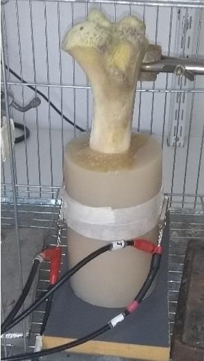
the patient or for the healthcare environment. The shown in Figure 1.
possibility of replacing this classical method with a Figure 2 shows the internal image of both phantoms
produced by X-ray radiology.
20190218 BONE FRACTURE DETECTION BY ELECTRICAL BIOIMPEDANCE - final.docx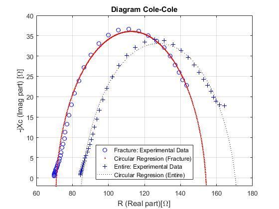
bioRxiv preprint first posted online Apr. 30, 2019; doi: http://dx.doi.org/10.1101/622936. The copyright holder for this preprint
(which was not peer-reviewed) is the author/funder, who has granted bioRxiv a license to display the preprint in perpetuity.
                              It is made available under a CC-BY-NC-ND 4.0 International license.

                                                                                                                                                     2

                                                                              which was, respectively, of 14, 12, 10 and 8 cm, i.e. an
                                                                              entire space interval that comprised that of the fracture
                                                                              level.

                                                                                                          III.   RESULTS
                                                                                 Figure 3 shows the Cole Cole diagram of the
                                                                              measurements, made at the same plane from the base, in
                                                                              both phantoms. From the experimentally assessed data, a
                                                                              circular regression was applied, obtaining characteristic
                                                                              values of the Cole function: Ro and R∞. Interestingly,
                                                                              experimental data in both phantoms shows a very good
                                                                              compliance with the ideal circumference arch of the
                                                                              corresponding regression. This occurrence reinforces our
                                                                              idea for a good possibility of use the Cole Cole diagram for
                      Fig. 1 Phantom of entire bone                           the bioimpedance studies also in ex vivo bones. Data for
                                                                              the phantom with the entire bone and with the fractured
                                                                              bone for all measurement heights are presented in Table 1.

Fig. 2 Internal image of both phantoms: on the right radiography is evident
 the sharp fracture produced by a thin hacksaw. Both femurs show similar
                             height and width.
                                                                              Fig. 3 Cole Cole diagram of both phantom with the electrode at 14 cm. The
                                                                              curves drawn with continuous lines represent the circumferential arc
B. Measurements                                                               calculated by the regression while the points represent the real values.

   The EIS measurements on the samples were made with a                          The Table 1 shows that in the fractured phantom the
configuration of bipolar electrodes with a Frequency                          extreme values of the real part of the impedance were
Response Analyzer Solartron 1250/87 (Solartron Metrology                      smaller in magnitude than in the whole bone one.
Ltd, UK) that injected the 1mA current with a frequency
sweep from 1Hz to 65 kHz. The electrodes were disposable
electrocardiograph electrodes (3M Ltd, United State) of
Ag/AgCl and their diameter was 9 mm. The measurement
protocol used was to place the electrodes facing in the axial
plane of the phantom, at a certain vertical distance of these
respects to the support surface (called height). In each
phantom, four EIS measurements were repeated by placing
the electrodes couple at a distance from the support surface

20190218 BONE FRACTURE DETECTION BY ELECTRICAL BIOIMPEDANCE - final.docx
bioRxiv preprint first posted online Apr. 30, 2019; doi: http://dx.doi.org/10.1101/622936. The copyright holder for this preprint
(which was not peer-reviewed) is the author/funder, who has granted bioRxiv a license to display the preprint in perpetuity.
It is made available under a CC-BY-NC-ND 4.0 International license.
3
Table 1 Values of both Ro and R∞ for each Cole-Cole diagram agar gel in that phantom. In a next measurement, both
phantoms should be constructed with the bones completely
Height Entire bone Fracture bone
submerged in the agar solution, equaling the volume and the
[cm] Ro [Ω] R∞ [Ω] Ro [Ω] R∞ [Ω]
height in both cases, to confirm that, due to the very high
14 170,4 85,4 154,0 71,2
conductivity of the agar gel, dispersion of currents (which in
the present work we can’t evaluate) prove to be negligible if
12 162,2 79,6 146,6 68,5 the Agar gel volume is diverse among phantoms.
At present, there is no bioimpedance or EIS analysis with
10 172,7 82,8 162,0 75,0 non-invasive surface electrodes (invasive surgical pins in
bone tissue and limb had been used) on living beings to
8 181,8 95,0 167,0 72,0
correlate bioimpedance values with bone limbs integrity
[12], though attempts of application of Electrical Impedance
Tomography (EIT) [14] to generate a diagnostic image has
IV. CONCLUSIONS
been made, but these had not reached a quality to make a
The most important observation in sight, from the results, diagnosis. So this work could represent the first approach,
is obtained with the Cole Cole diagram which is the from a simplified model such as phantoms built, to take
representative graph of any measurement of EIS on a surface measurements; obtaining coherent data with respect
biological system[10], [13]. Surface measurements - to the basic concepts of the EIS (Cole Cole diagram) and
regardless of the height at which the electrodes are located- compatible with the physiopathological reality of bone
show a total decrease in the resistive part of the phantom fracture (increase in conductivity from the absence of a
impedance that the bone fracture condition presents with tissue of greater resistivity as it is the bone that is replaced
respect to the entire condition. This could be explained by blood and extravascular/high conductive fluids) [7].
because the cross section of the osteotomy in the bone is Reasonably, this work gives the first indications of
occupied by a substance with high conductivity (saline feasibility to specify the detection of a fracture of a long
solution). Even if in a simplified way, this experimental bone in a limb in human beings through bioimpedance
condition might simulate what happens in a mammalian measurements, just strictly after the accident happens. Even
bone fracture. In fact, when the fracture occurs (no matter though it is a quick analysis of these measurements in which
what kind of fracture it is), in its proximal tissues there is an the precise height of the fracture and the geometric
increase in blood flow with localized oedema due to the measurements of the bones, among other factors to be
inflammatory reaction and to the process of healing and analyzed, are not contemplated, any way this does not
healing itself bone reconstruction. All of these occurrences detract this proposed first conclusion from this preliminary
imply a diffusion of material with low electrical resistance analysis.
inside the fracture section, i.e. a mirror condition like that
simulated in our fractured bone phantom. Moreover, the ACKNOWLEDGMENT
relatively large resistance reduction, observed along the
longitudinal axis of the sawn bone (about 6 cm), with The authors thank Dr. Martín Zamora, LAMEIN,
respect to the entire bone, could be due to the considerable Argentina, for his collaboration in the development of
porosity of the trabecular structure of the bone contained in phantoms, and Dr. Andrea Fois, CEO of the Nomadyca Ltd,
the phantom, a condition, this, that can not be kept very Italy, for his contribution in the analysis of the experimental
unlike what could happen around an in vivo bone data.
immediately after a fracture. The first author was partially supported by a Grant of
The limitations present in this work are that the Ministero degli Affari Esteri e della Cooperazione
measurements were taken on only two phantoms in two Internazionale (MAECI) of Italy, CONICET of República
different conditions (entire bone and fractured bone), which Argentina, IDEI Universidad Nacional de Tierra del Fuego
doesn’t allow having a number of experimental samples to and Gobierno de la Provincia de Tierra del Fuego. He
have a determining conclusion. Neither the bones belong to thanks LAMEIN Argentina, 2C TECHNOLOGIES S.R.L.
the same animal although both are femur of adult and very and Università di Cagliari for the kind hospitality where
similarly structured cow; the comparison between fractured part of this work has been done.
and entire bone would have been ideal to be from the same
animal to have an anatomy with greater similarity.
Moreover, due to the needs of mechanically secure
together both portion of the fractured bone, we extended the
20190218 BONE FRACTURE DETECTION BY ELECTRICAL BIOIMPEDANCE - final.docxbioRxiv preprint first posted online Apr. 30, 2019; doi: http://dx.doi.org/10.1101/622936. The copyright holder for this preprint
(which was not peer-reviewed) is the author/funder, who has granted bioRxiv a license to display the preprint in perpetuity.
It is made available under a CC-BY-NC-ND 4.0 International license.
4
CONFLICT OF INTEREST 7. Robbins, R. S. Cotran, V. Kumar, and C. T., Patologia
estructural y funcional, 6th ed. España: Interamericana, 2000.
8. R. McRae, Tratamiento pr ctico de fracturas, 5th ed. Barcelona,
The authors declare that they have no conflict of interest. España: Elsevier Health Sciences Spain, 2010.
9. J. G. Webster, E. R. Ritenour, T. Slavik, and N. Kwan-Hoong,
Webb’s physics of Medical Imaging, 2nd ed. New York, USA:
REFERENCES CRC Press, 2011.
10. M. Valentinuzzi, J. P. Morucci, B. Riguad, and C. J. Felice,
1. F. Simini, E. Santos, and M. Arregui, “Electrical Impedance “Critical Reviews in Biomedical Engineering,” p. Volume 24,
Tomography to Detect Trends in Pulmonary Oedema,” in Issues 4-6, 1996.
Bioimpedance in Biomedical Applications and Research, 1st 11. K. S. Cole and R. H. Cole, “Dispersion and absorption in
ed., F. Simini and P. Bertemes-Filho, Eds. Springer dielectrics I. Alternating current characteristics,” J. Chem.
International Publishing, 2017, pp. 45–64. Phys., vol. 9, no. 4, pp. 341–351, 1941.
2. A. Crisafulli, S. Salis, F. Tocco, F. Melis, R. Milia, G. Pittau, M. 12. A. H. Dell’Osa, F. Simini, and C. J. Felice, “Bioimpedance and
A. Caria, R. Solinas, L. Meloni, P. Pagliaro, and A. Concu, bone fracture detection : A state of the art,” in III Latin-
“Impaired central hemodynamic response and exaggerated American Conference on Bioimpedance, Manizales-Caldas,
vasoconstriction during muscle metaboreflex activation in heart Colombia, October 3rd - 5th, 2018.
failure patients,” Am. J. Physiol. (Heart Circ. Physiol.), vol. 13. S. Grimnes and Ø. Martinsen, Bioimpedance and bioelectricity
292, pp. 2988–2996, 2007. basics, 3rd ed. Oslo, Noruega: Academic Publics, 2014.
3. R. P. Braun, J. Mangana, S. Goldinger, L. French, R. Dummer, 14. H. C. Jongschaap, R. Wytch, J. M. Hutchison, and V. Kulkarni,
and A. A. Marghoob, “Electrical Impedance Spectroscopy in “Electrical impedance tomography: a review of current
Skin Cancer Diagnosis,” Dermatol. Clin., vol. 35, no. 4, pp. literature.,” Eur. J. Radiol., vol. 18, no. 3, pp. 165–74, 1994.
489–493, 2017.
4. L. Nescolarde, J. Yanguas, J. Terricabras, H. Lukaski, X. Author: Antonio Héctor Dell’Osa
Alomar, J. Rosell-Ferrer, and G. Rodas, “Detection of muscle Institute: Instituto de Desarrollo Económico e Innovación - Universidad
gap by L-BIA in muscle injuries : clinical prognosis,” Physiol. Nacional de Tierra del Fuego.
Meas., vol. 21, no. 38(7), pp. L1–L9, 2017. Street: Fuegia Basket 251 (9410).
5. P. Bertemes-Filho and F. Simini, Bioimpedance in Biomedical City: Ushuaia – Tierra del Fuego.
Applications and Research. Montevideo, Uruguay: Springer Country: Argentina.
International Publishing, 2018. Email: ahdellosa@untdf.edu.ar
6. Dirección de Estadísticas e Información de Salud - Ministerio de
Salud de la República Argetina, “Egresos hospitalarios del
sector oficial, según edad por grupos de diagnósticos | Deis,”
2014. http://www.deis.msal.gov.ar/index.php/causas-egresos/.
20190218 BONE FRACTURE DETECTION BY ELECTRICAL BIOIMPEDANCE - final.docxYou can also read