Cardiovascular Disease Complicating COVID-19 in the Elderly
←
→
Page content transcription
If your browser does not render page correctly, please read the page content below
medicina
Review
Cardiovascular Disease Complicating COVID-19 in the Elderly
Christopher Dayaramani, Joshua De Leon and Allison B. Reiss *

                                          Department of Medicine and Biomedical Research Institute, NYU Long Island School of Medicine,
                                          Mineola, NY 11501, USA; cdayaramani@gmail.com (C.D.); Joshua.DeLeon@NYULangone.org (J.D.L.)
                                          * Correspondence: Allison.Reiss@NYULangone.org

                                          Abstract: SARS-CoV-2, a single-stranded RNA coronavirus, causes an illness known as coronavirus
                                          disease 2019 (COVID-19). The highly transmissible virus gains entry into human cells primarily by
                                          the binding of its spike protein to the angiotensin-converting enzyme 2 receptor, which is expressed
                                          not only in lung tissue but also in cardiac myocytes and the vascular endothelium. Cardiovascular
                                          complications are frequent in patients with COVID-19 and may be a result of viral-associated systemic
                                          and cardiac inflammation or may arise from a virus-induced hypercoagulable state. This prothrom-
                                          botic state is marked by endothelial dysfunction and platelet activation in both macrovasculature and
                                          microvasculature. In patients with subclinical atherosclerosis, COVID-19 may incite atherosclerotic
                                          plaque disruption and coronary thrombosis. Hypertension and obesity are common comorbidities
                                          in COVID-19 patients that may significantly raise the risk of mortality. Sedentary behaviors, poor
                                          diet, and increased use of tobacco and alcohol, associated with prolonged stay-at-home restrictions,
                                          may promote thrombosis, while depressed mood due to social isolation can exacerbate poor self-care.
                                          Telehealth interventions via smartphone applications and other technologies that document nutrition
                                          and offer exercise programs and social connections can be used to mitigate some of the potential
                                damage to heart health.


Citation: Dayaramani, C.; De Leon,        Keywords: COVID-19; atherosclerosis; cardiovascular disease; hypertension; inflammation; cytokines
J.; Reiss, A.B. Cardiovascular Disease
Complicating COVID-19 in the
Elderly. Medicina 2021, 57, 833.
https://doi.org/10.3390/                  1. Introduction
medicina57080833
                                               Cardiovascular disease (CVD) is the leading cause of death worldwide [1,2]. Its
                                          incidence increases sharply with age, and the elderly bear a disproportionate burden of
Academic Editor: Manfredi Rizzo
                                          CVD morbidity and mortality [3–5]. Coronavirus disease 2019 (COVID-19), caused by
                                          severe acute respiratory distress syndrome coronavirus 2 (SARS-CoV-2), has also caused
Received: 6 July 2021
                                          significant mortality, specifically amongst the elderly, who are the most likely patient
Accepted: 12 August 2021
Published: 17 August 2021
                                          population to be hospitalized and die from the infection [6–8].
                                               Factors that likely contribute to a complicated course and higher death rate in COVID-19
Publisher’s Note: MDPI stays neutral
                                          patients with underlying CVD are the hypercoagulable state that can result from COVID-19
with regard to jurisdictional claims in
                                          infection, as well as polypharmacy (an indicator of comorbidities) [9–12]. Furthermore, the
published maps and institutional affil-
                                          pandemic can disrupt lifestyle, leading to poorer diet and inactivity [13]. Another obstacle
iations.                                  for older CVD patients is the avoidance of medical care from fear of contracting COVID-19.
                                          Hypertension, diabetes, and obesity, which often accompany CVD, are themselves estab-
                                          lished risk factors for severe COVID-19 that require careful management [14]. There is
                                          also a higher prevalence of cancer within the elderly population [15]. In addition to being
                                          immunocompromised from the cancer itself, cancer patients are frequently treated with
Copyright: © 2021 by the authors.
Licensee MDPI, Basel, Switzerland.
                                          immunosuppressants and cardiotoxic chemotherapies, making them especially susceptible
This article is an open access article
                                          to both viral illnesses and secondary cardiovascular complications. A particularly high risk
distributed under the terms and
                                          of poor outcome is seen in those who have undergone recent bone marrow or stem cell
conditions of the Creative Commons        transplantation and those exposed to poly ADP ribose polymerase (PARP) inhibitors [16,17].
Attribution (CC BY) license (https://     The aforementioned conditions are more prevalent in those over age 65 and extremely
creativecommons.org/licenses/by/          common in those over age 85. Low vitamin D levels, common in the obese state, may add
4.0/).                                    further risk [18].

Medicina 2021, 57, 833. https://doi.org/10.3390/medicina57080833                                       https://www.mdpi.com/journal/medicina
Medicina 2021, 57, 833 2 of 22
Excess production of cytokines and cytokine storms are central to many of the sequelae
of COVID-19, including damage to the cardiovascular system via pathways that involve di-
rect cardiotoxicity and through inflammation-induced myocarditis and pericarditis [19,20].
The effects of cytokines, particularly of interleukin (IL)-6, will be discussed in the sections
to follow.
Since persons with CVD are susceptible to poor COVID-19 outcomes, targeted treat-
ment and removal of barriers to care are crucial for this population [21]. These may include
the use of telemedicine, adjusting antihypertensive regimens, and online activity tracking.
In this review, focused on persons over age 65, we explore the interactions between CVD
and COVID-19 that lead to increased vulnerability to serious illness and death. We then
describe strategies to mitigate these risks.
2. Hypertension, the Immune System, and COVID-19
About 20% of COVID-19 patients are hypertensive, but some older COVID-19 popula-
tions have high blood pressure (HBP) rates exceeding 50%, consistent with the prevalence
of hypertension in persons of advancing age (Table 1) [22–24]. The status of hypertension
as an independent risk factor for morbidity and mortality in COVID-19 is unclear. Data
from China indicate a link between HBP and more severe COVID-19 infection [25]. A
meta-analysis of 9 studies from China examining COVID-19 comorbidities found a signifi-
cant correlation between COVID-19 severity and history of hypertension [26,27]. Based
on pooled data from another meta-analysis from China, hypertension conferred a 2.5-fold
increase in risk of severe COVID-19 illness and a 2.4-fold increase in mortality risk [28]. This
relationship between hypertension and COVID-19 was also found, but to a lesser degree,
in a community cohort study of persons aged 65 and older in England, where hypertension
was found in 59.6% of 502 hospitalized COVID-19 patients and associated with a 1.38-fold
greater risk for hospitalization but was not significantly associated with mortality [29]. A
cross-sectional study of over 210,000 COVID-19 patients from Mexico found that a number
of comorbidities, including hypertension, increased mortality [30]. Of these cases, over 20%
were hypertensive, and hypertension increased the risk of death (adjusted odds ratio 1.24).
Among older hospitalized COVID-19 patients in Italy, a retrospective study of 87 persons
with an average age of 72 years found that over 50% were hypertensive but that hyper-
tension did not predict mortality [31]. A case series of 5700 adults, median age 63 years,
hospitalized in the New York City area for COVID-19, found that 56.6% of the patients
had HBP, but the report did not address differences in outcome based on comorbidities
or medications [18]. A study of 184 COVID-19 patients with a mean age of 64.7 years,
admitted to a New York City hospital, found a positive correlation between hypertension
and mortality [32]. In the United States, a multicenter cohort study of 2215 patients hospi-
talized in ICUs for COVID-19 (59.7% with hypertension, mean age 60.5 years) found that
HBP did not increase the risk of death [33].
Numerous confounding variables make it difficult to link hypertension directly to
COVID-19 severity [38]. Bajgain et al. recently posited that comorbidities such as hyper-
tension may increase disease severity without escalating mortality [34]. Hypertension
may increase hospital readmission rates after discharge for COVID-19 patients, with re-
hospitalization most often due to respiratory distress or thrombotic episodes [35].
Although a contribution to COVID-19 mortality from hypertension is unconfirmed,
increased susceptibility to severe illness may result from a chronic hypertension-induced
inflammatory state causing immune dysregulation [39]. Basal levels of proinflammatory
cytokines are increased in patients with hypertension, where ILs-6, -8, -18, transforming
growth factor (TGF)-β, and tumor necrosis factor (TNF)-α are in disequilibrium with
anti-inflammatory cytokines (ILs-4 and -10) [40].Medicina 2021, 57, 833 3 of 22
Table 1. Hypertension and COVID-19 studies, with age included.
By First Author Reference Age Range Details
Mean age 51 years; 95% confidence interval,
Baradaran et al. [22] Meta-analysis
49–54 years
Median age 63 years; interquartile range, 5700 subjects, New York City
Richardson et al. [23]
52–75 years area, USA
73,274 subjects, European
Ahrenfeldt et al. [24] Mean age 68 years; age range 50–104 years
region
Median age 51 years; interquartile range,
Wu et al. [25] 201 subjects, Wuhan China
43–60 years
Systematic review,
Chen et al. [26] Unspecified
1936 subjects
Median age 52.4 years; age range
32.5–64.0 years. Severe group older than Meta-analysis, 10,948 subjects,
Liu et al. [27]
non-severe group (60.9 years, range most from China
45.0–74.0 years)
COVID-19 severity associated with
Systematic review, 2893
Lippi et al. [28] hypertension observed only in subjects over
subjects, China
age 60 years
UK Biobank Cohort, 507
Atkins et al. [29] Mean age 74.3 years (SD 4.5) COVID-19 positive subjects,
hospitalized
Cross-sectional study,
Hernández-Galdamez et al. [30] 45.7 (SD 16.3)
211,003 subjects, Mexico
Median age 72 years; interquartile range, Retrospective, single-center
De Vito et al. [31]
62.5–83.5 years study, 87 subjects, Italy
184 subjects, New York City
Mani et al. [32] Mean age 64.72 years (SD 14.87)
area, USA
Mean age (all) 60.5 years (SD 14.5). Mean age Multicenter cohort study,
Gupta et al. [33]
(died) 66.0 years (SD 13.3) 2215 subjects, USA
Median age 56 years; interquartile range, Systematic review,
Bajgain et al. [34]
48.25–67.4 years 22,753 subjects
Median age 61 years; interquartile range, 339 patients, Rhode Island,
Atalla et al. [35]
49–74 years USA
Cohort study, 68 patients,
Zheng et al. [36] Median age 47.13 years; range, 11–84 years.
China
Median age of hypertensive patients 69 years; Single-center, retrospective
Pan et al. [37]
interquartile range, 62–76 years study, 996 patients, China
SD: standard deviation.
Hypertension affects CD8+ T-cells, essential components of cell-mediated immunity
that protect against acute and persistent viral infections [41]. Hypertension stimulates CD8+
overproduction of cytokines, most notably, interferon (IFN)-γ and TNF-α [42,43]. These
cytokines can adversely affect renal epithelium and raise blood pressure by increasing
oxidative stress and renal sodium reabsorption [44,45]. Lymphopenia, due to reductions in
both CD4+ and CD8+ T-cells, is well-documented in severe COVID-19 and may further
disable antiviral defenses [36]. Pan and colleagues found that hypertensive COVID-19 pa-
tients have lower CD8+ counts, which may compromise the defense against the damaging
flood of cytokines, characteristic of COVID-19 infection [37,38].
Another key potential link between COVID-19 and hypertension is IL-6, a pleiotropic
cytokine found at high levels in cytokine storms, which are responsible for stimulating
the production of acute-phase proteins [46,47]. Plasma IL-6 levels strongly correlate withthe production of acute-phase proteins [46,47]. Plasma IL-6 levels strongly correlate w
hypertension in humans [48]. In the initial phase of inflammation, IL-6, together with T
β, is necessary for the development of Th17 cells, which help induce a state of neutroph
Medicina 2021, 57, 833
inflammation [49]. Chronic sustained IL-6 elevation can result in cardiac fibrosis 4 of 22
and
ventricular hypertrophy [50–52]. High IL-6 levels are associated with an increased risk
death from Covid-19 infection [53–55]. Whether the IL-6 level rather than blood press
is thehypertension
independent variable
in humans [48].determining Covid-19
In the initial phase severity IL-6,
of inflammation, risk together
is unresolved
with TGF- [56].
The
β, possibility
is necessary that
for the IL-6 worsens
development of Th17Covid-19 outcomes
cells, which hasa state
help induce led to of anti-IL-6
neutrophilictherapy
als with tocilizumab and sarilumab, but the results have been mixed [57–60].
inflammation [49]. Chronic sustained IL-6 elevation can result in cardiac fibrosis and left A rec
meta-analysis showed a significant reduction in all-cause mortality with arisk
ventricular hypertrophy [50–52]. High IL-6 levels are associated with an increased of
combination
death from COVID-19 infection [53–55]. Whether the IL-6 level rather than blood pressure
tocilizumab and dexamethasone in patients of varying ages, with the median age of
is the independent variable determining COVID-19 severity risk is unresolved [56].
proximately late 50s to
The possibility early
that IL-6 60s [61].COVID-19
worsens In a randomized study,
outcomes has led toboth tocilizumab
anti-IL-6 therapy and s
lumab were
trials witheffective
tocilizumab in and
improving
sarilumab,thebutsurvival
the resultsofhave
severely ill ICU
been mixed patients
[57–60]. (mean ag
A recent
meta-analysis showed a significant reduction in all-cause mortality
about 60 years] with Covid-19 [62]. It is possible that IL-6 either suppresses or increa with a combination
viralofreplication
tocilizumab and dexamethasone in patients of varying ages, with the median age of
and persistence, depending on multiple factors [63–65].
approximately late 50s to early 60s [61]. In a randomized study, both tocilizumab and
sarilumab were effective in improving the survival of severely ill ICU patients (mean age of
3. Angiotensin-Converting
about 60 years) with COVID-19 Enzyme
[62]. It Inhibitors/Angiotensin Receptor
is possible that IL-6 either suppresses Blockers
or increases (ARB
in Covid-19
viral replication and persistence, depending on multiple factors [63–65].
The
3. renin–angiotensin Enzyme
Angiotensin-Converting systemInhibitors/Angiotensin
(RAS) plays a key role in blood
Receptor pressure
Blockers (ARB) regulat
and in COVID-19
sodium balance [66]. In the classical RAS pathway, renin cleaves the proenzyme
The renin–angiotensin
giotensinogen system (RAS)
to form angiotensin (Ang)I. plays
AngIa key role in blood
is further pressure
cleaved regulation
by the angiotensin-c
and sodium balance [66]. In the classical RAS pathway, renin cleaves the proenzyme
verting enzyme (ACE) to yield AngII, which then acts through angiotensin receptor ty
angiotensinogen to form angiotensin (Ang)I. AngI is further cleaved by the angiotensin-
1 and 2 (AT1,enzyme
converting AT2). (ACE)
The AngII/AT1
to yield AngII,receptor axisacts
which then mediates vasoconstriction,
through angiotensin receptor while
effects of 1AT2
types and 2are often
(AT1, AT2).antagonistic
The AngII/AT1 toreceptor
AT1 (Figure 1). Vasoconstriction
axis mediates of the
vasoconstriction, while thepulmon
vasculature can lead to right ventricular hypertrophy [67,68]. AngII also increases blo
effects of AT2 are often antagonistic to AT1 (Figure 1). Vasoconstriction of the pulmonary
vasculature
pressure can lead
indirectly bytostimulating
right ventricular hypertrophy
aldosterone [67,68]. AngII
production, also increases
which blood
increases sodium a
pressure indirectly by stimulating aldosterone production, which increases sodium and
water reabsorption [69].
water reabsorption [69].
Figure 1. Figure 1. Renin–angiotensin-aldosterone
Renin–angiotensin-aldosterone system(RAS)
system (RAS) and
andACE
ACE inhibitors.
inhibitors.The The
precursor molecule
precursor angiotensinogen
molecule is
angiotensinogen is
cleaved bycleaved
reninbytorenin
formto angiotensin
form angiotensin 1 in
1 in response to
response to low
lowsodium-chloride,
sodium-chloride, as sensed by the macula
as sensed by thedensa.
macula Angiotensin 1 is
densa. Angiotensin 1
then further modified by the angiotensin-converting enzyme (ACE) to form angiotensin II. The ACE is
is then further modified by the angiotensin-converting enzyme (ACE) to form angiotensin II. The ACE is the target of ACE the target of ACE
inhibitors.inhibitors. Angiotensin II has direct effects on the vasculature, adrenal cortex, and brain, causing vasoconstriction and the
Angiotensin II has direct effects on the vasculature, adrenal cortex, and brain, causing vasoconstriction and the
secretion of aldosterone and the anti-diuretic hormone (ADH), respectively. An alternate pathway also exists, whereby
secretion of aldosterone and the anti-diuretic hormone (ADH), respectively. An alternate pathway also exists, whereby
angiotensin II is converted to angiotensins 1–9 and then angiotensins 1–7. A key enzyme in the formation of the latter is
angiotensin II is converted to angiotensins 1–9 and then angiotensins 1–7. A key enzyme in the formation of the latter is
ACE2. Angiotensins 1–7, in contrast to angiotensin II, cause vasodilation and inhibit vasoconstriction.
ACE2. Angiotensins 1–7, in contrast to angiotensin II, cause vasodilation and inhibit vasoconstriction.Medicina 2021, 57, 833 5 of 22
ACE2, homologous to ACE, cleavages AngII via an alternative pathway to form
Angiotensin 1–7. Angiotensin 1–7 has vasodilatory effects and protect against acute res-
piratory distress syndrome (ARDS) [70]. ACE2 is constitutively expressed in the lungs,
kidney, gastrointestinal tract, and endothelium. The COVID-19 particle enters host cells
via the full-length membrane-bound ACE2, which has an extracellular domain that acts as
a receptor for the SARS-COV-2 spike protein [71]. The spike transmembrane glycoprotein
is the target of many mRNA vaccines against COVID-19 [72].
Cleavage of the viral S-spike protein by host proprotein convertases generates S1 and
S2 segments. The S1 (globular) subunit contains the receptor-binding domain that allows
the attachment of COVID-19 to the ACE2 receptor. Once attached, COVID-19 fuses with
the host cell membrane via the S2 subunit, enters the cell, and subsequently replicates
in the cytoplasm [73,74]. Hypertension may also increase ACE2 enzyme concentration,
which initially sparked concern that hypertension might increase viral entry into cells,
amplifying the severity of infection [75]. Long-term use of ACE inhibitors (ACEIs) or ARBs
can upregulate ACE enzymes. ACEIs are not known to increase pulmonary or serum
ACE2 levels but do increase ACE2 in the brain, heart, and urine [76,77]. Overall, there
exists no definitive evidence that ACEIs or ARBs increase COVID-19 risk [78–83]. In fact,
a preponderance of evidence indicates ACEIs and ARBs are not harmful to COVID-19
patients and may even provide some benefit [84–86]. In a cohort from France of over
2 million hypertensive patients (mean age of 63 years), the outcome in patients with
COVID-19 on ACEIs and ARBs was compared to those on calcium channel blockers. The
ACEI and ARB groups showed a lower risk of COVID-19 hospitalization and a lower risk
of intubation/death [87].
A retrospective single-center study from Long Island, New York, found that 614 of
over 6000 COVID-19 patients were hypertensive and required hospitalization. These pa-
tients were categorized as taking and not taking ACEIs/ARBs prior to admission. Those
who had been on ACEIs/ARBs and discontinued these medications during their hospital
stay were twice as likely to be admitted to the ICU as those who continued their established
treatment. The authors speculate that, although the medications may cause ACE2 recep-
tor upregulation, discontinuing hypertensive medication can worsen illness because the
benefits of controlling blood pressure outweigh other factors and, possibly, ACEIs/ARBs
may mitigate the effects of cytokine storms [88]. This does not, however, indicate whether
a change in medication prior to contraction of illness or onset of symptoms would confer
benefit. Genetic variations in RAS genes and their expression profiles may also influence
how pharmacotherapies affect COVID-19 outcomes; this is an area of active study [89,90].
The current recommendation is that patients should not change their regimen, as directed
by the European and American Societies of Cardiology as well as the AHA [75,81,91–93].
4. Pulmonary Arterial Hypertension and Right Ventricular Hypertrophy
Pulmonary arterial hypertension (PAH), defined as a mean pressure above 25 mmHg
in the pulmonary circulation at rest (normal = 24/12), confers susceptibility to vascular
remodeling, fibrosis, and pulmonary edema [94]. Severe and sustained PAH can manifest
as pulmonary edema and right-sided hypertrophy and/or heart failure. The incidence of
COVID-19 in persons with PAH is comparable to that in the general population, and based
on their documented propensity to worse outcomes during hospitalization in general,
they may have greater mortality during COVID-19-related hospitalization [95,96]. In
24 COVID-19 patients with PAH, Pagnesi et al. determined that PAH specifically, and not
right ventricular hypertrophy, was associated with more severe COVID-19 illness and
worse clinical outcomes [97].
The RAS is of clinical importance in both PAH and COVID-19. Within the RAS, ACE2
may offer vasoprotective effects. ACE2 supplements are being considered as a treatment for
several lung diseases [98]. Such supplements have ameliorating effects in PAH via increased
Angiotensin 1–7 and nitric oxide bioavailability. Inhaled nitric oxide was shown to be
effective in the 2003 SARS epidemic, causing the reversal of PAH and decreased lengths ofMedicina 2021, 57, 833 6 of 22
ventilator support. Clinical trials are evaluating this therapy in COVID-19 [99–111]. PAH
is characterized by increased levels of the vasoconstrictor endothelin-1, and, therefore,
along with increasing nitric oxide, the antagonism of the endothelin-1 receptor is effective
PAH therapy that may be beneficial in COVID-19 infection [102]. Endothelin-1 may
decrease ACE2, augmenting vasoconstriction via higher levels of AngII from lung epithelial
cells [103–105]. Endothelin-1 may also promote endothelial dysfunction, platelet activation,
and IL-6 secretion, all detrimental in the setting of COVID-19 [106,107].
Overall, COVID-19 is known to induce microthrombosis and hemorrhage in the
alveoli and lung interstitium, and this may be exacerbated by pre-existing hypertension or
increased vascular stress brought on by COVID-19-associated PAH. [108].
5. Coagulopathy in COVID-19: Mechanisms, Manifestations, and Treatment
A state of hypercoagulability frequently accompanies COVID-19, especially in severe
disease [109,110]. Coagulopathy leaves patients vulnerable to thrombotic complications, in-
cluding venous thromboembolism, pulmonary embolism, and disseminated intravascular
coagulation [111–115].
Pro-inflammatory mediators produced during COVID-19 infection cause the release
of tissue factor, an initiator of blood coagulation, from mononuclear cells [116,117]. IL-6,
a key mediator elevated in the COVID-19 setting, can raise tissue factor levels and may
also stimulate platelet production in bone marrow and lungs [118–120]. The panoply
of inflammatory factors also activates endothelial cells, increasing their expression of
adhesion molecules and leading to the release of the von Willebrand factor, thus promoting
a procoagulant endothelial phenotype, excessive activity in the coagulation cascade, and
multiple thrombotic complications.
Coagulation abnormalities are detected in laboratory tests as increased serum concen-
trations of the procoagulants fibrinogen and D-dimers as well as decreased antithrombin
and prolonged prothrombin time (Table 2) [121,122].
Table 2. Early Markers Associated with Poor Outcomes in COVID-19 Patients.
Definition of
Marker Mean Level Time of Measurement Reference
Poor Outcome
IL-6 7.39 pg/mL On admission ARDS [20]
Fibrinogen 5.16 g/L On admission Death [113]
D-dimer ≥1 µg/mL Outpatient Death [119]
LDH 445 µg/mL On admission Ventilation [121]
CAC score ≥400 During hospitalization Death [123]
CRP >40 mg/L On admission Death/ARDS [124]
Ferritin >950 ng/L On admission ARDS [125]
ARDS: acute respiratory distress syndrome; CAC: coronary artery calcium; CRP: C-reactive protein; LDH: lactate
dehydrogenase; IL-6: interleukin-6.
As a result of vascular injury, the propeptide fibrinogen is cleaved to fibrin, and high
circulating fibrin levels are common in the early phase of COVID-19 infection. Either
hyper- or hypofibrinolysis can occur in the setting of COVID-19, with hypofibrinolysis
causing susceptibility to thrombus formation and hyperfibrinolysis causing susceptibility
to bleeding [126]. The hypofibrinolytic state may be attributed to the elevated production
of plasminogen activator inhibitor 1 (PAI-1) by epithelial and endothelial cells in the
inflamed lung [127]. D-dimers, produced during the degradation of crosslinked fibrin, are
below 0.5 µg/mL under normal physiologic conditions. An increase in fibrinogen and
D-dimers is associated with the risk of microthrombus formation in COVID-19 patients
and subsequent emboli and/or organ failure [128,129]. Fibrinogen levels were, on average,
higher in patients who developed severe versus less severe illness (5.16 vs. 4.51 g/L) [130].
D-dimer levels over 1 µg/mL can identify patients with poorer prognoses early in theMedicina 2021, 57, x FOR PEER REVIEW 7 of 22
Medicina 2021, 57, 833 7 of 22
course of disease and may signal the need for admission to critical care. Elevated D-dimer
course
appears to of
bedisease and may risk
an independent signal the need
factor for admission
for death [130,131]. to
In critical care.D-dimer
evaluating Elevatedlevels,
D-dimer
the appears to be an of
implementation independent
age adjustmentrisk factor forofdeath
instead [130,131].
a fixed In evaluating
cutoff may increase the D-dimer levels,
accuracy
the implementation
of clinical assessment [132]. of age adjustment instead of a fixed cutoff may increase the accuracy
ofSepsis-induced
clinical assessment [132].
coagulopathy due to Covid-19 infection can lead to thrombotic stroke
Sepsis-induced coagulopathy
and myocardial infarction (MI) [133–136]. due toLosartan,
COVID-19 infection can
previously lead to thrombotic
mentioned stroke
for its ability
and myocardial
to normalize levels ofinfarction
ACE2, is(MI) [133–136].
believed Losartan, previously
to be protective mentioned
against strokes, offering for its ability
another
to normalize
reason for its uselevels
in placeof ACE2,
of ACEIsis believed
[137]. to be protective against strokes, offering another
reason for its use
Addressing in place of ACEIs [137].
the hypercoagulability risk at all levels of Covid-19 severity and in dif-
ferent age groups is the
Addressing hypercoagulability
challenging. risk at allmay
Hypercoagulability levels of COVID-19
worsen severity
in the setting and in
of this
different age groups is challenging. Hypercoagulability may worsen
pandemic via decreased activity, decreased exercise, and less movement in general under in the setting of this
pandemic via decreased activity, decreased exercise, and less movement
quarantine restrictions. Particularly affected are the elderly, with more limited movement in general under
quarantine restrictions. Particularly affected are the elderly, with more
capability due to age and co-morbidities. Venous stasis that accompanies inactivity, com- limited movement
capability
bined due to age and co-morbidities.
with hypercoagulability, sets the stageVenous
for twostasis that accompanies
of three predisposinginactivity,
factors de- com-
bined with hypercoagulability, sets the stage for two of three predisposing factors described
scribed in Virchow’s triad for vascular thrombosis (blood flow alterations, endothelial in-
in Virchow’s triad for vascular thrombosis (blood flow alterations, endothelial injury, and
jury, and hypercoagulability) [138]. Hypertension predisposes to endothelial injury, the
hypercoagulability) [138]. Hypertension predisposes to endothelial injury, the last remain-
last remaining factor in Virchow’s triad. As discussed above, ACEIs should be used cau-
ing factor in Virchow’s triad. As discussed above, ACEIs should be used cautiously in
tiously in Covid-19 patients. Anti-inflammatory drugs, cytokine inhibitors, and statins
COVID-19 patients. Anti-inflammatory drugs, cytokine inhibitors, and statins may be con-
may be considered to protect the endothelium while simultaneously working against viral
sidered to protect the endothelium while simultaneously working against viral replication.
replication. Thromboprophylaxis with low molecular weight heparin could decrease D-
Thromboprophylaxis with low molecular weight heparin could decrease D-dimer levels,
dimer levels, and heparin can decrease fibrosis in those suffering from Covid-19-induced
and heparin can decrease fibrosis in those suffering from COVID-19-induced ARDS [130].
ARDS [130]. At this time, there are different approaches to coagulopathy in the field, with
At this time, there are different approaches to coagulopathy in the field, with the evaluation
the evaluation of efficacy still ongoing. We await the results of major clinical trials regard-
of efficacy still ongoing. We await the results of major clinical trials regarding the dosage,
ing the dosage, class, and timeline of the use of anticoagulation therapy [139].
class, and timeline of the use of anticoagulation therapy [139].
6. Myocardial Injury:
6. Myocardial Subclinical
Injury: Atherosclerosis
Subclinical andand
Atherosclerosis Acute Coronary
Acute Syndrome
Coronary Syndrome
Covid-19 cardiac
COVID-19 manifestations
cardiac maymay
manifestations include myocarditis,
include cardiac
myocarditis, arrhythmias,
cardiac arrhythmias,andand
newnew or worsening heart failure, which may be particularly damaging to
or worsening heart failure, which may be particularly damaging to patients with patients with a a
history of CVD [140,141]. The mechanisms underlying cardiac injury may
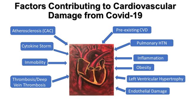
history of CVD [140,141]. The mechanisms underlying cardiac injury may be multifactorial be multifacto-
rial (Figure
(Figure 2). Inflammation
Inflammationand andthrombosis
thrombosisare areknown
known culprits
culprits[142]. Infection
[142]. Infectionincreases
increases
overall metabolic
overall demand
metabolic and heart
demand and rate.
heartThis
rate.intensifies oxygen expenditure
This intensifies while short-
oxygen expenditure while
ening the fillingthe
shortening time in the
filling diastole
time and
in the limiting
diastole andcoronary
limitingperfusion.
coronaryInfection-mediated
perfusion. Infection-
vasoconstriction and ventilation/perfusion
mediated vasoconstriction mismatch negatively
and ventilation/perfusion mismatch affecting blood
negatively oxygena-
affecting blood
tionoxygenation
can exacerbate canthe oxygen deficit,
exacerbate leading
the oxygen to myocardial
deficit, leading toischemia
myocardial [143,144].
ischemia Internal-
[143,144].
izedInternalized
virus loads virus
within cardiomyocytes
loads may directly
within cardiomyocytes maydamage thedamage
directly heart. the heart.
Figure 2. Factors contributing to cardiovascular damage in COVID-19. The white text in blue bubbles
Figure 2. Factors contributing to cardiovascular damage in Covid-19. The white text in blue bubbles
represents some of the pathologies that are associated with CVD in patients with COVID-19. Arrows
represents some of the pathologies that are associated with CVD in patients with Covid-19. Arrows
are drawn from each factor to the central image of the heart and associated vasculature to emphasize
are drawn from each factor to the central image of the heart and associated vasculature to emphasize
their effect on the cardiovascular system specifically. CAC—Coronary Artery Calcium score; IL-6:
interleukin-6; CVD: cardiovascular disease.Medicina 2021, 57, 833 8 of 22
Immune-inflammatory-mediated injury to the heart from COVID-19 is more likely
in severe cases and in those with HBP and can be monitored via the release of certain
injury biomarkers, including cardiac-specific troponin and creatine kinase-MB [145–148].
Elevated lactate dehydrogenase (LDH) may be of cardiac origin but is nonspecific and can
result from damage to other organs [149]. In COVID-19, LDH may be released from the
lung since this is a key site for inflammatory processes [150]. LDH levels above 445 µg/mL
on admission can be predictive of more severe COVID-19 [133,151–153]. An analysis of
353 COVID-19 patients, 79 (22.4%) of whom presented with myocardial injury, revealed
more frequent elevations in LDH (mean level: 244 U/L (without MI) vs. 655 U/L (with
MI) and creatinine (71 µmol/L (without MI) vs. 155 µmol/L (with MI) in the MI group
during their hospitalization [154]. In addition to acute MI, differential diagnoses for
increased serum cardiac biomarkers include stress-induced cardiomyopathy as well as
myocarditis [155–157].
Evidence is accumulating that cardiac injury combined with COVID-19 infection,
whether the myocardial injury is pre-existing or occurs during the infection, is associated
with poorer outcomes [158–160]. A prospective, multicenter cohort study in Spain found
that in patients with acute MI, COVID-19 is an independent risk factor for in-hospital
mortality [161]. A small study of 77 COVID-19 patients who died in Wuhan, China, in
early 2020 found that heart disease was present in 32%, and heart disease patients were
more likely to be in the short-term survival group and to die within 14 days of COVID-19
onset [153].
COVID-19-associated coagulopathy can cause both large and small vessel embolic phe-
nomena [123,162]. Postmortem histological evaluation has revealed signs of microvascular
thrombosis in both skin and lungs [163]. Coronary arteries and coronary microvascu-
lature may be affected by COVID-19 because this viral infection can directly activate
the endothelium via inflammatory cytokines and indirectly by causing hypoxic condi-
tions [124,164,165]. Further, the virus can directly infect endothelial cells, leading to an
inflamed endothelium [166]. Coronary microvascular endothelial inflammation could lead
to myocardial injury [125]. The potential for COVID-19 to compromise the patency of
coronary bypass grafts has been suggested [167].
Subclinical atherosclerosis can impact the course of COVID-19. Coronary artery calci-
fication (CAC), a specific imaging marker of coronary atherosclerosis that correlates with
the plaque burden, can reveal previously undiagnosed CVD in COVID-19 patients [168]. In
a cross-sectional study of 209 consecutively admitted COVID-19 patients without known
CVD, assessed for CAC, CAC was detected in 106 (Table 2) (50.7%). Half of those positive
patients required mechanical ventilation, extracorporeal membrane oxygenation, or died,
whereas only 17.5% of patients negative for CAC had such poor outcomes [169]. A separate
study by Nai Fovino et al. from Italy also found that high CAC as a surrogate for subclinical
atherosclerosis was a marker for worse outcomes [170]. In this study, 75% of patients with
high CAC either died or were admitted to the ICU, in contrast with only 20% of the group
with lower CAC scores. Patients with a high score were also more likely to experience
an MI.
C-reactive protein (CRP), an acute-phase reactant and marker of chronic low-grade
systemic inflammation, has prognostic value as a predictor of cardiovascular risk. A
number of studies have shown an association between elevated CRP and higher rates of
COVID-19-related hospitalization and mortality (Table 2) [171,172]. While moderate CRP
elevation in the 3–10 mg/L range is useful in cardiovascular risk prediction, CRP values
in COVID-19 may exceed 40 mg/L, reflecting an acute inflammatory response unrelated
to the heart [173,174]. Ferritin, critical for iron homeostasis, is also associated with a high
risk of arterial hypertension and has utility in predicting the risk of ARDS in COVID-19
patients [175,176]. Ferritin is used to monitor systemic inflammation and may also be
considered a marker of a hypercoagulable state [177]. Hypoxia is also a factor in elevating
ferritin levels, and, thus, high ferritin may be a consequence of poor oxygenation [178].Medicina 2021, 57, 833 9 of 22
Ferritin-targeted therapies are promising as a means of limiting the hypercoagulable state
in the early phases of COVID-19 [179,180].
Patients with pre-existing CVD and atherosclerotic plaque containing inflammatory
cells may be vulnerable to plaque activation upon exposure to cytokines such as IL-6.and
IFN-γ [24,181–183]. Leukocyte infiltration and the activation of the endothelium contribute
to the host synthesis of metalloproteinases and peptidases that break down collagen
and weaken and destabilize plaque [184,185]. Furthermore, acute infection induces a
prothrombotic, hypercoagulable state that can incite the formation of platelet thrombi [186].
7. Lifestyle
In response to the COVID-19 public health emergency, many governments imposed
lockdown measures, stay-at-home orders, and quarantine. Curfews are in effect in many
parts of the world, and social distancing is strongly recommended or mandated [187–190].
Quarantine refers to the separation of individuals or communities who may have been
exposed to an infectious disease, while lockdown is a restriction in movement, with the
closing of non-essential businesses and activities.
Quarantine and lockdown are effective in preventing disease transmission but can
increase sedentary behavior both, directly through impeded access to exercise facilities
and outdoor activities and the shutdown of interactive sports as well as indirectly through
an increased prevalence of anxiety, depression, and insomnia that drain energy [191–195].
Sedentary behavior and low physical activity are of particular concern in those with
CVD as these behaviors are highly associated with increased mortality in both males and
females [196–198]. Physical activity has numerous favorable effects on the cardiovascular
system, while lack of physical activity has long been linked to atherosclerosis [199,200].
Exercise delays the loss of age-associated endothelium-dependent vasodilation and reduces
systemic inflammation and oxidative stress [201–204]. It also improves cardiac function
and the lipid profile and decreases the resting heart rate [205–207]. Moderate to vigorous
physical activity, 60 min per day, 5–7 days per week, is recommended to combat the
physical and mental toll of COVID-19 isolation. Outdoor activities may include walking or
jogging; if the workout is indoor only, home-based stairclimbing, walking with step-counts,
or an exercise bicycle are good options [208,209].
Another lifestyle factor of concern for these patients during the pandemic is diet. To
compound the issue, quality of diet and level of physical activity are linked, and people
who spend a lot of time in front of screens also tend to eat more processed foods [210,211].
Pandemic constraints can lead to increased consumption of less healthy foods [212,213].
Boredom, anxiety, hesitation to venture outside, and fear of food shortages can drive the
purchase of more salty, sugar-laden foods with high trans-fat content and longer shelf
life. Poor dietary choices may result in weight gain and worsen pre-existing diabetes and
hypertension [214]. A survey conducted in Italy found that 48% of participants noted
weight gain during quarantine [215].
A balanced, nutritious diet containing fresh fruits and vegetables is important for the
maintenance of healthy body weight, serum lipid profiles, glucose metabolism, and blood
pressure levels [216–220]. Grocery delivery services are often available and particularly
useful to susceptible populations, such as those suffering from CVD and the elderly. Of
note, an anti-inflammatory diet, high in fiber from legumes, vegetables, and whole grains,
can promote the production of short-chain fatty acids that may offer protection via the
inhibition of cytokine production. Overall, this diet could potentially reduce the risk
of cardiovascular complications of COVID-19 by promoting healthy weight, improving
glucose control, and regulating metabolism [221]. Specific nutraceuticals of benefit to the
cardiovascular system in COVID-19 are not yet defined, but the area is under study [222].
Quarantine may introduce stressors into life such as the loss of childcare resources,
unemployment, death of a loved one, or strained relationships. Increased stress may lead
to substance abuse. A study conducted in Poland showed that alcohol consumption in-Medicina 2021, 57, 833 10 of 22
creased in 14.6% of survey respondents, and 45% of smokers acknowledged smoking more.
Furthermore, 43% of respondents reported increased food consumption/snacking [223].
Lastly, quarantine and feelings of isolation can increase depression and anxiety [224].
Among 1593 participants, Lei et al. found the total prevalence of anxiety and depression
to be approximately 8.3% and 14.6%, respectively, and the prevalence in the isolated
group (12.9%, 22.4%) was significantly higher than that in the group that did not undergo
quarantine (6.7%, 11.9%) [198]. The length of quarantine is also highly associated with the
development of mental health issues, with persons under quarantine conditions for more
than 10 days more likely to develop post-traumatic stress than those quarantined for less
than 10 days [190]. Mental well-being can be impacted by fear of contracting the illness,
fear of the financial burden associated with quarantine, restricted availability of food, lack
of information regarding quarantine specifics, and excessive time spent on COVID-19
related news (>1 h) [225–227].
Quarantine can lead to increased suicide risk, particularly among the elderly, who are
more likely to live alone and perceive themselves as a burden [228]. People with underlying
CVD who are subjected to extended periods of quarantine may experience stress-related
worsening of their cardiac status [229,230].
8. Delivering Care
Care delivery to patients with COVID-19 in office or hospital settings comes with
the risk of transmission and spread to physicians, staff, and other patients. Telemedicine
has been slowly gaining acceptance for the advantages it offers in access to care and cost-
effectiveness. Now, the pandemic has accelerated its growth because it brings the key
benefit of increased safety through the circumvention of direct physical contact [231]. This
safety comes with a downside as fewer in-person visits to medical personnel can lead to
decreased patient engagement [232]. The Technology Acceptance model states that the
willingness of the patient to utilize technology depends on two key factors: perceived
usefulness and perceived ease of use [233]. Therefore, barriers to use, such as lack of
familiarity with technology, privacy concerns, or preference for in-person communication,
may impact patient interaction and satisfaction with telemedicine [234,235].
Interestingly, in the case of hypertension, several studies have shown that virtual
approaches are as effective or more effective than traditional treatments in achieving blood
pressure goals [236–238]. ECG transmission systems may allow the detection of acute MI
in real-time [239,240]. It is important to note that cardiac emergencies require in-person
evaluation and treatment without delay.
9. Tailoring CVD Treatment and Vaccination in the Context of COVID-19
As of this writing, our understanding of the pathophysiologic mechanisms of Sars-
Cov-2 infection is rapidly changing, and evidence thus far obtained continues to include
potentially conflicting hypotheses [241,242]. As such, identifying targets for therapy remain
elusive, and no definitive recommendations can be made. In addition, previously used
therapies and some therapies under investigation may have adverse cardiovascular effects,
while their clinical efficacy for combating COVID-19 is unconfirmed [243]. However, once
specific cardiovascular complications have been identified, such as acute coronary syn-
dromes, acute heart failure, or myocarditis, it is reasonable to employ standard treatments
and therapies. Additionally, regardless of the exact mechanism(s) resulting in cardiovas-
cular involvement, the severe inflammatory state associated with this disease pinpoints
targeting this state as an important therapeutic goal. As demonstrated in their review,
Samidurai and Das [244] note that the precise mechanisms linking CVD and worsened
prognosis or higher mortality rate in COVID-19 patients remain unknown, but it is pre-
sumed that the pathophysiology of this virus’ method of infection, because it may directly
involve the cardiovascular system, predicts high rates of cardiovascular involvement and
higher morbidity and mortality in patients with pre-existing CVD.Medicina 2021, 57, 833 11 of 22
As noted previously, it is generally accepted within the medical community that ACE
inhibitors and ARBs should be continued as treatment in hypertensive patients who have
been diagnosed with COVID-19 [245,246]. In those with diabetes, COVID-19 may increase
blood glucose levels, and, therefore, strict monitoring and blood sugar control may be
indicated [247,248].
In those with CVD, who may be vulnerable to severe effects of COVID-19, prevention
of illness by vaccination is preferable to treatment after infection. At the time of this
writing, mRNA vaccines and adenoviral vaccines targeting the spike protein are being
administered widely [249]. The American College of Cardiology recommends that persons
with CVD be prioritized for vaccination and, thus far, the vaccine is well-tolerated by
these patients [250,251]. A small study of 86 mostly young subjects from Rome found that
hypertension, central obesity, and smoking were each associated with lower antibody titers
in individuals vaccinated with the Pfizer-BioNTech mRNA vaccine and suggested that
individuals with these risk factors may benefit from a booster vaccine dose [252].
One caveat is the possible link between COVID-19 mRNA vaccination and myocardi-
tis, but this is largely seen in younger vaccine recipients with a median age of about
26 years [253,254]. A major concern is the diminished effectiveness of vaccination in those
who have undergone solid organ transplants, including heart transplants. These patients
may fail to mount a robust immune response to vaccination, and this lack of response is
more likely in older persons above age 60. [255,256]. A booster vaccine dose may improve
protection against illness in these patients [257].
10. Conclusions
For the reasons discussed above, the elderly with preexisting CVD are vulnerable
to multiple cardiovascular manifestations that can lead to adverse outcomes. Table 3
highlights the most pressing issues facing older persons with CVD during the COVID-19
pandemic. It is our hope that with the release of highly effective vaccines, prevention will
render treatment needs less urgent in the future, but as the virus evolves, new variants
may prolong the pandemic [258]. Identification of effective therapies and novel therapeutic
targets remain a central focus for research and development [259]. Preventive strategies
such as advanced technological tools, including artificial intelligence and machine learning,
can accelerate the diagnosis/screening of patients for the virus [260]. Rapid COVID-
19 testing and self-testing are becoming more accessible [261]. In addition, analysis of
available literature and identification of potential therapeutic targets and other specific
clinical features can help address this contagion and perhaps form the foundation for
addressing future pandemics.
Table 3. Summary of key issues for older COVID-19 patients with CVD.
Issue Mitigating Actions
Blood pressure control, do not discontinue ACEI or
Hypertension
ARB medications
Diabetes Early glycemic control
Consider pharmacological thromboprophylaxis, especially
Risk of coagulopathy
if hospitalized
Virtual approach as necessary, family involvement
Access to healthcare
if possible
Consequences of isolation: Vaccination, nutritious diet, exercise program, social
sedentary, poor diet, depression, engagement (virtual or in person), mental and behavioral
anxiety, stress healthcare and monitoringMedicina 2021, 57, 833 12 of 22
Author Contributions: Conceptualization, A.B.R., J.D.L., and C.D.; writing—original draft prepara-
tion, A.B.R. and C.D.; writing review and editing, A.B.R. and J.D.L.; design of the figures, C.D. and
A.B.R. All authors have read and agreed to the published version of the manuscript.
Funding: This research received no external funding.
Institutional Review Board Statement: Not applicable. This study involved no humans or animals.
Informed Consent Statement: Not applicable.
Acknowledgments: The authors would also like to thank Charles Fuschillo, Bert E. Brodsky and The
Alzheimer’s Foundation of America. We thank Robert Buescher and Edmonds Bafford. The original
art in Figure 2 is by Samantha M. Steiner (oil pastel on heavy paper). This paper is dedicated to the
memory of Bill E.C. Chester.
Conflicts of Interest: The authors declare no conflict of interest.
References
1. Benjamin, E.J.; Muntner, P.; Alonso, A.; Bittencourt, M.S.; Callaway, C.W.; Carson, A.P.; Chamberlain, A.M.; Chang, A.R.; Cheng, S.;
Das, S.R.; et al. American Heart Association Council on Epidemiology and Prevention Statistics Committee and Stroke Statistics
Subcommittee. Heart disease and stroke statistics-2019 update: A report from the American Heart Association. Circulation 2019,
139, e56–e528. [CrossRef] [PubMed]
2. GBD 2015 Mortality and Causes of Death Collaborators. Global, regional, and national life expectancy, all-cause mortality, and
cause-specific mortality for 249 causes of death, 1980–2015: A systematic analysis for the Global Burden of Disease Study 2015.
Lancet 2016, 388, 1459–1544. [CrossRef]
3. Go, A.S.; Mozaffarian, D.; Roger, V.L.; Benjamin, E.J.; Berry, J.D.; Blaha, M.J.; Dai, S.; Ford, E.S.; Fox, C.S.; Franco, S.; et al. Heart
Disease and Stroke Statistics-2014 update. Circulation 2014, 129, e28–e292. [CrossRef] [PubMed]
4. Savji, N.; Rockman, C.B.; Skolnick, A.H.; Guo, Y.; Adelman, M.A.; Riles, T.; Berger, J.S. Association between advanced age and
vascular disease in different arterial territories: A population database of over 3.6 million subjects. J. Am. Coll. Cardiol. 2013, 61,
1736–1743. [CrossRef]
5. Rodgers, J.L.; Jones, J.; Bolleddu, S.I.; Vanthenapalli, S.; Rodgers, L.E.; Shah, K.; Karia, K.; Panguluri, S.K. Cardiovascular Risks
Associated with Gender and Aging. J. Cardiovasc. Dev. Dis. 2019, 6, 19. [CrossRef]
6. Wu, Z.; McGoogan, J.M. Characteristics of and important lessons from the coronavirus disease 2019 (COVID-19) outbreak in
China: Summary of a report of 72,314 cases from the Chinese Center for Disease Control and Prevention. JAMA 2020, 323,
1239–1242. [CrossRef] [PubMed]
7. Neumann-Podczaska, A.; Chojnicki, M.; Karbowski, L.M.; Al-Saad, S.R.; Hashmi, A.A.; Chudek, J.; Tobis, S.; Kropinska, S.;
Mozer-Lisewska, I.; Suwalska, A.; et al. Clinical characteristics and survival analysis in a small sample of older COVID-19 patients
with defined 60-day outcome. Int. J. Environ. Res. Public Health 2020, 17, E8362. [CrossRef] [PubMed]
8. Grasselli, G.; Zangrillo, A.; Zanella, A.; Antonelli, M.; Cabrini, L.; Castelli, A.; Cereda, D.; Coluccello, A.; Foti, G.; Fumagalli, R.;
et al. Baseline characteristics and outcomes of 1591 patients infected with SARS-CoV-2 admitted to ICUs of the Lombardy region,
Italy. JAMA 2020, 323, 1574–1581. [CrossRef]
9. Yang, C.; Liu, F.; Liu, W.; Cao, G.; Liu, J.; Huang, S.; Zhu, M.; Tu, C.; Wang, J.; Xiong, B. Myocardial injury and risk factors for
mortality in patients with COVID-19 pneumonia. Int. J. Cardiol. 2021, 326, 230–236. [CrossRef]
10. Nadkarni, G.N.; Lala, A.; Bagiella, E.; Chang, H.L.; Moreno, P.R.; Pujadas, E.; Arvind, V.; Bose, S.; Charney, A.W.; Chen, M.D.;
et al. Anticoagulation, bleeding, mortality, and pathology in hospitalized patients with COVID-19. J. Am. Coll. Cardiol. 2020, 76,
1815–1826. [CrossRef]
11. Awortwe, C.; Cascorbi, I. Meta-analysis on outcome-worsening comorbidities of COVID-19 and related potential drug-drug
interactions. Pharmacol. Res. 2020, 161, 105250. [CrossRef]
12. McQueenie, R.; Foster, H.M.E.; Jani, B.D.; Katikireddi, S.V.; Sattar, N.; Pell, J.P.; Ho, F.K.; Niedzwiedz, C.L.; Hastie, C.E.; Anderson,
J.; et al. Multimorbidity, polypharmacy, and COVID-19 infection within the UK Biobank cohort. PLoS ONE 2020, 15, e0238091.
[CrossRef] [PubMed]
13. Sánchez-Sánchez, E.; Ramírez-Vargas, G.; Avellaneda-López, Y.; Orellana-Pecino, J.I.; García-Marín, E.; Díaz-Jimenez, J. Eating
habits and physical activity of the Spanish population during the COVID-19 pandemic period. Nutrients 2020, 12, 2826. [CrossRef]
[PubMed]
14. Gao, Y.D.; Ding, M.; Dong, X.; Zhang, J.J.; Azkur, A.K.; Azkur, D.; Gan, H.; Sun, Y.L.; Fu, W.; Li, W.; et al. Risk factors for severe
and critically ill COVID-19 patients: A review. Allergy 2021, 76, 428–455. [CrossRef] [PubMed]
15. Nolen, S.C.; Evans, M.A.; Fischer, A.; Corrada, M.M.; Kawas, C.H.; Bota, D.A. Cancer-Incidence, prevalence and mortality in the
oldest-old. A comprehensive review. Mech. Ageing Dev. 2017, 164, 113–126. [CrossRef]
16. Quagliariello, V.; Bonelli, A.; Caronna, A.; Conforti, G.; Iovine, M.; Carbone, A.; Berretta, M.; Botti, G.; Maurea, N. SARS-CoV-2
Infection and Cardioncology: From Cardiometabolic Risk Factors to Outcomes in Cancer Patients. Cancers 2020, 12, 3316.
[CrossRef]Medicina 2021, 57, 833 13 of 22
17. Huang, Y.; Hu, Z.; Hu, D.; Quan, Z.; Zhou, X.; Fan, G.; Chen, X.; Liu, X.; Zhang, Z.; Chen, G.; et al. Clinical characteristics, risk
factors and cardiac manifestations of cancer patients with COVID-19. J. Appl. Physiol. 2021. [CrossRef]
18. Gonçalves, T.; Gonçalves, S.; Guarnieri, A.; Risegato, R.C.; Guimarães, M.P.; de Freitas, D.C.; Razuk-Filho, A.; Benedito Junior,
P.B.; Parrillo, E.F. Prevalence of obesity and hypovitaminosis D in elderly with severe acute respiratory syndrome coronavirus 2
(SARS-CoV-2). Clin. Nutr. ESPEN 2020, 40, 110–114. [CrossRef]
19. Vanderbeke, L.; Van Mol, P.; Van Herck, Y.; De Smet, F.; Humblet-Baron, S.; Martinod, K.; Antoranz, A.; Arijs, I.; Boeckx, B.;
Bosisio, F.M.; et al. Monocyte-driven atypical cytokine storm and aberrant neutrophil activation as key mediators of COVID-19
disease severity. Nat. Commun. 2021, 12, 4117. [CrossRef]
20. Basso, C.; Leone, O.; Rizzo, S.; De Gaspari, M.; van der Wal, A.C.; Aubry, M.C.; Bois, M.C.; Lin, P.T.; Maleszewski, J.J.; Stone, J.R.
Pathological features of COVID-19-associated myocardial injury: A multicentre cardiovascular pathology study. Eur. Heart J.
2020, 41, 3827–3835. [CrossRef]
21. Tai, S.; Tang, J.; Yu, B.; Tang, L.; Wang, Y.; Zhang, H.; Zhu, W.; Xiao, K.; Wen, C.; Tan, C.; et al. Association between cardiovascular
burden and requirement of intensive care among patients with mild COVID-19. Cardiovasc. Ther. 2020, 2020, 9059562. [CrossRef]
22. Baradaran, A.; Ebrahimzadeh, M.H.; Baradaran, A.; Kachooei, A.R. Prevalence of comorbidities in COVID-19 patients: A
systematic review and meta-analysis. Arch. Bone Jt. Surg. 2020, 8, 247–255. [PubMed]
23. Richardson, S.; Hirsch, J.S.; Narasimhan, M.; Crawford, J.M.; McGinn, T.; Davidson, K.W. Northwell COVID-19 Research
Consortium. Presenting characteristics, comorbidities, and outcomes among 5700 patients hospitalized with COVID-19 in the
New York City area. JAMA 2020, 323, 2052–2059. [CrossRef] [PubMed]
24. Ahrenfeldt, L.J.; Nielsen, C.R.; Möller, S.; Christensen, K.; Lindahl-Jacobsen, R. Burden and prevalence of risk factors for severe
COVID-19 disease in the ageing European population–A SHARE-based analysis. Z. Gesundh Wiss. 2020, 11, 1–10.
25. Wu, C.; Chen, X.; Cai, Y.; Xia, J.; Zhou, X.; Xu, S.; Huang, H.; Zhang, L.; Zhou, X.; Du, C.; et al. Risk factors associated with acute
respiratory distress syndrome and death in patients with coronavirus disease 2019 pneumonia in Wuhan, China. JAMA Intern.
Med. 2020, 180, 934–943. [CrossRef]
26. Chen, Y.; Gong, X.; Wang, L.; Guo, J. Effects of hypertension, diabetes and coronary heart disease on COVID-19 diseases severity:
A systematic review and meta-analysis. medRxiv 2020. [CrossRef]
27. Liu, H.; Chen, S.; Liu, M.; Nie, H.; Lu, H. Comorbid chronic diseases are strongly correlated with disease severity among
COVID-19 patients: A systematic review and meta-analysis. Aging Dis. 2020, 11, 668–678. [CrossRef] [PubMed]
28. Lippi, G.; Wong, J.; Henry, B.M. Hypertension in patients with coronavirus disease 2019 (COVID-19): A pooled analysis. Pol.
Arch. Intern. Med. 2020, 130, 304–309.
29. Atkins, J.L.; Masoli, J.; Delgado, J.; Pilling, L.C.; Kuo, C.L.; Kuchel, G.A.; Melzer, D. Preexisting comorbidities predicting
COVID-19 and mortality in the UK Biobank Community Cohort. J. Gerontol. A Biol. Sci. Med. Sci 2020, 75, 2224–2230. [CrossRef]
30. Hernández-Galdamez, D.R.; González-Block, M.Á.; Romo-Dueñas, D.K.; Lima-Morales, R.; Hernández-Vicente, I.A.; Lumbreras-
Guzmán, M.; Méndez-Hernández, P. Increased risk of hospitalization and death in patients with COVID-19 and pre-existing
noncommunicable diseases and modifiable risk factors in Mexico. Arch. Med. Res. 2020, 51, 683–689. [CrossRef] [PubMed]
31. De Vito, A.; Geremia, N.; Fiore, V.; Princic, E.; Babudieri, S.; Madeddu, G. Clinical features, laboratory findings and predictors of
death in hospitalized patients with COVID-19 in Sardinia, Italy. Eur. Rev. Med. Pharmacol. Sci. 2020, 24, 7861–7868. [PubMed]
32. Mani, V.R.; Kalabin, A.; Valdivieso, S.C.; Murray-Ramcharan, M.; Donaldson, B. At the epicenter of the American coronavirus
outbreak—New York inner city hospital COVID-19 experience and current data: A retrospective analysis. J. Med. Internet Res.
2020, 22, e20548. [CrossRef] [PubMed]
33. Gupta, S.; Hayek, S.S.; Wang, W.; Chan, L.; Mathews, K.S.; Melamed, M.L.; Brenner, S.K.; Leonberg-Yoo, A.; Schenck, E.J.; Radbel,
J.; et al. Factors associated with death in critically ill patients with coronavirus disease 2019 in the US. JAMA Intern. Med. 2020,
180, 1436–1447. [CrossRef] [PubMed]
34. Bajgain, K.T.; Badal, S.; Bajgain, B.B.; Santana, M.J. Prevalence of comorbidities among individuals with COVID-19: A rapid
review of current literature. Am. J. Infect. Control 2021, 49, 238–246. [CrossRef] [PubMed]
35. Atalla, E.; Kalligeros, M.; Giampaolo, G.; Mylona, E.K.; Shehadeh, F.; Mylonakis, E. Readmissions among patients with COVID-19.
Int. J. Clin. Pract. 2021, 75, e13700. [CrossRef]
36. Zheng, M.; Gao, Y.; Wang, G.; Song, G.; Liu, S.; Sun, D.; Xu, Y.; Tian, Z. Functional exhaustion of antiviral lymphocytes in
COVID-19 patients. Cell. Mol. Immunol. 2020, 17, 533–535. [CrossRef]
37. Pan, W.; Zhang, J.; Wang, M.; Ye, J.; Xu, Y.; Shen, B.; He, H.; Wang, Z.; Ye, D.; Zhao, M.; et al. Clinical features of COVID-19 in
patients with essential hypertension and the impacts of renin-angiotensin-aldosterone system inhibitors on the prognosis of
COVID-19 patients. Hypertension 2020, 76, 732–741. [CrossRef]
38. Kreutz, R.; Algharably, E.A.E.; Azizi, M.; Dobrowolski, P.; Guzik, T.; Januszewicz, A.; Persu, A.; Prejbisz, A.; Riemer, T.G.;
Wang, J.-G.; et al. Hypertension, the renin-angiotensin system, and the risk of lower respiratory tract infections and lung injury:
Implications for COVID-19. Cardiovasc. Res. 2020, 116, 1688–1699. [CrossRef]
39. Caillon, A.; Schiffrin, E.L. Role of inflammation and immunity in hypertension: Recent epidemiological, laboratory, and clinical
evidence. Curr. Hypertens. Rep. 2016, 18, 21. [CrossRef]
40. De Miguel, C.; Rudemiller, N.P.; Abais, J.M.; Mattson, D.L. Inflammation and hypertension: New understandings and potential
therapeutic targets. Curr. Hypertens. Rep. 2015, 17, 507. [CrossRef]You can also read